Review

Pharmaceutical Modulation of Intracranial Aneurysm Development and Rupture

1
Department of Neurological Surgery, University of Pittsburgh, Pittsburgh, PA 15213, USA
2
Department of Neurosurgery, Yale School of Medicine, New Haven, CT 06510, USA
*
Author to whom correspondence should be addressed.
J. Clin. Med. 2024, 13(11), 3324; https://doi.org/10.3390/jcm13113324
Submission received: 17 April 2024 / Revised: 23 May 2024 / Accepted: 31 May 2024 / Published: 5 June 2024
(This article belongs to the Special Issue Advances in Diagnosis and Treatment of Intracranial Aneurysms)

Abstract

:
Management of intracranial aneurysms (IAs) is determined by patient age, risk of rupture, and comorbid conditions. While endovascular and microsurgical interventions offer solutions to mitigate the risk of rupture, pharmacological management strategies may complement these approaches or serve as alternatives in appropriate cases. The pathophysiology of IAs allows for the targeting of inflammation to prevent the development and rupture of IAs. The aim of this review is to provide an updated summary of different pharmaceutical management strategies for IAs. Acetylsalicylic acid and renin-angiotensin-aldosterone system (RAAS) inhibitor antihypertensives have some evidence supporting their protective effect. Studies of selective cyclooxygenase-2 (COX-2) inhibitors, statins, ADP inhibitors, and other metabolism-affecting drugs have demonstrated inconclusive findings regarding their association with aneurysm growth or rupture. In this manuscript, we highlight the evidence supporting each drug’s effectiveness.

1. Introduction

Intracranial aneurysms (IAs) are isolated regions of cerebral vessel dilation that carry the risk of rupture and subsequently a potentially lethal subarachnoid hemorrhage (SAH). The most common type of IA is a saccular aneurysm which is distinguished by a “berry-shaped” evagination of the vessel wall [1]. Fusiform aneurysms are dilatations of the entire vessel circumference [1]. Inherent risk factors for IAs include a family history of IAs and female sex, while acquired risk factors include age, hypertension, smoking, and alcohol consumption [2]. An aging population coupled with more frequent use of imaging techniques (CT, MRI) has led to a higher rate of incidental aneurysm diagnoses [3]. IAs are estimated to occur in 3% of the population and are the most common cause of non-traumatic SAH [4,5]. A recent meta-analysis reported a 1.1% rate of rupture in small IAs (≤10 mm) over a mean of 3.7 years of follow-up when treated without endovascular or surgical intervention [6]. In aneurysms where growth is detected, the absolute risk of rupture at two years has been reported to be 6.0% and the mortality of SAH to be 29.0% at five years [7,8]. Treatment of an IA requires thoughtful risk stratification by the managing physician to balance the risk of operative intervention against the risk of conservative treatment to prevent morbidity and mortality in this patient population.
Endovascular and microsurgical techniques carry a risk of morbidity and mortality. In combination with advancing endovascular technology, a pharmacologic option for slowing the progression of aneurysms or preventing recurrent IAs may be a powerful and accessible tool to add to the arsenal of treatment options for patients. Several medications have been investigated for their protective role in IA development and rupture via observational studies; however, the data for most drugs have shown mixed results in clinical settings (Table 1). The purpose of this review is to synthesize recent literature evaluating the effects of pharmacological interventions on the risk of aneurysm development and rupture.
It is important to recognize that pharmaceutical treatment for the prevention of IA progression will likely be most helpful in mitigating the risk of rupture of moderately sized aneurysms. Smaller aneurysms are less likely to rupture and may be surveilled if deemed appropriate by the treating physician. Meanwhile, large aneurysms with a greater risk of rupture may be treated definitively with endovascular or surgical correction, depending on patient presentation [3]. Moderately sized aneurysms are more difficult to assess for surveillance or intervention. If medical therapies were available that could reduce rupture risk, patients with moderately sized aneurysms would benefit greatly.

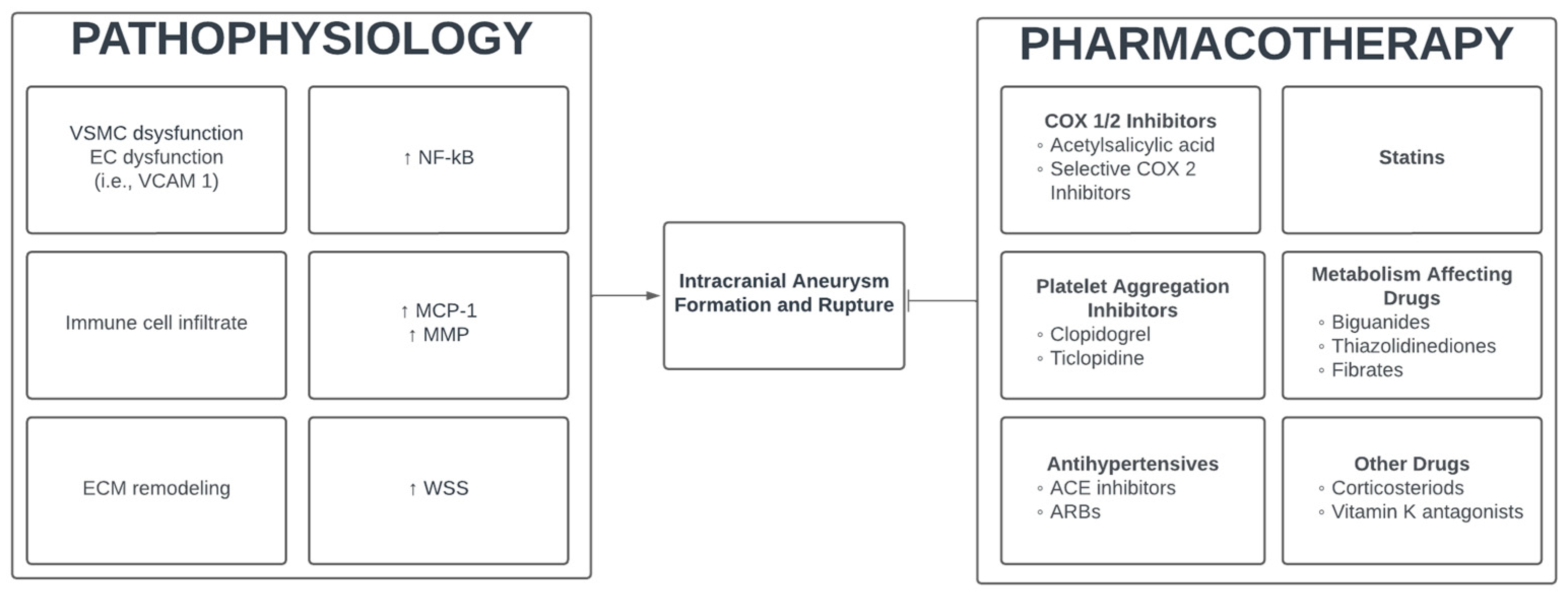
2. Pharmaceutical Modulation of Intracranial Aneurysms

2.1. Pathogenesis of Intracranial Aneurysms

Aneurysm development depends on a variety of factors including endothelial cell (EC) dysfunction, vessel wall shear stress (WSS), vascular smooth muscle cell (VSMC) dysfunction, extracellular matrix (ECM) remodeling, and immune cell infiltration [9]. WSS has been reported to be higher at branch points in the Circle of Willis where aneurysms frequently develop [10]. Mechanical stress can upregulate the expression of nuclear factor-kβ (NF-kB) which may further induce the expression of cell adhesion molecules, increase chemokines such as monocyte chemoattractant protein-1 (MCP-1), and downregulate important contractile proteins [9]. The resultant endothelial and VSMC dysfunction can lead to apoptosis and immune cell infiltration, creating an ideal inflammatory microenvironment for aneurysm formation. M1 macrophages can further promote degradation of the ECM via matrix metalloproteinase (MMP) production [11]. VSMC death and disruption of the extracellular matrix, including degradation of the elastic lamina, leads to a weakened vessel susceptible to aneurysm formation. Central to aneurysm development is NF-kB, a mediator of inflammation (Figure 1). Several clinical studies have demonstrated greater expression of NF-kB protein in the vessel wall of IAs compared to normal vascular tissue [12,13,14]. Further, many animal models of IA have confirmed the involvement of NF-kB, demonstrating increased mRNA and protein expression [12,15,16]. Thus, in addition to the aforementioned mechanisms of IA pathogenesis, NF-kB is an inflammatory mediator that may be a potential target for drug-based therapy.
In summary, aneurysms develop when shear stress at branch points in arterial circulation leads to the upregulation of maladaptive genes, such as NF-kB [9]. Consequently, the increased expression of chemokines and cell adhesion molecules leads to the development of an inflammatory local environment. These processes together can cause the degradation of important structural components of the vessel wall, including vascular smooth muscle and extracellular matrix [9].

2.2. Cyclooxygenase-1 and 2 Inhibitor (NSAIDS)

Attenuation of the NF-kB pathway and reduction of inflammatory mediators is a key goal of IA pharmacotherapy. Acetylsalicylic acid (ASA), commonly referred to as aspirin, has been the most frequently studied pharmacotherapy for the management of IAs. Aspirin has both antipyretic and anti-platelet activity, with its primary clinical benefit stemming from the latter. It inhibits cyclooxygenase-1 (COX1) and COX2, causing the reduced synthesis of prostaglandin H2 and thromboxane A2 [17]. This mitigates the standard platelet aggregation response leading to antithrombotic effects [17]. Additionally, aspirin has been shown to have some benefits outside the realm of its typical antithrombotic utility. Several in vitro and animal models have shown anti-oxidative and anti-inflammatory properties of aspirin, including the inhibition of macrophage populations, tumor necrosis factor-a (TNF-∝), NF-kB expression, and inhibitor of nuclear factor kappa-B kinase subunit beta (IKK-B) [18,19,20]. IKK-B is one of two kinases that facilitate the movement of NF-kB to the nucleus, leading to pro-inflammatory gene activation. ASA-mediated inhibition of IKK-B may prevent such downstream activation of inflammatory pathways [18]. Taken together, these anti-inflammatory properties may reduce matrix degradation, immune cell recruitment, and endothelial dysfunction in aneurysm development. Investigating the precise effects of ASA on IA pathogenesis may be limited by the biochemical properties of eicosanoids, a class of lipid-derived signaling compounds that includes prostaglandins [21]. Precise measurement is limited by short plasma half-lives and the multitude of eicosanoids with similar structures [21,22,23]. However, ASA has broad anti-inflammatory effects that have been demonstrated by measuring other inflammatory markers. Human trials have shown that aspirin may reduce total oxidant status, oxidized low-density lipoprotein (LDL), and C-reactive protein (CRP) [24,25]. LDL accumulation and oxidization may be associated with the degeneration of the aneurysm vessel wall, and CRP is associated with endothelial dysfunction [26,27]. Therefore, a reduction in LDL and CRP secondary to ASA use may have protective effects against aneurysmal development and rupture.
A quantifiable, protective effect of ASA on IA rupture has yet to be concretely determined, although there are some clinical data supporting the use of ASA in a prophylactic manner. The effects of ASA are best understood as (1) aneurysmal growth prevention and (2) reduced risk of aneurysm rupture. Zanaty et al. analyzed data from patients with multiple small aneurysms (≤5 mm) and found that aspirin had a significant association with a decreased aneurysm growth rate during observation of 146 patients with 229 aneurysms for a minimum of five years follow-up (OR 0.19, CI 0.05–0.63, p = 0.007) [28]. In this study, aspirin use was defined as ≥81 mg daily, and adherence to the aspirin regimen was documented in the medical record [28]. A more recent prospective cohort study of 272 patients with unruptured IAs less than 7 mm also found that standard or low dose aspirin greater than three times weekly was associated with a low risk of IA growth (HR 0.29, CI 0.11–0.77, p = 0.013) [29]. Importantly, both studies reflect populations with small IAs, and smaller aneurysm size is associated with lower rates of rupture [6].
In 2011, Hasan et al. conducted a nested case-control study with subjects from the International Study of Unruptured Intracranial Aneurysms (ISUIA). The logistic regression results showed that in patients with unruptured IA, aspirin use a minimum of three times weekly had a negative correlation with hemorrhage (aOR = 0.27, 95% CI 0.11–0.67, p = 0.03) [30]. Further, a study of 717 patients with cerebral aneurysms reported a much lower frequency of hemorrhagic presentation in patients taking ASA (40% vs. 28%, p = 0.016) [31]. More recently, a multicenter study in the UK (n = 2334) with either unruptured IA or aneurysmal SAH found that aspirin use was negatively associated with rupture (OR 0.28, CI 0.20–0.40, p < 0.001) [32]. A separate case-control study of 4619 patients with IA reported a significant inverse association of aspirin with IA rupture (OR 0.60, CI 0.45–0.80, p < 0.01) [33]. This same study demonstrated a dose-response curve among aspirin users and showed that higher aspirin doses were associated with a decreased risk of IA rupture (unweighted OR 0.72, 95% CI 0.60–0.85; weighted OR 0.65, 95% CI 0.53–0.81; p < 0.01) [33]. Lastly, a 2013 study that included 1797 cases of intracerebral hemorrhage (ICH) and 1340 cases of SAH showed no association between ASA and ICH risk (OR 1.06, CI 0.93–1.21) but did show an inverse association between ASA and SAH (OR 0.82, CI 0.67–1.00) [34]. Further regression analysis of 34 of 144 patients with a SAH and a history of aspirin use who were considered long-term users (≥3 years) found a stronger negative correlation with SAH risk (OR 0.63, CI 0.45–0.90), implying that prolonged ASA use could be protective in unruptured IA populations [34].
Further research has also investigated the relationship between aspirin and macrophage infiltration using ferumoxytol-enhanced MRI. Ferumoxytol-enhanced MRIs demonstrate vascular retention properties and can be quantified as a surrogate marker for inflammation [35]. After 3 months of daily aspirin, the signal intensity corresponding to ferumoxytol uptake by macrophages in the vessel wall was decreased from baseline in five patients with IA [35]. This trend was confirmed in later experiments where the signal intensity was stable in a control group of five patients and reduced in six patients treated with aspirin [36]. The authors of this study suggest that the decreased ferumoxytol uptake by macrophages may indicate that aspirin attenuates inflammation in aneurysm walls and therefore may reduce rupture risk [36].
Not all studies have supported the beneficial role of aspirin. A 2015 nationwide study in Denmark found that taking aspirin for three or more years was not associated with any increased or decreased risk of SAH (OR 1.13, CI 0.86–1.49), but taking aspirin for less than a month was associated with increased SAH risk (OR 1.75, CI 1.28–2.40) [37]. In a nested case-control study of 2065 patients with SAH selected from the German Pharmacoepidemiological Research Database, aspirin use was associated with an increased risk of SAH (OR 1.5, CI 1.2–2.0, p = 0.001) [38]. Lastly, a recent study using subjects from the UK Biobank found no relationship between aspirin use and aneurysmal SAH (HR 1.15, CI 0.91–1.47, p = 0.24) but did show that aspirin use was associated with fatal SAH (HR 1.69, CI 1.14–2.51) [39]. The results of these studies raise concerns over the benefit of ASA in patients with IAs and contradict several studies that imply that ASA use is associated with a lower risk of aneurysm growth and rupture. It is important to note that two studies that present negative data are population-level analyses from national databanks. Notably, the cohort study of the UK Biobank defined aspirin use as a binary measure and did not account for dosage or frequency and the nested case-control study which used subjects from a German databank defined aspirin use as current if the patient’s last prescription overlapped with the 7 days preceding hospital admission for SAH [38,39]. Conversely, several of the studies that support aspirin use reflect data from patients whose medication frequency was specified as at least three times weekly. Variability in follow-up criteria, dosing protocols, and patient selection persist as challenges to universal conclusions on ASA prophylaxis in this patient population.

2.3. Selective Cyclooxygenase-2 Inhibitors

Non-steroidal anti-inflammatory drugs (NSAIDs)’ mechanism of action involves the inhibition of the COX1 and COX2 enzymes to mitigate inflammation. Concomitant inhibition of COX1 and COX2 can precipitate unfavorable side effects for patients, including gastric ulcers, hypertension, and acute renal failure [40]. Selective COX2 inhibitors such as celecoxib aim to prevent side effects by avoiding the inhibition of the constitutively expressed COX1 which plays a role in maintaining gastric mucosa [41].
The basis of using selective COX2 inhibitors for the treatment and prevention of intracranial aneurysms is supported by animal model data, suggesting these medications attenuate the inflammatory response implicated in IAs. COX-mediated synthesis of prostaglandin E2 causes downstream activation of NF-kB, which has been implicated in aneurysm development [9,42]. Targeting of this pathway is understood to decrease aneurysmal development. Interestingly, animal studies of aortic aneurysms have demonstrated that COX2 inhibition can reduce measures of aneurysm growth and rupture [43,44]. Focusing on intracranial aneurysms, a study by Aoki et al. found that human IA tissues had greater staining of COX2 compared to controls and that in a rat model of IA, there was a significant increase in COX2 mRNA and protein in cerebral artery tissue, 3 months after induction of aneurysm (p < 0.01) [45]. Further, inhibition of COX2 prevented aneurysm formation in the same animal model (p < 0.05). The authors of this study concluded that COX2 may participate in a positive feedback loop with NF-kB [45]. Moreover, Hasan et al. found that in tissues from patients with IA, COX2 expression was significantly greater in both ruptured and unruptured aneurysms compared to an arterial control (p = 0.001) [46].
Given the preclinical data specifically implicating COX2 in the pathophysiology of aneurysms it may be reasonable to investigate COX2-specific inhibitors as prophylaxis for aneurysm development. That said, clinical research in this area has been limited, with the focus on aspirin, a nonspecific inhibitor of COX enzymes that has shown significant promise. A recent study of 1419 patients with saccular IAs found that neither treatment with COX2 selective inhibitors (HR 0.63, CI 0.29–1.39, p = 0.249) nor NSAIDS (HR 1.11, CI 0.50–2.45, p = 0.805) reduced de novo IA formation [47]. Risselada et al. found that the use of COX2 selective inhibitors was positively correlated with SAH (OR 2.35, CI 1.27–4.36) [48]. Additionally, a meta-analysis found that non-aspirin NSAIDS (OR 1.73, CI 1.07–2.79) and selective COX2 inhibitors (OR 2.04, CI 1.24–3.35) were positively associated with aneurysmal SAH [49]. There is an apparent disconnect between pre-clinical and human trials, and it is important to note that the efficacy of COX2 inhibition is limited by its inhibition of prostacyclin which has beneficial vasodilatory effects and limits vascular remodeling [50]. The current body of research is limited, and prospective investigations can delineate the protective effect of COX2 inhibition.

2.4. Platelet Aggregation Inhibitors

Clopidogrel, a thienopyridine, acts by selectively inhibiting adenosine diphosphate-mediated platelet aggregation [51]. More specifically, these medications are antagonists of the P2Y12 platelet receptor and harbor some anti-inflammatory properties. In a rat model of renal inflammation, treatment with clopidogrel reduced the expression of many pro-inflammatory factors including MCP-1 (p < 0.05) [52]. MCP-1 may be a pharmacologic target in the modulation of IA formation as animal models have reaffirmed the role of MCP-1 in cerebral IA development [53].
A recent retrospective study of 921 patients with IAs found that exposure to clopidogrel reduced the likelihood of rupture (6.6% vs. 23.5%, p = 0.001) [54]. A case-control study of 1004 SAH cases found a positive correlation between platelet aggregation inhibitors and risk of SAH (OR 1.32, CI 1.02–1.70) [48]. However, this association was not significant in a further case-crossover analysis and the study did not differentiate between classes of platelet-aggregation inhibitors, thus clopidogrel, ticlopidine, aspirin, dipyridamole, and carbasalate calcium were all included together [48]. To add to the conflicting nature of current data, a nationwide case-control study from Denmark (n = 5834) reported data indicating that clopidogrel was only associated with an increased risk of SAH in the short term [37]. There was a positive association between clopidogrel use and risk of SAH in patients taking the drug for less than a month (OR 2.33, CI 1.02–5.35) and between 2 and 3 months (OR 2.40, CI 1.20–4.81); however, this association was not significant beyond 3 months of use [37].
There is no clear answer on whether ADP receptor inhibitors have a protective or harmful effect on IA aneurysm rupture. The current data available are a small mix of observational studies demonstrating conflicting data. Higher strength evidence is needed in the form of high-powered case-control studies and if possible, randomized controlled trials, to delineate what role, if any, ADP receptor inhibitors can play in aneurysm management.

2.5. Antihypertensives

Hypertension is one of the most prominent risk factors for IA development and rupture [55,56,57]. Additionally, hemodynamic stress and renin–angiotensin–aldosterone system (RAAS) activation-induced inflammatory responses can perpetrate IA pathophysiology [55]. Chronic hypertension is accompanied by a variety of histopathological changes including altered intimal layer thickness, tunica media necrosis, and degeneration of the internal elastic lamina, which all compromise the structural integrity of the cerebral vasculature [58]. Hypertensive patients have a 2.6 times greater risk of ruptured IA when compared to non-hypertensive patients per a cross-sectional study of 29 patients [59]. A larger retrospective study of 3965 patients also found hypertension to be significantly associated with increased IA rupture risk (OR 2.55, CI 2.16–3.03) [60]. Anti-hypertensive medications have emerged as successful pharmacotherapies to reduce rupture risk in IA patients. A mouse model of intracranial aneurysms demonstrated that controlling blood pressure with hydralazine significantly reduced IA rupture rate (p < 0.05) [55]. Moreover, this study showed a dose-dependent relationship between hydralazine and IA rupture rate. Furthermore, initiation of captopril (angiotensin-converting enzyme inhibitor) reduced the incidence of rupture (p < 0.05) and improved survival (p < 0.05) in mice with IAs. Lastly, this study also investigated the roles of the local RAAS on vascular wall integrity. The results demonstrated that angiotensin-converting enzyme (ACE) inhibitors and angiotensin II type 1 receptor antagonists (ARB) reduced IA rupture rate without altering systemic hypertension, which further suggests the activation of a regional vascular RAAS system in IA rupture [55].
In a recent multi-center analysis (n = 3044), RAAS inhibitors were significantly associated with reduced rupture risk versus non-RAAS inhibitors (OR: 0.490, 95% CI: 0.402–0.597, p = 0.000) [61]. Individually, treatment with ACE inhibitors (OR: 0.559, 95% CI: 0.442–0.709, p = 0.000) or ARBs (OR: 0.414, 95% CI: 0.315–0.542, p = 0.000) suggested a lower risk of rupture. Notably, the association between RAAS inhibitors and reduced rupture risk was independent of blood pressure control, suggesting that RAAS inhibitors are the optimal blood pressure medications to prevent IA rupture in hypertensive patient populations [61].

2.6. Hydroxymethylglutaryl-CoA Reductase Inhibitors (Statins)

Hydroxymethylglutaryl-CoA (HMG-CoA) reductase inhibitors are a class of medications commonly referred to as statins. In addition to lowering LDL-cholesterol via synthesis inhibition, it has been proposed that statins may improve endothelial function through anti-inflammatory properties [62]. On a pathophysiological level, a trial of 17,802 healthy participants with elevated CRP levels treated with 20 mg of rosuvastatin daily showed significantly reduced levels of CRP (37% reduction, p < 0.001) [63]. CRP is associated with reduced endothelial vasoreactivity, and pharmacologic lowering of this marker could possibly reduce endothelial dysfunction, a process implicated in aneurysm development and rupture [27,63]. Landmesser et al. further demonstrated that compared to ezetimibe (selective cholesterol-absorption inhibitor), simvastatin had superior effects on arterial dilation. Here, improved arterial dilation demonstrated that statins disrupt aberrant endothelial function and that the mechanism of action in cholesterol-lowering drugs dictates the impact on vascular response [64]. An in vitro model demonstrated that incubation of monocytes in cerivastatin reduced adhesion to vascular endothelium [65]. Lastly, a clinical trial of 146 patients found that MCP-1 was reduced by 28% from baseline in patients treated with 1 mg/day pitavastatin and reduced by 11% in patients treated with 5 mg/day atorvastatin [66].
There are a few studies that provide data supporting statin use in patients with IAs. A multicenter study from Japan selected 117 cases of ruptured IAs and found an inverse relationship between statin use and aneurysmal rupture (aOR 0.30, CI 0.14–0.66) [67]. Another study of 4701 patients with IAs reported that the use of lipid-lowering medications was found to be inversely correlated with rupture (OR 0.58, CI 0.47–0.71, p < 0.01) [68]. Importantly, of the 4701 patients, 1129 were using lipid-lowering agents, and 937 of those patients were using statins only [68]. To add to the evidence backing statin use in IA patients, a cross-sectional study of 310 patients with ruptured IA and 887 patients with unruptured IA reported an inverse association of statin usage with rupture (OR 0.54, CI 0.38–0.77, p = 0.0008) [69].
Further research has sought to examine the effects of statin use on the pathogenesis of aneurysm development in patients with IA. Aneurysm wall enhancement (WE), measured via MRI, has emerged as one such technique to quantify the effect of treatments on aneurysmal stability. Aneurysmal WE is indicative of wall inflammation and confers an increased risk of rupture [70]. A recent study assessed 127 fusiform aneurysms and found that statin use was significantly correlated with a decreased WE (β = −0.236, p = 0.007) [71]. Moreover, a randomized controlled trial of 60 patients with unruptured IAs larger than 3 mm found that 20 mg of atorvastatin daily led to a decrease in WE as compared to baseline (p = 0.039) and a placebo group (p = 0.006), after 6 months of treatment [72]. Interestingly, there was no difference in aneurysm size in atorvastatin or placebo-treated groups [72].
Despite these studies which support the clinical use of statins in patients with IAs, several data suggest otherwise. One study examined the combined effect of statin and aspirin treatment on 194 ruptured IAs and 214 unruptured IAs and found that combined medical therapy was correlated with unruptured status (aOR 5.01, CI 1.37–18.33, p = 0.015), but statins alone were not found to have any association with IA rupture (aOR:1.65, CI 0.83–3.31, p = 0.155) [73]. A randomized controlled trial (n = 209) investigating the impact of 10 mg of atorvastatin daily, found no significant difference in aneurysm growth, rupture, or “new bleb formation” over a mean imaging follow-up period of 32.4 months (Log-rank p = 0.359) [74]. Of note, the systolic pressure, a known risk factor for aneurysm rupture, was significantly higher in the statin-treated group (p = 0.03) [74,75]. Further analysis is needed to delineate the impact of statins on aneurysmal growth while controlling for confounding comorbidities. A case-control study of 1200 patients with confirmed IA found no correlation between statin use and IA development (OR 1.08, CI 0.69–1.69) [76]. A separate study of patients with unruptured IAs (n = 28,931) reported that statin use was not associated with the risk of SAH (OR 1.03, CI 0.86–1.23, p = 0.730) [77]. Lastly, a recent study compared a patient cohort with IAs (n = 1960) to matched controls and found that statins were associated with a higher risk of IA development (aOR 1.34, CI 1.02–1.78) [78]. These results should be interpreted cautiously though, considering the likelihood of patients who are prescribed statins having other risk factors for aneurysm development (i.e., hypertension). Conversely, in the same study, a multivariate analysis of the IA patient cohort alone found that patients with ruptured IAs were less likely to be treated with statins (aOR 0.62, CI 0.47–0.81) [78]. Conflicting conclusions surrounding the use of statin use for IA management are complicated by the burden of comorbid conditions and medication compliance. A refined understanding of the role of statins in aneurysmal rupture prevention will require further clinical investigation with judicious controls on comorbid conditions.

2.7. Metabolism Affecting Drugs

Biguanides are a class of medications used to treat diabetes mellitus. Included in this class of drugs is metformin, the most prescribed anti-diabetic medication worldwide [79]. The precise mechanism of biguanides is yet unclear; however, it is evident that these drugs inhibit hepatic gluconeogenesis [80]. Further, given the pleiotropic effects of metformin, many groups have sought to investigate its potential benefit in different disease models. In a rat model of elastase-induced IA, it was found that metformin decreased the rate of IA rupture (p < 0.05) [81]. Furthermore, a meta-analysis examined the data from two studies assessing the correlation between biguanides and aneurysmal SAH, and found an inverse association with SAH (OR 0.57, CI 0.34–0.96) [49].
Thiazolidinediones (TZDs) are medications also used to treat type 2 diabetes mellitus that reverse insulin resistance and reduce the progression of metabolic syndrome. TZDs decrease hepatic gluconeogenesis, lower insulin levels, and decrease triglycerides [82]. On a cellular level, TZDs are peroxisome proliferator-activated regulator-y (PPAR-y) agonists [82]. It is understood that PPAR-y is expressed by both macrophages and VSMCs which are implicated in the pathophysiology of IA development [82]. Current research in animal models suggests that dysregulated PPAR-y may be implicated in pro-inflammatory states and ultimately, aneurysm development. In a mouse model of intracranial aneurysm, a PPAR-y antagonist increased the incidence of subarachnoid hemorrhage (p < 0.05) [83]. The same study also showed that transgenic mice with a dominant-negative smooth muscle-specific PPAR-y mutation, had significantly greater rates of aneurysm formation (p < 0.05) and rupture (p = 0.05) [83]. From a pharmacotherapy perspective, pioglitazone reduced the incidence of ruptured aneurysms compared to vehicle controls (p < 0.05) [84]. Further, in a mouse model of abdominal aortic aneurysm, rosiglitazone inhibited fatal aneurysm rupture (p = 0.0013) and reduced the maximal aortic dilatation (p < 0.0001) [85].
Fibrates are medications used to reduce serum triglyceride levels. They induce lipolysis, hepatic fatty acid uptake and increase reverse cholesterol transport [86]. Fibrates are also activators of peroxisome proliferator-activated receptor-a (PPAR-a) [87]. While there is limited literature on the effects of fibrates in cerebral aneurysms, a few basic science and clinical studies have investigated their use for aortic aneurysms and suggested a beneficial effect [87,88]. Two randomized controlled trials examining fibrate use in abdominal aneurysm patients have also been conducted [89,90]. Further investigation is required to characterize the effect of fibrates on IA development and rupture risk.

2.8. Other Medications

Synthetic glucocorticoids have potent anti-inflammatory effects that are mediated through the binding of the glucocorticoid receptor (GR) [91]. The ligand–receptor complex translocates to the nucleus and subsequently suppresses or elicits transcription of various genes. A few pro-inflammatory factors that are negatively regulated include factors implicated in IA pathogenesis, such as COX2, inducible nitric oxide synthase (iNOS), and TNF-a [91]. The role of corticosteroids in the pharmacological modulation of IA is underreported but they may modulate the inflammatory factors contributing to IA development and rupture. An observational study of 1158 patients presenting with SAH and confirmed IA found that SAH was significantly associated with the composite outcome of a history of corticosteroid use or a medical condition that may require the use of corticosteroids (OR 1.67, CI 1.09–2.54, p = 0.0l6) [92]. Additionally, autoimmune conditions modulate pro-inflammatory states and are also associated with smaller ruptured aneurysm size (p = 0.03) [93].
Table 1. Summary of clinical studies examining pharmaceutical modulation of intracranial aneurysms.
Study Author (Year)No. of PtsMedicationAssociation
Zanaty et al. (2020) [28]N = 146ASADecreased IA growth (OR 0.19, CI 0.05–0.63, p = 0.007)
Weng et al. (2020) [29]N = 272ASADecreased IA growth (HR 0.29, CI 0.11–0.77, p = 0.013)
Hasan et al. (2011) [30]N = 271ASADecreased odds of hemorrhage (aOR 0.27, CI 0.11–0.67, p = 0.03)
Gross et al. (2014) [31]N = 717ASADecreased rate of hemorrhage (40% vs. 28%, p = 0.016)
Hostettler et al. (2018) [32]N = 2334ASANegative association with rupture (OR 0.28, CI 0.20–0.40, p < 0.001)
Can et al. (2018) [33]N = 4619ASANegative association with rupture (OR 0.60, CI 0.45–0.80, p < 0.01)
Dose response (OR 0.65, CI 0.53–0.81, p < 0.01)
Garcia-Rodriguez et al. (2013) [34]N = 1340ASA > 3 yearsDecreased risk of SAH (OR 0.63, CI 0.45–0.90)
Garbe et al. (2013) [38]N = 2065ASAIncreased risk of SAH (OR 1.5, CI 1.2–2.0, p = 0.001)
Ewbank et al. (2023) [39]N = 541ASANo association with SAH (HR 1.15, CI 0.91–1.47, p = 0.24)
Pottegård et al. (2015) [37]N = 5834ASA < 1 monthIncreased risk of SAH (OR 1.75, CI 1.28–2.40)
ASA > 3 yearsNo association with SAH (OR 1.13, CI 0.86–1.49)
Raisanen et al. (2022) [47]N = 1419COX2iNo association with IA formation (HR 0.63, CI 0.29–1.39, p = 0.249)
Risselada et al. (2011) [48]N = 1004COX2iPositive association with SAH (OR 2.35, CI 1.27–4.36)
Pottegård et al. (2015) [37]N = 5834Clopidogrel < 1 monthPositive association with SAH (OR 2.33, CI 1.02–5.35), no significant relationship in long-term users.
Hudson et al. (2023) [54]N = 921ClopidogrelLower likelihood of rupture (6.6% vs. 23.5%, p = 0.001)
Risselada et al. (2011) [48]N = 1004Platelet Aggregation InhibitorsPositive association with SAH (OR 1.32, CI 1.02–1.70) in case-control study, significance lost in case-crossover analysis.
Zhong et al. (2022) [61]N = 3044ACEiNegative association with rupture (OR 0.559, CI 0.442–0.709, p = 0.000)
ARBsNegative association with rupture (OR 0.414, CI 0.315–0.542, p = 0.000)
Yoshimura et al. (2014) [67]N = 421StatinsNegative association with rupture (aOR 0.30, CI 0.14–0.66)
Shimizu et al. (2021) [69]N = 1197StatinsNegative association with rupture (OR 0.54, CI 0.38–0.77, p = 0.0008)
Terceno et al. (2021) [73]N = 368StatinsNo association with rupture (aOR 1.65, CI 0.83–3.31, p = 0.155)
Yoshida et al. (2021) [74]N = 209StatinsNo difference in IA growth, rupture, or “new bleb formation” (Log-rank p = 0.359)
Marbacher et al. (2012) [76]N = 300StatinsNo association with IA formation (OR 1.08, CI 0.69–1.69, p = 0.74)
Bekelis et al. (2015) [77]N = 28,931StatinsNo association with SAH (OR 1.03, CI 0.86–1.23, p = 0.730)
Jabbarli et al. (2023) [78]N = 1960StatinsPositive association with IA formation (aOR 1.34, CI 1.02–1.78)
N = 2446StatinsNegative association with rupture (aOR 0.62, CI 0.47–0.81)
Can et al. (2018) [68]N = 4701Lipid-Lowering MedicationsNegative association with rupture (OR 0.58, CI 0.47–0.71, p < 0.01)
Ruigrok et al. (2006) [92]N = 1158CorticosteroidsComposite outcome of corticosteroids or a medical condition that may be treated with corticosteroids had a positive association with SAH (OR 1.67, CI 1.09–2.54, p = 0.016)
Pottegård et al. (2015) [37]N = 5834Vitamin-K AntagonistsNo association with SAH (OR 1.24, CI 0.86–1.77) in long-term users (>3 years)
Garbe et al. (2013) [38]N = 2065Vitamin-K AntagonistsPositive association with SAH (OR 1.7, CI 1.3–2.3, p < 0.001)
Risselada et al. (2011) [48]N = 1004Vitamin-K AntagonistsPositive association with SAH (OR 2.90, CI 1.27–6.65) in case-crossover, not significant in case-control
Lastly, vitamin K antagonists are anti-coagulant medications that act by inhibiting the vitamin K epoxide reductase complex [94]. The prevention of the gamma-carboxylation of vitamin-K-dependent coagulation factors reduces thrombosis risk [94]. A 2011 study of 1004 SAH cases found no association between the risk of SAH and vitamin K antagonist use (OR 1.29, CI 0.89–1.87) in a case-control analysis but did find a significant positive association in a case-crossover analysis (OR 2.90, CI 1.27–6.65) [48]. Conversely, a later case-control study of 2065 cases of SAH found that the risk of SAH was positively associated with phenprocoumon, a vitamin K antagonist (OR 1.7, CI 1.3–2.3, p < 0.001) [38]. Lastly, Pottegard et al. found that vitamin K antagonists were not significantly correlated with SAH with long-term (>3 years) use (aOR 1.24, CI 0.86–1.77) [37].
Recent research on the forefront of aneurysm pathophysiology has demonstrated a possible role for the tyrosine kinase inhibitor, sunitinib [95]. The authors produced a mouse model of basilar artery aneurysm by overexpressing a mutated platelet-derived growth factor B, and further showed that treatment with sunitinib significantly reduced arterial cross-sectional area (p < 0.05) [95]. Similarly, in a rat model of intracranial aneurysm, erlotinib was shown to reduce the rate of aneurysm development [96].
The utility of corticosteroids and vitamin-K antagonists in the prevention of IA development and rupture is unclear and future studies that adequately control for underlying disease states are best suited to characterize their impact on IA development and rupture. Pharmaceuticals tested in the pre-clinical space such as tyrosine kinase inhibitors may lead to new discoveries in the pathogenesis of IAs which could further the use of prophylactic medications for IA progression.

3. Conclusions

While there is an abundance of literature examining the effects of acetylsalicylic acid exposure on aneurysm growth and rupture, the data are inconsistent. Robust clinical studies of populations with medium-sized aneurysms may reveal a more niche role for ASA in the treatment of IAs. Further research is needed to fully elucidate the effects of statins, COX2 inhibitors, and ADP inhibitors, among other metabolism-affecting drugs, on the development, growth, and rupture of intracranial aneurysms.

Author Contributions

Conceptualization, A.C. and R.M.S.; writing—original draft preparation, A.C. and R.M.S.; writing—review and editing, R.M.S., J.S.H., K.W.N., Z.C.G., P.A., R.C.J., M.J.L. and B.G.; All authors have read and agreed to the published version of the manuscript.

Funding

This research received no external funding.

Conflicts of Interest

The authors declare no conflicts of interest.

References

  1. Gasparotti, R.; Liserre, R. Intracranial aneurysms. Eur. Radiol. 2005, 15, 441–447. [Google Scholar] [CrossRef]
  2. Keedy, A. An overview of intracranial aneurysms. McGill J. Med. 2006, 9, 141–146. [Google Scholar] [CrossRef]
  3. Ajiboye, N.; Chalouhi, N.; Starke, R.M.; Zanaty, M.; Bell, R. Unruptured Cerebral Aneurysms: Evaluation and Management. Sci. World J. 2015, 2015, 954954. [Google Scholar] [CrossRef]
  4. Ziu, E.; Khan Suheb, M.Z.; Mesfin, F.B. Subarachnoid Hemorrhage. In StatPearls; StatPearls Publishing: Treasure Island, FL, USA, 2023. [Google Scholar]
  5. Cras, T.Y.; Bos, D.; Ikram, M.A.; Vergouwen, M.D.I.; Dippel, D.W.J.; Voortman, T.; Adams, H.H.H.; Vernooij, M.W.; Roozenbeek, B. Determinants of the Presence and Size of Intracranial Aneurysms in the General Population: The Rotterdam Study. Stroke 2020, 51, 2103–2110. [Google Scholar] [CrossRef]
  6. Chandra, R.V.; Maingard, J.; Slater, L.-A.; Cheung, N.K.; Lai, L.T.; Gall, S.L.; Thrift, A.G.; Phan, T.G. A Meta-Analysis of Rupture Risk for Intracranial Aneurysms 10 mm or Less in Size Selected for Conservative Management without Repair. Front. Neurol. 2022, 12, 743023. [Google Scholar] [CrossRef]
  7. Roquer, J.; Cuadrado-Godia, E.; Guimaraens, L.; Conesa, G.; Rodriguez-Campello, A.; Capellades, J.; García-Arnillas, M.P.; Fernández-Candil, J.L.; Avellaneda-Gómez, C.; Giralt-Steinhauer, E.; et al. Short- and long-term outcome of patients with aneurysmal subarachnoid hemorrhage. Neurology 2020, 95, e1819–e1829. [Google Scholar] [CrossRef]
  8. van der Kamp, L.T.; Rinkel, G.J.E.; Verbaan, D.; van den Berg, R.; Vandertop, W.P.; Murayama, Y.; Ishibashi, T.; Lindgren, A.; Koivisto, T.; Teo, M.; et al. Risk of Rupture after Intracranial Aneurysm Growth. JAMA Neurol. 2021, 78, 1228–1235. [Google Scholar] [CrossRef]
  9. Liu, Z.; Ajimu, K.; Yalikun, N.; Zheng, Y.; Xu, F. Potential Therapeutic Strategies for Intracranial Aneurysms Targeting Aneurysm Pathogenesis. Front. Neurosci. 2019, 13, 1238. [Google Scholar] [CrossRef]
  10. Alnæs, M.S.; Isaksen, J.; Mardal, K.-A.; Romner, B.; Morgan, M.K.; Ingebrigtsen, T. Computation of Hemodynamics in the Circle of Willis. Stroke 2007, 38, 2500–2505. [Google Scholar] [CrossRef]
  11. Signorelli, F.; Sela, S.; Gesualdo, L.; Chevrel, S.; Tollet, F.; Pailler-Mattei, C.; Tacconi, L.; Turjman, F.; Vacca, A.; Schul, D.B. Hemodynamic Stress, Inflammation, and Intracranial Aneurysm Development and Rupture: A Systematic Review. World Neurosurg. 2018, 115, 234–244. [Google Scholar] [CrossRef]
  12. Lai, X.L.; Deng, Z.F.; Zhu, X.G.; Chen, Z.H. Apc gene suppresses intracranial aneurysm formation and rupture through inhibiting the NF-κB signaling pathway mediated inflammatory response. Biosci. Rep. 2019, 39, BSR20181909. [Google Scholar] [CrossRef]
  13. Cheng, W.T.; Wang, N. Correlation between MMP-2 and NF-κ B expression of intracranial aneurysm. Asian Pac. J. Trop. Med. 2013, 6, 570–573. [Google Scholar] [CrossRef]
  14. Fan, X.J.; Zhao, H.D.; Yu, G.; Zhong, X.L.; Yao, H.; Yang, Q.D. Role of inflammatory responses in the pathogenesis of human cerebral aneurysm. Genet. Mol. Res. 2015, 14, 9062–9070. [Google Scholar] [CrossRef]
  15. Jin, T.; An, Q.; Qin, X.; Qin, X.; Hu, Y.; Hu, J.; Zhou, B.; Leng, B. Resveratrol inhibits cerebral aneurysms in mice via downregulating the NF-κB pathway. Acta Biochim. Pol. 2022, 69, 613–618. [Google Scholar] [CrossRef]
  16. Li, S.; Tian, Y.; Huang, X.; Zhang, Y.; Wang, D.; Wei, H.; Dong, J.; Jiang, R.; Zhang, J. Intravenous transfusion of endothelial colony-forming cells attenuates vascular degeneration after cerebral aneurysm induction. Brain Res. 2014, 1593, 65–75. [Google Scholar] [CrossRef]
  17. Awtry, E.H.; Loscalzo, J. Aspirin. Circulation 2000, 101, 1206–1218. [Google Scholar] [CrossRef]
  18. Yin, M.-J.; Yamamoto, Y.; Gaynor, R.B. The anti-inflammatory agents aspirin and salicylate inhibit the activity of IκB kinase-β. Nature 1998, 396, 77–80. [Google Scholar] [CrossRef]
  19. Chen, C.M.; Tung, Y.T.; Wei, C.H.; Lee, P.Y.; Chen, W. Anti-Inflammatory and Reactive Oxygen Species Suppression through Aspirin Pretreatment to Treat Hyperoxia-Induced Acute Lung Injury in NF-kappaB-Luciferase Inducible Transgenic Mice. Antioxidants 2020, 9, 429. [Google Scholar] [CrossRef]
  20. Jorda, A.; Aldasoro, M.; Aldasoro, C.; Guerra-Ojeda, S.; Iradi, A.; Vila, J.M.; Campos-Campos, J.; Valles, S.L. Action of low doses of Aspirin in Inflammation and Oxidative Stress induced by aβ(1-42) on Astrocytes in primary culture. Int. J. Med. Sci. 2020, 17, 834–843. [Google Scholar] [CrossRef]
  21. Chhonker, Y.S.; Bala, V.; Murry, D.J. Quantification of eicosanoids and their metabolites in biological matrices: A review. Bioanalysis 2018, 10, 2027–2046. [Google Scholar] [CrossRef]
  22. Dickinson, J.S.; Murphy, R.C. Mass spectrometric analysis of leukotriene A4 and other chemically reactive metabolites of arachidonic acid. J. Am. Soc. Mass Spectrom. 2002, 13, 1227–1234. [Google Scholar] [CrossRef]
  23. Gréen, K. Metabolism and pharmacokinetics of prostaglandin analogs in man. In Prostaglandins and Fertility Regulation; Toppozada, M.K., Bygdeman, M., Hafez, E.S.E., Eds.; Springer: Dordrecht, The Netherlands, 1984; pp. 11–19. [Google Scholar]
  24. Kurban, S.; Mehmetoglu, I. Effects of acetylsalicylic acid on serum paraoxonase activity, Ox-LDL, coenzyme Q10 and other oxidative stress markers in healthy volunteers. Clin. Biochem. 2010, 43, 287–290. [Google Scholar] [CrossRef]
  25. Berg, K.; Langaas, M.; Ericsson, M.; Pleym, H.; Basu, S.; Nordrum, I.S.; Vitale, N.; Haaverstad, R. Acetylsalicylic acid treatment until surgery reduces oxidative stress and inflammation in patients undergoing coronary artery bypass grafting. Eur. J. Cardiothorac. Surg. 2013, 43, 1154–1163. [Google Scholar] [CrossRef]
  26. Frösen, J.; Tulamo, R.; Heikura, T.; Sammalkorpi, S.; Niemelä, M.; Hernesniemi, J.; Levonen, A.-L.; Hörkkö, S.; Ylä-Herttuala, S. Lipid accumulation, lipid oxidation, and low plasma levels of acquired antibodies against oxidized lipids associate with degeneration and rupture of the intracranial aneurysm wall. Acta Neuropathol. Commun. 2013, 1, 71. [Google Scholar] [CrossRef]
  27. Fichtlscherer, S.; Rosenberger, G.; Walter, D.H.; Breuer, S.; Dimmeler, S.; Zeiher, A.M. Elevated C-Reactive Protein Levels and Impaired Endothelial Vasoreactivity in Patients With Coronary Artery Disease. Circulation 2000, 102, 1000–1006. [Google Scholar] [CrossRef]
  28. Zanaty, M.; Roa, J.A.; Nakagawa, D.; Chalouhi, N.; Allan, L.; Al Kasab, S.; Limaye, K.; Ishii, D.; Samaniego, E.A.; Jabbour, P.; et al. Aspirin associated with decreased rate of intracranial aneurysm growth. J. Neurosurg. JNS 2020, 133, 1478–1485. [Google Scholar] [CrossRef]
  29. Weng, J.-C.; Wang, J.; Li, H.; Jiao, Y.-M.; Fu, W.-L.; Huo, R.; Yan, Z.-H.; Xu, H.-Y.; Zhan, J.; Wang, S.; et al. Aspirin and Growth of Small Unruptured Intracranial Aneurysm. Stroke 2020, 51, 3045–3054. [Google Scholar] [CrossRef]
  30. Hasan, D.M.; Mahaney, K.B.; Brown, R.D., Jr.; Meissner, I.; Piepgras, D.G.; Huston, J.; Capuano, A.W.; Torner, J.C. Aspirin as a promising agent for decreasing incidence of cerebral aneurysm rupture. Stroke 2011, 42, 3156–3162. [Google Scholar] [CrossRef]
  31. Gross, B.A.; Rosalind Lai, P.M.; Frerichs, K.U.; Du, R. Aspirin and aneurysmal subarachnoid hemorrhage. World Neurosurg. 2014, 82, 1127–1130. [Google Scholar] [CrossRef]
  32. Hostettler, I.C.; Alg, V.S.; Shahi, N.; Jichi, F.; Bonner, S.; Walsh, D.; Bulters, D.; Kitchen, N.; Brown, M.M.; Houlden, H.; et al. Characteristics of Unruptured Compared to Ruptured Intracranial Aneurysms: A Multicenter Case–Control Study. Neurosurgery 2018, 83, 43–52. [Google Scholar] [CrossRef]
  33. Can, A.; Rudy, R.F.; Castro, V.M.; Yu, S.; Dligach, D.; Finan, S.; Gainer, V.; Shadick, N.A.; Savova, G.; Murphy, S.; et al. Association between aspirin dose and subarachnoid hemorrhage from saccular aneurysms: A case-control study. Neurology 2018, 91, e1175–e1181. [Google Scholar] [CrossRef]
  34. García-Rodríguez, L.A.; Gaist, D.; Morton, J.; Cookson, C.; González-Pérez, A. Antithrombotic drugs and risk of hemorrhagic stroke in the general population. Neurology 2013, 81, 566–574. [Google Scholar] [CrossRef]
  35. Hasan, D.M.; Chalouhi, N.; Jabbour, P.; Magnotta, V.A.; Kung, D.K.; Young, W.L. Imaging aspirin effect on macrophages in the wall of human cerebral aneurysms using ferumoxytol-enhanced MRI: Preliminary results. J. Neuroradiol. 2013, 40, 187–191. [Google Scholar] [CrossRef]
  36. Hasan, D.M.; Chalouhi, N.; Jabbour, P.; Dumont, A.S.; Kung, D.K.; Magnotta, V.A.; Young, W.L.; Hashimoto, T.; Winn, H.R.; Heistad, D. Evidence that acetylsalicylic acid attenuates inflammation in the walls of human cerebral aneurysms: Preliminary results. J. Am. Heart Assoc. 2013, 2, e000019. [Google Scholar] [CrossRef]
  37. Pottegård, A.; García Rodríguez, L.A.; Poulsen, F.R.; Hallas, J.; Gaist, D. Antithrombotic drugs and subarachnoid haemorrhage risk. A nationwide case-control study in Denmark. Thromb. Haemost. 2015, 114, 1064–1075. [Google Scholar] [CrossRef]
  38. Garbe, E.; Kreisel, S.H.; Behr, S. Risk of subarachnoid hemorrhage and early case fatality associated with outpatient antithrombotic drug use. Stroke 2013, 44, 2422–2426. [Google Scholar] [CrossRef]
  39. Ewbank, F.; Birks, J.; Gaastra, B.; Hall, S.; Galea, I.; Bulters, D. Aspirin and Subarachnoid Haemorrhage in the UK Biobank. Transl. Stroke Res. 2023, 14, 490–498. [Google Scholar] [CrossRef]
  40. Vonkeman, H.E.; van de Laar, M.A. Nonsteroidal anti-inflammatory drugs: Adverse effects and their prevention. Semin. Arthritis Rheum. 2010, 39, 294–312. [Google Scholar] [CrossRef]
  41. Ghlichloo, I.; Gerriets, V. Nonsteroidal Anti-Inflammatory Drugs (NSAIDs). In StatPearls; StatPearls Publishing: Treasure Island, FL, USA, 2023. [Google Scholar]
  42. Aoki, T.; Kataoka, H.; Shimamura, M.; Nakagami, H.; Wakayama, K.; Moriwaki, T.; Ishibashi, R.; Nozaki, K.; Morishita, R.; Hashimoto, N. NF-κB Is a Key Mediator of Cerebral Aneurysm Formation. Circulation 2007, 116, 2830–2840. [Google Scholar] [CrossRef]
  43. Miralles, M.; Wester, W.; Sicard, G.A.; Thompson, R.; Reilly, J.M. Indomethacin inhibits expansion of experimental aortic aneurysms via inhibition of the cox2 isoform of cyclooxygenase. J. Vasc. Surg. 1999, 29, 884–892; discussion 892–893. [Google Scholar] [CrossRef]
  44. Ghoshal, S.; Loftin, C.D. Cyclooxygenase-2 inhibition attenuates abdominal aortic aneurysm progression in hyperlipidemic mice. PLoS ONE 2012, 7, e44369. [Google Scholar] [CrossRef]
  45. Aoki, T.; Nishimura, M.; Matsuoka, T.; Yamamoto, K.; Furuyashiki, T.; Kataoka, H.; Narumiya, S. PGE(2) -EP(2) signalling in endothelium is activated by haemodynamic stress and induces cerebral aneurysm through an amplifying loop via NF-κB. Br. J. Pharmacol. 2011, 163, 1237–1249. [Google Scholar] [CrossRef]
  46. Hasan, D.; Hashimoto, T.; Kung, D.; Macdonald, R.L.; Winn, H.R.; Heistad, D. Upregulation of cyclooxygenase-2 (COX-2) and microsomal prostaglandin E2 synthase-1 (mPGES-1) in wall of ruptured human cerebral aneurysms: Preliminary results. Stroke 2012, 43, 1964–1967. [Google Scholar] [CrossRef]
  47. Räisänen, S.; Huttunen, J.; Huuskonen, T.J.; von Und Zu Fraunberg, M.; Koivisto, T.; Jääskeläinen, J.E.; Lindgren, A.; Frösen, J. Risk factor management matters more than pharmaceutical cyclooxygenase-2 inhibition in the prevention of de novo intracranial aneurysms. Eur. J. Neurol. 2022, 29, 2734–2743. [Google Scholar] [CrossRef]
  48. Risselada, R.; Straatman, H.; Van Kooten, F.; Dippel, D.W.J.; Van Der Lugt, A.; Niessen, W.J.; Firouzian, A.; Herings, R.M.C.; Sturkenboom, M.C.J.M. Platelet aggregation inhibitors, vitamin K antagonists and risk of subarachnoid hemorrhage. J. Thromb. Haemost. 2011, 9, 517–523. [Google Scholar] [CrossRef]
  49. Shimizu, K.; Aoki, T.; Etminan, N.; Hackenberg, K.A.M.; Tani, S.; Imamura, H.; Kataoka, H.; Sakai, N. Associations Between Drug Treatments and the Risk of Aneurysmal Subarachnoid Hemorrhage: A Systematic Review and Meta-analysis. Transl. Stroke Res. 2022, 14, 833–841. [Google Scholar] [CrossRef]
  50. Smyth, E.M.; Grosser, T.; Wang, M.; Yu, Y.; FitzGerald, G.A. Prostanoids in health and disease. J. Lipid. Res. 2009, 50, S423–S428. [Google Scholar] [CrossRef]
  51. Iqbal, A.M.; Lopez, R.A.; Hai, O. Antiplatelet Medications. In StatPearls; StatPearls Publishing LLC: Treasure Island, FL, USA, 2023. [Google Scholar]
  52. Tu, X.; Chen, X.; Xie, Y.; Shi, S.; Wang, J.; Chen, Y.; Li, J. Anti-inflammatory renoprotective effect of clopidogrel and irbesartan in chronic renal injury. J. Am. Soc. Nephrol. 2008, 19, 77–83. [Google Scholar] [CrossRef]
  53. Aoki, T.; Kataoka, H.; Ishibashi, R.; Nozaki, K.; Egashira, K.; Hashimoto, N. Impact of monocyte chemoattractant protein-1 deficiency on cerebral aneurysm formation. Stroke 2009, 40, 942–951. [Google Scholar] [CrossRef]
  54. Hudson, J.S.; Nowicki, K.W.; Lucke-Wold, B.; Gersey, Z.C.; Dodd, W.S.; Alattar, A.; McCarthy, D.J.; Agarwal, P.; Mehdi, Z.; Lang, M.J.; et al. Clopidogrel Is Associated with Reduced Likelihood of Aneurysmal Subarachnoid Hemorrhage: A Multi-Center Matched Retrospective Analysis. Transl. Stroke Res. 2023. [Google Scholar] [CrossRef]
  55. Tada, Y.; Wada, K.; Shimada, K.; Makino, H.; Liang, E.I.; Murakami, S.; Kudo, M.; Kitazato, K.T.; Nagahiro, S.; Hashimoto, T.; et al. Roles of hypertension in the rupture of intracranial aneurysms. Stroke 2014, 45, 579–586. [Google Scholar] [CrossRef]
  56. Schievink, W.I. Intracranial Aneurysms. N. Engl. J. Med. 1997, 336, 28–40. [Google Scholar] [CrossRef]
  57. Connolly, E.S., Jr.; Rabinstein, A.A.; Carhuapoma, J.R.; Derdeyn, C.P.; Dion, J.; Higashida, R.T.; Hoh, B.L.; Kirkness, C.J.; Naidech, A.M.; Ogilvy, C.S.; et al. Guidelines for the management of aneurysmal subarachnoid hemorrhage: A guideline for healthcare professionals from the American Heart Association/american Stroke Association. Stroke 2012, 43, 1711–1737. [Google Scholar] [CrossRef]
  58. Inci, S.; Spetzler, R.F. Intracranial aneurysms and arterial hypertension: A review and hypothesis. Surg. Neurol. 2000, 53, 530–540; discussion 540–542. [Google Scholar] [CrossRef]
  59. Fatmawati, H.S.A.; Santoso, A.G. Hypertension as a Determining Factor in the Rupture of Intracranial Aneurysms, Diagnosed by 64-MDCT Angiography. Makara J. Health Res. 2017, 21, 49–53. [Google Scholar] [CrossRef]
  60. Zhong, P.; Lu, Z.; Li, T.; Lan, Q.; Liu, J.; Wang, Z.; Chen, S.; Huang, Q. Association Between Regular Blood Pressure Monitoring and the Risk of Intracranial Aneurysm Rupture: A Multicenter Retrospective Study with Propensity Score Matching. Transl. Stroke Res. 2022, 13, 983–994. [Google Scholar] [CrossRef]
  61. Zhong, P.; Lu, Z.; Li, Z.; Li, T.; Lan, Q.; Liu, J.; Chen, S.; Huang, Q. Effect of Renin-Angiotensin-Aldosterone System Inhibitors on the Rupture Risk Among Hypertensive Patients With Intracranial Aneurysms. Hypertension 2022, 79, 1475–1486. [Google Scholar] [CrossRef]
  62. Ward, N.C.; Watts, G.F.; Eckel, R.H. Statin Toxicity. Circ. Res. 2019, 124, 328–350. [Google Scholar] [CrossRef] [PubMed]
  63. Ridker, P.M.; Danielson, E.; Fonseca, F.A.; Genest, J.; Gotto, A.M., Jr.; Kastelein, J.J.; Koenig, W.; Libby, P.; Lorenzatti, A.J.; MacFadyen, J.G.; et al. Rosuvastatin to prevent vascular events in men and women with elevated C-reactive protein. N. Engl. J. Med. 2008, 359, 2195–2207. [Google Scholar] [CrossRef]
  64. Landmesser, U.; Bahlmann, F.; Mueller, M.; Spiekermann, S.; Kirchhoff, N.; Schulz, S.; Manes, C.; Fischer, D.; de Groot, K.; Fliser, D.; et al. Simvastatin versus ezetimibe: Pleiotropic and lipid-lowering effects on endothelial function in humans. Circulation 2005, 111, 2356–2363. [Google Scholar] [CrossRef]
  65. Yoshida, M.; Sawada, T.; Ishii, H.; Gerszten, R.E.; Rosenzweig, A.; Gimbrone, M.A., Jr.; Yasukochi, Y.; Numano, F. Hmg-CoA reductase inhibitor modulates monocyte-endothelial cell interaction under physiological flow conditions in vitro: Involvement of Rho GTPase-dependent mechanism. Arterioscler. Thromb. Vasc. Biol. 2001, 21, 1165–1171. [Google Scholar] [CrossRef] [PubMed]
  66. Nakagomi, A.; Shibui, T.; Kohashi, K.; Kosugi, M.; Kusama, Y.; Atarashi, H.; Shimizu, W. Differential Effects of Atorvastatin and Pitavastatin on Inflammation, Insulin Resistance, and the Carotid Intima-Media Thickness in Patients with Dyslipidemia. J. Atheroscler. Thromb. 2015, 22, 1158–1171. [Google Scholar] [CrossRef] [PubMed]
  67. Yoshimura, Y.; Murakami, Y.; Saitoh, M.; Yokoi, T.; Aoki, T.; Miura, K.; Ueshima, H.; Nozaki, K. Statin use and risk of cerebral aneurysm rupture: A hospital-based case-control study in Japan. J. Stroke Cerebrovasc. Dis. 2014, 23, 343–348. [Google Scholar] [CrossRef] [PubMed]
  68. Can, A.; Castro, V.M.; Dligach, D.; Finan, S.; Yu, S.; Gainer, V.; Shadick, N.A.; Savova, G.; Murphy, S.; Cai, T.; et al. Lipid-Lowering Agents and High HDL (High-Density Lipoprotein) Are Inversely Associated With Intracranial Aneurysm Rupture. Stroke 2018, 49, 1148–1154. [Google Scholar] [CrossRef]
  69. Shimizu, K.; Imamura, H.; Tani, S.; Adachi, H.; Sakai, C.; Ishii, A.; Kataoka, H.; Miyamoto, S.; Aoki, T.; Sakai, N. Candidate drugs for preventive treatment of unruptured intracranial aneurysms: A cross-sectional study. PLoS ONE 2021, 16, e0246865. [Google Scholar] [CrossRef] [PubMed]
  70. Matouk, C.C.; Mandell, D.M.; Günel, M.; Bulsara, K.R.; Malhotra, A.; Hebert, R.; Johnson, M.H.; Mikulis, D.J.; Minja, F.J. Vessel wall magnetic resonance imaging identifies the site of rupture in patients with multiple intracranial aneurysms: Proof of principle. Neurosurgery 2013, 72, 492–496; discussion 496. [Google Scholar] [CrossRef]
  71. Xia, J.; Peng, F.; Chen, X.; Yang, F.; Feng, X.; Niu, H.; Xu, B.; Liu, X.; Guo, J.; Zhong, Y.; et al. Statins may Decrease Aneurysm wall Enhancement of Unruptured Fusiform Intracranial Aneurysms: A high-resolution 3T MRI Study. Transl. Stroke Res. 2023. [Google Scholar] [CrossRef]
  72. Kang, H.; Tian, D.-C.; Yang, X.; Zhang, Y.; Li, W.; Sui, B.; Duan, Y.; Zhang, Y.; Liu, J.; Wang, K.; et al. A Randomized Controlled Trial of Statins to Reduce Inflammation in Unruptured Cerebral Aneurysms. JACC Cardiovasc. Imaging 2022, 15, 1668–1670. [Google Scholar] [CrossRef]
  73. Terceño, M.; Remollo, S.; Silva, Y.; Bashir, S.; Werner, M.; Vera-Monge, V.A.; Serena, J.; Castaño, C. Effect of combined acetylsalicylic acid and statins treatment on intracranial aneurysm rupture. PLoS ONE 2021, 16, e0247153. [Google Scholar] [CrossRef]
  74. Yoshida, K.; Uwano, I.; Sasaki, M.; Takahashi, O.; Sakai, N.; Tsuruta, W.; Nakase, H.; Ogasawara, K.; Osato, T.; Takahashi, J.C.; et al. Small Unruptured Aneurysm Verification-prevention Effect against Growth of Cerebral Aneurysm Study Using Statin. Neurol. Med. Chir. 2021, 61, 442–451. [Google Scholar] [CrossRef]
  75. Sandvei, M.S.; Romundstad, P.R.; Müller, T.B.; Vatten, L.; Vik, A. Risk Factors for Aneurysmal Subarachnoid Hemorrhage in a Prospective Population Study. Stroke 2009, 40, 1958–1962. [Google Scholar] [CrossRef] [PubMed]
  76. Marbacher, S.; Schläppi, J.A.; Fung, C.; Hüsler, J.; Beck, J.; Raabe, A. Do statins reduce the risk of aneurysm development? A case-control study. J. Neurosurg. 2012, 116, 638–642. [Google Scholar] [CrossRef]
  77. Bekelis, K.; Smith, J.; Zhou, W.; MacKenzie, T.A.; Roberts, D.W.; Skinner, J.; Morden, N.E. Statins and subarachnoid hemorrhage in Medicare patients with unruptured cerebral aneurysms. Int. J. Stroke 2015, 10, 38–45. [Google Scholar] [CrossRef] [PubMed]
  78. Jabbarli, R.; Darkwah Oppong, M.; Chihi, M.; Dinger, T.F.; Said, M.; Rodemerk, J.; Dammann, P.; Schmidt, B.; Deuschl, C.; Guberina, N.; et al. Regular medication as a risk factor for intracranial aneurysms: A comparative case-control study. Eur. Stroke J. 2023, 8, 251–258. [Google Scholar] [CrossRef] [PubMed]
  79. Damanhouri, Z.A.; Alkreathy, H.M.; Alharbi, F.A.; Abualhamail, H.; Ahmad, M.S. A Review of the Impact of Pharmacogenetics and Metabolomics on the Efficacy of Metformin in Type 2 Diabetes. Int. J. Med. Sci. 2023, 20, 142–150. [Google Scholar] [CrossRef] [PubMed]
  80. Di Magno, L.; Di Pastena, F.; Bordone, R.; Coni, S.; Canettieri, G. The Mechanism of Action of Biguanides: New Answers to a Complex Question. Cancers 2022, 14, 3220. [Google Scholar] [CrossRef] [PubMed]
  81. Li, S.; Shi, Y.; Liu, P.; Song, Y.; Liu, Y.; Ying, L.; Quan, K.; Yu, G.; Fan, Z.; Zhu, W. Metformin inhibits intracranial aneurysm formation and progression by regulating vascular smooth muscle cell phenotype switching via the AMPK/ACC pathway. J. Neuroinflamm. 2020, 17, 191. [Google Scholar] [CrossRef]
  82. Hsueh, W.A.; Law, R.E. PPARγ and Atherosclerosis. Arterioscler.Thromb. Vasc. Biol. 2001, 21, 1891–1895. [Google Scholar] [CrossRef] [PubMed]
  83. Hasan, D.M.; Starke, R.M.; Gu, H.; Wilson, K.; Chu, Y.; Chalouhi, N.; Heistad, D.D.; Faraci, F.M.; Sigmund, C.D. Smooth Muscle Peroxisome Proliferator–Activated Receptor γ Plays a Critical Role in Formation and Rupture of Cerebral Aneurysms in Mice In Vivo. Hypertension 2015, 66, 211–220. [Google Scholar] [CrossRef]
  84. Shimada, K.; Furukawa, H.; Wada, K.; Korai, M.; Wei, Y.; Tada, Y.; Kuwabara, A.; Shikata, F.; Kitazato, K.T.; Nagahiro, S.; et al. Protective Role of Peroxisome Proliferator-Activated Receptor-γ in the Development of Intracranial Aneurysm Rupture. Stroke 2015, 46, 1664–1672. [Google Scholar] [CrossRef]
  85. Jones, A.; Deb, R.; Torsney, E.; Howe, F.; Dunkley, M.; Gnaneswaran, Y.; Gaze, D.; Nasr, H.; Loftus, I.M.; Thompson, M.M.; et al. Rosiglitazone Reduces the Development and Rupture of Experimental Aortic Aneurysms. Circulation 2009, 119, 3125–3132. [Google Scholar] [CrossRef]
  86. Staels, B.; Dallongeville, J.; Auwerx, J.; Schoonjans, K.; Leitersdorf, E.; Fruchart, J.-C. Mechanism of Action of Fibrates on Lipid and Lipoprotein Metabolism. Circulation 1998, 98, 2088–2093. [Google Scholar] [CrossRef] [PubMed]
  87. Golledge, J.; Cullen, B.; Rush, C.; Moran, C.S.; Secomb, E.; Wood, F.; Daugherty, A.; Campbell, J.H.; Norman, P.E. Peroxisome proliferator-activated receptor ligands reduce aortic dilatation in a mouse model of aortic aneurysm. Atherosclerosis 2010, 210, 51–56. [Google Scholar] [CrossRef]
  88. Krishna, S.M.; Seto, S.W.; Moxon, J.V.; Rush, C.; Walker, P.J.; Norman, P.E.; Golledge, J. Fenofibrate increases high-density lipoprotein and sphingosine 1 phosphate concentrations limiting abdominal aortic aneurysm progression in a mouse model. Am. J. Pathol. 2012, 181, 706–718. [Google Scholar] [CrossRef]
  89. Moxon, J.V.; Rowbotham, S.E.; Pinchbeck, J.L.; Lazzaroni, S.M.; Morton, S.K.; Moran, C.S.; Quigley, F.; Jenkins, J.S.; Reid, C.M.; Cavaye, D.; et al. A Randomised Controlled Trial Assessing the Effects of Peri-operative Fenofibrate Administration on Abdominal Aortic Aneurysm Pathology: Outcomes From the FAME Trial. Eur. J. Vasc. Endovasc. Surg. 2020, 60, 452–460. [Google Scholar] [CrossRef] [PubMed]
  90. Pinchbeck, J.L.; Moxon, J.V.; Rowbotham, S.E.; Bourke, M.; Lazzaroni, S.; Morton, S.K.; Matthews, E.O.; Hendy, K.; Jones, R.E.; Bourke, B.; et al. Randomized Placebo-Controlled Trial Assessing the Effect of 24-Week Fenofibrate Therapy on Circulating Markers of Abdominal Aortic Aneurysm: Outcomes From the FAME -2 Trial. J. Am. Heart Assoc. 2018, 7, e009866. [Google Scholar] [CrossRef] [PubMed]
  91. Williams, D.M. Clinical Pharmacology of Corticosteroids. Respir. Care 2018, 63, 655–670. [Google Scholar] [CrossRef]
  92. Ruigrok, Y.M.; Dekkers, P.J.; Bromberg, J.E.; Algra, A.; Rinkel, G.J. Corticosteroid use and risk of aneurysmal subarachnoid haemorrhage. J. Neurol. 2006, 253, 496–499. [Google Scholar] [CrossRef] [PubMed]
  93. Matur, A.V.; Yamani, A.S.; Robinson, M.W.; Smith, M.S.; Shirani, P.; Grossman, A.W.; Prestigiacomo, C.J. Association between underlying autoimmune disease and small aneurysm size at rupture. J. Neurosurg. 2023, 138, 701–708. [Google Scholar] [CrossRef]
  94. Patel, S.; Singh, R.; Preuss, C.V.; Patel, N. Warfarin. In StatPearls; StatPearls Publishing LLC: Treasure Island, FL, USA, 2023. [Google Scholar]
  95. Shima, Y.; Sasagawa, S.; Ota, N.; Oyama, R.; Tanaka, M.; Kubota-Sakashita, M.; Kawakami, H.; Kobayashi, M.; Takubo, N.; Ozeki, A.N.; et al. Increased PDGFRB and NF-κB signaling caused by highly prevalent somatic mutations in intracranial aneurysms. Sci. Transl. Med. 2023, 15, eabq7721. [Google Scholar] [CrossRef]
  96. Luo, Y.; Tang, H.; Zhang, Z.; Zhao, R.; Wang, C.; Hou, W.; Huang, Q.; Liu, J. Pharmacological inhibition of epidermal growth factor receptor attenuates intracranial aneurysm formation by modulating the phenotype of vascular smooth muscle cells. CNS Neurosci. Ther. 2022, 28, 64–76. [Google Scholar] [CrossRef]
Figure 1. Summary of pathophysiology and pharmacotherapy that could modulate IA formation and rupture.
Jcm 13 03324 g001
Disclaimer/Publisher’s Note: The statements, opinions and data contained in all publications are solely those of the individual author(s) and contributor(s) and not of MDPI and/or the editor(s). MDPI and/or the editor(s) disclaim responsibility for any injury to people or property resulting from any ideas, methods, instructions or products referred to in the content.

