Article

Exercise Intolerance Is Associated with Cardiovascular Dysfunction in Long COVID-19 Syndrome

by
Angelos Vontetsianos
1,†,
Nikolaos Chynkiamis
1,2,*,†,
Maria Ioanna Gounaridi
3,†,
Christina Anagnostopoulou
1,
Christiana Lekka
1,
Stavroula Zaneli
1,
Nektarios Anagnostopoulos
1,
Evangelos Oikonomou
3,
Manolis Vavuranakis
3,
Nikoletta Rovina
4,
Andriana I. Papaioannou
1,
Georgios Kaltsakas
1,5,6,
Nikolaos Koulouris
1 and
Ioannis Vogiatzis
1,7
1
Rehabilitation Unit, 1st Respiratory Medicine Department, “Sotiria” Hospital, National and Kapodistrian University of Athens, 11527 Athens, Greece
2
Thorax Research Foundation, 11521 Athens, Greece
3
3rd Department of Cardiology, Sotiria Chest Disease Hospital, Medical School, National and Kapodistrian University of Athens, 11527 Athens, Greece
4
1st Respiratory Medicine Department, “Sotiria” Hospital, National and Kapodistrian University of Athens, 11527 Athens, Greece
5
Lane Fox Respiratory Service, Guy’s and St Thomas’ NHS Foundation Trust, London SE1 7EH, UK
6
Centre of Human and Applied Physiological Sciences, Faculty of Life Sciences and Medicine, King’s College London, London SE1 1UL, UK
7
Department of Sport, Exercise and Rehabilitation, Faculty of Health and Life Sciences, Northumbria University Newcastle, Newcastle upon Tyne NE1 8ST, UK
*
Author to whom correspondence should be addressed.
These authors contributed equally to this work.
J. Clin. Med. 2024, 13(14), 4144; https://doi.org/10.3390/jcm13144144
Submission received: 21 May 2024 / Revised: 3 July 2024 / Accepted: 11 July 2024 / Published: 16 July 2024
(This article belongs to the Section Cardiovascular Medicine)

Abstract

:
Background/Objectives: Cardiorespiratory complications are commonly reported among patients with long COVID-19 syndrome. However, their effects on exercise capacity remain inconclusive. We investigated the impact of long COVID-19 on exercise tolerance combining cardiopulmonary exercise testing (CPET) with resting echocardiographic data. Methods: Forty-two patients (55 ± 13 years), 149 ± 92 days post-hospital discharge, and ten healthy age-matched participants underwent resting echocardiography and an incremental CPET to the limit of tolerance. Left ventricular global longitudinal strain (LV-GLS) and the left ventricular ejection fraction (LVEF) were calculated to assess left ventricular systolic function. The E/e’ ratio was estimated as a surrogate of left ventricular end-diastolic filling pressures. Tricuspid annular systolic velocity (SRV) was used to assess right ventricular systolic performance. Through tricuspid regurgitation velocity and inferior vena cava diameter, end-respiratory variations in systolic pulmonary artery pressure (PASP) were estimated. Peak work rate (WRpeak) and peak oxygen uptake (VO2peak) were measured via a ramp incremental symptom-limited CPET. Results: Compared to healthy participants, patients had a significantly (p < 0.05) lower LVEF (59 ± 4% versus 49 ± 5%) and greater left ventricular end-diastolic diameter (48 ± 2 versus 54 ± 5 cm). In patients, there was a significant association of E/e’ with WRpeak (r = −0.325) and VO2peak (r = −0.341). SRV was significantly associated with WRpeak (r = 0.432) and VO2peak (r = 0.556). LV-GLS and PASP were significantly correlated with VO2peak (r = −0.358 and r = −0.345, respectively). Conclusions: In patients with long COVID-19 syndrome, exercise intolerance is associated with left ventricular diastolic performance, left ventricular end-diastolic pressure, PASP and SRV. These findings highlight the interrelationship of exercise intolerance with left and right ventricular performance in long COVID-19 syndrome.

1. Introduction

The COVID-19 pandemic has affected more than 650 million people [1], imposing a major health and socioeconomic burden on healthcare systems globally. Even though most patients survive the acute phase of the disease [2], a significant number of them present with persistent symptoms for more than three months after hospital discharge—a condition known as long COVID-19 syndrome. The most commonly reported symptoms include fatigue, dyspnoea and exercise intolerance [3].
A recently published systematic review and meta-analysis reported that exercise capacity, assessed via cardiopulmonary exercise testing (CPET), was reduced in a significant number of patients with long COVID-19 [4]. The potential cardiovascular mechanisms associated with reduced exercise tolerance included chronotropic incompetence and a reduced stroke volume [4].
During the acute phase of COVID-19, the cardiovascular system is affected through multiple mechanisms involving microcirculation injury, endothelial dysfunction, thrombosis activation, inflammation and immune activation [5]. During the long COVID-19 phase, several cardiovascular manifestations have been described to persist for up to one year following convalescence from acute COVID-19. Among other features, cardiovascular involvement in the long phase of the disease may be present with autonomic dysregulation, related to orthostatic hypotension and high or irregular heart rhythms, endothelial dysfunction, impaired left ventricular function, ventriculoarterial uncoupling and exercise intolerance [6,7,8].
Based on such evidence, it was hypothesised that cardiovascular dysfunction could be a potential contributing mechanism to exercise intolerance in patients with long COVID-19 syndrome [9]. The aim of our study was to investigate the association between right and left ventricular performance and exercise tolerance in patients with long COVID-19 syndrome by combining the diagnostic results of cardiopulmonary exercise testing with resting echocardiography.

2. Materials and Methods

2.1. Study Design

This was an exploratory, single-centre, cross-sectional study, investigating the association between cardiac function and exercise intolerance in patients with long COVID-19 syndrome. This study took place at Sotiria General Hospital for Chest Diseases, Athens, Greece, from August 2022 to December 2022. All patients were diagnosed with long COVID-19 based on the persistence of fatigue for a minimum of three months following hospital discharge due to SARS-CoV-2 infection [9,10]. Healthy participants that matched with patients with long COVID-19 for age and BMI served as controls. This study’s flowchart is presented in Figure 1. Exclusion criteria included participation in another clinical trial, history of myocardial infarction, hospitalisation for unstable angina, stroke, coronary artery bypass graft, implantation of a cardiac resynchronisation therapy device, active treatment for cancer, advanced heart failure and heart failure decongestion, acute psychosis, major psychiatric disorder, neurological disease, musculoskeletal problems or substance abuse within 3 months prior to informed consent. Following long COVID-19 diagnosis, informed written consent was obtained from all study participants and a ramp incremental CPET and resting echocardiography test were performed on the same day. The investigations were carried out according to the rules of the Declaration of Helsinki of 1975, and this study was approved by the Sotiria Hospital Ethics Committee (Protocol ID-24633). The present investigation comprises a sub-study of a research project assessing the effects of rehabilitation on CPET outcomes in patients with long COVID-19 syndrome, registered at ClinicalTrials.gov with identifier number NCT05736939.

2.2. Respiratory Function Assessment

Standard spirometry was performed with a metabolic cart (Vmax Encore 22: Sensor Medics, Yorba Linda, CA, USA) using the “fast inspiratory manoeuvre” [11]. Static lung volumes were determined by the multiple nitrogen washout technique (Vmax Encore 22 apparatus) [12]. The Diffusing Capacity Of The Lungs For Carbon Monoxide (DLCO) was measured via the single-breath technique (Vmax Encore 22 apparatus) [13]. Predicted values for spirometry, static lung volumes and the DLco were in accordance with the European Community for Coal and Steel [14].

2.3. Chronic Dyspnoea and Fatigue Assessment

The assessment of chronic dyspnoea was performed using the modified Medical Research Council Dyspnoea Scale (Mmrc), scale 0–4. Chronic fatigue was assessed via the FACIT questionnaire [15].

2.4. Ramp Incremental Cardiopulmonary Exercise Test

Participants performed a ramp incremental CPET to the limit of tolerance on an electromagnetically braked cycle ergometer (Vyaire Medical GmbH, Hoechberg, Germany) with breath-by-breath gas exchange measurements. The exercise protocol was divided into three phases as follows: (a) 3 min of resting baseline measurements, (b) 3 min warm-up of unloaded pedalling and (c) ramp incremental exercise where the work rate was increased by 10–25 Watt every minute to the limit of tolerance. The phase of the incremental exercise did not exceed 10–12 min. The pedalling frequency was set at 50–60 revolutions/min.
Throughout the tests, patients breathed through a pneumotachograph, which was connected to a metabolic cart (Vyntus ONE, Vyaire Medical GmbH, Hoechberg, Germany) for recording the physiological parameters of breathing on a breath-by-breath basis. During each test, arterial oxygen saturation (SpO2%) was recorded by an ear pulse oximeter connected to the metabolic cart (Vyntus ONE, Vyaire Medical GmbH, Hoechberg, Germany). Cardiac function was assessed via a 12-lead ECG (Marquette Max, Marquette Hellige GmbH, Freiburg Im Breisgau, Germany), and blood pressure was monitored via an automatic cuff connected to the metabolic cart at predetermined time points. The modified Borg 1–10 scale [16] was used to rate the magnitude of perceived dyspnoea and leg discomfort every 3 min throughout the test and at the end of the exercise test.

2.5. Resting Echocardiography

Transthoracic echocardiography tests were conducted with all participants in the left lateral decubitus position, using the Philips EPIQ CVx system (Philips, Andover, MA, USA) and performed by the same expert operator. All Echo studies were analysed using the Philips Q station 3.9 ultrasound software. The Echo protocol included all standard views and measurements according to the American Society of Echocardiography and the European Society of Cardiovascular Imaging [17]. Left ventricular systolic function was evaluated by calculating the left ventricular ejection fraction (LVEF) according to Simpson’s Biplane Method using 4-chamber and 2-chamber views [17]. Concurrently, an automated 2D Cardiac QuantificationA.I system was used. The estimation of the left ventricular global longitudinal strain (LV-GLS) was performed with the Philips Automated Cardiac Motion QuantificationA.I system (aCQM), using the apical 4-, 3- and 2-chamber views, and subsequent to manual adjustments, a deformation assessment through a 2D speckle tracking technology was achieved. Left ventricular end-diastolic filling pressures were estimated through the ratio of early trans-mitral flow velocity to early mitral annular velocity (E/e’) [18]. Right ventricular systolic performance was evaluated using tissue Doppler imaging by calculating the peak tricuspid annular systolic velocity (SRV). End-respiratory variation systolic pulmonary artery pressure (PASP) was calculated with the simplified Bernoulli equation using the maximum tricuspid regurgitation velocity and inferior vena cava diameter end-respiratory variation [19].

2.6. Statistical Analysis

Categorical variables were presented as absolute values. Continuous variables were tested for normality with the Kolmogorov–Smirnov test and by plotting p-p plots and visual inspection. Accordingly, normally distributed continuous variables were presented as the mean ± SD. For continuous variables, comparisons between the two studied groups were performed using an independent sample t-test. For categorical variables, differences were tested with the chi square test. Correlation analysis was performed using the Pearson correlation coefficient. All calculations were based on two-sided tests. A p-value < 0.05 was considered significant. Statistical analysis was performed using IBM SPSS 22 statistical software.

3. Results

3.1. Study Population

The demographic characteristics of the patients and healthy controls are presented in Table 1. Patients with long COVID-19 had normal lung function and increased levels of chronic fatigue and dyspnoea (Table 1).

3.2. Exercise Tolerance

Data from CPET are presented in Table 2. Patients with long COVID-19 syndrome presented with lower exercise tolerance compared to healthy age-matched individuals.

3.3. Echocardiographic Findings

Regarding echocardiographic parameters, patients with long COVID-19 had a lower left ventricular ejection fraction, impaired LV-GLS and greater left ventricular end-diastolic diameter (Table 3). Among other examined echocardiographic parameters, there was no difference between patients and healthy controls in the indices of the right ventricular structure and function, size of the left atrium and indices of increased left ventricular end-diastolic pressure. Overall, 30% of the patients presented with impaired left ventricular performance (LV-GLS > −20%), 12% had impaired right ventricular performance (PASP > 30 mmHg) and 10% presented with both right and left ventricular performance impairment, whilst 48% presented with normal cardiac function.

3.4. Associations between Exercise Tolerance and Cardiovascular Function

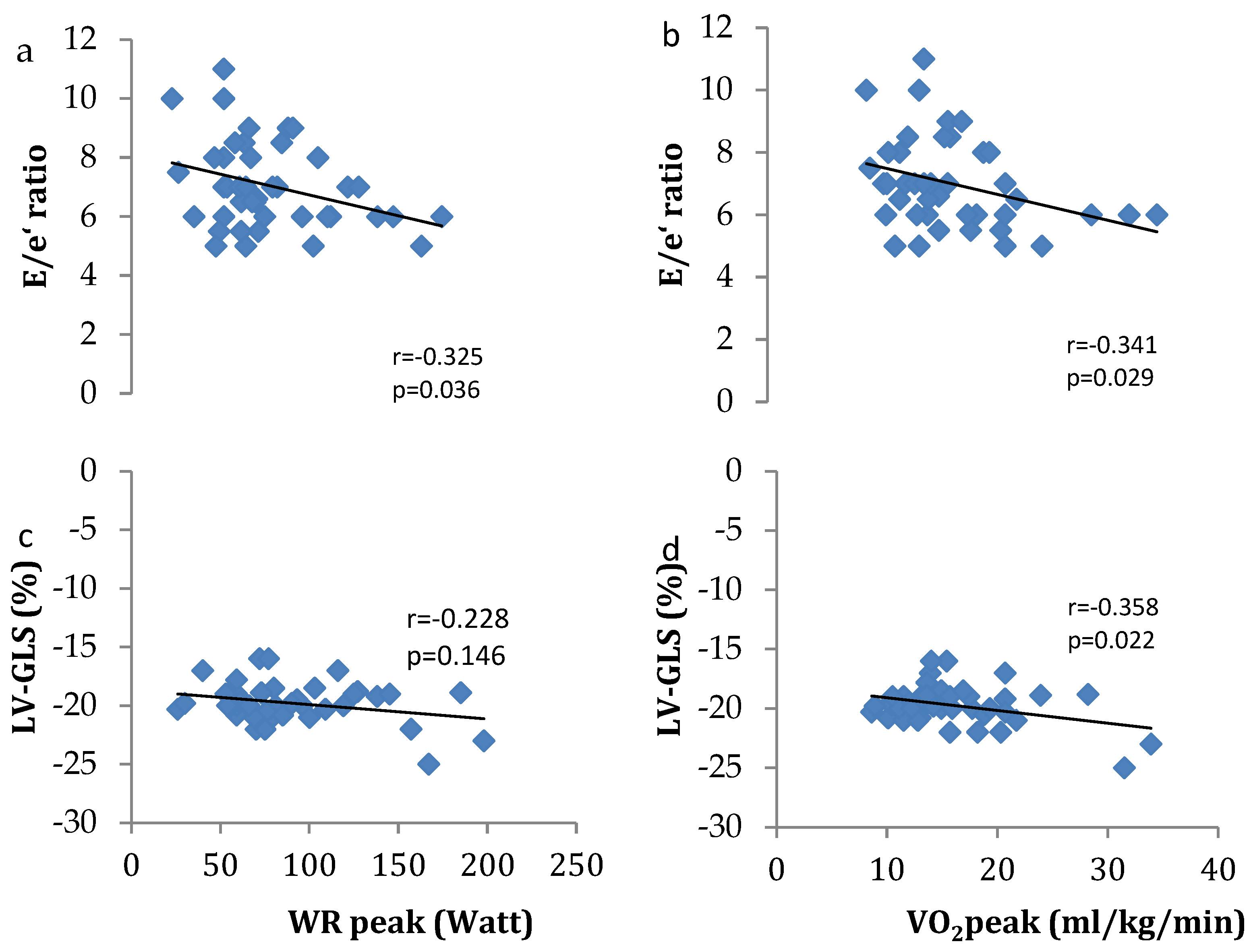
Associations between right heart function and exercise tolerance outcomes are presented in Figure 2. PASP was associated with peak oxygen consumption (VO2peak) (r = −0.345; p = 0.029), whilst SRV was associated with peak work rate (WRpeak) (r = 0.432; p = 0.004) and VO2peak (r = 0.556; p = 0.001). Associations between left heart function are presented in Figure 3. The E/e’ ratio and LV-GLS were both associated with VO2peak (r = −0.341; p = 0.029 and r = −0.358; p = 0.022, respectively), whilst only the E/e’ ratio was associated with WRpeak (r = −0.325; p = 0.036). No significant associations were reported in the healthy controls between right and left heart function and exercise tolerance outcomes.

4. Discussion

In this study, we have demonstrated that cardiovascular dysfunction at rest was associated with exercise intolerance in long COVID-19 patients. Specifically, impaired left and right ventricular performance was associated with reduced peak VO2 during incremental exercise in patients with long COVID-19 syndrome when compared with age-matched healthy controls.
The magnitude of abnormally low peak VO2 in the present study is comparable to that reported in recent studies in COVID-19 survivors at 3, 6 and 11 months post-hospital discharge [20,21,22,23]. However, these studies found no major pulmonary or cardiac limitations in patients with long COVID-19 syndrome, thereby suggesting that cardiac limitations were unlikely to limit exercise tolerance [20,21,22]. Instead, these studies rather suggested that peripheral factors affecting oxygen extraction and utilisation combined with autonomic dysregulation were likely responsible for reduced VO2 [20,21,22,23]. In contrast, Szekely and colleagues reported that reduced exercise tolerance was associated with an attenuated heart rate and stroke volume reserve in patients recovering from COVID-19 [24]. Th authors suggested that the inability to increase the stroke volume could result from either left or right ventricular dysfunction [24,25] and diastolic dysfunction [26]. Failure to augment the stroke volume as a potential mechanism of exercise intolerance has also been reported using magnetic resonance-augmented CPET [27] in COVID-19 survivors. Our findings are in line with these studies [24,27], when considering the significant associations between left ventricular function and peak values for WR and VO2.
In a recent systematic review and meta-analysis [4], several potential mechanisms were identified to limit cardiopulmonary exercise tolerance in long COVID-19 patients including (i) alterations in systemic oxygen delivery and peripheral muscle oxygen extraction (due to direct myopathic changes from the virus and/or mitochondrial dysfunction) [23], (ii) chronotropic incompetence [26,28] and preload failure [26,29], (iii) dysfunctional breathing [30] and (iv) deconditioning [30]. According to Durstenfeld et al. [4], autonomic and endothelial dysfunction caused by SARS-CoV-2, chronic inflammation and autoimmunity could be responsible for the aforementioned pathophysiologic mechanisms limiting exercise tolerance in long COVID-19 syndrome. Recent evidence has consistently shown that chronotropic incompetence is a key limitation to exercise capacity in patients with heart failure with preserved ejection fraction [31,32]. Similarly, long COVID-19 patients may experience chronotropic insufficiency, which could further impair their exercise tolerance. Our cohort study found that long COVID-19 patients with cardiac dysfunction, albeit without heart failure, exhibited reduced exercise tolerance, which may be attributed to chronotropic incompetence.
In regard to the cardiovascular system, COVID-19 convalescent patients have been reported to present with impaired left and right ventricular systolic performance, impaired endothelial function, increased arterial stiffness and impaired ventriculoarterial coupling [5,6,33,34]. Left ventricular systolic dysfunction is often subclinical without an overt reduction in LVEF, and a number of studies have described the use of cardiac magnetic resonance and LV-GLS in identifying potential subclinical left ventricular dysfunction in long COVID-19 patients [35,36]. Our results are in line with the existing literature as we report that patients with long COVID-19 syndrome presented with impaired left ventricular performance, assessed via LVEDD, LVEF and LV-GLS, compared to healthy individuals, despite that the aforementioned variables were not associated with exercise tolerance [6]. Interestingly, within the three patients with pre-existing coronary artery disease, LV-GLS was only slightly reduced in two patients, and PASP was slightly increased in one patient. As the values in the aforementioned parameters were not outliers and thus did not affect the outcome of the analysis, we decided to include them in the analysis.
The capacity of the cardiovascular system to adapt to increased metabolic requirements, such as those during incremental exercise, cannot be assessed based on a single cardiovascular parameter since the whole cardiovascular performance is based on rather complicated multifactorial mechanisms [37].
LV-GLS has been evaluated as an early index of systolic impairment before any change in the left ventricular ejection fraction is observed [38]. Indeed, investigations have shown that LV-GLS is not only a prognostic indicator but is also associated with the functional performance of the patients [39].
Furthermore, during exercise, oxygen delivered to the tissues should suffice for their metabolic requirements, which is another drive to increase cardiac output. Venous return is also increased during exercise. The required increase in cardiac output is further facilitated if the systolic performance of the right ventricle and its ability to forward blood through the pulmonary circulation are intact [40].
There is evidence of the impact of impaired SRV on exercise tolerance, during the 6 min walking test and cardiopulmonary exercise test [41]. Indeed, in the present study, SRV in patients with long COVID-19 syndrome is correlated with exercise workload. Taken together, these findings strengthen the role of SRV evaluation both at rest and during exercise testing in convalescent COVID-19 patients and may suggest that right ventricular systolic impairment may act as an additional mechanism underlying the poor performance status of long COVID-19 patients.
Furthermore, the adequacy of the left ventricular filling during the diastolic phase of the cardiac cycle determines the left ventricular preload and contributes to enhanced left ventricular performance and hence cardiac output [42]. The increase in heart rate during exercise shortens the diastolic period, and in conjunction with a stiff and non-compliant left ventricle, left ventricular filling is achieved at the cost of higher energy consumption; the subsequent increase in the left atrial pressure is transmitted to the pulmonary circulation causing pulmonary hypertension and/or pulmonary interstitial oedema, thus further compromising respiratory capacity [37]. Left ventricular diastolic impairment and an increased ratio of E/e’—the surrogate index of left ventricular filling pressures—have been reported in post-COVID-19 patients [43,44,45]. Importantly, in the present study, we have reported that an increase in the E/e’ ratio and PASP is correlated with reduced exercise tolerance in long COVID-19 patients.
In our cohort, baseline measurements for FEV1, FVC and FEV1/FVC were within the normal range in both groups, despite the fact that statistical differences were noted. In fact, neither an obstructive nor restrictive pattern was observed in the patient group. Moreover, SpO2 at the limit of tolerance was not different between the two groups even though DLco was mildly reduced in the patient group compared to the healthy individuals. Hence, the reduced DLco is unlikely to account for the differences in exercise tolerance between the two groups.
The ventilatory equivalent for carbon dioxide output (VE/VCO2) corresponds to the ventilation required to expire a certain amount of carbon dioxide. The VE/VCO2 ratio is defined as pathologic when it is greater than 40 in peak exercise, and this could result from increased dead space in the context of either interstitial lung disease or pulmonary hypertension [46]. In our cohort, the observed differences in the two groups in VE/VCO2 were not statistically significant and were within the normal range. In combination with the results from the pulmonary function testing, we deduced that patients did not show signs of interstitial lung disease (normal TLC and mildly reduced DLco). In terms of PASP measurements assessed via resting echocardiography, we found that the values for PASP were slightly increased in both groups, and the differences between the patient group and the controls were not statistically significant.
The healthy group exhibited a more pronounced cardiovascular response to exercise, characterised by an increased heart rate and mean arterial pressure during peak exercise, as compared to the patient group. This enhanced response is likely attributed to the superior exercise capacity demonstrated by the healthy individuals, as reflected by their higher achieved work rates.

4.1. Clinical Implications and Future Directions

Our study contributes to the growing body of evidence supporting the notion that the causes of exercise intolerance in long COVID-19 syndrome are complex and multifactorial and that different clusters of symptoms arise from diverse mechanisms [4]. Impaired left and right ventricular performance may reduce exercise capacity in a subgroup of long COVID-19 patients. Regular follow-up and timely interventions by heart and lung specialists are important in addressing possible complications and reassuring patients about the benignity of the condition. Future studies should investigate the effect of cardiopulmonary rehabilitation programmes on cardiovascular function in patients with long COVID-19.

4.2. Study Limitations

A few limitations should be taken into consideration while interpreting the results. The population of the control group is relatively too small to provide a practical comparison that would strengthen statistical significance, especially in an older population where there may coexist several physiological factors that limit exercise capacity. Echocardiography was not performed during the CPET, and thus, heart function during exercise could not be assessed in the present study. Additionally, pulmonary artery systolic pressure was estimated via echocardiography to avoid the use of a pulmonary arterial catheter that would have provided a direct measurement. Another limitation was that this was a cross-sectional study, and thus, it was not possible to observe changes over time. Hence, we missed the potential longitudinal effects of COVID-19 on cardiovascular and exercise performance. Finally, data on CPET and echocardiography in recovered COVID-19 patients without long-term symptoms were lacking, preventing us from distinguishing whether the observed cardiovascular dysfunction was a direct result of long COVID-19 or other confounding factors. Consequently, comparisons between survivors with and without long COVID-19 syndrome were not feasible, thereby impeding this study from establishing a clear cause–effect relationship.

5. Conclusions

In patients with long COVID-19 syndrome, exercise tolerance is reduced, as evidenced by lower WRpeak and VO2peak levels. Additionally, cardiac function is compromised, with impairments noted in both left and right ventricular systolic performance, PASP and diastolic function indices. These cardiac abnormalities are most likely linked to the decreased exercise capacity observed in these patients. It is important to identify the various phenotypes of long COVID-19 and understand the associated underlying pathophysiologic mechanisms. These findings may enable the development and implementation of new therapeutic interventions.

Author Contributions

Conceptualisation, A.V., N.C., I.V., M.V., N.R. and N.K.; Methodology, A.V., N.C., M.I.G., C.A., C.L., S.Z., N.A., E.O. and A.I.P.; Validation, E.O., M.V., N.R., G.K., N.K. and I.V.; Formal Analysis, A.V., N.C., M.I.G., C.A., C.L., S.Z., N.A., E.O. and A.I.P.; Investigation, A.V., N.C., M.I.G., C.A., C.L., S.Z., N.A., E.O. and A.I.P.; Resources, A.V., N.C., M.I.G., C.A., C.L., S.Z., N.A., E.O. and A.I.P.; Data Curation, A.V., N.C., M.I.G., C.A., C.L., S.Z., N.A., E.O. and A.I.P.; Writing—Original Draft Preparation, A.V., N.C., M.I.G., C.A., C.L. and E.O.; Writing—Review and Editing, E.O., M.V., N.R., G.K., N.K. and I.V.; Visualisation, E.O., M.V., N.R., G.K., N.K. and I.V.; Supervision, E.O., M.V., N.R., G.K., N.K. and I.V. All authors have read and agreed to the published version of the manuscript.

Funding

This research received no external funding.

Institutional Review Board Statement

This study was conducted in accordance with the Declaration of Helsinki and approved by the Institutional Review Board (or Ethics Committee) of the Sotiria Hospital Ethics Committee (Protocol ID-24633, 22 May 2022).

Informed Consent Statement

Informed consent was obtained from all subjects involved in the study.

Data Availability Statement

The data presented in this study are available upon request from the corresponding author.

Acknowledgments

We are grateful to the lung function physiologist of the 1st Respiratory Medicine Department at Sotiria Hospital for their support during the course of the study. We also thank our patients and healthy participants for their cooperation during the study.

Conflicts of Interest

The authors declare no conflicts of interest.

References

  1. Johns Hopkins University. Coronavirus Resource Centre. Available online: https://coronavirus.jhu.edu/map.html (accessed on 3 October 2023).
  2. WHO. Available online: https://covid19.who.int/region/euro/country/gr (accessed on 4 October 2023).
  3. Vaes, A.W.; Goërtz, Y.M.J.; Van Herck, M.; Machado, F.V.C.; Meys, R.; Delbressine, J.M.; Houben-Wilke, S.; Gaffron, S.; Maier, D.; Burtin, C.; et al. Recovery from COVID-19: A sprint or marathon? 6-month follow-up data from online long COVID-19 support group members. ERJ Open Res. 2021, 7, 00141-2021. [Google Scholar] [CrossRef] [PubMed]
  4. Durstenfeld, M.S.; Sun, K.; Tahir, P.; Peluso, M.J.; Deeks, S.G.; Aras, M.A.; Grandis, D.J.; Long, C.S.; Beatty, A.; Hsue, P.Y. Use of Cardiopulmonary Exercise Testing to Evaluate Long COVID-19 Symptoms in Adults: A Systematic Review and Meta-analysis. JAMA Netw. Open 2022, 5, e2236057. [Google Scholar] [CrossRef] [PubMed]
  5. Cenko, E.; Badimon, L.; Bugiardini, R.; Claeys, M.J.; De Luca, G.; de Wit, C.; Derumeaux, G.; Dorobantu, M.; Duncker, D.J.; Eringa, E.C.; et al. Cardiovascular disease and COVID-19: A consensus paper from the ESC Working Group on Coronary Pathophysiology & Microcirculation, ESC Working Group on Thrombosis and the Association for Acute CardioVascular Care (ACVC), in collaboration with the European Heart Rhythm Association (EHRA). Cardiovasc. Res. 2021, 117, 2705–2729. [Google Scholar] [CrossRef] [PubMed]
  6. Oikonomou, E.; Lampsas, S.; Theofilis, P.; Souvaliotis, N.; Papamikroulis, G.A.; Katsarou, O.; Kalogeras, K.; Pantelidis, P.; Papaioannou, T.G.; Tsatsaragkou, A.; et al. Impaired left ventricular deformation and ventricular-arterial coupling in post-COVID-19: Association with autonomic dysregulation. Heart Vessel. 2023, 38, 381–393. [Google Scholar] [CrossRef] [PubMed]
  7. Oikonomou, E.; Souvaliotis, N.; Lampsas, S.; Siasos, G.; Poulakou, G.; Theofilis, P.; Papaioannou, T.G.; Haidich, A.B.; Tsaousi, G.; Ntousopoulos, V.; et al. Endothelial dysfunction in acute and long standing COVID-19: A prospective cohort study. Vasc. Pharmacol. 2022, 144, 106975. [Google Scholar] [CrossRef] [PubMed]
  8. Theofilis, P.; Lampsas, S.; Oikonomou, E.; Siasos, G.; Vavuranakis, M.A.; Marinos, G.; Tsioufis, K.; Vavuranakis, M.; Tousoulis, D. Endothelial dysfunction in convalescent COVID-19 patients: Systematic review and meta-analysis. Arch. Cardiovasc. Dis. 2022, 115, 675–677. [Google Scholar] [CrossRef] [PubMed]
  9. NICE. COVID-19 Rapid Guideline: Managing the Longterm Effects of COVID-19. Available online: https://www.nice.org.uk/guidance/ng188/resources/covid19-rapid-guideline-managing-the-longterm-effects-of-covid19-pdf-51035515742 (accessed on 25 October 2023).
  10. Venkatesan, P. NICE guideline on long COVID. Lancet Respir. Med. 2021, 9, 129. [Google Scholar] [CrossRef]
  11. D’Angelo, E.; Prandi, E.; Marazzini, L.; Milic-Emili, J. Dependence of maximal flow-volume curves on time course of preceding inspiration in patients with chronic obstruction pulmonary disease. Am. J. Respir. Crit. Care Med. 1994, 150, 1581–1586. [Google Scholar] [CrossRef]
  12. Darling, R.C.; Cournand, A.; Richards, D.W. Studies on the intrapulmonary mixture of gases. III. An open circuit method for measuring residual air. J. Clin. Investig. 1940, 19, 609–618. [Google Scholar] [CrossRef]
  13. Macintyre, N.; Crapo, R.O.; Viegi, G.; Johnson, D.C.; van der Grinten, C.P.; Brusasco, V.; Burgos, F.; Casaburi, R.; Coates, A.; Enright, P.; et al. Standardisation of the single-breath determination of carbon monoxide uptake in the lung. Eur. Respir. J. 2005, 26, 720–735. [Google Scholar] [CrossRef]
  14. Quanjer, P.H.; Tammeling, G.; Cotes, J.E.; Pedersen, O.F.; Peslin, R.; Yernault, J.C. Standardized lung function testing. Official statement of the European Respiratory Society. Eur. Respir. J. 1993, 6 (Suppl. 16), 1–100. [Google Scholar]
  15. Ilsley, T.; Howden, E.J. Clinimetrics: Functional Assessment of Chronic Illness Therapy-Fatigue (FACIT-Fatigue). J. Physiother. 2023, 69, 273–274. [Google Scholar] [CrossRef] [PubMed]
  16. Borg, G.A. Psychophysical bases of perceived exertion. Med. Sci. Sports Exerc. 1982, 14, 377–381. [Google Scholar] [CrossRef] [PubMed]
  17. Lang, R.M.; Badano, L.P.; Mor-Avi, V.; Afilalo, J.; Armstrong, A.; Ernande, L.; Flachskampf, F.A.; Foster, E.; Goldstein, S.A.; Kuznetsova, T.; et al. Recommendations for cardiac chamber quantification by echocardiography in adults: An update from the American Society of Echocardiography and the European Association of Cardiovascular Imaging. J. Am. Soc. Echocardiogr. Off. Publ. Am. Soc. Echocardiogr. 2015, 28, 1–39.e14. [Google Scholar] [CrossRef]
  18. Nagueh, S.F.; Smiseth, O.A.; Appleton, C.P.; Byrd, B.F., 3rd; Dokainish, H.; Edvardsen, T.; Flachskampf, F.A.; Gillebert, T.C.; Klein, A.L.; Lancellotti, P.; et al. Recommendations for the Evaluation of Left Ventricular Diastolic Function by Echocardiography: An Update from the American Society of Echocardiography and the European Association of Cardiovascular Imaging. Eur. Heart J. Cardiovasc. Imaging 2016, 17, 1321–1360. [Google Scholar] [CrossRef] [PubMed]
  19. Rudski, L.G.; Lai, W.W.; Afilalo, J.; Hua, L.; Handschumacher, M.D.; Chandrasekaran, K.; Solomon, S.D.; Louie, E.K.; Schiller, N.B. Guidelines for the echocardiographic assessment of the right heart in adults: A report from the American Society of Echocardiography endorsed by the European Association of Echocardiography, a registered branch of the European Society of Cardiology, and the Canadian Society of Echocardiography. J. Am. Soc. Echocardiogr. Off. Publ. Am. Soc. Echocardiogr. 2010, 23, 685–713, quiz 786–688. [Google Scholar] [CrossRef]
  20. Baratto, C.; Caravita, S.; Faini, A.; Perego, G.B.; Senni, M.; Badano, L.P.; Parati, G. Impact of COVID-19 on exercise pathophysiology: A combined cardiopulmonary and echocardiographic exercise study. J. Appl. Physiol. 2021, 130, 1470–1478. [Google Scholar] [CrossRef]
  21. Schwendinger, F.; Knaier, R.; Radtke, T.; Schmidt-Trucksäss, A. Low Cardiorespiratory Fitness Post-COVID-19: A Narrative Review. Sports Med. 2023, 53, 51–74. [Google Scholar] [CrossRef] [PubMed]
  22. Vannini, L.; Quijada-Fumero, A.; Martín, M.P.R.; Pina, N.C.; Afonso, J.S.H. Cardiopulmonary exercise test with stress echocardiography in COVID-19 survivors at 6 months follow-up. Eur. J. Intern. Med. 2021, 94, 101–104. [Google Scholar] [CrossRef]
  23. Singh, I.; Joseph, P.; Heerdt, P.M.; Cullinan, M.; Lutchmansingh, D.D.; Gulati, M.; Possick, J.D.; Systrom, D.M.; Waxman, A.B. Persistent Exertional Intolerance after COVID-19: Insights from Invasive Cardiopulmonary Exercise Testing. Chest 2022, 161, 54–63. [Google Scholar] [CrossRef]
  24. Szekely, Y.; Lichter, Y.; Sadon, S.; Lupu, L.; Taieb, P.; Banai, A.; Sapir, O.; Granot, Y.; Hochstadt, A.; Friedman, S.; et al. Cardiorespiratory Abnormalities in Patients Recovering from Coronavirus Disease 2019. J. Am. Soc. Echocardiogr. Off. Publ. Am. Soc. Echocardiogr. 2021, 34, 1273–1284.e9. [Google Scholar] [CrossRef] [PubMed]
  25. Churchill, T.W.; Bertrand, P.B.; Bernard, S.; Namasivayam, M.; Churchill, J.; Crousillat, D.; Davis, E.F.; Hung, J.; Picard, M.H. Echocardiographic Features of COVID-19 Illness and Association with Cardiac Biomarkers. J. Am. Soc. Echocardiogr. Off. Publ. Am. Soc. Echocardiogr. 2020, 33, 1053–1054. [Google Scholar] [CrossRef] [PubMed]
  26. Szekely, Y.; Lichter, Y.; Taieb, P.; Banai, A.; Hochstadt, A.; Merdler, I.; Gal Oz, A.; Rothschild, E.; Baruch, G.; Peri, Y.; et al. Spectrum of Cardiac Manifestations in COVID-19: A Systematic Echocardiographic Study. Circulation 2020, 142, 342–353. [Google Scholar] [CrossRef] [PubMed]
  27. Brown, J.T.; Saigal, A.; Karia, N.; Patel, R.K.; Razvi, Y.; Constantinou, N.; Steeden, J.A.; Mandal, S.; Kotecha, T.; Fontana, M.; et al. Ongoing Exercise Intolerance Following COVID-19: A Magnetic Resonance-Augmented Cardiopulmonary Exercise Test Study. J. Am. Heart Assoc. 2022, 11, e024207. [Google Scholar] [CrossRef]
  28. Abdallah, S.J.; Voduc, N.; Corrales-Medina, V.F.; McGuinty, M.; Pratt, A.; Chopra, A.; Law, A.; Garuba, H.A.; Thavorn, K.; Reid, R.E.R.; et al. Symptoms, Pulmonary Function, and Functional Capacity Four Months after COVID-19. Ann. Am. Thorac. Soc. 2021, 18, 1912–1917. [Google Scholar] [CrossRef] [PubMed]
  29. Mancini, D.M.; Brunjes, D.L.; Lala, A.; Trivieri, M.G.; Contreras, J.P.; Natelson, B.H. Use of Cardiopulmonary Stress Testing for Patients with Unexplained Dyspnea Post-Coronavirus Disease. JACC Heart Fail. 2021, 9, 927–937. [Google Scholar] [CrossRef] [PubMed]
  30. Ladlow, P.; O’Sullivan, O.; Bennett, A.N.; Barker-Davies, R.; Houston, A.; Chamley, R.; May, S.; Mills, D.; Dewson, D.; Rogers-Smith, K.; et al. The effect of medium-term recovery status after COVID-19 illness on cardiopulmonary exercise capacity in a physically active adult population. J. Appl. Physiol. 2022, 132, 1525–1535. [Google Scholar] [CrossRef] [PubMed]
  31. Mueller, S.; Haller, B.; Feuerstein, A.; Winzer, E.B.; Beckers, P.; Haykowsky, M.J.; Gevaert, A.B.; Hommel, J.; Azevedo, L.F.; Duvinage, A.; et al. Peak O(2)-pulse predicts exercise training-induced changes in peak VO(2) in heart failure with preserved ejection fraction. ESC Heart Fail. 2022, 9, 3393–3406. [Google Scholar] [CrossRef] [PubMed]
  32. Pandey, A.; Khera, R.; Park, B.; Haykowsky, M.; Borlaug, B.A.; Lewis, G.D.; Kitzman, D.W.; Butler, J.; Berry, J.D. Relative Impairments in Hemodynamic Exercise Reserve Parameters in Heart Failure with Preserved Ejection Fraction: A Study-Level Pooled Analysis. JACC Heart Fail. 2018, 6, 117–126. [Google Scholar] [CrossRef]
  33. Erdem, K.; Duman, A. Pulmonary artery pressures and right ventricular dimensions of post-COVID-19 patients without previous significant cardiovascular pathology. Heart Lung J. Crit. Care 2023, 57, 75–79. [Google Scholar] [CrossRef]
  34. Gounaridi, M.I.; Vontetsianos, A.; Oikonomou, E.; Theofilis, P.; Chynkiamis, N.; Lampsas, S.; Anastasiou, A.; Papamikroulis, G.A.; Katsianos, E.; Kalogeras, K.; et al. The Role of Rehabilitation in Arterial Function Properties of Convalescent COVID-19 Patients. J. Clin. Med. 2023, 12, 2233. [Google Scholar] [CrossRef]
  35. Caiado, L.D.C.; Azevedo, N.C.; Azevedo, R.R.C.; Caiado, B.R. Cardiac involvement in patients recovered from COVID-19 identified using left ventricular longitudinal strain. J. Echocardiogr. 2022, 20, 51–56. [Google Scholar] [CrossRef] [PubMed]
  36. Cremonesi, M.; Felicetta, A.; Cannata, F.; Serio, S.; van Beek, J.J.P.; Bombace, S.; My, I.; Zanon, V.; Catalano, C.; Papadopoulou, V.; et al. Long COVID-19 Cardiac Complications Are Associated with Autoimmunity to Cardiac Self-Antigens Sufficient to Cause Cardiac Dysfunction. Circulation 2023, 148, 504–507. [Google Scholar] [CrossRef] [PubMed]
  37. Del Buono, M.G.; Arena, R.; Borlaug, B.A.; Carbone, S.; Canada, J.M.; Kirkman, D.L.; Garten, R.; Rodriguez-Miguelez, P.; Guazzi, M.; Lavie, C.J.; et al. Exercise Intolerance in Patients with Heart Failure: JACC State-of-the-Art Review. J. Am. Coll. Cardiol. 2019, 73, 2209–2225. [Google Scholar] [CrossRef] [PubMed]
  38. Donal, E.; Coquerel, N.; Bodi, S.; Kervio, G.; Schnell, F.; Daubert, J.C.; Carré, F. Importance of ventricular longitudinal function in chronic heart failure. Eur. J. Echocardiogr. J. Work. Group Echocardiogr. Eur. Soc. Cardiol. 2011, 12, 619–627. [Google Scholar] [CrossRef] [PubMed]
  39. Hasselberg, N.E.; Haugaa, K.H.; Sarvari, S.I.; Gullestad, L.; Andreassen, A.K.; Smiseth, O.A.; Edvardsen, T. Left ventricular global longitudinal strain is associated with exercise capacity in failing hearts with preserved and reduced ejection fraction. Eur. Heart J. Cardiovasc. Imaging 2015, 16, 217–224. [Google Scholar] [CrossRef] [PubMed]
  40. Hellsten, Y.; Nyberg, M. Cardiovascular Adaptations to Exercise Training. Compr. Physiol. 2015, 6, 1–32. [Google Scholar] [CrossRef] [PubMed]
  41. Sista, A.K.; Miller, L.E.; Kahn, S.R.; Kline, J.A. Persistent right ventricular dysfunction, functional capacity limitation, exercise intolerance, and quality of life impairment following pulmonary embolism: Systematic review with meta-analysis. Vasc. Med. 2017, 22, 37–43. [Google Scholar] [CrossRef] [PubMed]
  42. LaCombe, P.; Jose, A.; Basit, H.; Lappin, S.L. Physiology, Starling Relationships. In StatPearls; StatPearls Publishing LLC.: Treasure Island, FL, USA, 2023. [Google Scholar]
  43. Hadzibegovic, S.; Lena, A.; Churchill, T.W.; Ho, J.E.; Potthoff, S.; Denecke, C.; Rösnick, L.; Heim, K.M.; Kleinschmidt, M.; Sander, L.E.; et al. Heart failure with preserved ejection fraction according to the HFA-PEFF score in COVID-19 patients: Clinical correlates and echocardiographic findings. Eur. J. Heart Fail. 2021, 23, 1891–1902. [Google Scholar] [CrossRef]
  44. Tudoran, C.; Tudoran, M.; Pop, G.N.; Giurgi-Oncu, C.; Cut, T.G.; Lazureanu, V.E.; Oancea, C.; Parv, F.; Ciocarlie, T.; Bende, F. Associations between the Severity of the Post-Acute COVID-19 Syndrome and Echocardiographic Abnormalities in Previously Healthy Outpatients Following Infection with SARS-CoV-2. Biology 2021, 10, 469. [Google Scholar] [CrossRef]
  45. Zaccone, G.; Tomasoni, D.; Italia, L.; Lombardi, C.M.; Metra, M. Myocardial Involvement in COVID-19: An Interaction Between Comorbidities and Heart Failure with Preserved Ejection Fraction. A Further Indication of the Role of Inflammation. Curr. Heart Fail. Rep. 2021, 18, 99–106. [Google Scholar] [CrossRef]
  46. Balady, G.J.; Arena, R.; Sietsema, K.; Myers, J.; Coke, L.; Fletcher, G.F.; Forman, D.; Franklin, B.; Guazzi, M.; Gulati, M.; et al. Clinician’s Guide to cardiopulmonary exercise testing in adults: A scientific statement from the American Heart Association. Circulation 2010, 122, 191–225. [Google Scholar] [CrossRef]
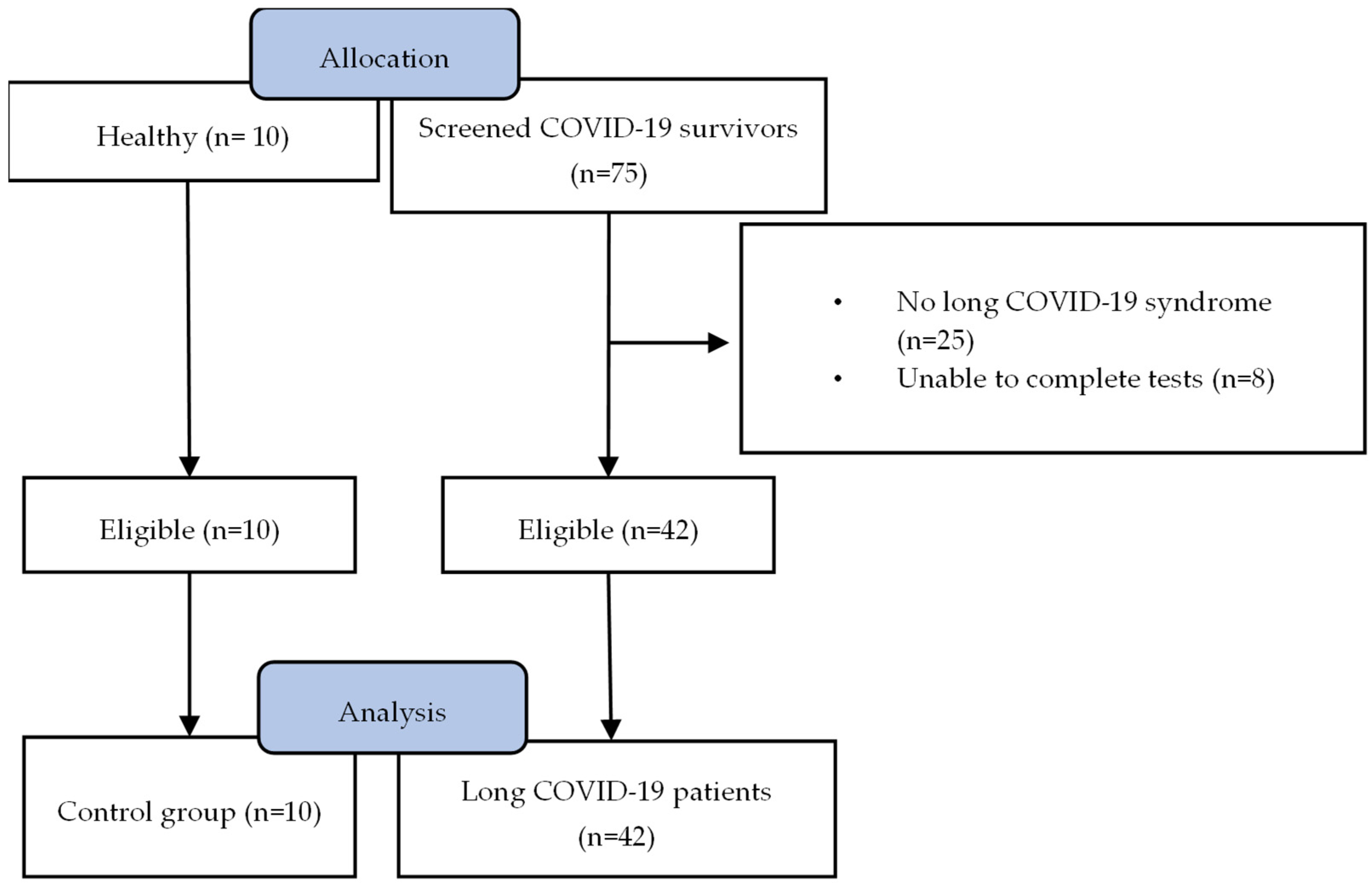
Figure 1. Study flowchart. Following the initial screening, 42 patients and 10 healthy age-matched individuals underwent cardiopulmonary exercise testing (CPET) and resting echocardiography. All participants were included in the final analysis.
Jcm 13 04144 g001
Figure 2. Association between (a) pulmonary artery systolic pressure (PASP) and peak work rate (WR), (b) PASP and peak oxygen consumption (VO2), (c) tricuspid annular systolic velocity (SRV) and WR and (d) SRV and VO2.
Jcm 13 04144 g002
Figure 3. Association between (a) trans-mitral flow velocity/early mitral annular velocity (E/e’) and peak work rate (WR), (b) E/e’ and peak oxygen consumption (VO2), (c) left ventricular global longitudinal strain (LV-GLS) and WR and (d) LV-GLS and VO2.
Jcm 13 04144 g003
Table 1. Patient demographic characteristics.
VariableLong COVID-19 PatientsHealthy Controlp Value
Gender (Male/Female)19/234/60.76
Age (years)55 ± 1352 ± 80.58
Height (cm)166 ± 11170 ± 120.293
Weight (kg)80.8 ± 17.884.4 ± 20.00.579
BMI29.0 ± 5.228.8 ± 3.70.916
Days from discharge149 ± 92--
Smoking status
  Current50
  Ex630.57
  Never317
FEV1 (% predicted)98 ± 19110 ± 170.065
FVC (% predicted)96 ± 21121 ± 200.002
FEV1/FVC84 ± 677 ± 60.002
TLC (% predicted)92 ± 27100 ± 30.339
Comorbidities
  Dyslipidaemia8
  Hypertension6
  Coronary artery disease3
  Diabetes mellitus4
  Asthma2
  Anxiety–Depression4
DLco (% predicted)73 ± 2198 ± 30.002
mMRC2.1 ± 1.20.4 ± 0.50.001
FACIT24 ± 1048 ± 40.001
DLco, Diffusing Capacity for Carbon Monoxide; FACIT, Functional Assessment of Chronic Illness Therapy scale; FEV1, Forced Expiratory Volume at the 1st second; FVC, Forced Vital Capacity; mMRC, modified Medical Research Council Dyspnoea Scale; TLC, Total Lung Capacity; values are mean ± standard deviation (SD).
Table 2. Cardiopulmonary exercise testing data at the limit of tolerance.
VariableLong COVID-19 PatientsHealthy Controlp Value
WRpeak (watts)88 ± 40148 ± 630.001
WRpeak (%pred)74 ± 27115 ± 270.001
VO2peak (mL/kg/min)16.1 ± 5.721.1 ± 4.30.014
VO2peak (%pred)67 ± 1889 ± 130.001
RER1.12 ± 0.101.26 ± 0.110.001
VE/VCO234 ± 637 ± 60.116
O2 pulse (mL/kg/beat)10.4 ± 3.511.4 ± 4.00.465
O2 pulse (%pred)90 ± 2195 ± 120.495
Heart rate (beats/minute)126 ± 18159 ± 170.001
Heart rate (%pred)79 ± 1895 ± 100.007
Mean arterial pressure (mmHg)108 ± 17126 ± 140.003
SpO2 (%)96 ± 599 ± 10.078
Dyspnoea3.8 ± 2.34.4 ± 2.80.476
Fatigue5.9 ± 2.56.0 ± 2.30.890
Reason for termination
(Dyspnoea/Fatigue/Both) (%)
4 (10%)/34 (80%)/4 (10%)4 (40%)/6 (60%)/0 (0%)-
RER, respiratory exchange ratio; SpO2, oxygen saturation; VCO2, carbon dioxide output; VE, minute ventilation; VO2, oxygen consumption; WR, work rate; values are mean ± SD.
Table 3. Resting echocardiographic data.
VariableLong COVID-19 PatientsHealthy Controlp Value
LVEDD (mm)54 ± 548 ± 20.001
LVEF (%)54 ± 559 ± 40.010
LVmass (g)135 ± 25143 ± 290.412
LAvol (ml)45 ± 1145 ± 100.986
E/e’ ratio7.0 ± 1.56.4 ± 1.00.230
RV diameter (mm) 31 ± 331 ± 40.706
SRV (cm/s)12.0 ± 1.612.6 ± 1.80.325
PASP (mmHg)28 ± 627 ± 40.892
LV-GLS (%)−20 ± 2−21 ± 10.020
LVOT (mm)22.8 ± 2.322.2 ± 2.40.521
SV (ml)75 ± 972 ± 70.325
E/e’, early trans-mitral flow velocity/early mitral annular velocity; LAvol, Left atrium volume; LV-GLS, left ventricular global longitudinal strain; LVEDD, left ventricular end-diastolic diameter; LVEF, left ventricular ejection fraction; LVmass, Left ventricle mass; LVOT, Left ventricle outflow tract; PASP, pulmonary artery systolic pressure; RV, right ventricle; SRV, tricuspid annular systolic velocity; SV, stroke volume; values are mean ± SD.
Disclaimer/Publisher’s Note: The statements, opinions and data contained in all publications are solely those of the individual author(s) and contributor(s) and not of MDPI and/or the editor(s). MDPI and/or the editor(s) disclaim responsibility for any injury to people or property resulting from any ideas, methods, instructions or products referred to in the content.