
Hamstring Muscle Stiffness in Athletes with and without Anterior Cruciate Ligament Reconstruction History: A Retrospective Study
 ,
,
Abstract
:1. Introduction
2. Methodology
2.1. Research Design and Participants
2.2. Sample Size
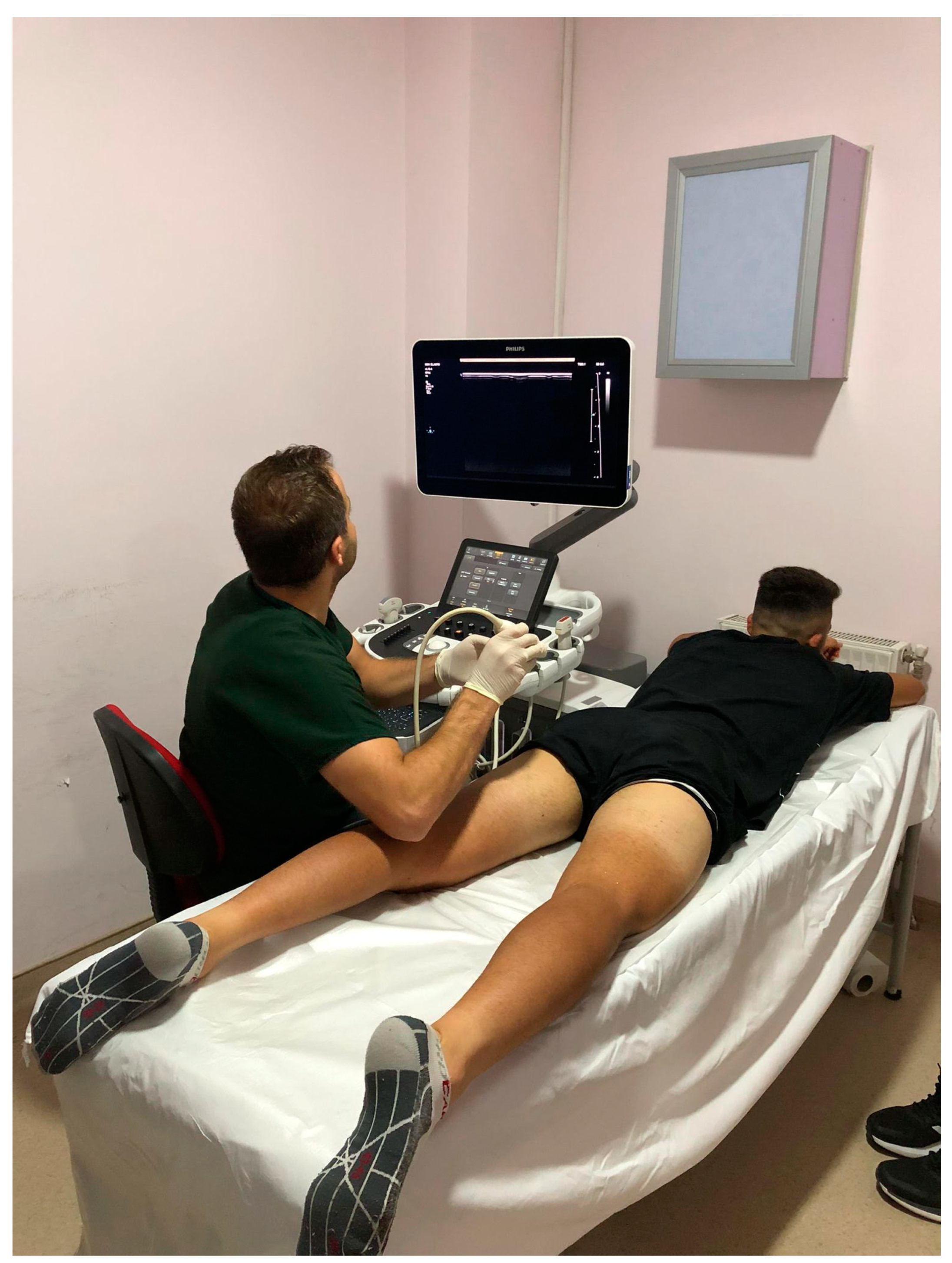
2.3. Measurement Procedures
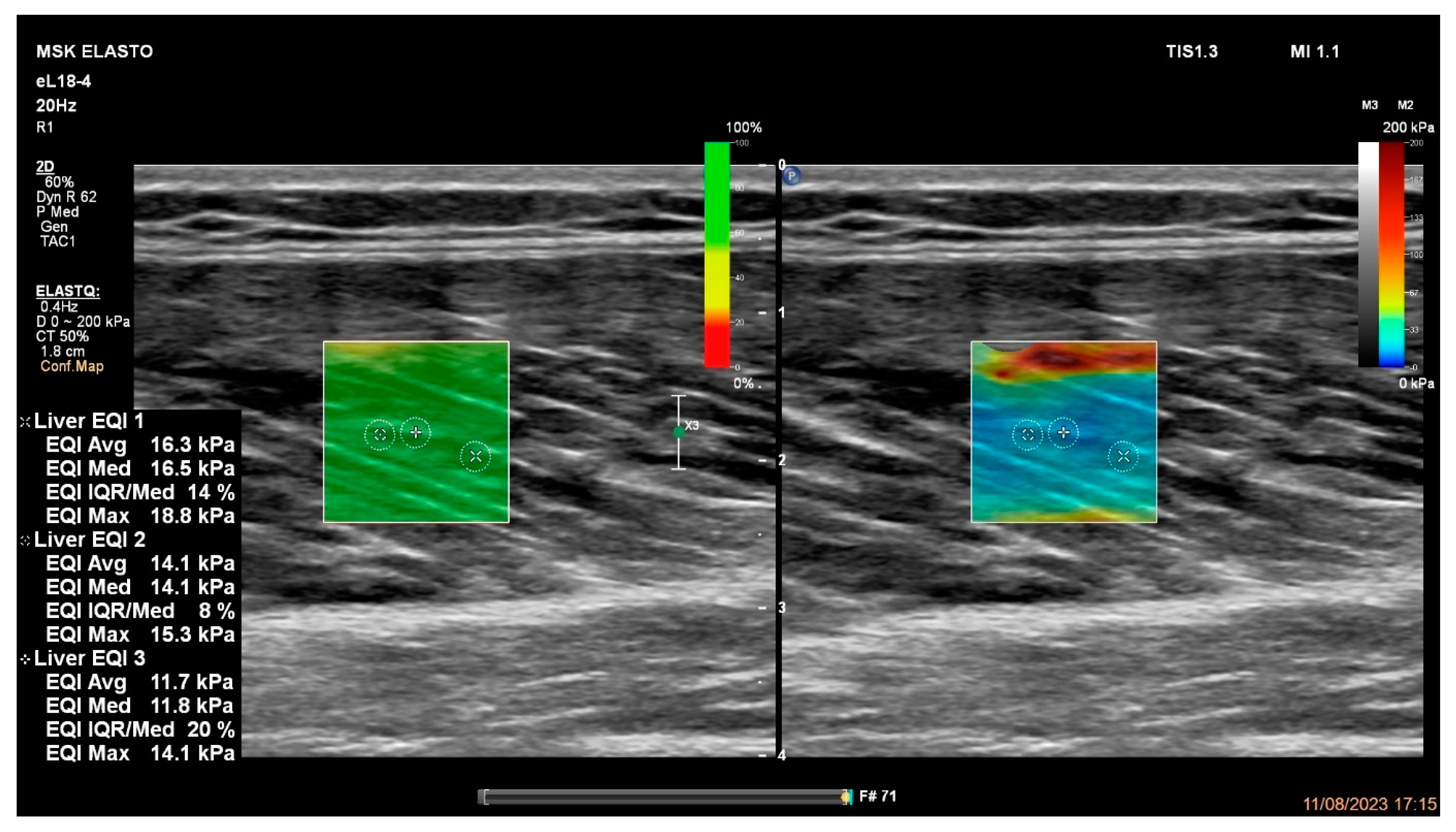
2.4. Hamstring Muscles’ Passive Stiffness Measurements
2.5. Statistical Analyses
3. Results
4. Discussion
5. Conclusions
Supplementary Materials
Author Contributions
Funding
Institutional Review Board Statement
Informed Consent Statement
Data Availability Statement
Acknowledgments
Conflicts of Interest
References
- Brooks, J.H.; Fuller, C.W.; Kemp, S.P.; Reddin, D.B. Epidemiology of injuries in English professional rugby union: Part 1 match injuries. Br. J. Sports Med. 2005, 39, 757–766. [Google Scholar] [CrossRef]
- Ekstrand, J.; Hägglund, M.; Waldén, M. Injury incidence and injury patterns in professional football: The UEFA injury study. Br. J. Sports Med. 2011, 45, 553–558. [Google Scholar] [CrossRef] [PubMed]
- Feeley, B.T.; Kennelly, S.; Barnes, R.P.; Muller, M.S.; Kelly, B.T.; Rodeo, S.A.; Warren, R.F. Epidemiology of National Football League Training Camp Injuries from 1998 to 2007. Am. J. Sports Med. 2008, 36, 1597–1603. [Google Scholar] [CrossRef] [PubMed]
- Opar, D.A.; Drezner, J.; Shield, A.; Williams, M.; Webner, D.; Sennett, B.; Kapur, R.; Cohen, M.; Ulager, J.; Cafengiu, A.; et al. Acute hamstring strain injury in track-and-field athletes: A 3-year observational study at the Penn Relay Carnival. Scand. J. Med. Sci. Sports 2014, 24, e254–e259. [Google Scholar] [CrossRef]
- Orchard, J.W.; Seward, H.; Orchard, J.J. Results of 2 decades of injury surveillance and public release of data in the Australian Football League. Am. J. Sports Med. 2013, 41, 734–741. [Google Scholar] [CrossRef]
- Dorn, T.W.; Schache, A.G.; Pandy, M.G. Muscular strategy shift in human running: Dependence of running speed on hip and ankle muscle performance. J. Exp. Biol. 2012, 215, 1944–1956. [Google Scholar] [CrossRef] [PubMed]
- Rahnama, N.; Lees, A.; Bambaecichi, E. Comparison of muscle strength and flexibility between the preferred and non-preferred leg in English soccer players. Ergonomics 2005, 48, 1568–1575. [Google Scholar] [CrossRef]
- Schache, A.G.; Blanch, P.D.; Dorn, T.W.; Brown, N.A.; Rosemond, D.; Pandy, M.G. Effect of running speed on lower limb joint kinetics. Med. Sci. Sports Exerc. 2011, 43, 1260–1271. [Google Scholar] [CrossRef] [PubMed]
- Walsh, M.; Boling, M.C.; McGrath, M.; Blackburn, J.T.; Padua, D.A. Lower extremity muscle activation and knee flexion during a jump-landing task. J. Athl. Train. 2012, 47, 406–413. [Google Scholar] [CrossRef]
- Chu, S.K.; Rho, M.E. Hamstring Injuries in the Athlete: Diagnosis, Treatment, and Return to Play. Curr. Sports Med. Rep. 2016, 15, 184–190. [Google Scholar] [CrossRef]
- Ekstrand, J.; Bengtsson, H.; Waldén, M.; Davison, M.; Khan, K.M.; Hägglund, M. Hamstring injury rates have increased during recent seasons and now constitute 24% of all injuries in men’s professional football: The UEFA Elite Club Injury Study from 2001/02 to 2021/22. Br. J. Sports Med. 2022, 57, 292–298. [Google Scholar] [CrossRef] [PubMed]
- Ekstrand, J.; Krutsch, W.; Spreco, A.; van Zoest, W.; Roberts, C.; Meyer, T.; Bengtsson, H. Time before return to play for the most common injuries in professional football: A 16-year follow-up of the UEFA Elite Club Injury Study. Br. J. Sports Med. 2020, 54, 421–426. [Google Scholar] [CrossRef] [PubMed]
- Ekstrand, J.; Gillquist, J. Soccer injuries and their mechanisms: A prospective study. Med. Sci. Sports Exerc. 1983, 15, 267–270. [Google Scholar] [CrossRef]
- Seward, H.; Orchard, J.; Hazard, H.; Collinson, D. Football injuries in Australia at the élite level. Med. J. Aust. 1993, 159, 298–301. [Google Scholar] [CrossRef]
- Opar, D.A.; Williams, M.D.; Shield, A.J. Hamstring strain injuries: Factors that lead to injury and re-injury. Sports Med. 2012, 42, 209–226. [Google Scholar] [CrossRef]
- Arnason, A.; Andersen, T.E.; Holme, I.; Engebretsen, L.; Bahr, R. Prevention of hamstring strains in elite soccer: An intervention study. Scand. J. Med. Sci. Sports 2008, 18, 40–48. [Google Scholar] [CrossRef]
- Petersen, J.; Thorborg, K.; Nielsen, M.B.; Budtz-Jørgensen, E.; Hölmich, P. Preventive effect of eccentric training on acute hamstring injuries in men’s soccer: A cluster-randomized controlled trial. Am. J. Sports Med. 2011, 39, 2296–2303. [Google Scholar] [CrossRef] [PubMed]
- van der Horst, N.; Smits, D.W.; Petersen, J.; Goedhart, E.A.; Backx, F.J. The preventive effect of the nordic hamstring exercise on hamstring injuries in amateur soccer players: A randomized controlled trial. Am. J. Sports Med. 2015, 43, 1316–1323. [Google Scholar] [CrossRef]
- Liu, H.; Garrett, W.E.; Moorman, C.T.; Yu, B. Injury rate, mechanism, and risk factors of hamstring strain injuries in sports: A review of the literature. J. Sport Health Sci. 2012, 1, 92–101. [Google Scholar] [CrossRef]
- Kenneally-Dabrowski, C.J.B.; Brown, N.A.T.; Lai, A.K.M.; Perriman, D.; Spratford, W.; Serpell, B.G. Late swing or early stance? A narrative review of hamstring injury mechanisms during high-speed running. Scand. J. Med. Sci. Sports 2019, 29, 1083–1091. [Google Scholar] [CrossRef]
- Chumanov, E.S.; Heiderscheit, B.C.; Thelen, D.G. Hamstring musculotendon dynamics during stance and swing phases of high-speed running. Med. Sci. Sports Exerc. 2011, 43, 525–532. [Google Scholar] [CrossRef] [PubMed]
- Kujala, U.M.; Orava, S.; Järvinen, M. Hamstring injuries. Current trends in treatment and prevention. Sports Med. 1997, 23, 397–404. [Google Scholar] [CrossRef] [PubMed]
- Thelen, D.; Chumanov, E.; Hoerth, D.; Best, T.; Swanson, S.; Li, L.; Youn, M.; Heiderscheit, B.C. Hamstring Muscle Kinematics during Treadmill Sprinting. Med. Sci. Sports Exerc. 2005, 37, 108–114. [Google Scholar] [CrossRef] [PubMed]
- Roig Pull, M.; Ranson, C. Eccentric muscle actions: Implications for injury prevention and rehabilitation. Phys. Ther. Sport 2007, 8, 88–97. [Google Scholar] [CrossRef]
- Clark, R.A. Hamstring injuries: Risk assessment and injury prevention. Ann. Acad. Med. Singap. 2008, 37, 341–346. [Google Scholar] [CrossRef] [PubMed]
- Green, B.; Bourne, M.N.; van Dyk, N.; Pizzari, T. Recalibrating the risk of hamstring strain injury (HSI): A 2020 systematic review and meta-analysis of risk factors for index and recurrent hamstring strain injury in sport. Br. J. Sports Med. 2020, 54, 1081. [Google Scholar] [CrossRef] [PubMed]
- Arnason, A.; Sigurdsson, S.B.; Gudmundsson, A.; Holme, I.; Engebretsen, L.; Bahr, R. Risk factors for injuries in football. The Am. J. Sports Med. 2004, 32, 5–16. [Google Scholar] [CrossRef] [PubMed]
- Dauty, M.; Menu, P.; Fouasson-Chailloux, A. Cutoffs of isokinetic strength ratio and hamstring strain prediction in professional soccer players. Scand. J. Med. Sci. Sports 2018, 28, 276–281. [Google Scholar] [CrossRef] [PubMed]
- Engebretsen, A.H.; Myklebust, G.; Holme, I.; Engebretsen, L.; Bahr, R. Intrinsic risk factors for hamstring injuries among male soccer players: A prospective cohort study. Am. J. Sports Med. 2010, 38, 1147–1153. [Google Scholar] [CrossRef]
- Fousekis, K.; Tsepis, E.; Poulmedis, P.; Athanasopoulos, S.; Vagenas, G. Intrinsic risk factors of non-contact quadriceps and hamstring strains in soccer: A prospective study of 100 professional players. Br. J. Sports Med. 2011, 45, 709–714. [Google Scholar] [CrossRef]
- Henderson, G.; Barnes, C.A.; Portas, M.D. Factors associated with increased propensity for hamstring injury in English Premier League soccer players. J. Sci. Med. Sport 2010, 13, 397–402. [Google Scholar] [CrossRef] [PubMed]
- Bourne, M.N.; Opar, D.A.; Williams, M.D.; Shield, A.J. Eccentric knee flexor strength and risk of hamstring injuries in rugby union: A prospective study. Am. J. Sports Med. 2015, 43, 2663–2670. [Google Scholar] [CrossRef] [PubMed]
- Opar, D.; Williams, M.; Timmins, R.; Hickey, J.; Duhig, S.; Shield, A. Eccentric hamstring strength and hamstring injury risk in Australian footballers. Med. Sci. Sports Exerc. 2015, 47, 857–865. [Google Scholar] [CrossRef] [PubMed]
- Orchard, J.; Seward, H.; Orchard, J.; Driscoll, T. The speed-fatigue trade off in hamstring aetiology: Analysis of 2011 AFL injury data. Sport Health 2012, 30, 53. [Google Scholar]
- Orchard, J.W. Intrinsic and extrinsic risk factors for muscle strains in Australian football. Am. J. Sports Med. 2001, 29, 300–303. [Google Scholar] [CrossRef] [PubMed]
- Sugiura, Y.; Saito, T.; Sakuraba, K.; Sakuma, K.; Suzuki, E. Strength deficits identified with concentric action of the hip extensors and eccentric action of the hamstrings predispose to hamstring injury in elite sprinters. J. Orthop. Sports Phys. Ther. 2008, 38, 457–464. [Google Scholar] [CrossRef] [PubMed]
- Freckleton, G.; Cook, J.; Pizzari, T. The predictive validity of a single leg bridge test for hamstring injuries in Australian Rules Football Players. Br. J. Sports Med. 2014, 48, 713–717. [Google Scholar] [CrossRef]
- Verrall, G.M.; Slavotinek, J.P.; Barnes, P.G.; Fon, G.T.; Spriggins, A.J. Clinical risk factors for hamstring muscle strain injury: A prospective study with correlation of injury by magnetic resonance imaging. Br. J. Sports Med. 2001, 35, 435–440. [Google Scholar] [CrossRef] [PubMed]
- Malliaropoulos, N.; Bikos, G.; Meke, M.; Vasileios, K.; Valle, X.; Lohrer, H.; Maffulli, N.; Padhiar, N. Higher frequency of hamstring injuries in elite track and field athletes who had a previous injury to the ankle-a 17 years observational cohort study. J. Foot Ankle Res. 2018, 11, 7. [Google Scholar] [CrossRef]
- De Vos, R.-J.; Reurink, G.; Goudswaard, G.-J.; Moen, M.H.; Weir, A.; Tol, J.L. Clinical findings just after return to play predict hamstring re-injury, but baseline MRI findings do not. Br. J. Sports Med. 2014, 48, 1377–1384. [Google Scholar] [CrossRef]
- Goossens, L.; Witvrouw, E.; Vanden Bossche, L.; De Clercq, D. Lower eccentric hamstring strength and single leg hop for distance predict hamstring injury in PETE students. Eur. J. Sport Sci. 2015, 15, 436–442. [Google Scholar] [CrossRef] [PubMed]
- Schuermans, J.; Van Tiggelen, D.; Danneels, L.; Witvrouw, E. Susceptibility to hamstring injuries in soccer: A prospective study using muscle functional magnetic resonance imaging. Am. J. Sports Med. 2016, 44, 1276–1285. [Google Scholar] [CrossRef]
- Venturelli, M.; Schena, F.; Zanolla, L.; Bishop, D. Injury risk factors in young soccer players detected by a multivariate survival model. J. Sci. Med. Sport 2011, 14, 293–298. [Google Scholar] [CrossRef] [PubMed]
- Smith, M.M.F.; Bonacci, J.; Mendis, M.D.; Christie, C.; Rotstein, A.; Hides, J.A. Gluteus medius activation during running is a risk factor for season hamstring injuries in elite footballers. J. Sci. Med. Sport 2017, 20, 159–163. [Google Scholar] [CrossRef]
- Schuermans, J.; Danneels, L.; Van Tiggelen, D.; Palmans, T.; Witvrouw, E. Proximal neuromuscular control protects against hamstring injuries in male soccer players: A prospective study with electromyography time-series analysis during maximal sprinting. Am. J. Sports Med. 2017, 45, 1315–1325. [Google Scholar] [CrossRef] [PubMed]
- Duhig, S.; Shield, A.J.; Opar, D.; Gabbett, T.J.; Ferguson, C.; Williams, M. Effect of high-speed running on hamstring strain injury risk. Br. J. Sports Med. 2016, 50, 1536–1540. [Google Scholar] [CrossRef] [PubMed]
- Ruddy, J.D.; Pollard, C.W.; Timmins, R.G.; Williams, M.D.; Shield, A.J.; Opar, D.A. Running exposure is associated with the risk of hamstring strain injury in elite Australian footballers. Br. J. Sports Med. 2018, 52, 919–928. [Google Scholar] [CrossRef] [PubMed]
- Schuermans, J.; Van Tiggelen, D.; Palmans, T.; Danneels, L.; Witvrouw, E. Deviating running kinematics and hamstring injury susceptibility in male soccer players: Cause or consequence? Gait Posture 2017, 57, 270–277. [Google Scholar] [CrossRef] [PubMed]
- Timmins, R.G.; Bourne, M.N.; Shield, A.J.; Williams, M.D.; Lorenzen, C.; Opar, D.A. Short biceps femoris fascicles and eccentric knee flexor weakness increase the risk of hamstring injury in elite football (soccer): A prospective cohort study. Br. J. Sports Med. 2016, 50, 1524–1535. [Google Scholar] [CrossRef]
- Watsford, M.L.; Murphy, A.J.; McLachlan, K.A.; Bryant, A.L.; Cameron, M.L.; Crossley, K.M.; Makdissi, M. A prospective study of the relationship between lower body stiffness and hamstring injury in professional Australian rules footballers. Am. J. Sports Med. 2010, 38, 2058–2064. [Google Scholar] [CrossRef]
- Abe, T.; Fukashiro, S.; Harada, Y.; Kawamoto, K. Relationship between sprint performance and muscle fascicle length in female sprinters. J. Physiol. Anthropol. Appl. Human Sci. 2001, 20, 141–147. [Google Scholar] [CrossRef] [PubMed]
- Abe, T.; Kojima, K.; Stager, J.M. Skeletal muscle mass and muscular function in master swimmers is related to training distance. Rejuvenation Res. 2014, 17, 415–421. [Google Scholar] [CrossRef] [PubMed]
- Abe, T.; Loenneke, J.P.; Thiebaud, R.S. Morphological and functional relationships with ultrasound measured muscle thickness of the lower extremity: A brief review. Ultrasound 2015, 23, 166–173. [Google Scholar] [CrossRef] [PubMed]
- Akima, H.; Kano, Y.; Enomoto, Y.; Ishizu, M.; Okada, M.; Oishi, Y.; Katsuta, S.; Kuno, S. Muscle function in 164 men and women aged 20—84 yr. Med. Sci. Sports Exerc. 2001, 33, 220–226. [Google Scholar] [CrossRef] [PubMed]
- Brechue, W.F.; Abe, T. The role of FFM accumulation and skeletal muscle architecture in powerlifting performance. Eur. J. Appl. Physiol. 2002, 86, 327–336. [Google Scholar] [CrossRef] [PubMed]
- Freilich, R.J.; Kirsner, R.L.; Byrne, E. Isometric strength and thickness relationships in human quadriceps muscle. Neuromuscul. Disord. 1995, 5, 415–422. [Google Scholar] [CrossRef] [PubMed]
- Fukunaga, T.; Miyatani, M.; Tachi, M.; Kouzaki, M.; Kawakami, Y.; Kanehisa, H. Muscle volume is a major determinant of joint torque in humans. Acta Physiol. Scand. 2001, 172, 249–255. [Google Scholar] [CrossRef] [PubMed]
- Fukunaga, T.; Roy, R.R.; Shellock, F.G.; Hodgson, J.A.; Day, M.K.; Lee, P.L.; Kwong-Fu, H.; Edgerton, V.R. Physiological cross-sectional area of human leg muscles based on magnetic resonance imaging. J. Orthop. Res. 1992, 10, 928–934. [Google Scholar] [CrossRef] [PubMed]
- Fukunaga, T.; Roy, R.R.; Shellock, F.G.; Hodgson, J.A.; Edgerton, V.R. Specific tension of human plantar flexors and dorsiflexors. J. Appl. Physiol. (1985) 1996, 80, 158–165. [Google Scholar] [CrossRef]
- Ikai, M.; Fukunaga, T. Calculation of muscle strength per unit cross-sectional area of human muscle by means of ultrasonic measurement. Int. Z. Angew. Physiol. 1968, 26, 26–32. [Google Scholar] [CrossRef]
- Ikebukuro, T.; Kubo, K.; Okada, J.; Yata, H.; Tsunoda, N. The relationship between muscle thickness in the lower limbs and competition performance in weightlifters and sprinters. Jpn. J. Phys. Fit. Sports Med. 2011, 60, 401–411. [Google Scholar]
- Kumagai, K.; Abe, T.; Brechue, W.; Ryushi, T.; Takano, S.; Mizuno, M. Sprint performance is related to muscle fascicle length in male 100-m sprinters. J. Appl. Physiol. 2000, 88, 811–816. [Google Scholar] [CrossRef] [PubMed]
- Lieber, R.L. Skeletal muscle structure and function. In Inplications for Rehabilitation and Sports Medicine; Williams and Wilkins: Maryland, MD, USA, 1992. [Google Scholar]
- Lieber, R.L.; Fridén, J. Functional and clinical significance of skeletal muscle architecture. Muscle Nerve 2000, 23, 1647–1666. [Google Scholar] [CrossRef] [PubMed]
- Mangine, G.T.; Fukuda, D.H.; LaMonica, M.B.; Gonzalez, A.M.; Wells, A.J.; Townsend, J.R.; Jajtner, A.R.; Fragala, M.S.; Stout, J.R.; Hoffman, J.R. Influence of gender and muscle architecture asymmetry on jump and sprint performance. J. Sports Sci. Med. 2014, 13, 904–911. [Google Scholar] [PubMed]
- Mangine, G.T.; Fukuda, D.H.; Townsend, J.R.; Wells, A.J.; Gonzalez, A.M.; Jajtner, A.R.; Bohner, J.D.; LaMonica, M.; Hoffman, J.R.; Fragala, M.S.; et al. Sprinting performance on the Woodway Curve 3.0TM is related to muscle architecture. Eur. J. Sport Sci. 2015, 15, 606–614. [Google Scholar] [CrossRef] [PubMed]
- Maughan, R.J.; Watson, J.S.; Weir, J. Strength and cross-sectional area of human skeletal muscle. J. Physiol. 1983, 338, 37–49. [Google Scholar] [CrossRef] [PubMed]
- Moreau, N.G.; Simpson, K.N.; Teefey, S.A.; Damiano, D.L. Muscle architecture predicts maximum strength and is related to activity levels in cerebral palsy. Phys. Ther. 2010, 90, 1619–1630. [Google Scholar] [CrossRef]
- Narici, M.V.; Landoni, L.; Minetti, A.E. Assessment of human knee extensor muscles stress from in vivo physiological cross-sectional area and strength measurements. Eur. J. Appl. Physiol. Occup. Physiol. 1992, 65, 438–444. [Google Scholar] [CrossRef] [PubMed]
- Nasirzade, A.; Ehsanbakhsh, A.; Ilbeygi, S.; Sobhkhiz, A.; Argavani, H.; Aliakbari, M. Relationship between sprint performance of front crawl swimming and muscle fascicle length in young swimmers. J. Sports Sci. Med. 2014, 13, 550–556. [Google Scholar]
- Nasirzadeh, A.; Sadeghi, H.; Sobhkhiz, A.; Mohammadian, K.; Nikouei, A.; Baghaian, M.; Fattahi, A. Multivariate analysis of 200-m front crawl swimming performance in young male swimmers. Acta Bioeng. Biomech. 2015, 17, 137–143. [Google Scholar] [CrossRef]
- Nimphius, S.; McGuigan, M.R.; Newton, R.U. Changes in muscle architecture and performance during a competitive season in female softball players. J. Strength Cond. Res. 2012, 26, 2655–2666. [Google Scholar] [CrossRef]
- Shephard, R.J.; Bouhlel, E.; Vandewalle, H.; Monod, H. Muscle mass as a factor limiting physical work. J. Appl. Physiol. (1985) 1988, 64, 1472–1479. [Google Scholar] [CrossRef]
- Yagiz, G.; Akaras, E.; Kubis, H.-P.; Owen, J.A. The Effects of Resistance Training on Architecture and Volume of the Upper Extremity Muscles: A Systematic Review of Randomised Controlled Trials and Meta-Analyses. Appl. Sci. 2022, 12, 1593. [Google Scholar] [CrossRef]
- Hides, J.; Frazer, C.; Blanch, P.; Grantham, B.; Sexton, C.; Mendis, M.D. Clinical utility of measuring the size of the lumbar multifidus and quadratus lumborum muscles in the Australian football league setting: A prospective cohort study. Phys. Ther. Sport 2020, 46, 186–193. [Google Scholar] [CrossRef]
- Hides, J.A.; Brown, C.T.; Penfold, L.; Stanton, W.R. Screening the lumbopelvic muscles for a relationship to injury of the quadriceps, hamstrings, and adductor muscles among elite Australian Football League players. J. Orthop. Sports Phys. Ther. 2011, 41, 767–775. [Google Scholar] [CrossRef]
- Hides, J.A.; Stanton, W.R. Can motor control training lower the risk of injury for professional football players? Med. Sci. Sports Exerc. 2014, 46, 762–768. [Google Scholar] [CrossRef]
- Hides, J.A.; Stanton, W.R. Predicting football injuries using size and ratio of the multifidus and quadratus lumborum muscles. Scand. J. Med. Sci. Sports 2017, 27, 440–447. [Google Scholar] [CrossRef]
- Hides, J.A.; Stanton, W.R.; Mendis, M.D.; Franettovich Smith, M.M.; Sexton, M.J. Small Multifidus Muscle Size Predicts Football Injuries. Orthop. J. Sports Med. 2014, 2, 2325967114537588. [Google Scholar] [CrossRef]
- Jeon, J.Y.; Kang, H.W.; Kim, D.Y.; Kim, Y.T.; Lee, D.Y.; Lee, D.-O. Relationship between calf muscle cross-sectional area and ankle fracture. Foot Ankle Surg. 2020, 27, 860–864. [Google Scholar] [CrossRef]
- Lindström, M.; Strandberg, S.; Wredmark, T.; Felländer-Tsai, L.; Henriksson, M. Functional and muscle morphometric effects of ACL reconstruction. A prospective CT study with 1 year follow-up. Scand. J. Med. Sci. Sports 2013, 23, 431–442. [Google Scholar] [CrossRef]
- Mangine, G.T.; Hoffman, J.R.; Gonzalez, A.M.; Jajtner, A.R.; Scanlon, T.; Rogowski, J.P.; Wells, A.J.; Fragala, M.S.; Stout, J.R. Bilateral differences in muscle architecture and increased rate of injury in national basketball association players. J. Athl. Train. 2014, 49, 794–799. [Google Scholar] [CrossRef] [PubMed]
- Shida, N.; Yagiz, G.; Yamada, T. The Effects of Exergames on Muscle Architecture: A Systematic Review and Meta-Analysis. Appl. Sci. 2021, 11, 10325. [Google Scholar] [CrossRef]
- Yagiz, G.; Akaras, E.; Kubis, H.-P.; Owen, J.A. Heterogeneous effects of eccentric training and nordic hamstring exercise on the biceps femoris fascicle length based on ultrasound assessment and extrapolation methods: A systematic review of randomised controlled trials with meta-analyses. PLoS ONE 2021, 16, e0259821. [Google Scholar] [CrossRef]
- Yagiz, G.; Dayala, V.K.; Williams, K.; Owen, J.A.; Kubis, H.-P. Alterations in biceps femoris long head fascicle length, Eccentric hamstring strength qualities and single-leg hop distance throughout the ninety minutes of TSAFT90 simulated football match. PLoS ONE 2022, 17, e0278222. [Google Scholar] [CrossRef]
- Yagiz, G.; Fredianto, M.; Ulfa, M.; Ariani, I.; Agustin, A.D.; Shida, N.; Moore, E.W.; Kubis, H.P. A retrospective comparison of the biceps femoris long head muscle structure in athletes with and without hamstring strain injury history. PLoS ONE 2024, 19, e0298146. [Google Scholar] [CrossRef]
- Eby, S.F.; Song, P.; Chen, S.; Chen, Q.; Greenleaf, J.F.; An, K.N. Validation of shear wave elastography in skeletal muscle. J. Biomech. 2013, 46, 2381–2387. [Google Scholar] [CrossRef]
- Le Sant, G.; Ates, F.; Brasseur, J.L.; Nordez, A. Elastography Study of Hamstring Behaviors during Passive Stretching. PLoS ONE 2015, 10, e0139272. [Google Scholar] [CrossRef]
- Nin, D.Z.; Pain, M.T.G.; Lim, Y.H.; Kong, P.W. Hamstring Muscle Architecture and Viscoelastic Properties: Reliability and Retrospective Comparison between Previously Injured and Uninjured Athletes. J. Mech. Med. Biol. 2021, 21, 2150007. [Google Scholar] [CrossRef]
- Šarabon, N.; Kozinc, Ž.; Podrekar, N. Using shear-wave elastography in skeletal muscle: A repeatability and reproducibility study on biceps femoris muscle. PLoS ONE 2019, 14, e0222008. [Google Scholar] [CrossRef] [PubMed]
- Yagiz, G.; Shida, N.; Kuruma, H.; Furuta, M.; Morimoto, K.; Yamada, M.; Uchiyama, T.; Kubis, H.P.; Owen, J.A. Rugby Players Exhibit Stiffer Biceps Femoris, Lower Biceps Femoris Fascicle Length to Knee Extensors, and Knee Flexors to Extensors Muscle Volume Ratios Than Active Controls. Int. J. Sports Physiol. Perform. 2023, 18, 1030–1037. [Google Scholar] [CrossRef] [PubMed]
- He, X.; Qiu, J.; Cao, M.; Ho, Y.C.; Leong, H.T.; Fu, S.C.; Ong, M.T.; Fong, D.T.; Yung, P.S. Effects of Deficits in the Neuromuscular and Mechanical Properties of the Quadriceps and Hamstrings on Single-Leg Hop Performance and Dynamic Knee Stability in Patients After Anterior Cruciate Ligament Reconstruction. Orthop. J. Sports Med. 2022, 10, 23259671211063893. [Google Scholar] [CrossRef]
- Kuszewski, M.T.; Gnat, R.; Szlachta, G.; Kaczyńska, M.; Knapik, A. Passive stiffness of the hamstrings and the rectus femoris in persons after an ACL reconstruction. Phys. Sportsmed. 2019, 47, 91–95. [Google Scholar] [CrossRef]
- Davis, D.S.; Ashby, P.E.; McCale, K.L.; McQuain, J.A.; Wine, J.M. The effectiveness of 3 stretching techniques on hamstring flexibility using consistent stretching parameters. J. Strength Cond. Res. 2005, 19, 27–32. [Google Scholar] [CrossRef]
- Kuszewski, M.; Gnat, R.; Saulicz, E. Stability training of the lumbo-pelvo-hip complex influence stiffness of the hamstrings: A preliminary study. Scand. J. Med. Sci. Sports 2009, 19, 260–266. [Google Scholar] [CrossRef]
- Association, W.M. World Medical Association Declaration of Helsinki: Ethical Principles for Medical Research Involving Human Subjects. JAMA 2013, 310, 2191–2194. [Google Scholar] [CrossRef]
- Brandenburg, J.E.; Eby, S.F.; Song, P.; Zhao, H.; Brault, J.S.; Chen, S.; An, K.N. Ultrasound elastography: The new frontier in direct measurement of muscle stiffness. Arch. Phys. Med. Rehabil. 2014, 95, 2207–2219. [Google Scholar] [CrossRef]
- Kot, B.C.; Zhang, Z.J.; Lee, A.W.; Leung, V.Y.; Fu, S.N. Elastic modulus of muscle and tendon with shear wave ultrasound elastography: Variations with different technical settings. PLoS ONE 2012, 7, e44348. [Google Scholar] [CrossRef] [PubMed]
- Nakao, G.; Kodesho, T.; Kato, T.; Yokoyama, Y.; Saito, Y.; Ohsaki, Y.; Watanabe, K.; Katayose, M.; Taniguchi, K. Relationship between shear elastic modulus and passive muscle force in human hamstring muscles using a Thiel soft-embalmed cadaver. J. Med. Ultrason. 2023, 50, 275–283. [Google Scholar] [CrossRef] [PubMed]
- Frankewycz, B.; Henssler, L.; Weber, J.; Silva, N.; Koch, M.; Jung, E.M.; Docheva, D.; Alt, V.; Pfeifer, C.G. Changes of Material Elastic Properties during Healing of Ruptured Achilles Tendons Measured with Shear Wave Elastography: A Pilot Study. Int. J. Mol. Sci. 2020, 21, 3427. [Google Scholar] [CrossRef]
- DeJong, H.; Abbott, S.; Zelesco, M.; Spilsbury, K.; Martin, L.; Sanderson, R.; Ziman, M.; Kennedy, B.F.; Wood, F.M. A Novel, Reliable Protocol to Objectively Assess Scar Stiffness Using Shear Wave Elastography. Ultrasound. Med. Biol. 2020, 46, 1614–1629. [Google Scholar] [CrossRef] [PubMed]
- Tsai, W.Y.; Hsueh, Y.Y.; Chen, P.Y.; Hung, K.S.; Huang, C.C. High-Frequency Ultrasound Elastography for Assessing Elastic Properties of Skin and Scars. IEEE Trans. Ultrason. Ferroelectr. Freq. Control 2022, 69, 1871–1880. [Google Scholar] [CrossRef] [PubMed]
- Eriksson, K.; Larsson, H.; Wredmark, T.; Hamberg, P. Semitendinosus tendon regeneration after harvesting for ACL reconstruction. Knee Surg. Sports Traumatol. Arthrosc. 1999, 7, 220–225. [Google Scholar] [CrossRef] [PubMed]
- Stevanović, V.; Blagojević, Z.; Petković, A.; Glišić, M.; Sopta, J.; Nikolić, V.; Milisavljević, M. Semitendinosus tendon regeneration after anterior cruciate ligament reconstruction: Can we use it twice? Int. Orthop. 2013, 37, 2475–2481. [Google Scholar] [CrossRef] [PubMed]
- Dulgheriu, T.; Muntean, D.; Solomon, C.; Dudea, S.M. Comparison of Ultrasound Elastography Values between: Different Machines, Transducers, Acquisition Depths, Roi Diameters and Examiners, on a Biological Tissue in Vitro Study—Preliminary Results. Ultrasound Med. Biol. 2022, 48, S27. [Google Scholar] [CrossRef]
- McPherson, A.L.; Nagai, T.; Schilaty, N.D.; Hale, R.; Hewett, T.E.; Bates, N.A. High school male basketball athletes exhibit greater hamstring muscle stiffness than females as assessed with shear wave elastography. Skelet. Radiol. 2020, 49, 1231–1237. [Google Scholar] [CrossRef] [PubMed]
- Avrillon, S.; Lacourpaille, L.; Hug, F.; Le Sant, G.; Frey, A.; Nordez, A.; Guilhem, G. Hamstring muscle elasticity differs in specialized high-performance athletes. Scand. J. Med. Sci. Sports 2020, 30, 83–91. [Google Scholar] [CrossRef]
- Nakamura, M.; Hasegawa, S.; Umegaki, H.; Nishishita, S.; Kobayashi, T.; Fujita, K.; Tanaka, H.; Ibuki, S.; Ichihashi, N. The difference in passive tension applied to the muscles composing the hamstrings—Comparison among muscles using ultrasound shear wave elastography. Man. Ther. 2016, 24, 1–6. [Google Scholar] [CrossRef]
- Miyamoto, N.; Hirata, K.; Kanehisa, H. Effects of hamstring stretching on passive muscle stiffness vary between hip flexion and knee extension maneuvers. Scand. J. Med. Sci. Sports 2017, 27, 99–106. [Google Scholar] [CrossRef] [PubMed]
- Paluch, Ł.; Nawrocka-Laskus, E.; Wieczorek, J.; Mruk, B.; Frel, M.; Walecki, J. Use of Ultrasound Elastography in the Assessment of the Musculoskeletal System. Pol. J. Radiol. 2016, 81, 240–246. [Google Scholar] [CrossRef]
- Eby, S.F.; Cloud, B.A.; Brandenburg, J.E.; Giambini, H.; Song, P.; Chen, S.; LeBrasseur, N.K.; An, K.N. Shear wave elastography of passive skeletal muscle stiffness: Influences of sex and age throughout adulthood. Clin. Biomech. 2015, 30, 22–27. [Google Scholar] [CrossRef] [PubMed]
- Kennedy, P.; Barnhill, E.; Gray, C.; Brown, C.; van Beek, E.J.R.; Roberts, N.; Greig, C.A. Magnetic resonance elastography (MRE) shows significant reduction of thigh muscle stiffness in healthy older adults. Geroscience 2020, 42, 311–321. [Google Scholar] [CrossRef]
- Miyamoto, N.; Miyamoto-Mikami, E.; Hirata, K.; Kimura, N.; Fuku, N. Association analysis of the ACTN3 R577X polymorphism with passive muscle stiffness and muscle strain injury. Scand. J. Med. Sci. Sports 2018, 28, 1209–1214. [Google Scholar] [CrossRef] [PubMed]
- Freitas, S.R.; Radaelli, R.; Oliveira, R.; Vaz, J.R. Hamstring Stiffness and Strength Responses to Repeated Sprints in Healthy Nonathletes and Soccer Players with versus without Previous Injury. Sports Health 2023, 15, 824–834. [Google Scholar] [CrossRef]
- Lee, Y.; Kim, M.; Lee, H. The Measurement of Stiffness for Major Muscles with Shear Wave Elastography and Myoton: A Quantitative Analysis Study. Diagnostics 2021, 11, 524. [Google Scholar] [CrossRef]
- Alfuraih, A.M.; Alhowimel, A.; Alghanim, S.; Khayat, Y.; Aljamaan, A.; Alsobayel, H.I. The Association between Tensiomyography and Elastography Stiffness Measurements in Lower Limb Skeletal Muscles. Sensors 2022, 22, 1206. [Google Scholar] [CrossRef]
- Ham, S.; Kim, S.; Choi, H.; Lee, Y.; Lee, H. Greater Muscle Stiffness during Contraction at Menstruation as Measured by Shear-Wave Elastography. Tohoku J. Exp. Med. 2020, 250, 207–213. [Google Scholar] [CrossRef] [PubMed]
- Liu, J.; Wang, K.; Wu, J.; Miao, H.; Qian, Z.; Ren, L.; Ren, L. In Vivo Assessment of Lower Limb Muscle Stress State Based on Shear Wave Elastography. IEEE Access 2020, 8, 122185–122196. [Google Scholar] [CrossRef]
- Cornelson, S.M.; Ruff, A.N.; Wells, C.; Sclocco, R.; Kettner, N.W. Sonographic measures and sensory threshold of the normal sciatic nerve and hamstring muscles. J. Ultrasound. 2022, 25, 47–57. [Google Scholar] [CrossRef] [PubMed]
- Vatovec, R.; Marušič, J.; Marković, G.; Šarabon, N. Effects of Nordic hamstring exercise combined with glider exercise on hip flexion flexibility and hamstring passive stiffness. J. Sports Sci. 2021, 39, 2370–2377. [Google Scholar] [CrossRef]
- Sanders, T.L.; Maradit Kremers, H.; Bryan, A.J.; Larson, D.R.; Dahm, D.L.; Levy, B.A.; Stuart, M.J.; Krych, A.J. Incidence of Anterior Cruciate Ligament Tears and Reconstruction: A 21-Year Population-Based Study. Am. J. Sports Med. 2016, 44, 1502–1507. [Google Scholar] [CrossRef]

| Injured Thighs (n = 13) (mean ± SD) | Uninjured Contralateral Thighs (n = 13) (Mean ± SD) | Control Thighs (n = 40) (Mean ± SD) | p-Values -Injured vs. Contralateral -Injured vs. Control -Contralateral vs. Control | |
|---|---|---|---|---|
| BFlh | 26.19 ± 5.28 | 26.16 ± 7.41 | 27.64 ± 5.58 | Injured vs. contralateral: p = 1 Injured vs. control: p = 1 Contralateral vs. control: p = 1 |
| Semimembranosus | 24.35 ± 5.58 | 24.65 ± 8.35 | 22.83 ± 5.67 | Injured vs. contralateral: p = 1 Injured vs. control: p = 1 Contralateral vs. control: p = 1 |
| Semitendinosus | 22.45 ± 7 | 25.52 ± 7 | 22.54 ± 4.4 | Injured vs. contralateral: p = 0.487 Injured vs. control: p = 1 Contralateral vs. control: p = 0.291 |
Disclaimer/Publisher’s Note: The statements, opinions and data contained in all publications are solely those of the individual author(s) and contributor(s) and not of MDPI and/or the editor(s). MDPI and/or the editor(s) disclaim responsibility for any injury to people or property resulting from any ideas, methods, instructions or products referred to in the content. |
© 2024 by the authors. Licensee MDPI, Basel, Switzerland. This article is an open access article distributed under the terms and conditions of the Creative Commons Attribution (CC BY) license (https://creativecommons.org/licenses/by/4.0/).
Share and Cite
Kepir, E.; Demiral, F.; Akaras, E.; Paksoy, A.E.; Sevindik Aktas, B.; Yilmaz Cankaya, B.; Oztop, B.; Yagiz, G.; Owen, J.A. Hamstring Muscle Stiffness in Athletes with and without Anterior Cruciate Ligament Reconstruction History: A Retrospective Study. J. Clin. Med. 2024, 13, 4370. https://doi.org/10.3390/jcm13154370
Kepir E, Demiral F, Akaras E, Paksoy AE, Sevindik Aktas B, Yilmaz Cankaya B, Oztop B, Yagiz G, Owen JA. Hamstring Muscle Stiffness in Athletes with and without Anterior Cruciate Ligament Reconstruction History: A Retrospective Study. Journal of Clinical Medicine. 2024; 13(15):4370. https://doi.org/10.3390/jcm13154370
Chicago/Turabian StyleKepir, Ersagun, Furkan Demiral, Esedullah Akaras, Ahmet Emre Paksoy, Buket Sevindik Aktas, Bahar Yilmaz Cankaya, Bilgehan Oztop, Gokhan Yagiz, and Julian Andrew Owen. 2024. "Hamstring Muscle Stiffness in Athletes with and without Anterior Cruciate Ligament Reconstruction History: A Retrospective Study" Journal of Clinical Medicine 13, no. 15: 4370. https://doi.org/10.3390/jcm13154370