
Artificial Intelligence Application in a Case of Mandibular Third Molar Impaction: A Systematic Review of the Literature

 , , ,
, , ,
Abstract
:1. Introduction
2. Methodology
2.1. Specification of the Problem
2.2. Inclusion and Exclusion Criteria
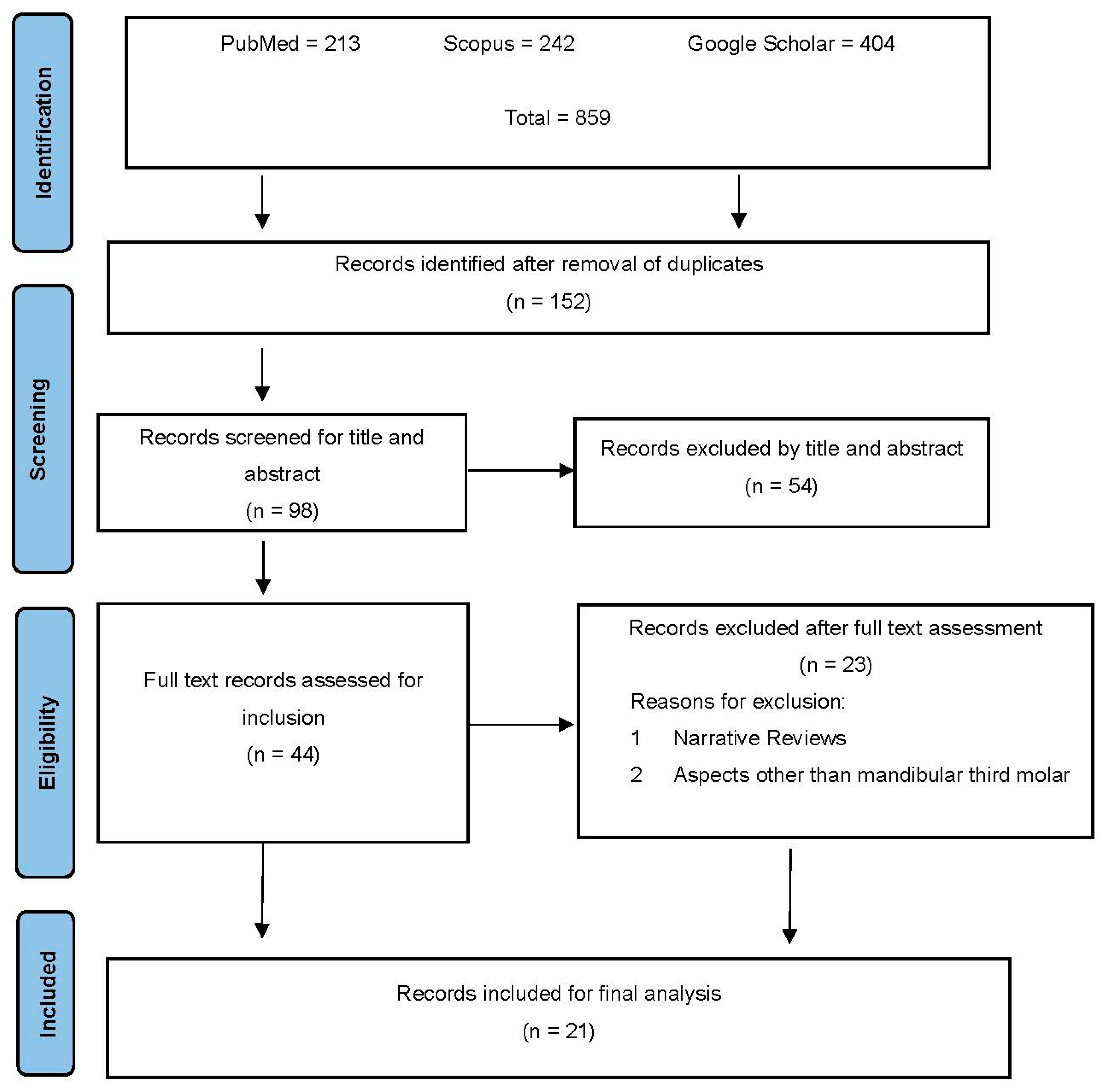
2.3. Search Strategy
2.4. Retrieval of Studies from the Databases
2.5. Data Collection
2.6. Evaluation of the Risk of Bias
3. Results
4. Discussion
4.1. Prediction of Third Molar Impaction
4.2. Determination of the Position of the Third Molar
4.3. Relationship of the Third Molar with the Inferior Dental Nerve
4.4. Extraction Difficulties
4.5. Strengths and Limitations
5. Conclusions
Funding
Conflicts of Interest
References
- Stone, P.; Brooks, R.; Brynjolfsson, E.; Calo, R.; Etzioni, O.; Hager, G.; Hirschberg, J.; Kalyanakrishnan, S.; Kamar, E.; Kraus, S.; et al. Artificial Intelligence and Life in 2030: The One Hundred Year Study on Artificial Intelligence. arXiv 2022, arXiv:2211.06318. [Google Scholar] [CrossRef]
- Mellit, A.; Kalogirou, S.A. Artificial intelligence techniques for photovoltaic applications: A review. Prog. Energy Combust. Sci. 2008, 34, 574–632. [Google Scholar] [CrossRef]
- Futyma-Gąbka, K.; Rózyło-Kalinowska, I. The use of artificial intelligence in radiological diagnosis and detection of dental caries: A systematic review. J. Stomatol. 2021, 74, 262–266. [Google Scholar] [CrossRef]
- Soori, M.; Arezoo, B.; Dastres, R. Artificial intelligence, machine learning and deep learning in advanced robotics, a review. Cogn. Robot. 2023, 3, 54–70. [Google Scholar] [CrossRef]
- Khanagar, S.B.; Al-Ehaideb, A.; Maganur, P.C.; Vishwanathaiah, S.; Patil, S.; Baeshen, H.A.; Sarode, S.C.; Bhandi, S. Developments, application, and performance of artificial intelligence in dentistry—A systematic review. J. Dent. Sci. 2021, 16, 508–522. [Google Scholar] [CrossRef] [PubMed]
- Pauwels, R.; Brasil, D.M.; Yamasaki, M.C.; Jacobs, R.; Bosmans, H.; Freitas, D.Q.; Haiter-Neto, F. Artificial Intelligence for detection of periapical lesions on intraoral radiographs: Comparison between convolutional neural networks and human observers. Oral Surg. Oral Med. Oral Pathol. Oral Radiol. 2021, 131, 610–616. [Google Scholar] [CrossRef] [PubMed]
- Yang, H.; Jo, E.; Kim, H.J.; Cha, I.H.; Jung, Y.S.; Nam, W.; Kim, J.Y.; Kim, J.K.; Kim, Y.H.; Oh, T.G.; et al. Deep Learning for Automated Detection of Cyst and Tumors of the Jaw in Panoramic Radiographs. J. Clin. Med. 2020, 9, 1839. [Google Scholar] [CrossRef] [PubMed]
- Mertens, S.; Krois, J.; Cantu, A.G.; Arsiwala, L.T.; Schwendicke, F. Artificial intelligence for caries detection: Randomized trial. J. Dent. 2021, 115, 103849. [Google Scholar] [CrossRef]
- Rai, S.; Misra, D.; Misra, A.; Tomar, H.; Dhawan, A.; Gupta, R. Reliability of Grayscale Value for Bone Density Determination in Oral Rehabilitation using Dental Implants. Int. J. Appl. Basic Med. Res. 2023, 13, 143–148. [Google Scholar] [CrossRef]
- Park, C.S.; Kang, S.R.; Kim, J.E.; Huh, K.H.; Lee, S.S.; Heo, M.S.; Han, J.J.; Yi, W.J. Validation of bone mineral density measurement using quantitative CBCT image based on deep learning. Sci. Rep. 2023, 13, 11921. [Google Scholar] [CrossRef]
- Farajollahi, M.; Safarian, M.S.; Hatami, M.; Esmaeil Nejad, A.; Peters, O.A. Applying artificial intelligence to detect and analyse oral and maxillofacial bone loss—A scoping review. Aust. Endod. J. 2023, 49, 720–734. [Google Scholar] [CrossRef] [PubMed]
- Uzun Saylan, B.C.; Baydar, O.; Yeşilova, E.; Kurt Bayrakdar, S.; Bilgir, E.; Bayrakdar, İ.Ş.; Çelik, Ö.; Orhan, K. Assessing the Effectiveness of Artificial Intelligence Models for Detecting Alveolar Bone Loss in Periodontal Disease: A Panoramic Radiograph Study. Diagnostics 2023, 13, 1800. [Google Scholar] [CrossRef] [PubMed]
- Xiao, Y.; Liang, Q.; Zhou, L.; He, X.; Lv, L.; Chen, J.; Endian, S.; Jianbin, G.; Wu, D.; Lin, L. Construction of a new automatic grading system for jaw bone mineral density level based on deep learning using cone beam computed tomography. Sci. Rep. 2022, 12, 12841. [Google Scholar] [CrossRef] [PubMed]
- Latt, M.; Chewpreecha, P.; Wongsirichat, N. Prediction of difficulty in impacted lower third molars extraction; review literature. Mahidol Dent. J. 2015, 35, 281–290. [Google Scholar]
- Pedro, F.L.; Bandéca, M.C.; Volpato, L.E.; Marques, A.T.; Borba, A.M.; Musis, C.D.; Borges, A.H. Prevalence of impacted teeth in a Brazilian subpopulation. J. Contemp. Dent. Pract. 2014, 15, 209–213. [Google Scholar] [CrossRef] [PubMed]
- Eshghpour, M.; Nezadi, A.; Moradi, A.; Shamsabadi, R.M.; Rezaei, N.M.; Nejat, A. Pattern of mandibular third molar impaction: A cross-sectional study in northeast of Iran. Niger. J. Clin. Pract. 2014, 17, 673–677. [Google Scholar] [CrossRef] [PubMed]
- Yoo, J.H.; Yeom, H.G.; Shin, W.; Yun, J.P.; Lee, J.H.; Jeong, S.H.; Lim, H.J.; Lee, J.; Kim, B.C. Deep learning based prediction of extraction difficulty for mandibular third molars. Sci. Rep. 2021, 11, 1954. [Google Scholar] [CrossRef] [PubMed]
- Vranckx, M.; Van Gerven, A.; Willems, H.; Vandemeulebroucke, A.; Ferreira Leite, A.; Politis, C.; Jacobs, R. Artificial Intelligence (AI)-Driven Molar Angulation Measurements to Predict Third Molar Eruption on Panoramic Radiographs. Int. J. Environ. Res. Public Health 2020, 17, 3716. [Google Scholar] [CrossRef] [PubMed]
- Orhan, K.; Bilgir, E.; Bayrakdar, I.S.; Ezhov, M.; Gusarev, M.; Shumilov, E. Evaluation of artificial intelligence for detecting impacted third molars on cone-beam computed tomography scans. J. Stomatol. Oral Maxillofac. Surg. 2021, 122, 333–337. [Google Scholar] [CrossRef]
- Moher, D.; Liberati, A.; Tetzlaff, J.; Altman, D.G. Preferred reporting items for systematic reviews and meta-analyses: The PRISMA statement. PLoS Med. 2009, 6, e1000097. [Google Scholar] [CrossRef]
- Celik, M.E. Deep Learning Based Detection Tool for Impacted Mandibular Third Molar Teeth. Diagnostics 2022, 12, 942. [Google Scholar] [CrossRef] [PubMed]
- Kempers, S.; van Lierop, P.; Hsu, T.M.H.; Moin, D.A.; Bergé, S.; Ghaeminia, H.; Xi, T.; Vinayahalingam, S. Positional assessment of lower third molar and mandibular canal using explainable artificial intelligence. J. Dent. 2023, 133, 104519. [Google Scholar] [CrossRef] [PubMed]
- Lahoud, P.; Diels, S.; Niclaes, L.; Van Aelst, S.; Willems, H.; Van Gerven, A.; Quirynen, M.; Jacobs, R. Development and validation of a novel artificial intelligence driven tool for accurate mandibular canal segmentation on CBCT. J. Dent. 2022, 116, 103891. [Google Scholar] [CrossRef] [PubMed]
- Ariji, Y.; Mori, M.; Fukuda, M.; Katsumata, A.; Ariji, E. Automatic visualization of the mandibular canal in relation to an impacted mandibular third molar on panoramic radiographs using deep learning segmentation and transfer learning techniques. Oral Surg. Oral Med. Oral Pathol. Oral Radiol. 2022, 134, 749–757. [Google Scholar] [CrossRef] [PubMed]
- Kim, J.Y.; Kahm, S.H.; Yoo, S.B.; Bae, S.; Kang, J.; Lee, S.H. The efficacy of supervised learning and semi-supervised learning in diagnosis of impacted third molar on panoramic radiographs through artificial intelligence model. DentoMaxilloFacial Radiol. 2023, 52, 20230030. [Google Scholar] [CrossRef] [PubMed]
- Fukuda, M.; Kise, Y.; Nitoh, M.; Ariji, Y.; Fujita, H.; Katsumata, A.; Ariji, E. Deep learning system to predict the three-dimensional contact status between the mandibular third molar and mandibular canal using panoramic radiographs. Oral Sci. Int. 2023, 21, 46–53. [Google Scholar] [CrossRef]
- Papasratorn, D.; Pornprasertsuk-Damrongsri, S.; Yuma, S.; Weerawanich, W. Investigation of the best effective fold of data augmentation for training deep learning models for recognition of contiguity between mandibular third molar and inferior alveolar canal on panoramic radiographs. Clin. Oral Investig. 2023, 27, 3759–3769. [Google Scholar] [CrossRef]
- Lei, Y.; Chen, X.; Wang, Y.; Tang, R.; Zhang, B. A Lightweight Knowledge-Distillation-Based Model for the Detection and Classification of Impacted Mandibular Third Molars. Appl. Sci. 2023, 13, 9970. [Google Scholar] [CrossRef]
- Joo, Y.; Moon, S.Y.; Choi, C. Classification of the Relationship Between Mandibular Third Molar and Inferior Alveolar Nerve Based on Generated Mask Images. IEEE Access 2023, 11, 81777–81786. [Google Scholar] [CrossRef]
- Zhu, T.; Chen, D.; Wu, F.; Zhu, F.; Zhu, H. Artificial Intelligence Model to Detect Real Contact Relationship between Mandibular Third Molars and Inferior Alveolar Nerve Based on Panoramic Radiographs. Diagnostics 2021, 11, 1664. [Google Scholar] [CrossRef]
- Lo Casto, A.; Spartivento, G.; Benfante, V.; Di Raimondo, R.; Ali, M.; Di Raimondo, D.; Tuttolomondo, A.; Stefano, A.; Yezzi, A.; Comelli, A. Artificial Intelligence for Classifying the Relationship between Impacted Third Molar and Mandibular Canal on Panoramic Radiographs. Life 2023, 13, 1441. [Google Scholar] [CrossRef] [PubMed]
- Kwon, D.; Ahn, J.; Kim, C.S.; Kang, D.O.; Paeng, J.Y. A deep learning model based on concatenation approach to predict the time to extract a mandibular third molar tooth. BMC Oral Health 2022, 22, 571. [Google Scholar] [CrossRef] [PubMed]
- Elborolosy, S.; Salem, W.; Hamed, M.; Sayed, A.; Helmy, B.; Elngar, A. Predicting Difficulty Level of Surgical Removal of Impacted Mandibular Third Molar Using Deep Learning Approaches. Res. Sq. 2022. [Google Scholar] [CrossRef]
- Takebe, K.; Imai, T.; Kubota, S.; Nishimoto, A.; Amekawa, S.; Uzawa, N. Deep learning model for the automated evaluation of contact between the lower third molar and inferior alveolar nerve on panoramic radiography. J. Dent. Sci. 2023, 18, 991–996. [Google Scholar] [CrossRef]
- Choi, E.; Lee, S.; Jeong, E.; Shin, S.; Park, H.; Youm, S.; Son, Y.; Pang, K. Artificial intelligence in positioning between mandibular third molar and inferior alveolar nerve on panoramic radiography. Sci. Rep. 2022, 12, 2456. [Google Scholar] [CrossRef] [PubMed]
- Vinayahalingam, S.; Xi, T.; Berge, S.; Maal, T.; De Jong, G. Automated detection of third molars and mandibular nerve by deep learning. Sci. Rep. 2019, 9, 9007. [Google Scholar] [CrossRef] [PubMed]
- Fukuda, M.; Ariji, Y.; Kise, Y.; Nozawa, M.; Kuwada, C.; Funakoshi, T.; Muramatsu, C.; Fujita, H.; Katsumata, A.; Ariji, E. Comparison of 3 deep learning neural networks for classifying the relationship between the mandibular third molar and the mandibular canal on panoramic radiographs. Oral Surg. Oral Med. Oral Pathol. Oral Radiol. 2020, 130, 336–343. [Google Scholar] [CrossRef] [PubMed]
- Lee, J.; Park, J.; Moon, S.Y.; Lee, K. Automated Prediction of Extraction Difficulty and Inferior Alveolar Nerve Injury for Mandibular Third Molar Using a Deep Neural Network. Appl. Sci. 2022, 12, 475. [Google Scholar] [CrossRef]
- Kwak, G.H.; Kwak, E.J.; Song, J.M.; Park, H.R.; Jung, Y.H.; Cho, B.H.; Hui, P.; Hwang, J.J. Automatic mandibular canal detection using a deep convolutional neural network. Sci. Rep. 2020, 10, 5711. [Google Scholar] [CrossRef]
- Guerrero, C.; la Hoz, F.; Alvis Guzmán, N. Calidad en revisiones sistemáticas de evaluaciones económicas de tecnologías en salud. Panor. Econ. 2019, 27, 581–597. [Google Scholar] [CrossRef]
- Chopra, S.; Vranckx, M.; Ockerman, A.; Östgren, P.; Krüger-Weiner, C.; Benchimol, D.; Shujaat, S.; Jacobs, R. A retrospective longitudinal assessment of artificial intelligence-assisted radiographic prediction of lower third molar eruption. Sci. Rep. 2024, 14, 994. [Google Scholar] [CrossRef] [PubMed]
- Lundervold, A.S.; Lundervold, A. An overview of deep learning in medical imaging focusing on MRI. Z Med. Phys. 2019, 29, 102–127. [Google Scholar] [CrossRef] [PubMed]
- Yilmaz, D.; Ataman-Duruel, E.T.; Beycioğlu, Z.; Goyushov, S.; Çimen, T.; Duruel, O.; Tözüm, T.F. The Radiological Evaluation of Mandibular Canal Related Variables in Mandibular Third Molar Region: A Retrospective Multicenter Study. J. Oral Maxillofac. Res. 2022, 13, e2. [Google Scholar] [CrossRef]
- Gong, Z.; Feng, W.; Su, X.; Choi, C. System for automatically assessing the likelihood of inferior alveolar nerve injury. Comput. Biol. Med. 2024, 169, 107923. [Google Scholar] [CrossRef]

| Database | Search Strategy |
|---|---|
| PUBMED | ((impacted tooth) OR (mandibular third molar impaction)) AND (artificial intelligence) |
| SCOPUS | TITLE-ABS-KEY (artificial AND intelligence) AND TITLE-ABS-KEY (impacted AND tooth) OR TITLE-ABS-KEY (mandibular third molar impaction) |
| Google Scholar | Artificial intelligence AND impacted tooth OR mandibular third molar impaction |
| Year and Author | Aims of the Study | Sample | Study Design | Imaging Tool | AI Application Method | Comparison if Applicable | Results |
|---|---|---|---|---|---|---|---|
| Vranckx et al. (2020) [18] | Prediction of impacted third molar eruption potential | 838 patients | Randomized clinical trial | OPG | ResNet-101 learning models | Expert human observer | The accuracy of the angulation measurements of the molars via AI was 79%. The second molar measurements were more accurate than the first molars. Prediction of third molar impaction eruption was reliable. |
| Orhan et al. (2021) [19] | Impaction location, number, and relation to the inferior alveolar nerve canal | 130 molars | Retrospective | CBCT | Deep CNN system | Human observers | A total of 112 impacted teeth, 99 canals, and a relationship between the third molar and maxillary sinus were detected. Accuracy values agreed with those of the human eye. |
| Celik ME (2022) [21] | Detection of the impaction and proposal of a new method of AI | 300 patients | Randomized clinical trial | OPG | Two phases: YOLOV3, Faster RCNN | Human observer | YOLOv3 demonstrated excellent performance in detecting impacted third molars in panoramic images. |
| Kempers et al. (2023) [22] | Relationship of impaction to the inferior alveolar canal | 863 patients | Case–control study | OPG | MobileNet-V2 precision model | Skeletonization algorithm and distance measurement | The MobileNet AI model automatically verified the skeletal, nerve, and dental anatomical relationships in the panoramic radiographs. |
| Lahoud et al. (2022) [23] | Mandibular canal shape direction and diameter determination | 235 patients | Retrospective study | CBCT | Machine learning model of algorithms | Expert observers | The total time for the automated segmentation of the mandibular canal and third molars was 21.2 s, 107 times faster than the manual segmentation performed by the expert. |
| Ariji et al. (2022) [24] | Relationship of impaction to the inferior alveolar canal and position of impaction | 3200 patients | Longitudinal study | OPG | U-Net neural network model | Expert observer; source IA model | The model creation time was 10.5 h. The transfer process was reduced by 90%. The learning models could be replicated and applied to different specimens for the location of impacted third molars. |
| Kim et al. (2023) [25] | Classification of mandibular third molar | 1625 patients | Retrospective | OPG | WideResNet and LaplaceNet model | Expert observer | AI models allowed up to 300 image masks to be collected for diagnosis. Even with fewer image masks, the models had higher accuracy. |
| Fukuda et al. (2023) [26] | Relationship of impaction to the inferior alveolar canal, and position of impaction | 800 patients | Case–control study | CT, CBCT, OPG | Deep learning models | Observer: specialists and residents | The machine learning training process was performed twice, compared with the performances of the observers. The diagnostic accuracy of the residents was 0.85, that of the specialist radiologists was 0.81, and that of the deep learning model was 0.81. |
| Papasratorn et al. (2023) [27] | Relationship of impaction to the inferior alveolar canal, and position of impaction | 1800 patients | Analytical | CBCT | Pre-trained AlexNet, VGG-16, GoogLeNet models | Expert observer | All models recognized the location of the third molar. VGG-16 demonstrated better performance than other trained models. |
| Lei et al. (2023) [28] | Classification of mandibular third molar | 2146 patients | Retrospective | OPG | Model YOLOv5 | Expert observer | The introduction of modified AI models increased the accuracy by 3% of the initial model. The amount of time spent on calculations and image processing was significantly reduced. |
| Joo et al. (2023) [29] | Relationship of impaction to the inferior alveolar canal and position of impaction | 5408 patients | Analytical | OPG, CBCT | AI models M3, YOLOv4 | Expert opinions | AI models allowed the generation of image masks of the lower dental nerve and third molar. The proposed system verified the classification performance with an improved accuracy of 0.833 using masked images. |
| Zhu et al. (2021) [30] | Relationship of impaction to the inferior alveolar canal | 503 patients | Experimental quantitative | CBCT, OPG | YOLOv4 (MM3-IANnet) deep learning model | CBCT, expert observer | The MM3-INnet demonstrated a higher detection protocol (83%) than the expert dentist (76%) in diagnosing impacted third molars. |
| Lo Casto et al. (2023) [31] | Diagnostic performance of two conventional neural networks in determining the impaction position | 83 patients | Randomized clinical trial | OPG | Two CNNs: ResNet-152 and VGG-19, | Expert (professional) and inexperienced (student) human observer | The student’s diagnostic performance was 62.53%, that of the expert observer was 85.28%, and RestNet achieved 88.86%. |
| Kwon et al. (2022) [32] | Timing of impaction extraction | 724 patients | Cross-sectional study | OPG | CNN + MLP neural network model | Professional experts | A predictive AI model for third molar extraction challenges can be developed with clinical and radiographic data. |
| Elborolosy et al. (2022) [33] | Difficulty of impacted third molar extraction | 2414 patients | Case studies | OPG | VGG-16, MobileNetV2, ResNet50 learning models | Expert observers | The accuracy of the machine learning models was 81% for VGG-16, 79% for MobileNetV2, and 44% for ResNet50. The prediction models and third molar anatomy of VGG1-6 through VGG-19 were comparable to those of the expert. |
| Takebe et al. (2023) [34] | Relationship of impaction to the inferior alveolar canal and position of impaction | 579 patients | Retrospective | OPG | YOLOv3 models | Expert observer | The accuracy rate of the YOLOv3 model was 0.89, while that of the expert professional was 0.6. |
| Choi et al. (2022) [35] | Relationship of impaction to the inferior alveolar canal | 571 patients | Case–control studies | OPG, CBCT | ResNet-50 model of AI | Expert observers | In terms of determining the buccolingual position, expert practitioners demonstrated an accuracy of 69%, and AIN models showed an accuracy of 80%. |
| Vinayahalingam et al. (2019) [36] | Relationship of impaction to the inferior alveolar canal | 81 patients | Randomized clinical trial | OPG | CNN model (M3, IAN) | Panoramic radiography | The CNN segmenters showed defects only in the apical region of the third molar. The training data guide the automated segmentation through AI CNN applications. |
| Fukuda et al. (2020) [37] | Relationship of impaction to the inferior alveolar canal | 600 patients | Case–control study | OPG | Three CNNs | Expert overlooker | The CNN layers determined the storage space and learned parameters. No difference in diagnostic performance was observed between the CNN patches. |
| Lee et al. (2022) [38] | Prediction of extraction difficulty of the impacted mandibular third molar | 5397 patients | Retrospective | OPG | Machine learning model of neural networks | Expert observer | Learning the region of interest (third molar) allowed for the prediction of the difficulty of extraction and the probability of injury to the lower dental nerve in 99% of the cases. An accuracy of 83% was obtained for the prediction of extraction difficulty. |
| Kwak et al. (2020) [39] | Detection and segmentation of the mandibular canal on CBCT images using various deep learning networks were attempted in this study to investigate the possibility of clinical application | 102 patients | Retrospective | OPG | VGGNet, SegNet learning network models | Expert observer | SegNet had an overall accuracy of 0.82, higher than other models without prior tooth segmentation. The learning channels gave higher accuracy in treatment planning. |
| Selected Article | SIGN Level |
|---|---|
| Orhan et al. (2021) [19] | 2++ |
| Celik (2022) [21] | 2+ |
| Lo casto et al. (2023) [31] | 2+ |
| Fukuda et al. (2020) [37] | 2+ |
| Zhu et al. (2021) [30] | 3 |
| Vinayahalingam et al. (2019) [36] | 2+ |
| Kwon et al. (2022) [32] | 3 |
| Kempers et al. (2023) [22] | 2+ |
| Choi et al. (2022) [35] | 2+ |
| Elborolosy et al. (2022) [33] | 3 |
| Vranckx et al. (2020) [18] | 2+ |
| Joo et al. (2023) [29] | 2++ |
| Ariji et al. (2022) [24] | 2+ |
| Lahoud et al. (2022) [23] | 3 |
| Papasratorn et al. (2023) [27] | 3 |
| Takebe et al. (2023) [34] | 2+ |
| Kim et al. (2023) [25] | 2+ |
| Lei et al. (2023) [28] | 3 |
| Fukuda et al. (2023) [26] | 3 |
| Lee et al. (2022) [38] | 3 |
| Kwak et al. (2020) [39] | 2+ |
Disclaimer/Publisher’s Note: The statements, opinions and data contained in all publications are solely those of the individual author(s) and contributor(s) and not of MDPI and/or the editor(s). MDPI and/or the editor(s) disclaim responsibility for any injury to people or property resulting from any ideas, methods, instructions or products referred to in the content. |
© 2024 by the authors. Licensee MDPI, Basel, Switzerland. This article is an open access article distributed under the terms and conditions of the Creative Commons Attribution (CC BY) license (https://creativecommons.org/licenses/by/4.0/).
Share and Cite
Assiri, H.A.; Hameed, M.S.; Alqarni, A.; Dawasaz, A.A.; Arem, S.A.; Assiri, K.I. Artificial Intelligence Application in a Case of Mandibular Third Molar Impaction: A Systematic Review of the Literature. J. Clin. Med. 2024, 13, 4431. https://doi.org/10.3390/jcm13154431
Assiri HA, Hameed MS, Alqarni A, Dawasaz AA, Arem SA, Assiri KI. Artificial Intelligence Application in a Case of Mandibular Third Molar Impaction: A Systematic Review of the Literature. Journal of Clinical Medicine. 2024; 13(15):4431. https://doi.org/10.3390/jcm13154431
Chicago/Turabian StyleAssiri, Hassan Ahmed, Mohammad Shahul Hameed, Abdullah Alqarni, Ali Azhar Dawasaz, Saeed Abdullah Arem, and Khalil Ibrahim Assiri. 2024. "Artificial Intelligence Application in a Case of Mandibular Third Molar Impaction: A Systematic Review of the Literature" Journal of Clinical Medicine 13, no. 15: 4431. https://doi.org/10.3390/jcm13154431