Review

Intrauterine Shaping of Fetal Microbiota

by
Norbert Dera
1,2,*,
Natalia Żeber-Lubecka
3,
Michał Ciebiera
2,4,
Katarzyna Kosińska-Kaczyńska
1,
Iwona Szymusik
1,
Diana Massalska
2,5,
Kacper Dera
6 and
Katarzyna Bubień
1
1
Department of Obstetrics, Perinatology and Neonatology, Center of Postgraduate Medical Education, 01-809 Warsaw, Poland
2
Warsaw Institute of Women’s Health, 00-189 Warsaw, Poland
3
Department of Gastroenterology, Hepatology and Clinical Oncology, Centre of Postgraduate Medical Education, 02-781 Warsaw, Poland
4
Department of Genetics, Maria Sklodowska-Curie National Research Institute of Oncology, 02-781 Warsaw, Poland
5
Second Department of Obstetrics and Gynecology, Centre of Postgraduate Medical Education, 00-189 Warsaw, Poland
6
Provincial Specialist Hospital in Olsztyn, 10-561 Olsztyn, Poland
*
Author to whom correspondence should be addressed.
J. Clin. Med. 2024, 13(17), 5331; https://doi.org/10.3390/jcm13175331
Submission received: 6 July 2024 / Revised: 18 August 2024 / Accepted: 29 August 2024 / Published: 9 September 2024
(This article belongs to the Section Clinical Pediatrics)

Abstract

:
Mechanisms resulting from the physiological immaturity of the digestive system in children delivered before 32 weeks of gestation and, in particular, different interactions between the microbiome and the body have not been fully elucidated yet. Next-generation sequencing methods demonstrated the presence of bacterial DNA in the placenta and amniotic fluid, which may reflect bacterial populations that initiate intestinal colonization in utero. Numerous studies confirmed the hypothesis stating that intestinal bacteria played an important role in the pathogenesis of necrotizing enterocolitis (NEC) early- and late-onset neonatal sepsis (EONS and LONS). The model and scale of disorders within the intestinal microbiome are the subject of active research in premature infants. Neonatal meconium was primarily used as an indicator defining the environment in utero, as it is formed before birth. Metagenomic results and previous data from microbiological bacterial cultures showed a correlation between the time from birth to sample collection and the detection of bacteria in the neonatal meconium. Therefore, it may be determined that the colonization of the newborn’s intestines is influenced by numerous factors, which may be divided into prenatal, perinatal, and postnatal, with particular emphasis put on the mode of delivery and contact with the parent immediately after birth. Background: The aim of this review was to collect available data on the intrauterine shaping of the fetal microbiota. Methods: On 13 March 2024, the available literature in the PubMed National Library of Medicine search engine was reviewed using the following selected keywords: “placental microbiome”, “intestinal bacteria in newborns and premature infants”, and “intrauterine microbiota”. Results: After reviewing the available articles and abstracts and an in-depth analysis of their content, over 100 articles were selected for detailed elaboration. We focused on the origin of microorganisms shaping the microbiota of newborns. We also described the types of bacteria that made up the intrauterine microbiota and the intestinal microbiota of newborns. Conclusions: The data presented in the review on the microbiome of both term newborns and those with a body weight below 1200 g indicate a possible intrauterine colonization of the fetus depending on the duration of pregnancy. The colonization occurs both via the vaginal and intestinal route (hematogenous route). However, there are differences in the demonstrated representatives of various types of bacteria, phyla Firmicutes and Actinobacteria in particular, taking account of the distribution in their abundance in the individual groups of pregnancy duration. Simultaneously, the distribution of the phyla Actinobacteria and Proteobacteria is consistent. Considering the duration of pregnancy, it may also be concluded that the bacterial flora of vaginal origin dominates in preterm newborns, while the flora of intestinal origin dominates in term newborns. This might explain the role of bacterial and infectious factors in inducing premature birth with the rupture of fetal membranes.

1. Introduction

The aim of this review was to collect available data on the intrauterine shaping of the fetal microbiota. In the first weeks and months of life, gut microbiome disorders affect the infant’s growth, development, and health status. Impaired intestinal microbiota formation during infant development results in an increased risk of immune and metabolic diseases that may persist in childhood and, potentially, continue into adulthood [1]. Most research on the development of the intestinal microbiome focused on term infants [2]. The physiological immaturity of the digestive system in infants delivered before 32 weeks of gestation was found to lead to several interactions between the microbiome and the body. The underlying mechanisms have not been fully elucidated yet [3].
The maturation of both intestinal barrier function and immunity occurs in the prenatal period. The fetal intestine is more permeable to macromolecules and less tolerant of antigens compared to the infant intestine after birth. The transfer of maternal immunoglobulin G (IgG) through the placenta and the absorption in the fetal intestine increase in the period close to the term of pregnancy, shaping the immune response of the newborn after birth [4]. Therefore, the intrauterine environment is able to shape health far beyond the fetal life and may influence long-term changes in health parameters. According to recent research, colonization with specific microorganisms in early life may predispose to the development of childhood diseases, including asthma and obesity [5,6]. Several teams of researchers, using methods based on next-generation sequencing, showed the presence of bacterial DNA in the placenta and amniotic fluid [7,8]. It was also suggested that it reflected bacterial populations that initiated intestinal colonization in utero. According to Bartnicka et al., the use of the latest molecular techniques allowed the confirmation of the presence of bacteria in the amniotic fluid, umbilical cord blood, placenta, and fetal membranes. In addition, the detection of individual bacterial species in the meconium (such as Escherichia coli, Enterococcus faecium, and Staphylococcus epidermidis) might result from their translocation via maternal bloodstream. The translocation hypothesis was confirmed by the fact that bacteria of the genera Enterococcus, Streptococcus, Staphylococcus, and the Propionibacterium species were isolated from the umbilical cord blood [9].
Although the presence and function of the placental microbiota have not been fully elucidated, it is known that the intestinal microflora during pregnancy is a key determinant of the health of the offspring. According to abundant evidence, the maternal intestinal microbiota during pregnancy might determine the development of atopy and autoimmune phenotypes in the offspring and affect the child’s immunity and predisposition to an occurrence of disease [10].
Two conceptual models were used to describe the development of the intestinal microbiome of premature infants. According to the first concept, the intestinal microbiome developed in an orderly manner, depending on the maturation of the child’s body, with the minor impact of environmental factors. The second concept assumed that external factors such as the diet, exposure to antibiotics, and the hospital environment played a major role in the development of the microbiome [11].
Postnatally, in early life, a balanced gut microbiome is crucial for the functioning of the body and the maturation of the immune system [12]. In recent years, a growing body of evidence has pointed to the importance of the gut microbiome in providing resistance to colonization by pathogens or opportunistic intestinal pathobionts. Disorders of intestinal microbiota maturation, i.e., intestinal dysbiosis, predispose newborns to the formation of NEC [13], nosocomial infections, or EONS and LONS [14,15,16,17].
Low weight gain in premature infants was found to be associated with disorders in the gut–brain axis [18], the composition of the intestinal microbiota [19,20], insufficient absorption of nutrients [21], and the existing diseases of neonatal age. Numerous studies confirmed the hypothesis that intestinal bacteria played an important role in the pathogenesis of NEC, EONS, and LONS. However, it was impossible to clearly identify one bacterial species as the causative agent. Therefore, in the case of NEC, an increase in the abundance of several microorganisms was observed, mainly from the phyla Firmicutes and Proteobacteria (Cronobacter sakazakii, Klebsiella sp., and E. coli) [22,23]. An increased abundance of Clostridium spp. and their toxins was detected in the stool of neonates with NEC [24,25,26].
Importantly, the colonization of the newborn’s intestine was found to be influenced by numerous factors, which might be divided into prenatal, perinatal, and postnatal, with particular emphasis put on the mode of delivery and contact with the parent immediately after birth [27,28] (Figure 1).
This review focuses on prenatal factors affecting the course of pregnancy and the condition of the fetus. They underlie the proper functioning in later life. Over ten times more bacteria than cells are found in the human body [29,30]. Sequencing the hypervariable regions of the bacterial 16S RNA gene enabled the taxonomic identification of bacteria at the level of the genus and species, but not at the subspecies level [31]. Shotgun metagenomics could identify bacteria at a lower level of taxonomic resolution, which means the detection of low- or very low-abundance microbial communities and could also be more effective in differentiating closely related species [32]. The development of next-generation sequencing made it possible to obtain an image of the reproductive microbiome. In 2015, Franasiak et al. were the first to determine the microbiota of the uterine cavity based on material collected from the end of the catheter during embryo transfer [31].
Various hypotheses regarding the origin of the microbiota of the uterus referred both to the pathway ascending from the vagina and the intestinal tract (through the filtration of the intestinal vessels into the peritoneal cavity with reabsorption through the fallopian tubes, and with the help of dendritic cells and leukocytes transporting hematogenous material to the uterus) [33]. DiGiulio et al. determined that the amniotic fluid from premature births could be colonized by the placental microbiome and by microorganisms ascending from the vagina, while maintaining the continuity of the amniotic membrane [34]. Some authors suggested that the vagina might be the source of microbes that reached the placenta, amniotic fluid, and fetus via translocation through the chorion [35,36]. It was also confirmed that, during pregnancy, the microbiota of the mother’s vagina affected the immunity of the fetus in the uterus, even before passing through the vaginal canal during childbirth.
Until 2014, it had been believed that the placenta had not contained its own microbiome. This was related to the research techniques used, which had not taken account of differences between live and dead bacteria and had not allowed for obtaining a maternal blood sample facilitating the determination whether the microbiome came from the maternal chorion or the fetus. Therefore, the results were questionable [37]. However, according to current research, infections are suggested to underlie PPROM and preterm birth [38,39,40]. The placenta was demonstrated to have its own healthy microbiome, mainly containing Firmicutes, Tenericutes, Proteobacteria, Bacteroidetes, and Fusobacteria. It was also found that the placental microbiome was the most similar to the human oral microbiome [41].
Establishing and maintaining the integrity and function of the placenta is critical to fetal growth, development, and survival [42]. In a cross-sectional study including 195 patients, Stout et al. demonstrated that intracellular Gram-positive and Gram-negative bacteria were present in the basal plate (a structure including a layer of tissue directly at the maternal-fetal interface and below).

2. Materials and Methods

On 13 March 2024, the available literature in the PubMed National Library of Medicine search engine was reviewed, using the following selected keywords: “placental microbiome”, “intestinal bacteria in newborns and premature infants”, and “intrauterine microbiota”. After reviewing the available articles and abstracts and an in-depth analysis of their content, over 100 articles were selected for detailed elaboration. We focused on the origin of microorganisms shaping the microbiota of newborns. We also described the types of bacteria that made up the intrauterine microbiota and the intestinal microbiota of newborns.

3. Discussion

3.1. Differentiation of Views—Sterile (Acquired at Birth)/Non-Sterile Environment

The present literature review draws attention to the evolution of views regarding the uterine environment. The original views assumed that the cervical mucus, which adhered to the vagina colonized by bacteria, maintained uterine sterility. At the same time, in 1996, Egbase et al. claimed that the microflora of the reproductive system might affect the results of in vitro fertilization [43]. In subsequent years and in the latest research, the above hypotheses regarding uterine sterility were repeatedly undermined [44,45,46,47]. The only differences consisted in the way and method of the colonization of the uterus and the most identified bacterial flora. The examination of the neonatal meconium constitutes an indirect confirmation of the lack of sterility of the uterine cavity. According to Jimmy Kok-Foo Lee et al., over 50% of the meconium in the population of preterm newborns contained bacteria, and the percentage increased with gestational age, which indicated the intrauterine acquisition of the bacterial flora [11]. This is an extremely important aspect in relation to the functioning of the extrauterine human body. It constitutes the basis for the analysis and identification of the adequate bacterial flora of the gastrointestinal tract as a protective factor that influences the proper development and functioning from the neonatal period to adulthood.

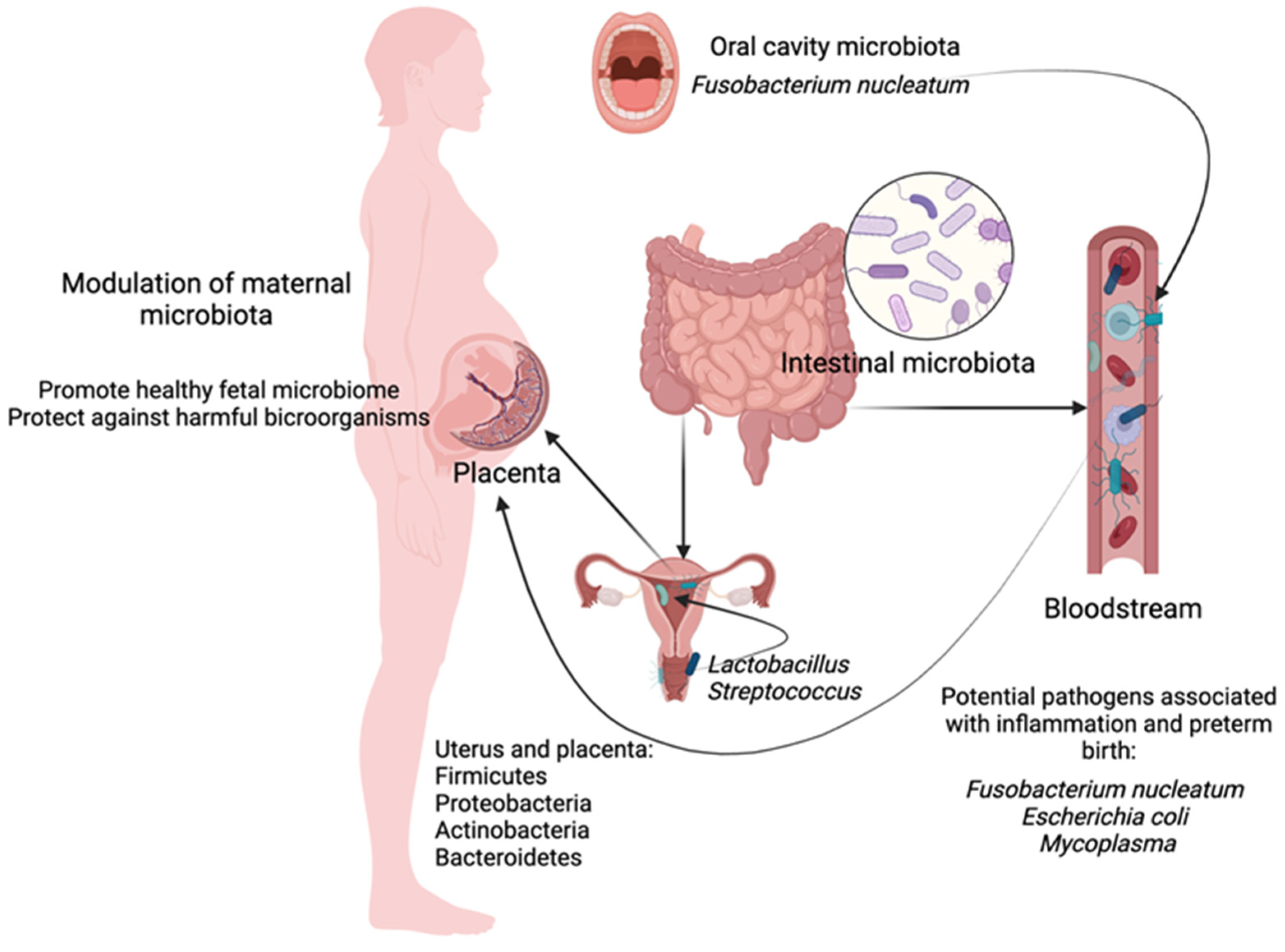
3.2. The Way of the Colonization of the Uterus: By Continuity, by Blood

According to the latest hypothesis, the uterine microbiota may ascend from the vagina and the intestinal tract. It occurs through the filtration of the intestinal vessels into the peritoneal cavity with reabsorption through the fallopian tubes and with the help of dendritic cells and leukocytes transporting hematogenous material to the uterus [33]. Jimenez et al. conducted animal studies and reported that after placing the genetically labeled Enterococcus faecium in the oral cavity of mice, it could be detected in the placenta. Similarly, they isolated the labeled Enterococcus faecium from the meconium of offspring after the oral inoculation of the strain to their pregnant mothers. It confirmed the above assumption that the intestinal microorganisms of mothers potentially penetrated the placenta into the intestines of their offspring [48].
Conversely, Goldenberg and Hanley suggested that the vagina might be the source of microbes that reached the placenta, amniotic fluid, and fetus via translocation through the chorion and amnion [35,36].
DiGiulio et al. determined that the amniotic fluid from premature births could be colonized by microorganisms ascending from the vagina while maintaining the continuity of the amniotic membrane [34]. The placental microbiome could also contribute to the colonization, which might be associated with the possible hematogenous translocation of the microbiome originating from the maternal oral cavity [41,49]. According to DiGiulio, species that had been long implicated in causing microbial invasion of the amniotic cavity (MIAC) remained among the common invaders (e.g., Ureaplasma spp., Mycoplasma spp., Fusobacterium spp., Streptococcus spp., Bacteroides spp., and Prevotella spp.). Similarly, cultivation-resistant anaerobes belonging to the family Fusobacteriaceae (particularly Sneathia sanguinegens and Leptotrichia spp.) and Candida spp. were commonly found in amniotic fluid [34]. Microbial invasion into the uterus was confirmed in 25–40% of preterm births [50,51], and in 7–12% of preterm births with intact membranes [52]. In the case of fetal membrane inflammation, the inflammatory process of the fetal surface of the placenta, the most frequently isolated pathogens included the following bacteria: Bacteroides, E. coli, Gardnerella vaginalis, Mycoplasma hominis, Peptostreptococci, Streptococci, and Ureaplasma urealyticum [53]. Therefore, it may be assumed that pathological bacteria might enter the amnion and chorion from the vagina. Some authors also hypothesized the transmission of an infection through the placenta from the oral cavity of patients with periodontal diseases [54]. Specific pathogenic bacteria of the oral cavity, including Fusobacterium nucleatum, Porphyromonas gingivalis, Filifactor alocis, and Campylobacter rectus, were associated with both periodontitis and the development of pregnancy diseases [54].

3.3. The Microbiota of the Oral Cavity and the Placenta of a Pregnant Woman

Research comparing the oral and placental microbiota performed on murine models also confirmed the similarity of the above locations. This may indicate the hematogenous origin of the placental microbiome [55,56,57,58]. The above was confirmed by the results of studies by Han et al., who found that a single Bergeyella strain was 100% identical at the 16S–23S rRNA sequence level between the patient’s periodontal subgingival plaque and the amniotic fluid. Both samples were 99.7% identical to the previously deposited oral Bergeyella sequence. None of the numerous vaginal samples collected from the patient revealed any Bergeyella species, and it was not known whether Bergeyella was commensal to any part of the body except the oral cavity [56]. Jimenez et al. demonstrated that the genetically labeled Enterococcus faecium placed in the oral cavity of mice could be later detected in the placenta. Importantly, it may provide numerous possibilities in the evaluation and modification of the bacterial flora of the maternal gastrointestinal tract. Considering the hematogenous origin of the placental microbiome, the proper supplementation of specific bacterial cultures in pregnant women might modulate the bacterial flora of the fetus and newborn with beneficial microbiome that might protect against colonization with harmful microorganisms.

3.4. Maternal Placental and Uterine Microbiome

Studies using the high-throughput sequencing technology confirmed the presence of the placental microbiome [59,60]. As regards the types of bacteria occurring in the placenta, Stout et al. demonstrated that both intracellular Gram-positive and Gram-negative bacteria were present in the basal plate (a structure including a layer of tissue directly at the maternal-fetal interface and below). They were observed in almost one-third of placental samples, with a high incidence in preterm births <28 weeks of gestation, but independently of the clinical or pathological features of fetal membrane inflammation [61]. Suggestions regarding the origin of intrauterine infections associated with premature birth, referring to the onset in the lower genital tract and penetration into the “sterile” intrauterine environment [50,62] contradicted the results of research using DNA-based technology, indicating commensal species commonly found in the oral cavity as the main causative factor of colonization [55,56,57,58]. The above was confirmed by the finding that oral microorganisms such as Fusobacterium nucleatum (Gram-negative oral anaerobes) might facilitate the hematogenous transmission of other commensal bacteria, i.e., Escherichia coli, during placental formation. It is related to their capability of binding to the vascular endothelium and changing the permeability [63].
Studies assessing the predominance of some species conducted by Moore et al. and Aagaard et al. revealed that E. coli was the most common microorganism in the placenta [41,64]. Simultaneously, they identified additional species of the oral microbiome, including Prevotella tannerae (gingival fissures) and non-pathogenic Neisseria species (mucosal surfaces). The above might be confirmed by research by Gosalbes et al., who revealed the abundance of E. coli in the meconium [65]. Conversely, Aagaard et al. demonstrated that the placenta had its own healthy microbiome, which mainly contained Firmicutes, Tenericutes, Proteobacteria, Bacteroidetes, and Fusobacteria. Overall, the profiles of the placental microbiome were the most similar (the Bray–Curtis dissimilarity < 0.3) to the one of the human oral microbiome [41]. At the same time, the intestinal bacterial flora was discussed. It included both Bacteroidetes and Firmicutes, which constituted 90% of the microflora, but also less abundant types, i.e., Proteobacteria, Actinobacteria, Fusobacteria, and Verrucomicrobia. Anaerobes constitute over 99% of normal intestinal microorganisms inhabiting mainly the distal part of the ileum and the colon [66,67]. To compare, four main types of bacteria, i.e., Firmicutes, Proteobacteria, Actinobacteria, and Bacteroidetes were present in the intestines of healthy newborns and premature infants with very low birth weight (VLBW) [68,69,70,71,72]. Nevertheless, premature infants and VLBW infants were characterized by lower microbial diversity, disturbed intestinal microbiome, and an increased colonization of potentially pathogenic nosocomial microorganisms compared to age-matched term infants [73].
Research on the uterine microbiome of healthy women also confirmed that the most consistent types included Firmicutes, Bacteroidetes, Proteobacteria, and Actinobacteria [31,74,75,76,77,78,79,80,81,82]. Conversely, Lactobacillus and Streptococcus, which may be found in the vagina and cervix, were the most commonly reported [83].
Regarding the dependence of individual bacterial species on the location, Brotman et al. found that each location in the body was characterized by one or several characteristic types, i.e., Firmicutes in the vagina, Actinobacteria in the skin of the retroauricular fold and the anterior nostrils, Proteobacteria and Firmicutes at all sites in the oral cavity, and Bacteroidetes in the feces. The placental microbiome was characterized by a greater abundance of Proteobacteria and the unique presence of Tenericutes, including the known intrauterine types of Mycoplasma and Ureaplasma [84].
The above studies showed that the intrauterine environment was not “sterile”. The origin of the microbiota may largely be associated with hematogenous transmission rather than the assumed ascending transmission from the vagina. At the same time, there are four main types of bacteria in the intestines of a healthy newborn that are characteristic and present in the normal microbiome of the placenta and uterus of healthy women. Conversely, based on the analysis of the bacterial flora of preterm infant intestines, which are characterized by lower diversity, with an increased colonization of potentially pathogenic microorganisms, it may be concluded that the dysbiosis of maternal body, gastrointestinal tract and, consequently, the uterus and placenta, constitutes the basis of the inflammatory process being the main causative factor for premature birth. Therefore, such a relationship, i.e., the assessment of the maternal microbiota and its modulation, may provide a powerful protective tool for the development of the embryo and fetus (Figure 2).

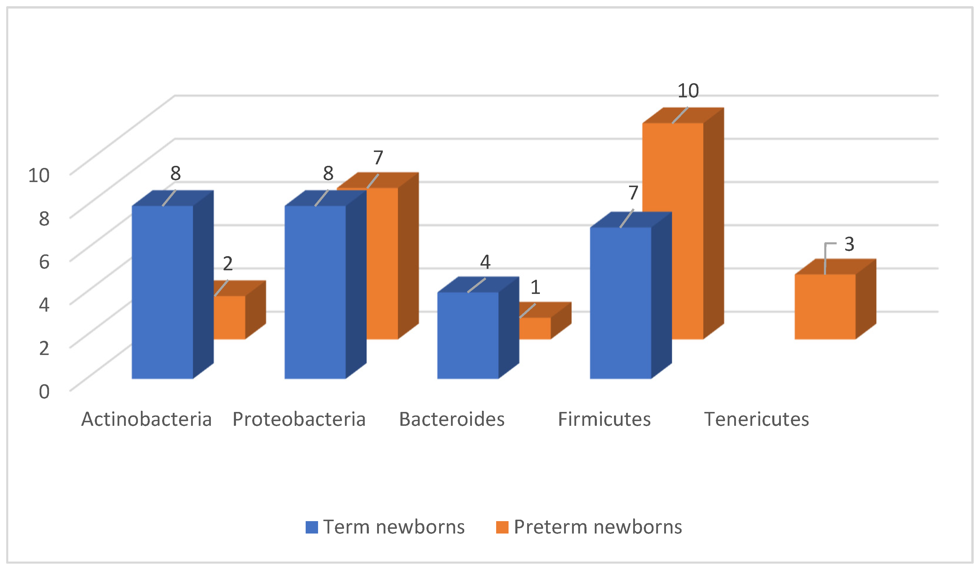
3.5. Meconium Microbiota Depending on Gestational Age and Specific Diseases

The neonatal meconium is formed before birth. Therefore, it was primarily used as an indicator defining the in utero environment [85]. However, the analyses did not comprise acquired microorganisms that appear during and/or immediately after birth. Metagenomic results and previous data from bacterial cultures showed a correlation between the time from birth to sample collection and the detection of bacteria in the neonatal meconium.
Studies in mice and humans revealed that the meconium was colonized by bacteria during pregnancy [14,86,87]. Jimenez et al. isolated the labeled Enterococcus faecium from the meconium of offspring after the oral inoculation of the strain with pregnant mothers. This indicated that the intestinal microorganisms of mothers potentially penetrated the placenta into the intestines of their offspring [58].
According to numerous authors, e.g., Gosalbes et al., the abundance of E. coli was confirmed in the meconium [65]. It was also the main factor contributing to the occurrence of early-onset sepsis in neonates with extremely low birth weight [84]. Aagaard et al. reported that the detection of commensal Escherichia in the meconium was associated with intrauterine colonization originating from the placenta [41]. According to Stoll et al., the acquisition of group B Streptococcus (GBS) and E. coli during delivery was identified as the main cause of EONS within 3 days after delivery in preterm infants [88]. In contrast, Dong et al. determined that skin or intestinal commensals, including Staphylococcus spp., E. coli, K. pneumoniae, or Candida spp., usually caused LONS [89]. Due to problems with the identification of a single causative agent, NEC was mainly associated with Firmicutes (coagulase-negative staphylococci) and Proteobacteria (Cronobacter sakazakii, Klebsiella sp., and E. coli) (Table 1) [88,89].
However, regarding significant correlations between the microbiota of the meconium and the duration of pregnancy, Ardissone et al. determined the relationships that were negatively linked to gestational age, with the exception of Oxalicibacterium [87]. The study revealed that the taxonomic families within the phylum Firmicutes that were correlated with gestational age included: Bacillaceae, Staphylococcaceae, Enterococcaceae, Lactobacilaceae, Leuconostocaceae, Clostridiaceae, Peptostreptococcaceae, Veillonellaceae, and Erysipelotrichaceae. At the genus level, the strongest correlations were assigned to Enterococcus and Lactobacillus, including an average of 8.67% versus 0.41% for Enterococcus and 0.82% versus 0.07% for Lactobacillus at <33 and >33 weeks, respectively. As regards Actinobacteria, Bifidobacterium was significantly correlated, with 5.47% versus 0.35% of all readings for <33 and >33 weeks, respectively. The phylum Proteobacteria that was significantly correlated with gestational age primarily included Enterobacteriaceae. The strongest correlations were identified for Enterobacter and Photorhabdus, with an average relative abundance of 6.35% versus 0.06% for Enterobacter and 0.98% versus 0.01% for Photorhabdus at <33 and >33 weeks, respectively. The abundance of Tannerella was lower and was correlated with low gestational age. Kang et al. determined that two classes, i.e., Bacteroidetes and Firmicutes, constituted the majority of the neonatal meconium microbiota. A significant increase in the relative abundance of Bacteroidetes and a decrease in Proteobacteria correlated with an increase in gestational age [90]. At the genus level, the dominant species included Prevotella and Bacteroides, with the relative abundance of Prevotella accounting for 20–30% of the intestinal microbiome. In healthy adults, two major classes, i.e., Firmicutes and Bacteroidetes, constituted over 90% of the gut microbiota, followed by Actinobacteria and Proteobacteria [91,92]. A study conducted in Germany by Klopp et al. also showed that gestational age was significantly linked to the meconium composition of extremely premature infants, and the most numerous phyla included Firmicutes, Bacteroidetes, Proteobacteria, and Actinobacteria (Table 2) [93].
The above research results, indicating a much lower diversity of bacterial populations, with an increased abundance within individual phyla, classes, and genera in preterm newborns, suggest a greater risk of the development of pathogenic flora through the dynamic multiplication and competitive displacement of protective microorganisms. Therefore, in combination with structural immaturity, the morbidity of newborns is inversely proportional to the age of pregnancy, i.e., the moment when significant environmental diversity becomes a protective factor, markedly contributing to the maintenance of the eubiotic state.

3.6. The Intestinal Microbiome in Newborns

A difference between the intestinal microbiome of a term newborn and one born prematurely was noted when using material from newborns (Table 3 and Table 4) [9,14,41,42,68,86,87,94]. Sood et al. determined that in the first week of life, the intestinal microbiome of term newborns was largely colonized by Actinobacteria (including Bifidobacterium), Proteobacteria, Bacteroides, and, to a much lesser extent, by Firmicutes (including Lactobacillus spp., which are dominant in the vaginal flora) [42,70]. According to Aagaard et al., the phyla Firmicutes and Tenericutes and a much smaller abundance of Actinobacteria dominated in newborns with a body weight < 1200 g, which was consistent with the previously cited data on the microbiome of the placenta [41]. Arboleya et al. stated that preterm infants were characterized by an increased abundance of Enterococcus, Enterobacter, Lactobacillus, and Staphylococcus bacteria and a reduced abundance of Bacteroides, Bifidobacterium, and Atopobium compared to term infants [68]. They also showed that the colonization of the intestine by Bifidobacterium was delayed in preterm infants [95]. A prospective study showed that gestational age was significantly linked to intestinal colonization by Bifidobacteria. Delivery at the gestational age of <33 weeks appeared to impair bifidobacterial colonization and predispose premature infants to infections and intestinal diseases. Conversely, Bartnicka et al. reported that bacteria belonging to the genera Bifidobacterium, Lactobacillus, and Streptococcus dominated in term newborns, while Enterobacteriaceae and Clostridium dominated in premature infants (Figure 3) [9].
The above data are summarized in Table 5.
Based on the analysis, a correlation between the intestinal microbiome of a newborn may be concluded, taking account of the birth age, and the potential microbiome of the mother, broken down by the site of colonization (Table 6 and Table 7).
It is obvious that the mode of delivery, activities and drugs used perinatally, and the first contact with the external environment postnatally are important factors regulating the composition of the intestinal microbiota.

3.7. Influence of the Intrauterine Microbiome on the Child’s Immune System

The immune aspect may not be ignored. It was confirmed that the microbiota of the mother’s vagina during pregnancy influenced the immunity of the fetus in the uterus, even before passing through the vaginal canal during childbirth. Over the past decade, studies involving both the 16S and metagenomics have shown that the human vagina mainly contains Lactobacillus spp. at concentrations reaching 107–109 per gram of vaginal secretion [77,96,97]. The resultant state of eubiotics is a protective factor against other species due to the competitive exclusion concept.
A pilot study conducted in 2016 showed that the percentage of pregnancies decreased by almost 40% in women without the dominance of Lactobacillus (non-lactobacillus-dominant, NLD) species in the uterus [74]. Recent research has suggested that an inflammatory response may be triggered in the endometrium by NLD-phenotype microbiota, affecting the success of embryo implantation. It may result from the regulation of inflammatory mediators during blastocyst adhesion to the epithelial wall of the endometrium [29]. Research showed that Lactobacillus bacteria were more prominent in women with endometrial polyps or chronic endometritis [78]. Conversely, chronic endometritis was found to be associated with recurrent pregnancy loss and the presence of plasma cells in endometrial biopsy [98,99,100,101]. Abundant bacteria were detected in such patients, including Neisseria gonorrhea, Chlamydia trachomatis, Escherichia coli, Streptococcus, Staphylococcus, and Enterococcus faecalis. Non-microbiological causes were also identified. It was also hypothesized that endometriosis changed the endometrium through the intensification of inflammation and resistance to progesterone. This might affect implantation and increase the risk of miscarriage and pregnancy complications, e.g., pregnancy-induced hypertension and preterm birth [102].
Neonates whose mothers were intragestationally vaginally colonized with Lactobacillus had a higher percentage of CD45RO+ cells and reduced IL-12 in the umbilical cord blood. Therefore, the authors stated that lactobacilli in the mother’s vagina influenced the development of fetal immunity [10,103].
The microflora during pregnancy initiates the immune programming of the offspring in various, interrelated ways that are not necessarily mutually exclusive. The above was confirmed by research by Hu et al., Madan et al., and Ardissone et al., who showed that the intestines of the fetus were colonized by bacteria during pregnancy [14,86,87]. Aagaard et al. and Collado et al. demonstrated that the placenta contained a unique microbiome [41,94]. Conversely, Ferretti et al. noted a greater stability of maternal intestinal strains in the intestines of newborns compared to vaginal and dermal strains [104]. The majority of authors indicated a disorder related to Proteobacteria and Firmicutes prior to the occurrence of NEC. Research also revealed that the diversity of gut bacteria was reduced in preterm infants at risk of LONS and NEC [11]. However, chorioamnionitis and fetal inflammatory response syndrome were linked to the development of numerous diseases in premature infants, i.e., cerebral palsy [105,106], intraventricular hemorrhage (IVH) [107,108,109], retinopathy of prematurity (ROP) [110], NEC [108,111] and EONS [109,112,113]. At the same time, it was shown that fetal membrane inflammation played a protective role in LONS by accelerating the maturation of the immune system [113].
The development of atopic disease in the offspring of mothers who used antibiotic therapy during pregnancy and, thus, modeled the maternal microflora (along with its metabolites) may be the evidence of the impact of this process on the modeling of the immune response [114].
During the first postnatal week, an increased abundance of the genera Bacteroides and Bifidobacterium was found in term newborns delivered vaginally [115,116]. Bifidobacterium and Bacteroides are genera that are thought to promote health and interact directly with the developing immune system during infancy [117,118,119]. It was found that Bacteroides fragilis produced a bacterial polysaccharide that assisted in the maturation of the immune system and the production of regulatory T lymphocytes [118,119,120]. A study conducted on the murine model, which is known for intestinal barrier defects, revealed that oral treatment with B. fragilis reduced intestinal permeability and changed the microbiological composition [121]. Research on the effect of antibiotic therapy on bacterial flora showed that early antibiotic use was associated with the reduced abundance of Bifidobacterium and Bacteroides in preterm infants. Therefore, it was suggested that early exposure to antibiotics might affect the future composition of intestinal bacteria in preterm infants.
Arboleya et al. demonstrated that antibiotics used perinatally affected the quantitative and qualitative structure of the intestinal microbiota. It was particularly manifested as the increased abundance of bacteria from the family Enterobacteriaceae and Clostridiaceae and the reduction in protective Bifidobacterium and Lactobacillus in infants [122].
This is of key importance from the viewpoint of the susceptibility to infections and their severity. At the same time, it translates into psychophysical development and metabolic disorders, which may predispose to the development of civilization diseases exacerbated by environmental factors.

3.8. Pregnancy Complications—Pre-Eclampsia and Its Spectrum

The participation of systemic inflammatory response in pregnancies complicated by pre-eclampsia (PEC4) and intrauterine growth restriction (IUGR) led to the development of a hypothesis that maternal infections might be an important factor in the pathogenesis of pregnancy complications.
It was also demonstrated that the risk of pre-eclampsia was increased in women with asymptomatic bacteriuria, urinary tract infection, and chronic pyelonephritis [123,124]. Den Hollander et al. confirmed that Helicobacter pylori, being a cause of chronic inflammation, was associated with an increased risk of PEC [125]. Moreover, Li Juan et al. demonstrated that pre-eclampsia was associated with a disturbance in the composition of intestinal microflora compared to women whose pregnancies were uncomplicated [126]. Dunn et al. published a review of a possible relationship between the microbiome and the development of pre-eclampsia, which showed that the PEC microbiome was studied by five groups of researchers [127]. Two studies analyzed placental location, and the remaining three examined the oral cavity, intestine, or intra-amniotic site. Some findings confirmed the link between pathogenic bacteria and PEC. However, specific pathogenic organisms were identified, so further research is justified. In a study conducted in 2015, placental tissue samples were collected from women with and without pre-eclampsia and tested for the presence of bacteria with the use of next-generation sequencing and PCR for 16s rRNA. A total of 12.7% of the tissues of women with PEC tested positive in the PCR test, while all the placentas in the control group tested negative [128].
In contrast, a study in which researchers carefully controlled for possible contaminants using multiple detection methods, including culture, qPCR, 16S rRNA gene sequencing, and shotgun metagenomics, revealed that no placental microbiota could be identified in the placenta.

4. Conclusions

The present review on the microbiome of both term newborns and premature infants indicates a possible intrauterine colonization of the fetus depending on the duration of pregnancy. The colonization occurs both via the vaginal and intestinal route (hematogenous route). The postnatal examination of the neonatal meconium clearly confirms the above hypothesis, especially if we consider the duration of pregnancies (the meconium of premature versus term neonates) and the continuity of fetal membranes. However, we noted differences in the demonstrated representatives of various types of bacteria, phyla Firmicutes and Actinobacteria in particular, taking account of the distribution in their abundance in individual groups of pregnancy duration. Simultaneously, the distribution of the phyla Actinobacteria and Proteobacteria was consistent. However, as regards the occurrence of specific species and genera, their similarity should be emphasized. Considering the duration of pregnancy, it can also be concluded that the bacterial flora of vaginal origin dominates in preterm newborns, while the flora of intestinal origin dominates in term newborns. This might explain the role of bacterial and infectious factors in inducing premature birth with the rupture of fetal membranes. The above conclusions suggest that appropriate shaping of the mother’s bacterial flora, comprising the microbiome of the digestive and the genital tract during pregnancy, may contribute to the prevention of premature births with the proper shaping of the fetal intestinal microbiota. This is an extremely important aspect, thanks to which it would be possible to avoid obstetric failures and, at the same time, influence the life and development of a person from conception to old age. This also translates into the financial aspect of the functioning of health care and the costs associated with the treatment of immunological and metabolic diseases associated with disturbed intestinal microbiota from the moment of conception. Our publication aimed to show this extremely important aspect, which obviously requires further research. Such research would constitute the basis for creating a scheme of action for the proper shaping of the microbiota of a pregnant woman.

Funding

The study was funded by the Center for Postgraduate Medical Education in Warsaw, grant no. 501-1-022-26-24 and grant no. 501-1-157-81-23/MG7.

Conflicts of Interest

The authors declare no conflict of interest.

References

  1. Gałęcka, M.; Basińska, A.M.; Bartnicka, A. Znaczenie mikrobioty jelitowej w kształtowaniu zdrowia człowieka—Implikacje w praktyce lekarza rodzinnego. Forum Med. Rodz. 2018, 12, 50–59. [Google Scholar]
  2. Healy, D.B.; Ryan, C.A.; Ross, R.P.; Stanton, C. Clinical implications of preterm infant gut microbiome development. Nat. Microbiol. 2022, 7, 22–33. [Google Scholar] [CrossRef]
  3. Indrio, F.; Neu, J.; Pettoello-Mantovani, M. Development of the Gastrointestinal Tract in Newborns as a Challenge for an Appropriate Nutrition: A Narrative Review. Nutrients 2022, 14, 1405. [Google Scholar] [CrossRef] [PubMed]
  4. Weström, B.; Sureda, E.A.; Pierzynowska, K. The Immature Gut Barrier and Its Importance in Establishing Immunity in Newborn Mammals. Front. Immunol. 2020, 11, 1153. [Google Scholar] [CrossRef]
  5. Milani, C.; Duranti, S.; Bottacini, F. The First Microbial Colonizers of the Human Gut: Composition, Activities, and Health Implications of the Infant Gut Microbiota. Microbiol. Mol. Biol. Rev. 2017, 81, e00036-17. [Google Scholar] [CrossRef]
  6. Lv, H.; Zhang, L.; Han, Y.; Wu, L.; Wang, B. The Development of Early Life Microbiota in Human Health and Disease. Engineering 2022, 12, 101–114. [Google Scholar] [CrossRef]
  7. Campisciano, G.; Quadrifoglio, M.; Comar, M.; De Seta, F. Evidence of bacterial DNA presence in chorionic villi and amniotic fluid in the first and second trimester of pregnancy. Future Microbiol. 2021, 16, 801–810. [Google Scholar] [CrossRef] [PubMed]
  8. Sharlandjieva, V.; Beristain, A.G.; Terry, J. Assessment of the human placental microbiome in early pregnancy. Front. Med. 2023, 10, 1096262. [Google Scholar] [CrossRef] [PubMed]
  9. Bartnicka, A.; Gałęcka, M.; Mazela, J. Wpływ czynników prenatalnych i postnatalnych na mikrobiotę jelitową noworodków. Stand. Med. Pediatr. 2016, 13, 112–116. [Google Scholar]
  10. Nyangahu, D.D.; Jaspan, H.B. Influence of maternal microbiota during pregnancy on infant immunity. Clin. Exp. Immunol. 2019, 198, 47–56. [Google Scholar] [CrossRef]
  11. Lee, J.K.-F.; Hern Tan, L.T.; Ramadas, A.; Ab Mutalib, N.-S.; Lee, L.-H. Exploring the Role of Gut Bacteria in Health and Disease in Preterm Neonates. Int. J. Environ. Res. Public. Health 2020, 17, 6963. [Google Scholar] [CrossRef] [PubMed]
  12. Staude, B.; Oehmke, F.; Lauer, T.; Behnke, J.; Gopel, W.; Schloter, M.; Schulz, H.; Krauss-Etschmann, S.; Ehrhardt, H. The Microbiome and Preterm Birth: A Change in Paradigm with Profound Implications for Pathophysiologic Concepts and Novel Therapeutic Strategies. Biomed. Res. Int. 2018, 2018, 7218187. [Google Scholar] [CrossRef] [PubMed]
  13. Claud, E.C.; Keegan, K.P.; Brulc, J.M.; Lu, L.; Bartels, D.; Glass, E.; Chang, E.B.; Meyer, F.; Antonopoulos, D.A. Bacterial community structure and functional contributions to emergence of health or necrotizing enterocolitis in preterm infants. Microbiome 2013, 1, 20. [Google Scholar] [CrossRef] [PubMed]
  14. Madan, J.C.; Salari, R.C.; Saxena, D.; Davidson, L.; O’Toole, G.A.; Moore, J.H.; Sogin, M.L.; Foster, J.A.; Edwards, W.H.; Palumbo, P.; et al. Gut microbial colonisation in premature neonates predicts neonatal sepsis. Arch. Dis. Child. Fetal Neonat. 2012, 97, 456–462. [Google Scholar] [CrossRef]
  15. Carl, M.A.; Ndao, I.M.; Springman, A.C.; Manning, S.D.; Johnson, J.R.; Johnston, B.D.; Burnham, C.A.; Weinstock, E.S.; Weinstock, G.M.; Wylie, T.N.; et al. Sepsis from the gut: The enteric habitat of bacteria that cause late-onset neonatal bloodstream infections. Clin. Infect. Dis. 2014, 58, 1211–1218. [Google Scholar] [CrossRef]
  16. Shaw, A.G.; Sim, K.; Randell, P.; Cox, M.J.; McClure, Z.E.; Li, M.S.; Donaldson, H.; Langford, P.R.; Cookson, W.O.; Moffatt, M.F.; et al. Late-Onset Bloodstream Infection and Perturbed Maturation of the Gastrointestinal Microbiota in Premature Infants. PLoS ONE 2015, 10, e0132923. [Google Scholar] [CrossRef]
  17. Mai, V.; Torrazza, R.M.; Ukhanova, M.; Wang, X.; Sun, Y.; Li, N.; Shuster, J.; Sharma, R.; Hudak, M.L.; Neu, J. Distortions in development of intestinal microbiota associated with late onset sepsis in preterm infants. PLoS ONE 2013, 8, e52876. [Google Scholar] [CrossRef]
  18. Groer, M.; Luciano, A.A.; Dishaw, L.; Ashmeade, T.; Miller, E.; Gilbert, J. Development of the preterm infant gut microbiome: A research priority. Microbiome 2014, 2, 38. [Google Scholar] [CrossRef]
  19. Younge, N.E.; Newgard, C.B.; Cotten, C.M.; Goldberg, R.N.; Muehlbauer, M.J.; Bain, J.R.; Stevens, R.D.; O’Connell, T.M.; Rawls, J.F.; Seed, P.C.; et al. Disrupted Maturation of the Microbiota and Metabolome among Extremely Preterm Infants with Postnatal Growth Failure. Sci. Rep. 2019, 9, 8167. [Google Scholar] [CrossRef]
  20. Henderickx, J.G.E.; Zwittink, R.D.; van Lingen, R.A.; Knol, J.; Belzer, C. The Preterm Gut Microbiota: An Inconspicuous Challenge in Nutritional Neonatal Care. Front. Cell. Infect. Microbiol. 2019, 9, 85. [Google Scholar] [CrossRef]
  21. Lee, J.K.; Yu, V.Y. Calorie intake in sick versus respiratory stable very low birthweight babies. Pediatr. Int. 1996, 38, 449–454. [Google Scholar] [CrossRef] [PubMed]
  22. Pammi, M.; Cope, J.; Tarr, P.I.; Warner, B.B.; Morrow, A.L.; Mai, V.; Gregory, K.E.; Kroll, J.S.; McMurtry, V.; Ferris, M.J.; et al. Intestinal dysbiosis in preterm infants preceding necrotizing enterocolitis: A systematic review and meta-analysis. Microbiome 2017, 5, 31. [Google Scholar] [CrossRef] [PubMed]
  23. Coggins, S.A.; Wynn, J.L.; Weitkamp, J.-H. Infectious causes of necrotizing enterocolitis. Clin. Perinatol. 2015, 42, 133–154. [Google Scholar] [CrossRef] [PubMed]
  24. Kosloske, A.M.; Ulrich, J.A. A bacteriologic basis for the clinical presentations of necrotizing enterocolitis. J. Pediatr. Surg. 1980, 15, 558–564. [Google Scholar] [CrossRef]
  25. Kosloske, A.; Ulrich, J.; Hoffman, H. Fulminant necrotising enterocolitis associated with clostridia. Lancet 1978, 312, 1014–1016. [Google Scholar] [CrossRef]
  26. Lindberg, T.P.; Caimano, M.J.; Hagadorn, J.I.; Bennett, E.M.; Maas, K.; Brownell, E.A.; Matson, A.P. Preterm infant gut microbial patterns related to the development of necrotizing enterocolitis. J. Matern.-Fetal Neonatal Med. 2020, 33, 349–358. [Google Scholar] [CrossRef]
  27. Jeong, S. Factors influencing development of the infant microbiota: From prenatal period to early infancy. Clin. Exp. Pediatr. 2022, 65, 438–447. [Google Scholar] [CrossRef]
  28. Houghteling, P.D.; Walker, W.A. Why is initial bacterial colonization of the intestine important to the infant’s and child’s health? J. Pediatr. Gastroenterol. Nutr. 2015, 60, 294–307. [Google Scholar] [CrossRef]
  29. Bardos, J.; Fiorentino, D.; Longman, R.E.; Paidas, M. Immunological Role of the Maternal Uterine Microbiome in Pregnancy: Pregnancies Pathologies and Alterated Microbiota. Front. Immunol. 2019, 10, 2823. [Google Scholar] [CrossRef]
  30. NIH Hmp Working Group; Peterson, J.; Garges, S.; Giovanni, M.; McInnes, P.; Wang, L.; Schloss, J.A.; Bonazzi, V.; McEwen, J.E.; Wetterstrand, K.A.; et al. The NIH human microbiome project. Genome Res. 2009, 19, 2317–2323. [Google Scholar] [CrossRef]
  31. Franasiak, J.M.; Werner, M.D.; Juneau, C.R.; Tao, X.; Landis, J.; Zhan, Y.; Treff, N.R.; Scott, R.T. Endometrial microbiome at the time of embryo transfer: Next-generation sequencing of the 16s ribosomal subunit. J. Assist. Reprod. Genet. 2016, 33, 129–136. [Google Scholar] [CrossRef] [PubMed]
  32. Li, F.; Chen, C.; Wei, W.; Wang, Z.; Dai, J.; Hao, L.; Song, L.; Zhang, X.; Zeng, L.; Du, H.; et al. The metagenome of the female upper reproductive tract. Gigascience 2018, 7, Giy107. [Google Scholar] [CrossRef] [PubMed]
  33. Donnet-Hughes, A.; Perez, P.F.; Doré, J.; Leclerc, M.; Levenez, F.; Benyacoub, J.; Serrant, P.; Segura-Roggero, I.; Schiffrin, E.J. Potential role of the intestinal microbiota of the mother in neonatal immune education. Proc. Nutr. Soc. 2010, 69, 407–415. [Google Scholar] [CrossRef] [PubMed]
  34. DiGiulio, D.B. Diversity of microbes in amniotic fluid. Semin. Fetal Neonatal Med. 2012, 17, 2–11. [Google Scholar] [CrossRef]
  35. Goldenberg, R.L.; Culhane, J.F.; Iams, J.D.; Romero, R. Epidemiology and causes of preterm birth. Lancet 2008, 371, 75–84. [Google Scholar] [CrossRef]
  36. Hanley, J. Child health neonatal infections: Group B streptococcus search date March 2007 prophylactic treatment of at-risk neonates: GBS child health neonatal infections: Group B streptococcus. Clin. Evid. 2008, 1, 1–6. [Google Scholar]
  37. Kliman, H.J. Comment on the placenta harbors a unique microbiome. Sci. Transl. Med. 2014, 6, 254le4. [Google Scholar] [CrossRef]
  38. Garmi, G.; Okopnik, M.; Keness, Y.; Zafran, N.; Berkowitz, E.; Salim, R. Correlation between clinical, placental histology and microbiological findings in spontaneous preterm births. Fetal Diagn. Ther. 2016, 40, 141–149. [Google Scholar] [CrossRef]
  39. Sweeney, E.L.; Kallapur, S.G.; Gisslen, T.; Lambers, D.S.; Chougnet, C.A.; Stephenson, S.-A.; Jobe, A.H.; Knox, C.L. Placental infection with ureaplasma species is associated with histologic chorioamnionitis and adverse outcomes in moderately preterm and late-preterm infants. J. Infect. Dis. 2016, 213, 1340–1347. [Google Scholar] [CrossRef]
  40. Quinn, P.A.; Butany, J.; Taylor, J.; Hannah, W. Chorioamnionitis: Its association with pregnancy outcome and microbial infection. Am. J. Obstet. Gynecol. 1987, 156, 379–387. [Google Scholar] [CrossRef]
  41. Aagaard, K.; Ma, J.; Antony, K.M.; Ganu, R.; Petrosino, J.; Versalovic, J. The placenta harbors a unique microbiome. Sci. Transl. Med. 2014, 6, 237ra65. [Google Scholar] [CrossRef]
  42. Sood, R.; Zehnder, J.L.; Druzin, M.L.; Brown, P.O. Gene expression patterns in human placenta. Proc. Natl. Acad. Sci. USA 2006, 103, 5478–5483. [Google Scholar] [CrossRef] [PubMed]
  43. Egbase, P.E.; al-Sharhan, M.; al-Othman, S.; al-Mutawa, M.; Udo, E.E.; Grudzinskas, J.G. Incidence of microbial growth from the tip of the embaryo transfer catheter after embryo transfer in relation to clinical pregnancy rate following in-vitro fertilization. Hum. Reprod. 1996, 11, 1687–1689. [Google Scholar] [CrossRef]
  44. Heinonen, P.K.; Teisala, K.; Punnonen, R.; Miettinen, A.; Lehtinen, M.; Paavonen, J. Anatomic sites of upper genital tract infection. Obstet. Gynecol. 1985, 66, 384–390. [Google Scholar]
  45. Eschenbach, D.A.; Rosene, K.; Tompkins, L.S.; Watkins, H.; Gravett, M.G. Endometrial cultures obtained by a triple-lumen method from afebrile and febrile postpartum women. J. Infect. Dis. 1986, 153, 1038–1045. [Google Scholar] [CrossRef] [PubMed]
  46. Hemsell, D.L.; Obregon, V.L.; Heard, M.C.; Nobles, B.J. Endometrial bacteria in asymptomatic, nonpregnant women. J. Reprod. Med. 1989, 34, 872–874. [Google Scholar] [PubMed]
  47. Moller, B.R.; Kristiansen, F.V.; Thorsen, P.; Frost, L.; Mogensen, S.C. Sterility of the uterine cavity. Acta Obstet. Gynecol. Scand. 1995, 74, 216–219. [Google Scholar] [CrossRef]
  48. Jiménez, E.; Marín, M.L.; Martín, R.; Odriozola, J.M.; Olivares, M.; Xaus, J.; Fernández, L.; Rodríguez, J.M. Is meconium from healthy newborns actually sterile? Res. Microbiol. 2008, 159, 187–189. [Google Scholar] [CrossRef]
  49. Leon, R.; Silva, N.; Ovalle, A.; Chaparro, A.; Ahumada, A.; Gajardo, M.; Martinez, M.; Gamonal, J. Detection of Porphyromonas gingivalis in the amniotic fluid in pregnant women with a diagnosis of threatened premature labor. J. Periodontol. 2007, 78, 1249–1255. [Google Scholar] [CrossRef]
  50. Goldenberg, R.L.; Hauth, J.C.; Andrews, W.W. Intrauterine infection and preterm delivery. N. Engl. J. Med. 2000, 342, 1500–1507. [Google Scholar] [CrossRef]
  51. Lahra, M.M.; Beeby, P.J.; Jeffery, H.E. Maternal versus fetal inflammation and respiratory distress syndrome: A 10-year hospital cohort study. Arch. Dis. Child. Fetal Neonatal Ed. 2009, 94, F13–F16. [Google Scholar] [CrossRef] [PubMed]
  52. Romero, R.; Miranda, J.; Chaiworapongsa, T.; Chaemsaithong, P.; Gotsch, F.; Dong, Z.; Ahmed, A.I.; Yoon, B.H.; Hassan, S.S.; Kim, C.J.; et al. A novel molecular microbiologic technique for the rapid diagnosis of microbial invasion of the amniotic cavity and intra-amniotic infection in preterm labor with intact membranes. Am. J. Reprod. Immunol. 2014, 71, 330–358. [Google Scholar] [CrossRef]
  53. Moreno, I.; Simon, C. Deciphering the effect of reproductive tract microbiota on human reproduction. Reprod. Med. Biol. 2019, 18, 40–50. [Google Scholar] [CrossRef]
  54. Cobb, C.M.; Kelly, P.J.; Williams, K.B.; Babbar, S.; Angolkar, M.; Derman, R.J. The oral microbiome and adverse pregnancy outcomes. Int. J. Womens Health 2017, 9, 551–559. [Google Scholar] [CrossRef]
  55. Fardini, Y.; Chung, P.; Dumm, R.; Joshi, N.; Han, Y.W. Transmission of diverse oral bacteria to murine placenta: Evidence for the oral microbiome as a potential source of intrauterine infection. Infect. Immun. 2010, 78, 1789–1796. [Google Scholar] [CrossRef]
  56. Han, Y.W.; Ikegami, A.; Bissada, N.F.; Herbst, M.; Redline, R.W.; Ashmead, G.G. Transmission of an uncultivated Bergeyella strain from the oral cavity to amniotic fluid in a case of preterm birth. J. Clin. Microbiol. 2006, 44, 1475–1483. [Google Scholar] [CrossRef]
  57. Han, Y.W.; Fardini, Y.; Chen, C.; Iacampo, K.G.; Peraino, V.A.; Shamonki, J.M.; Redline, R.W. Term stillbirth caused by oral Fusobacterium nucleatum. Obstet. Gynecol. 2010, 115, 442–445. [Google Scholar] [CrossRef]
  58. Han, Y.W.; Shen, T.; Chung, P.; Buhimschi, I.A.; Buhimschi, C.S. Uncultivated bacteria as etiologic agents of intra-amniotic inflammation leading to preterm birth. J. Clin. Microbiol. 2009, 47, 38–47. [Google Scholar] [CrossRef] [PubMed]
  59. Doyle, R.; Alber, D.; Jones, H.; Harris, K.; Fitzgerald, F.; Peebles, D.; Klein, N. Term and preterm labour are associated with distinct microbial community structures in placental membranes which are independent of mode of delivery. Placenta 2014, 35, 1099–1101. [Google Scholar] [CrossRef]
  60. Parnell, L.A.; Briggs, C.M.; Cao, B.; Delannoy-Bruno, O.; Schrieffer, A.E.; Mysorekar, I.U. Microbial communities in placentas from term normal pregnancy exhibit spatially variable profiles. Sci. Rep. 2017, 7, 11200. [Google Scholar] [CrossRef]
  61. Stout, M.J.; Conlon, B.; Landeau, M.; Lee, I.; Bower, C.; Zhao, Q.; Roehl, K.A.; Nelson, D.M.; Macones, G.A.; Mysorekar, I.U. Identification of intracellular bacteria in the basal plate of the human placenta in term and preterm gestations. Am. J. Obstet. Gynecol. 2013, 208, 226.e1–226.e7. [Google Scholar] [CrossRef] [PubMed]
  62. Romero, R.; Schaudinn, C.; Kusanovic, J.P.; Gorur, A.; Gotsch, F.; Webster, P.; Nhan-Chang, C.L.; Erez, O.; Kim, C.J.; Espinoza, J.; et al. Detection of a microbial biofilm in intraamniotic infection. Am. J. Obstet. Gynecol. 2008, 198, 135.e1–135.e5. [Google Scholar] [CrossRef]
  63. Fardini, Y.; Wang, X.; Témoin, S.; Nithianantham, S.; Lee, D.; Shoham, M.; Han, Y.W. Fusobacterium nucleatum adhesin FadA binds vascular endothelial cadherin and alters endothelial integrity. Mol. Microbiol. 2011, 82, 1468–1480. [Google Scholar] [CrossRef]
  64. Moore, L.V.; Johnson, J.L.; Moore, W.E. Descriptions of Prevotella tannerae sp. nov. and Prevotella enoeca sp. nov. from the human gingival crevice and emendation of the description of Prevotella zoogleoformans. Int. J. Syst. Bacteriol. 1994, 44, 599–602. [Google Scholar] [CrossRef]
  65. Gosalbes, M.J.; Llop, S.; Vallès, Y.; Moya, A.; Ballester, F.; Francino, M.P. Meconium microbiota types dominated by lactic acid or enteric bacteria are differentially associated with maternal eczema and respiratory problems in infants. Clin. Exp. Allergy 2013, 43, 198–211. [Google Scholar] [CrossRef] [PubMed]
  66. Rinninella, E.; Raoul, P.; Cintoni, M.; Franceschi, F.; Miggiano, G.A.D.; Gasbarrini, A.; Mele, M.C. What is the Healthy Gut Microbiota Composition? A Changing Ecosystem across Age, Environment, Diet, and Diseases. Microorganisms 2019, 7, 14. [Google Scholar] [CrossRef] [PubMed]
  67. Eckburg, P.B.; Bik, E.M.; Bernstein, C.N.; Purdom, E.; Dethlefsen, L.; Sargent, M.; Gill, S.R.; Nelson, K.E.; Relman, D.A. Diversity of the human intestinal microbial flora. Science 2005, 308, 1635. [Google Scholar] [CrossRef] [PubMed]
  68. Arboleya, S.; Binetti, A.; Salazar, N.; Fernandez, N.; Solis, G.; Hernandez-Barranco, A.; Margolles, A.; de Los Reyes-Gavilan, C.G.; Gueimonde, M. Establishment and development of intestinal microbiota in preterm neonates. FEMS Microbiol. Ecol. 2012, 79, 763–772. [Google Scholar] [CrossRef] [PubMed]
  69. Adlerberth, I.; Wold, A.E. Establishment of the gut microbiota in Western infants. Acta Paediatr. 2009, 98, 229–238. [Google Scholar] [CrossRef]
  70. Gritz, E.C.; Bhandari, V. The human neonatal gut microbiome: A brief review. Front. Pediatr. 2015, 3, 17. [Google Scholar]
  71. Unger, S.; Stintzi, A.; Shah, P.; Mack, D.; O’Connor, D.L. Gut microbiota of the very-low-birth-weight infant. Pediatr. Res. 2015, 77, 205–213. [Google Scholar] [CrossRef] [PubMed]
  72. Bjorkstrom, M.V.; Hall, L.; Soderlund, S.; Hakansson, E.G.; Hakansson, S.; Domellof, M. Intestinal flora in very low-birth weight infants. Acta Paediatr. 2009, 98, 1762–1767. [Google Scholar] [CrossRef] [PubMed]
  73. Costello, E.K.; Carlisle, E.M.; Bik, E.M.; Morowitz, M.J.; Relman, D.A. Microbiome assembly across multiple body sites in low-birthweight infants. mBio 2013, 4, e00782-13. [Google Scholar] [CrossRef] [PubMed]
  74. Moreno, I.; Codoñer, F.M.; Vilella, F.; Valbuena, D.; Martinez-Blanch, J.F.; Jimenez-Almazán, J.; Alonso, R.; Alamá, P.; Remohí, J.; Pellicer, A.; et al. Evidence that the endometrial microbiota has an effect on implantation success or failure. Am. J. Obstet. Gynecol. 2016, 215, 684–701. [Google Scholar] [CrossRef] [PubMed]
  75. Wade, W. Unculturable bacteria–the uncharacterized organisms that cause oral infections. J. R. Soc. Med. 2002, 95, 81–83. [Google Scholar] [CrossRef]
  76. Tao, X.; Franasiak, J.M.; Zhan, Y.; Scott, R.T.; Rajchel, J.; Bedard, J.; Newby, R.; Treff, N.R.; Chu, T. Characterizing the endometrial microbiome by analyzing the ultra-low bacteria from embryo transfer catheter tips in IVF cycles: Next generation sequencing (NGS) analysis of the 16s ribosomal gene. Hum. Microb. J. 2017, 3, 15–21. [Google Scholar] [CrossRef]
  77. Chen, C.; Song, X.; Wei, W.; Zhong, H.; Dai, J.; Lan, Z.; Li, F.; Yu, X.; Feng, Q.; Wang, Z.; et al. The microbiota continuum along the female reproductive tract and its relation to uterine-related diseases. Nat. Commun. 2017, 8, 875. [Google Scholar] [CrossRef]
  78. Fang, R.L.; Chen, L.X.; Shu, W.S.; Yao, S.Z.; Wang, S.W.; Chen, Y.Q. Barcoded sequencing reveals diverse intrauterine microbiomes in patients suffering with endometrial polyps. Am. J. Transl. Res. 2016, 8, 1581–1592. [Google Scholar]
  79. Verstraelen, H.; Vilchez-Vargas, R.; Desimpel, F.; Jauregui, R.; Vankeirsbilck, N.; Weyers, S.; Verhelst, R.; De Sutter, P.; Pieper, D.H.; Van De Wiele, T. Characterisation of the human uterine microbiome in non-pregnant women through deep sequencing of the V1-2 region of the 16s rRNA gene. PeerJ 2016, 4, E1602. [Google Scholar] [CrossRef]
  80. Walther-António, M.R.S.; Chen, J.; Multinu, F.; Hokenstad, A.; Distad, T.J.; Cheek, E.H.; Keeney, G.L.; Creedon, D.J.; Nelson, H.; Mariani, A.; et al. Potential contribution of the uterine microbiome in the development of endometrial cancer. Genome Med. 2016, 8, 122. [Google Scholar] [CrossRef]
  81. Miles, S.M.; Hardy, B.L.; Merrell, D.S. Investigation of the microbiota of the reproductive tract in women undergoing a total hysterectomy and bilateral salpingo-oopherectomy. Fertil. Steril. 2017, 107, 813–820.e1. [Google Scholar] [CrossRef] [PubMed]
  82. Khan, K.N.; Fujishita, A.; Masumoto, H.; Muto, H.; Kitajima, M.; Masuzaki, H.; Kitawaki, J. Molecular detection of intrauterine microbial colonization in women with endometriosis. Eur. J. Obstet. Gynecol. Reprod. Biol. 2016, 199, 69–75. [Google Scholar] [CrossRef]
  83. Koedooder, R.; Mackens, S.; Budding, A.; Fares, D.; Blockeel, C.; Laven, J.; Schoenmakers, S. Identyfikacja i ocena mikrobiomu w żeńskich i męskich drogach rozrodczych. Aktual. Hum. Reprod. 2019, 25, 298–325. [Google Scholar] [CrossRef]
  84. Brotman, R.M. Vaginal microbiome and sexually transmitted infections: An epidemiologic perspective. J. Clin. Investig. 2011, 121, 4610–4617. [Google Scholar] [CrossRef] [PubMed]
  85. Stinson, L.F.; Keelan, J.A.; Payne, M.S. Comparison of Meconium DNA Extraction Methods for Use in Microbiome Studies. Front. Microbiol. 2018, 9, 270. [Google Scholar] [CrossRef]
  86. Hu, J.; Nomura, Y.; Bashir, A.; Fernandez-Hernandez, H.; Itzkowitz, S.; Pei, Z.; Stone, J.; Loudon, H.; Peter, I. Diversified microbiota of meconium is affected by maternal diabetes status. PLoS ONE 2013, 8, e78257. [Google Scholar] [CrossRef]
  87. Ardissone, A.N.; De La Cruz, D.M.; Davis-Richardson, A.G.; Rechcigl, K.T.; Li, N.; Drew, J.C.; Murgas-Torrazza, R.; Sharma, R.; Hudak, M.L.; Triplett, E.W.; et al. Meconium microbiome analysis identifies bacteria correlated with premature birth. PLoS ONE 2014, 9, e90784. [Google Scholar] [CrossRef]
  88. Stoll, B.J.; Hansen, N.I.; Sánchez, P.J.; Faix, R.G.; Poindexter, B.B.; Van Meurs, K.P.; Bizzarro, M.J.; Goldberg, R.N.; Frantz, I.D.; Hale, E.C.; et al. Early Onset Neonatal Sepsis: The Burden of Group B Streptococcal and E. coli Disease Continues. Pediatrics 2011, 127, 817–826. [Google Scholar] [CrossRef] [PubMed]
  89. Dong, Y.; Speer, C.P. Late-onset neonatal sepsis: Recent developments. Arch. Dis. Child.-Fetal Neonatal Ed. 2015, 100, F257–F263. [Google Scholar] [CrossRef]
  90. Kang, H.M.; Kim, S.; Hwang-Bo, S.; Yoo, I.H.; Seo, Y.-M.; Oh, M.Y.; Im, S.-A.; Youn, Y.-A. Compositional Differences of Meconium Microbiomes of Preterm and Term Infants, and Infants That Developed Necrotizing Enterocolitis or Feeding Intolerance. Pathogens 2023, 12, 55. [Google Scholar] [CrossRef]
  91. Jalanka-Tuovinen, J.; Salonen, A.; Nikkilä, J.; Immonen, O.; Kekkonen, R.; Lahti, L.; Palva, A.; de Vos, W.M. Intestinal microbiota in healthy adults: Temporal analysis reveals individual and common core and relation to intestinal symptoms. PLoS ONE 2011, 6, e23035. [Google Scholar] [CrossRef] [PubMed]
  92. Claesson, M.J.; Jeffery, I.B.; Conde, S.; Power, S.E.; O’Connor, E.M.; Cusack, S.; Harris, H.M.; Coakley, M.; Lakshminarayanan, B.; O’Sullivan, O.; et al. Gut microbiota composition correlates with diet and health in the elderly. Nature 2012, 488, 178–184. [Google Scholar] [CrossRef] [PubMed]
  93. Klopp, J.; Ferretti, P.; Meyer, C.U.; Hilbert, K.; Haiß, A.; Marißen, J.; Henneke, P.; Hudalla, H.; Pirr, S.; Viemann, D.; et al. Meconium Microbiome of Very Preterm Infants across Germany. mSphere 2022, 7, e00808-21. [Google Scholar] [CrossRef]
  94. Collado, M.C.; Rautava, S.; Aakko, J.; Isolauri, E.; Salminen, S. Human gut colonisation may be initiated in utero by distinct microbial communities in the placenta and amniotic fluid. Sci. Rep. 2016, 6, 23129. [Google Scholar] [CrossRef] [PubMed]
  95. Butel, M.-J.; Suau, A.; Campeotto, F.; Magne, F.; Aires, J.; Ferraris, L.; Kalach, N.; Leroux, B.; Dupont, C. Conditions of Bifidobacterial Colonization in Preterm Infants: A Prospective Analysis. J. Pediatr. Gastroenterol. Nutr. 2007, 44, 577–582. [Google Scholar] [CrossRef] [PubMed]
  96. Srinivasan, S.; Liu, C.; Mitchell, C.M.; Fiedler, T.L.; Thomas, K.K.; Agnew, K.J.; Marrazzo, J.M.; Fredricks, D.N. Temporal variability of human vaginal bacteria and relationship with bacterial. PLoS ONE 2010, 5, e10197. [Google Scholar] [CrossRef]
  97. Kroon, S.J.; Ravel, J.; Huston, W.M. Cervicovaginal microbiota, womens health and reproductive outcomes. Fertil. Steril. 2018, 110, 327–336. [Google Scholar] [CrossRef]
  98. D’Ippolito, S.; Di Nicuolo, F.; Pontecorvi, A.; Gratta, M.; Scambia, G.; Di Simone, N. Endometrial microbes and microbiome: Recent insights on the inflammatory and immune players of the human endometrium. Am. J. Reprod. Immunol. 2018, 80, e13065. [Google Scholar] [CrossRef]
  99. McQueen, D.B.; Perfetto, C.O.; Hazard, F.K.; Lathi, R.B. Pregnancy outcomes in women with chronic endometritis and recurrent pregnancy loss. Fertil. Steril. 2015, 104, 927–931. [Google Scholar] [CrossRef]
  100. McQueen, D.B.; Bernardi, L.A.; Stephenson, M.D. Chronic endometritis in women with recurrent early pregnancy loss and/or fetal demise. Fertil. Steril. 2014, 101, 1026–1030. [Google Scholar] [CrossRef]
  101. Matteo, M.; Cicinelli, E.; Greco, P.; Massenzio, F.; Baldini, D.; Falagario, T.; Rosenberg, P.; Castellana, L.; Specchia, G.; Liso, A. Abnormal pattern of lymphocyte subpopulations in the endometrium of infertile women with chronic endometritis. Am. J. Reprod. Immunol. 2009, 61, 322–329. [Google Scholar] [CrossRef] [PubMed]
  102. Farland, L.V.S.; Prescott, J.; Sasamoto, N.; Tobias, D.K.S.; Gaskins, A.J.S.; Stuart, J.J.S.; Carusi, D.A.; Chavarro, J.E.M.; Horne, A.W.M.C.; Rich-Edwards, J.W.S.; et al. Endometriosis and risk of adverse pregnancy outcomes. Obstet. Gynecol. 2019, 134, 527–536. [Google Scholar] [CrossRef]
  103. Stencel-Gabriel, K.; Gabriel, I.; Wiczkowski, A.; Paul, M.; Olejek, A. Prenatal priming of cord blood T lymphocytes by microbiota in the maternal vagina. Am. J. Reprod. Immunol. 2009, 61, 246–252. [Google Scholar] [CrossRef] [PubMed]
  104. Ferretti, P.; Pasolli, E.; Tett, A.; Asnicar, F.; Gorfer, V.; Fedi, S.; Armanini, F.; Truong, D.T.; Manara, S.; Zolfo, M.; et al. Mother-to-infant microbial transmission from different body sites shapes the developing Infant gut microbiome. Cell Host Microbe 2018, 24, 133–145. [Google Scholar] [CrossRef]
  105. Wu, Y.W.; Colford, J.J.M. Chorioamnionitis as a Risk Factor for Cerebral Palsy: A Meta-analysis. JAMA 2000, 284, 1417–1424. [Google Scholar] [CrossRef]
  106. Shatrov, J.G.; Birch, S.C.M.; Lam, L.T.; Quinlivan, J.A.; McIntyre, S.; Mendz, G.L. Chorioamnionitis and Cerebral Palsy: A Meta-Analysis. Obstet. Gynecol. 2010, 116, 387–392. [Google Scholar] [CrossRef]
  107. Ogunyemi, D.; Murillo, M.; Jackson, U.; Hunter, N.; Alperson, B. The relationship between placental histopathology findings and perinatal outcome in preterm infants. J. Matern. Fetal Neonatal Med. 2003, 13, 102–109. [Google Scholar] [CrossRef] [PubMed]
  108. Andrews, W.W.; Goldenberg, R.L.; Faye-Petersen, O.; Cliver, S.; Goepfert, A.R.; Hauth, J.C. The Alabama Preterm Birth study: Polymorphonuclear and mononuclear cell placental infiltrations, other markers of inflammation, and outcomes in 23- to 32-week preterm newborn infants. Am. J. Obstet. Gynecol. 2006, 195, 803–808. [Google Scholar] [CrossRef] [PubMed]
  109. Soraisham, A.S.; Singhal, N.; McMillan, D.D.; Sauve, R.S.; Lee, S.K.; Canadian Neonatal, N. A multicenter study on the clinical outcome of chorioamnionitis in preterm infants. Am. J. Obstet. Gynecol. 2009, 200, e371–e376. [Google Scholar] [CrossRef]
  110. Dammann, O.; Brinkhaus, M.J.; Bartels, D.B.; Dordelmann, M.; Dressler, F.; Kerk, J.; Dork, T.; Dammann, C.E. Immaturity, perinatal inflammation, and retinopathy of prematurity: A multi-hit hypothesis. Early Hum. Dev. 2009, 85, 325–329. [Google Scholar] [CrossRef]
  111. Been, J.V.; Lievense, S.; Zimmermann, L.J.; Kramer, B.W.; Wolfs, T.G. Chorioamnionitis as a risk factor for necrotizing enterocolitis: A systematic review and meta-analysis. J. Pediatr. 2013, 162, 236–242. [Google Scholar] [CrossRef]
  112. Klinger, G.; Levy, I.; Sirota, L.; Boyko, V.; Reichman, B.; Lerner-Geva, L.; Israel Neonatal, N. Epidemiology and risk factors for early onset sepsis among very-low-birthweight infants. Am. J. Obstet. Gynecol. 2009, 201, 38.e1–38.e6. [Google Scholar] [CrossRef] [PubMed]
  113. Strunk, T.; Doherty, D.; Jacques, A.; Simmer, K.; Richmond, P.; Kohan, R.; Charles, A.; Burgner, D. Histologic chorioamnionitis is associated with reduced risk of late-onset sepsis in preterm infants. Pediatrics 2012, 129, e134–e141. [Google Scholar] [CrossRef] [PubMed]
  114. Willing, B.P.; Russell, S.L.; Finlay, B.B. Shifting the balance: Antibiotic effects on host–microbiota mutualism. Nat. Rev. Microbiol. 2011, 9, 233–243. [Google Scholar] [CrossRef] [PubMed]
  115. Chernikova, D.A.; Madan, J.C.; Housman, M.L.; Zain-Ul-Abideen, M.; Lundgren, S.N.; Morrison, H.G.; Sogin, M.L.; Williams, S.M.; Moore, J.H.; Karagas, M.R.; et al. The premature infant gut microbiome during the first 6 weeks of life differs based on gestational maturity at birth. Pediatr. Res. 2018, 84, 71–79. [Google Scholar] [CrossRef]
  116. Bäckhed, F.; Roswall, J.; Peng, Y.; Feng, Q.; Jia, H.; Kovatcheva-Datchary, P.; Li, Y.; Xia, Y.; Xie, H.; Zhong, H.; et al. Dynamics and stabilization of the human gut microbiome during the first year of life. Cell Host Microbe 2015, 17, 690–703. [Google Scholar] [CrossRef]
  117. Butel, M.J.; Roland, N.; Hibert, A.; Popot, F.; Favre, A.; Tessedre, A.C.; Bensaada, M.; Rimbault, A.; Szylit, O. Clostridial pathogenicity in experimental necrotising enterocolitis in gnotobiotic quails and protective role of bifidobacteria. J. Med. Microbiol. 1998, 47, 391–399. [Google Scholar] [CrossRef]
  118. Round, J.L.; Mazmanian, S.K. Inducible Foxp3+ regulatory T-cell development by a commensal bacterium of the intestinal microbiota. Proc. Natl. Acad. Sci. USA 2010, 107, 12204–12209. [Google Scholar] [CrossRef]
  119. Mazmanian, S.K.; Liu, C.H.; Tzianabos, A.O.; Kasper, D.L. An immunomodulatory molecule of symbiotic bacteria directs maturation of the host immune system. Cell 2005, 122, 107–118. [Google Scholar] [CrossRef]
  120. Telesford, K.M.; Yan, W.; Ochoa-Reparaz, J.; Pant, A.; Kircher, C.; Christy, M.A.; Begum-Haque, S.; Kasper, D.L.; Kasper, L.H. A commensal symbiotic factor derived from Bacteroides fragilis promotes human CD39+Foxp3+ T cells and Treg function. Gut Microbes 2015, 6, 234–242. [Google Scholar] [CrossRef]
  121. Hsiao, E.Y.; McBride, S.W.; Hsien, S.; Sharon, G.; Hyde, E.R.; McCue, T.; Codelli, J.A.; Chow, J.; Reisman, S.E.; Petrosino, J.F.; et al. Microbiota modulate behavioral and physiological abnormalities associated with neurodevelopmental disorders. Cell 2013, 155, 1451–1463. [Google Scholar] [CrossRef] [PubMed]
  122. Arboleya, S.; Sánchez, B.; Milani, C.; Duranti, S.; Solís, G.; Fernández, N.; de Los Reyes-Gavilan, C.G.; Ventura, M.; Margolles, A.; Gueimonde, M. Intestinal microbiota development in preterm neonates and effect of perinatal antibiotics. J. Pediatr. 2015, 166, 538–544. [Google Scholar] [CrossRef] [PubMed]
  123. Minassian, C.; Thomas, S.L.; Williams, D.J.; Campbell, O.; Smeeth, L. Acute maternal infection and risk of preeclampsia: A population based case control study. PLoS ONE 2013, 8, e73047. [Google Scholar] [CrossRef]
  124. Kalinderi, K.; Delkos, D.; Kalinderis, M.; Athanasiadis, A.; Kalogiannidis, I. Urinary tract infection during pregnancy: Current concepts on a common multifaceted problem. J. Obstet. Gynaecol. 2018, 38, 448–453. [Google Scholar] [CrossRef] [PubMed]
  125. Hollander, W.J.D.; Schalekamp-Timmermans, S.; Holster, I.L.; Jaddoe, V.W.; Hofman, A.; Moll, H.A.; Perez-Perez, G.I.; Blaser, M.J.; Steegers, E.A.P.; Kuipers, E.J. Helicobacter pylori colonization and pregnancies complicated by preeclampsia, spontaneous prematurity, and small for gestational age birth. Helicobacter 2017, 22, e12364. [Google Scholar] [CrossRef]
  126. Lv, L.-J.; Li, S.-H.; Li, S.-C.; Zhong, Z.-C.; Duan, H.-L.; Tian, C.; Li, H.; He, W.; Chen, M.-C.; He, T.-W.; et al. Early onset preeclampsia is associated with gut microbial alterations in antepartum and postpartum women. Front. Cell Infect. Microbiol. 2019, 9, 224. [Google Scholar] [CrossRef]
  127. Dunn, A.B.; Hanson, L.; VandeVusse, L.; Leslie, S. Through the microbial looking glass: Premature labor, preclampsia, and gestational diabetes: A scoping review. J. Perinat. Neonatal Nurs. 2019, 33, 35–51. [Google Scholar] [CrossRef]
  128. Amarasekara, R.; Jayasekara, R.W.; Senanayake, H.; Dissanayake, V.H. Microbiome of the placenta in pre-eclampsia supports the role of bacteria in the multifactorial cause of pre-eclampsia. J. Obstet. Gynaecol. Res. 2015, 41, 662–669. [Google Scholar] [CrossRef]
Figure 1. Complex interactions and outcomes related to the infant gut microbiome. During pregnancy, maternal gut microbiota and the placental microbiome influence fetal immunity through maternal IgG transfer [4]. Intrauterine bacterial translocation contributes to early-life gut colonization, shaping the infant gut microbiome. This microbiome development may lead to either a balanced gut microbiome, promoting proper functioning, immunity, and healthy growth, or intestinal dysbiosis, which is associated with conditions like NEC, EONS, LONS, nosocomial infections, and metabolic and immune disorders. Factors such as prenatal conditions, mode of delivery, and immediate postnatal contact with parents significantly affect microbiome development and subsequent infant health outcomes.
Jcm 13 05331 g001
Figure 2. Colonization of the uterus and the relationship between the maternal microbiome and fetal development. Microbiota can ascend from the vagina and the intestinal tract through pathways including the peritoneal cavity and fallopian tubes, as well as through hematogenous routes. Microbes from the oral cavity and intestines can enter the bloodstream and reach the placenta, suggesting a hematogenous origin for the placental microbiome. The uterus is shown as a non-sterile environment, harboring common bacteria such as Firmicutes, Proteobacteria, Actinobacteria, and Bacteroidetes. Specific pathogens like Fusobacterium nucleatum, Escherichia coli, and Mycoplasma are linked to inflammation and preterm birth. Modulating the maternal microbiome through probiotics or dietary changes can influence the fetal microbiome positively, potentially protecting against harmful microorganisms. This highlights the importance of maternal microbiota in fetal development and the potential for interventions to promote a healthy pregnancy.
Jcm 13 05331 g002
Figure 3. The diagram shows the types of bacteria that make up the gut microbiome according to their frequency of occurrence, divided into full-term and preterm newborns, as presented by the above-mentioned authors.
Jcm 13 05331 g003
Table 1. Microbiota differences depending on diseases.
DiseaseNECEONSLONS
Organism
Escherichia coli+++
Streptococcus agalactiae +
Staphylococcus spp.+ +
Klebsiella pneumoniae+ +
Cronobacter sakazakii+
Candida spp. +
Table 2. Correlation between meconium microbiota and the duration of pregnancy.
ResearcherArdissone et al. [87]Kang et al. [90]Klopp et al. [93]
Time of DeliveryPhylumOccurrence
<33 weeksFirmicutes8.67%
Enterococcus
0.82%
Lactobacillus
most of the neonatal meconium microbiota; the dominant ones:
Prevotella
Bacteroides
detected
ActinobacteriaBifidobacterium
5.47%
-detected
Proteobacteria6.35%
Enterobacter
0.98%
Photorhabdus
detecteddetected
Tannerellalow bacterial count--
Bacteroidetes-most of the neonatal meconium microbiotadetected
>33 weeksFirmicutes0.41%
Enterococcus
0.07%
Lactobacillus
most of the neonatal meconium microbiota-
Actinobacteria0.35%
Bifidobacterium
--
Proteobacteria0.06%
Enterobacter
0.01%
Photorhabdus
lower compared to newborns delivered at lower gestational ages-
Tannerellavery low bacterial count--
Bacteroidetes-higher compared to newborns delivered at lower gestational ages; the dominant ones:
Prevotella
Bacteroides
-
“-”—non-occurrence.
Table 3. The intestinal microbiome in term newborns.
ResearcherSood et al. [42]Bartnicka et al. [9]Ardissone et al. [87]Collado et al. [94]Jianzhong Hu et al. [86]
Taxa
Actinobacteria++
(including Bifidobacterium)
++ ++
Propionibacterium
++
Proteobacteria++ ++
Stenotrophomonas, Escherichia
++
Enterobacteriaceae, Escherichia, Shigella
++
Bacteroides++ ++
Firmicutes+
(including Lactobacillus)
++
Lactobacillus, Streptococcus
++
Streptococcus, Lactobacillus
++
The number of “+” signs is proportional to the size of the microbial population.
Table 4. The dominant intestinal microbiome of preterm infants.
ResearcherArboleya et al. [68]Bartnicka et al. [9]Aagaard et al. Intestinal Microbiome of a Newborn with the Body Weight < 1200 g [41]Ardissone et al. [87]Juliette Madan et al. [14]
Taxa
Actinobacteria ++
Tenericutes ++ +
Firmicutes++
Enterococcus
Lactobacillus
Staphylococcus
++
Clostridium
++++++
Lactobacillus
Staphylococcus
Proteobacteria++
Enterobacter
+
Enterobacteriaceae
++++
Enterobacteriales
Bacteroidetes +
The number of “+” signs is proportional to the size of the microbial population.
Table 5. The diversity of the microbiota in both full-term and premature infants.
Full-Term
versus
Premature
Newborn
The Intestinal Microbiome in Term NewbornsThe Intestinal Microbiome in Term and Preterm NewbornsThe Intestinal Microbiome in Preterm Newborns
Phylum
FirmicutesStreptococcusLactobacillusStaphylococcus
Enterococcus
Clostridium
ProteobacteriaEscherichia
Shigella
Stenotrophomonas
EnterobacteriaceaeEnterobacter
ActinobacteriaBifidobacterium
Propionibacterium
The authors did not perform species differentiationinsignificant amount
Bacteroidetesinsignificant amountThe authors did not perform species differentiationtrace amount
TenericuteslackThe authors did not perform species differentiationinsignificant amount
Table 6. Neonatal gut microbiome vs. potential microbiome of the gut, vagina, amniotic fluid, and placenta of the mother—term newborns.
SiteIntestineVaginaAmniotic Fluid and Placenta
Organism
Firmicutes++
including Streptococcus
+
including Lactobacillus
+
Lactobacillus, Streptococcus
Tenericutes++
Proteobacteria++
including E. Coli (+++),
non-pathogenic Neisseria species
++
Escherichia/Shigella, Enterobacter
Bacteroidetes++
including Prevotella tannerae
++
Fusobacteria++
Actinobacteria++
including Bifidobacterium
++
Propionibacterium
The number of “+” signs is proportional to the size of the microbial population.
Table 7. Neonatal gut microbiome vs. potential microbiome of the gut, vagina, amniotic fluid, and placenta of the mother—preterm newborns.
SiteIntestineVaginaAmniotic Fluid and Placenta
Organism
Firmicutes+++
Enterococcus, Staphylococcus,
Clostridium
+++
including Lactobacillus, Staphylococcus
++
Proteobacteria+++
Enterobacter
++
Bacteroidetes+++
Actinobacteria+
Bifidobacterium
+
Atopobium
+
The number of “+” signs is proportional to the size of the microbial population.
Disclaimer/Publisher’s Note: The statements, opinions and data contained in all publications are solely those of the individual author(s) and contributor(s) and not of MDPI and/or the editor(s). MDPI and/or the editor(s) disclaim responsibility for any injury to people or property resulting from any ideas, methods, instructions or products referred to in the content.

