
The Application of Mohs Micrographic Surgery in the Treatment of Acral Basal Cell Carcinoma: A Report of Two Cases

 , , ,
, , , 
Abstract
1. Introduction
2. Case Reports
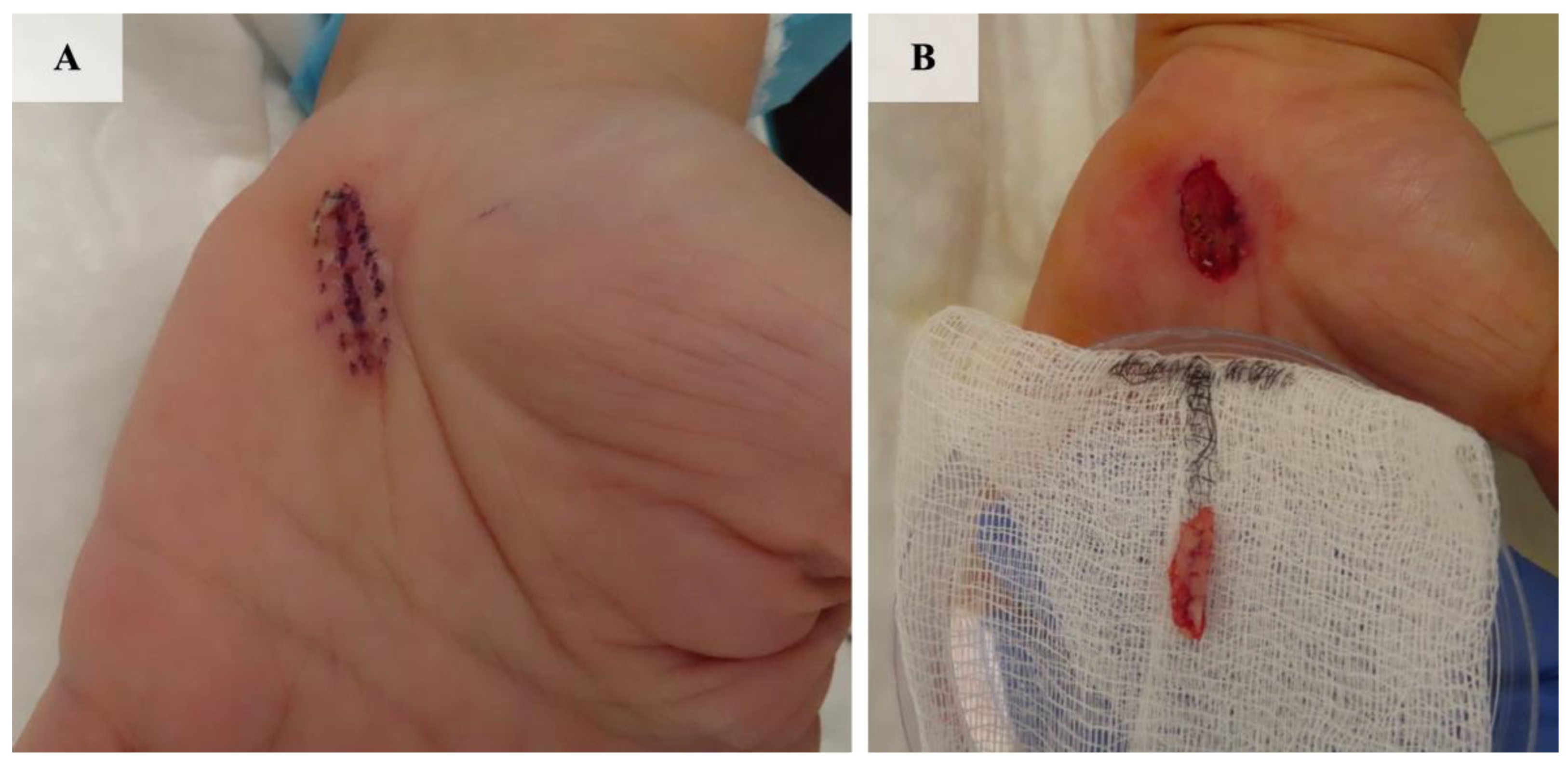
2.1. Patient 1
2.2. Patient 2
3. Discussion
4. Conclusions
Author Contributions
Funding
Institutional Review Board Statement
Informed Consent Statement
Data Availability Statement
Conflicts of Interest
References
- Mortada, H.; Aldihan, R.; Alhindi, N.; Abu Alqam, R.; Alnaim, M.F.; Kattan, A.E. Basal cell carcinoma of the hand: A systematic review and meta-analysis of incidence of recurrence. JPRAS Open 2023, 35, 42–57. [Google Scholar] [CrossRef] [PubMed]
- Loh, T.Y.; Rubin, A.G.; Brian Jiang, S.I. Basal Cell Carcinoma of the Dorsal Hand: An Update and Comprehensive Review of the Literature. Dermatol. Surg. 2016, 42, 464–470. [Google Scholar] [CrossRef] [PubMed]
- Barone, H.; Schaeffer, M.; Buckland, M.; LaFond, A.A.; Krach, K. Squamous Cell Carcinoma in Situ of the Nail Unit: Current Evidence and Recommendations for Patient Centered Treatment. J. Cutan. Med. Surg. 2023, 27, 51–59. [Google Scholar] [CrossRef] [PubMed]
- Seo, J.; Oh, Y.; Kim, S.K.; Roh, M.R.; Chung, K.Y. Slow Mohs Micrographic Surgery for Acral Melanoma Treatment in Korean Patients. Dermatol. Surg. 2021, 47, e42–e46. [Google Scholar] [CrossRef] [PubMed]
- Tang, G.T.; Elakis, J.; Scardamaglia, L. Cutaneous manifestations and treatment of arsenic toxicity: A systematic review. Ski. Health Dis. 2023, 3, e231. [Google Scholar] [CrossRef] [PubMed]
- Salerni, G.; Cecilia, N.; Cabrini, F.; Kolm, I.; Carrera, C.; Alos, L.; Malvehy, J.; Puig, S. Plantar basal cell carcinoma in a patient with xeroderma pigmentosum: Importance of dermoscopy for early diagnosis of nonpigmented skin cancer. Br. J. Dermatol. 2011, 165, 1143–1145. [Google Scholar] [CrossRef] [PubMed]
- Ee, H.L.; Tan, S.H.; Kumarasinghe, S.P. Plantar basal cell carcinoma: A possible eccrine origin. Clin. Exp. Dermatol. 2004, 29, 678–679. [Google Scholar] [CrossRef] [PubMed]
- Cabo, H.; Kolm, I.; Puig, S.; Malvehy, J. Palmar basal cell carcinoma in a patient with Gorlin-Goltz syndrome. Arch. Dermatol. 2007, 143, 813–814. [Google Scholar] [CrossRef] [PubMed]
- Sławińska, M.; Sikorska, M.; Biernat, W.; Nowicki, R.; Sobjanek, M. Significance of dermoscopy in early detection of acral basal cell carcinoma in patients with Gorlin-Goltz syndrome. Dermatol. Rev. Prz. Dermatol. 2018, 105, 87–90. [Google Scholar] [CrossRef]
- Hone, N.L.; Grandhi, R.; Ingraffea, A.A. Basal Cell Carcinoma on the Sole: An Easily Missed Cancer. Case Rep. Dermatol. 2016, 8, 283–286. [Google Scholar] [CrossRef] [PubMed]
- Alonso-Corral, M.J.; Gómez-Avivar, M.P.; Berenguel-Ibañez, M.M.; Ruiz-Villaverde, R. Palmar Basal Cell Carcinoma: An Unusual Site? Actas Dermo-Sifiliográficas (Engl. Ed.) 2014, 105, 623–624. [Google Scholar] [CrossRef]
- Peris, K.; Fargnoli, M.C.; Garbe, C.; Kaufmann, R.; Bastholt, L.; Seguin, N.B.; Bataille, V.; Marmol, V.D.; Dummer, R.; Harwood, C.A.; et al. Diagnosis and treatment of basal cell carcinoma: European consensus-based interdisciplinary guidelines. Eur. J. Cancer 2019, 118, 10–34. [Google Scholar] [CrossRef] [PubMed]
- Paoli, J.; Cogrel, O.; Geer, S.v.d.; Krekels, G.A.M.; Leeuw, J.d.; Moehrle, M.; Ostertag, J.U.; Buceta, L.R.; Sheth, N.; Läuchli, S.; et al. ESMS Position Document on the Use of Mohs Micrographic Surgery and Other Micrographic Surgery Techniques in Europe. 2019. Available online: https://esms-mohs.eu/fileadmin/user_upload/ESMS_Society_web/Resources_PDF/ESMS_Position_Paper_-_WEB.pdf (accessed on 1 October 2024).
- Nazzaro, G.; Benzecry, V.; Mattioli, M.A.; Denaro, N.; Beltramini, G.A.; Marzano, A.V.; Passoni, E. Sonidegib in Locally Advanced Basal Cell Carcinoma: A Monocentric Retrospective Experience and a Review of Published Real-Life Data. Cancers 2023, 15, 3621. [Google Scholar] [CrossRef] [PubMed]
- Dessinioti, C.; Stratigos, A.J. Immunotherapy and Its Timing in Advanced Basal Cell Carcinoma Treatment. Dermatol. Pract. Concept. 2023, 13, e2023252. [Google Scholar] [CrossRef] [PubMed]
- Mleczko, A.; Franke, I.; Pokrywka, A.; Gollnick, H.; Leverkus, M. BerEP4-negative basal cell carcinoma on the palm: Case report and review of the literature. J. Dtsch. Dermatol. Ges. 2011, 9, 140–143. [Google Scholar] [CrossRef] [PubMed]
- Natarelli, N.; Hanlon, K.; Chen, W.S.; Grichnik, J.M.; Zager, J.S.; Correa-Selm, L. Reflectance confocal microscopic visualization of melanocytic bodies in the stratum corneum overlying acral lentiginous melanoma. Lasers Surg. Med. 2023, 55, 253–256. [Google Scholar] [CrossRef] [PubMed]


Disclaimer/Publisher’s Note: The statements, opinions and data contained in all publications are solely those of the individual author(s) and contributor(s) and not of MDPI and/or the editor(s). MDPI and/or the editor(s) disclaim responsibility for any injury to people or property resulting from any ideas, methods, instructions or products referred to in the content. |
© 2024 by the authors. Licensee MDPI, Basel, Switzerland. This article is an open access article distributed under the terms and conditions of the Creative Commons Attribution (CC BY) license (https://creativecommons.org/licenses/by/4.0/).
Share and Cite
Żółkiewicz, J.; Banciu, L.; Sławińska, M.; Frumosu, M.; Tebeică, T.; Sobjanek, M.; Leventer, M. The Application of Mohs Micrographic Surgery in the Treatment of Acral Basal Cell Carcinoma: A Report of Two Cases. J. Clin. Med. 2024, 13, 6643. https://doi.org/10.3390/jcm13226643
Żółkiewicz J, Banciu L, Sławińska M, Frumosu M, Tebeică T, Sobjanek M, Leventer M. The Application of Mohs Micrographic Surgery in the Treatment of Acral Basal Cell Carcinoma: A Report of Two Cases. Journal of Clinical Medicine. 2024; 13(22):6643. https://doi.org/10.3390/jcm13226643
Chicago/Turabian StyleŻółkiewicz, Jakub, Laura Banciu, Martyna Sławińska, Mariana Frumosu, Tiberiu Tebeică, Michał Sobjanek, and Mihaela Leventer. 2024. "The Application of Mohs Micrographic Surgery in the Treatment of Acral Basal Cell Carcinoma: A Report of Two Cases" Journal of Clinical Medicine 13, no. 22: 6643. https://doi.org/10.3390/jcm13226643
APA StyleŻółkiewicz, J., Banciu, L., Sławińska, M., Frumosu, M., Tebeică, T., Sobjanek, M., & Leventer, M. (2024). The Application of Mohs Micrographic Surgery in the Treatment of Acral Basal Cell Carcinoma: A Report of Two Cases. Journal of Clinical Medicine, 13(22), 6643. https://doi.org/10.3390/jcm13226643