
Neurocognitive Changes in Patients with Post-COVID Depression
 , , , ,
Abstract
:1. Introduction
2. Materials and Methods
2.1. Study Participants and Clinical Assessment
2.2. Questionnaire to Assess Acute and Post-COVID Symptoms
2.3. Neuropsychological Assessment
2.3.1. Montreal Cognitive Assessment (MoCA)
2.3.2. Olfactory Testing
2.3.3. Stroop Color Word Test
2.3.4. Word Memory Test
2.3.5. Trail Making Test
2.4. Statistical Analysis
- (1)
- categorical predictors—sex (2 levels: male, female), diagnosed depression (2 levels: yes, no), and COVID-19 severity (2 levels: 1—mild, 2—moderate/severe/critical);
- (2)
- continuous predictors included years of education, age, time after the first and last COVID-19 infection, and number of acute and post-COVID symptoms.
3. Results
3.1. Clinical Assessment of the Patients with Post-COVID Depression
3.2. Acute and Post-COVID Symptoms
3.3. Results of Neuropsychological Testing
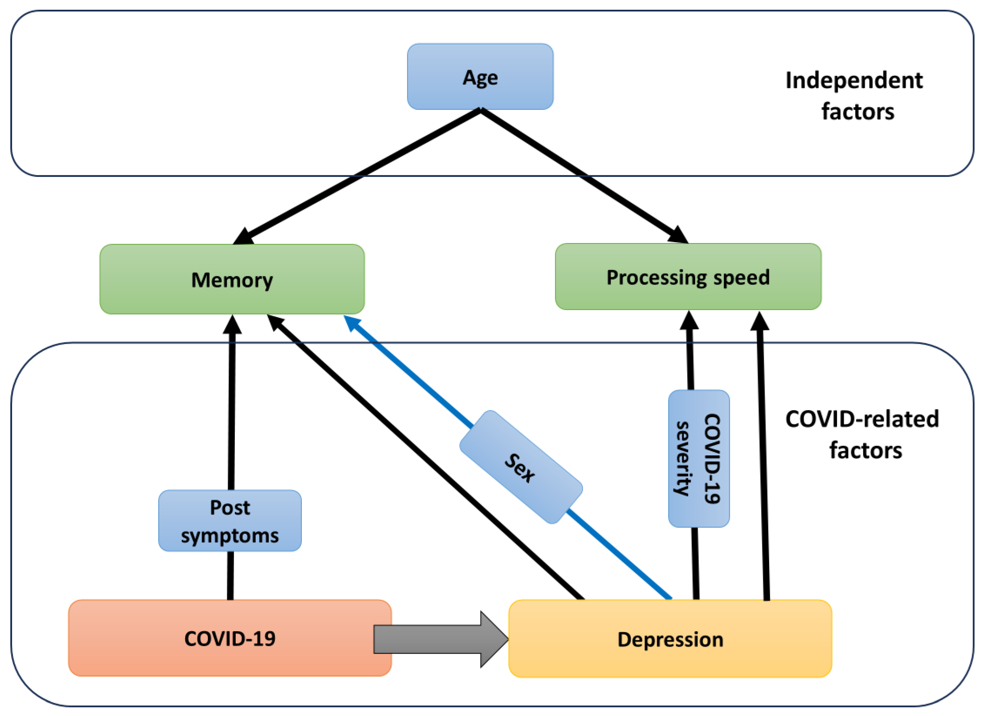
3.4. Associations between COVID-19-Related Parameters, Depression, and Neuropsychological Testing
4. Discussion
4.1. COVID-19 as the Cause of Depression and Cognitive Impairments
4.2. Comparison of Cognitive Changes in PCD Patients with Literature Data on MDD Patients
4.3. Gender Differences in Cognitive Performance
4.4. Possible Physiological Basis of COVID-19-Related Cognitive Changes in PCD Patients
5. Conclusions
6. Study Limitations
Author Contributions
Funding
Institutional Review Board Statement
Informed Consent Statement
Data Availability Statement
Conflicts of Interest
References
- Soriano, J.B.; Murthy, S.; Marshall, J.C.; Relan, P.; Diaz, J.V. Clinical Case Definition of Post-COVID-19 Condition by a Delphi Consensus. Lancet Infect. Dis. 2022, 22, e102–e107. [Google Scholar] [CrossRef] [PubMed]
- Ceban, F.; Ling, S.; Lui, L.M.W.; Lee, Y.; Gill, H.; Teopiz, K.M.; Rodrigues, N.B.; Subramaniapillai, M.; Di, J.D.; Cao, B.; et al. Fatigue and Cognitive Impairment in Post-COVID-19 Syndrome: A Systematic Review and Meta-Analysis. Brain Behav. Immun. 2020, 101, 93–135. [Google Scholar] [CrossRef] [PubMed]
- Taquet, M.; Geddes, J.R.; Husain, M.; Luciano, S.; Harrison, P.J. 6-Month Neurological and Psychiatric Outcomes in 236 379 Survivors of COVID-19: A Retrospective Cohort Study Using Electronic Health Records. Lancet Psychiatry 2021, 8, 416–427. [Google Scholar] [CrossRef] [PubMed]
- Lauria, A.; Carfì, A.; Benvenuto, F.; Bramato, G.; Ciciarello, F.; Rocchi, S.; Rota, E.; Salerno, A.; Stella, L.; Tritto, M.; et al. Neuropsychological Measures of Post-COVID-19 Cognitive Status. Front. Psychol. 2023, 14, 1136667. [Google Scholar] [CrossRef] [PubMed]
- Simonetti, A.; Bernardi, E.; Margoni, S.; Catinari, A.; Restaino, A.; Ieritano, V.; Palazzetti, M.; Mastrantonio, F.; Janiri, D.; Tosato, M.; et al. Mixed Depression in the Post-COVID-19 Syndrome: Correlation between Excitatory Symptoms in Depression and Physical Burden after COVID-19. Brain Sci. 2023, 13, 688. [Google Scholar] [CrossRef] [PubMed]
- Poletti, S.; Palladini, M.; Mazza, M.G.; De Lorenzo, R.; Irene, B.; Sara, B.; Beatrice, B.; Ceciclio, B.; Stefania, C.; Valentina, C.; et al. Long-Term Consequences of COVID-19 on Cognitive Functioning up to 6 Months after Discharge: Role of Depression and Impact on Quality of Life. Eur. Arch. Psychiatry Clin. Neurosci. 2022, 272, 773–782. [Google Scholar] [CrossRef] [PubMed]
- Brown, L.A.; Ballentine, E.; Zhu, Y.; McGinley, E.L.; Pezzin, L.; Abramoff, B. The Unique Contribution of Depression to Cognitive Impairment in Post-Acute Sequelae of SARS-CoV-2 Infection. Brain Behav. Immun.-Health 2022, 22, 100460. [Google Scholar] [CrossRef] [PubMed]
- Miskowiak, K.W.; Fugledalen, L.; Jespersen, A.E.; Sattler, S.M.; Podlekareva, D.; Rungby, J.; Porsberg, C.M.; Johnsen, S. Trajectory of Cognitive Impairments over 1 Year after COVID-19 Hospitalisation: Pattern, Severity, and Functional Implications. Eur. Neuropsychopharmacol. 2022, 59, 82–92. [Google Scholar] [CrossRef]
- Cheetham, N.J.; Penfold, R.; Giunchiglia, V.; Bowyer, V.; Sudre, C.H.; Canas, L.S.; Deng, J.; Murray, B.; Kerfoot, E.; Antonelli, M.; et al. The Effects of COVID-19 on Cognitive Performance in a Community-Based Cohort: A COVID Symptom Study Biobank Prospective Cohort Study. eClinicalMedicine 2023, 62, 102086. [Google Scholar] [CrossRef]
- Pistarini, C.; Fiabane, E.; Houdayer, E.; Vassallo, C.; Manera, M.R.; Alemanno, F. Cognitive and Emotional Disturbances Due to COVID-19: An Exploratory Study in the Rehabilitation Setting. Front. Neurol. 2021, 12, 643646. [Google Scholar] [CrossRef]
- Carola, V.; Vincenzo, C.; Morale, C.; Pelli, M.; Rocco, M.; Nicolais, G. Psychological Health in COVID-19 Patients after Discharge from an Intensive Care Unit. Front. Public Health 2022, 10, 951136. [Google Scholar] [CrossRef]
- Sobrino-Relaño, S.; Balboa-Bandeira, Y.; Peña, J.; Ibarretxe-Bilbao, N.; Zubiaurre-Elorza, L.; Ojeda, N. Neuropsychological Deficits in Patients with Persistent COVID-19 Symptoms: A Systematic Review and Meta-Analysis. Sci. Rep. 2023, 13, 10309. [Google Scholar] [CrossRef] [PubMed]
- Ma, Y.; Li, W.; Deng, H.; Wang, L.; Wang, Y.; Wang, P. Prevalence of Depression and Its Association with Quality of Life in Clinically Stable Patients with COVID-19. J. Affect. Disord. 2020, 275, 145–148. [Google Scholar] [CrossRef]
- Renaud-Charest, O.; Lui, L.M.W.; Eskander, S.; Ceban, F.; Ho, R.; Di Vincenzo, J.D.; Rosenblat, J.D.; Lee, Y.; Subramaniapillai, M.; McIntyre, R.S. Onset and Frequency of Depression in Post-COVID-19 Syndrome: A Systematic Review. J. Psychiatr. Res. 2021, 144, 129–137. [Google Scholar] [CrossRef] [PubMed]
- Herman, B.; Bruni, A.; Zain, E.; Dzulhadj, A.; Oo, A.C.; Viwattanakulvanid. Post-COVID Depression and Its Multiple Factors, Does Favipiravir Have a Protective Effect? A Longitudinal Study of Indonesia COVID-19 Patients. PLoS ONE 2022, 17, e0279184. [Google Scholar] [CrossRef]
- Pilotto, A.; Cristillo, V.; Cotti Piccinelli, S.; Zoppi, N.; Bonzi, G.; Sattin, D.; Schiavolin, S.; Raggi, A.; Canale, A.; Gipponi, S.; et al. Long-Term Neurological Manifestations of COVID-19: Prevalence and Predictive Factors. Neurol. Sci. 2021, 42, 4903–4907. [Google Scholar] [CrossRef]
- Gözen, E.D.; Aliyeva, C.; Tevetoğlu, F.; Karaali, R.; Balkan, İ.İ.; Yener, H.M.; Özdoğan, H.A. Evaluation of Olfactory Function With Objective Tests in COVID-19-Positive Patients: A Cross-Sectional Study. Ear Nose Throat J. 2021, 100, 169S–173S. [Google Scholar] [CrossRef]
- Biederman, J.; Petty, C.R.; Fried, R.; Black, S.; Faneuil, A.; Doyle, A.E.; Seidman, L.J.; Faraone, S.V. Discordance between Psychometric Testing and Questionnaire-Based Definitions of Executive Function Deficits in Individuals with ADHD. J. Atten. Disord. 2008, 12, 92–102. [Google Scholar] [CrossRef] [PubMed]
- Zigmond, A.S.; Snalth, R.P. The Hospital Anxiety and Depression Scale. Acta Psychiatr. Scand. 1983, 67, 361–370. [Google Scholar] [CrossRef]
- Hamilton, M. A Rating Scale for Depression. J. Neurol. Neurosurg. Psychiatry 1960, 23, 56–62. [Google Scholar] [CrossRef]
- Hamilton, M. Development of a Rating Scale for Depressive Illness. Br. J. Soc. Clin. Psychol. 1967, 6, 278–296. [Google Scholar] [CrossRef]
- Chan Sui Ko, A.; Candellier, A.; Mercier, M.; Joseph, C.; Schmit, J.L.; Lanoix, J.P.; Andrejak, C. Number of Initial Symptoms Is More Related to Long COVID-19 than Acute Severity of Infection: A Prospective Cohort of Hospitalized Patients. Int. J. Infect. Dis. 2022, 118, 220–223. [Google Scholar] [CrossRef] [PubMed]
- Fernández-de-las-Peñas, C.; Pellicer-Valero, O.J.; Navarro-Pardo, E.; Rodríguez-Jiménez, J.; Martín-Guerrero, J.D.; Cigarán-Méndezd, M. The Number of Symptoms at the Acute COVID-19 Phase Is Associated with Anxiety and Depressive Long-Term Post-COVID Symptoms: A Multicenter Study. J. Psychosom. Res. 2021, 150, 110625. [Google Scholar] [CrossRef] [PubMed]
- Durstenfeld, M.S.; Peluso, M.J.; Peyser, N.D.; Lin, F.; Knight, S.J.; Djibo, A.; Khatib, R.; Kitzman, H.; O’Brien, E.; Williams, N.; et al. Factors Associated with Long COVID Symptoms in an Online Cohort Study. Open Forum Infect. Dis. 2023, 10, ofad047. [Google Scholar] [CrossRef]
- Asadi-Pooya, A.A.; Akbari, A.; Emami, A.; Lotfi, M.; Rostamihosseinkhani, M.; Nemati, H.; Barzegar, Z.; Kabiri, M.; Zeraatpisheh, Z.; Farjoud-Kouhanjani, M.; et al. Risk Factors Associated with Long Covid Syndrome: A Retrospective Study. Iran. J. Med. Sci. 2021, 46, 428–436. [Google Scholar] [CrossRef] [PubMed]
- Nasreddine, Z.S.; Phillips, N.A.; Bedirian, V.; Charbonneau, S.; Whitehead, V.; Collin, I.; Cummings, J.L.; Chertkow, H. The Montreal Cognitive Assessment, MoCA: A Brief Screening Tool For Mild Cognitive Impairment. J. Am. Geriatr. Soc. 2005, 53, 695–699. [Google Scholar] [CrossRef]
- Green, P.; Montijo, J.; Brockhaus, R. High Specificity of the Word Memory Test and Medical Symptom Validity Test in Groups with Severe Verbal Memory Impairment. Appl. Neuropsychol. 2011, 18, 86–94. [Google Scholar] [CrossRef]
- Allen, M.; Bigler, E.; Larsen, J.; Goodrich-Hunsaker, N.; Hopkins, R. Functional Neuroimaging Evidence for High Cognitive Effort on the Word Memory Test in the Absence of External Incentives. Brain Inj. 2007, 21, 1425–1428. [Google Scholar] [CrossRef]
- Reitan, R.M. Validity of the Trail Making Test as an Indicator of Organic Brain Damage. Percept Mot Ski. 1958, 8, 271–276. [Google Scholar] [CrossRef]
- Stroop, J.R. Studies of Interference in Serial Verbal Reactions. J. Exp. Psychol. 1935, 18, 643–662. [Google Scholar] [CrossRef]
- Huang, C.L.-C. The Value of Patient-Administered Depression Rating Scale in Detecting Cognitive Deficits in Depressed Patients. J. Clin. Med. Res. 2010, 2, 27–33. [Google Scholar] [CrossRef]
- Freud, T.; Vostrikov, A.; Dwolatzky, T.; Punchik, B.; Press, Y. Validation of the Russian Version of the MoCA Test as a Cognitive Screening Instrument in Cognitively Asymptomatic Older Individuals and Those With Mild Cognitive Impairment. Front. Med. 2020, 7, 447. [Google Scholar] [CrossRef]
- Mahendran, R.; Chua, J.; Feng, L.; Kua, E.H.; Preedy, V.R. The Mini-Mental State Examination and Other Neuropsychological Assessment Tools for Detecting Cognitive Decline; Elsevier Inc.: Amsterdam, The Netherlands, 2015; ISBN 9780124079397. [Google Scholar]
- Srisurapanont, M.; Eurviriyanukul, K.; Suttajit, S.; Varnado, P. Internal Consistency and Concurrent Validity of the Montreal Cognitive Assessment in Individuals with Major Depressive Disorder. Psychiatry Res. 2017, 253, 333–337. [Google Scholar] [CrossRef]
- Hummel, T.; Konnerth, C.G.; Rosenheim, K.; Kobal, G. Screening of Olfactory Function with a Four-Minute Odor Identification Test: Reliability, Normative Data, and Investigations in Patients with Olfactory Loss. Ann. Otol. Rhinol. Laryngol. 2001, 110, 976–981. [Google Scholar] [CrossRef] [PubMed]
- Rumeau, C.; Nguyen, D.T.; Jankowski, R. How to Assess Olfactory Performance with the Sniffin’ Sticks Test®. Eur. Ann. Otorhinolaryngol. Head Neck Dis. 2016, 133, 203–206. [Google Scholar] [CrossRef] [PubMed]
- Schepens, E.J.A.; Stegeman, I.; Kamalski, D.M.A. Diagnostic Accuracy of the Screenings Sniffin’ Sticks Test (SST-12) in COVID-19 Induced Olfactory Disorders. PLoS ONE 2024, 19, e0295911. [Google Scholar] [CrossRef] [PubMed]
- Cousijn, J.; van Benthem, P.; van der Schee, E.; Spijkerman, R. Motivational and Control Mechanisms Underlying Adolescent Cannabis Use Disorders: A Prospective Study. Dev. Cogn. Neurosci. 2015, 16, 36–45. [Google Scholar] [CrossRef] [PubMed]
- Troyer, A.K.; Leach, L.; Strauss, E. Aging and Response Inhibition: Normative Data for the Victoria Stroop Test. Aging, Neuropsychol. Cogn. 2006, 13, 20–35. [Google Scholar] [CrossRef] [PubMed]
- Tremblay, M.-P.; Potvin, O.; Belleville, S.; Bier, N.; Gagnon, L.; Blanchet, S.; Domingues, N.-S.; Gaudreau, G.; Macoir, J.; Hudon, C. The Victoria Stroop Test: Normative Data in Quebec-French Adults and Elderly. Arch. Clin. Neuropsychol. 2016, 31, acw029. [Google Scholar] [CrossRef] [PubMed]
- Tydecks, S.; Merten, T.; Gubbay, J. The Word Memory Test and the One-in-Five-Test in an Analogue Study with Russian Speaking Participants. Int. J. Forensic Psychol. 2006, 1, 29–37. [Google Scholar]
- Wagner, S.; Helmreich, I.; Dahmen, N.; Lieb, K.; Tadi, A. Reliability of Three Alternate Forms of the Trail Making Tests A and B. Arch. Clin. Neuropsychol. 2011, 26, 314–321. [Google Scholar] [CrossRef]
- Ramani, C.; Davis, E.M.; Kim, J.S.; Provencio, J.J.; Enfield, K.B.; Kadl, A. Post-ICU COVID-19 Outcomes: A Case Series. Chest 2021, 159, 215–218. [Google Scholar] [CrossRef]
- Huang, S.; Zhou, X.; Zhao, W.; Du, Y.; Yang, D.; Huang, Y.; Chen, Y.; Zhang, H.; Yang, G.; Liu, J.; et al. Dynamic White Matter Changes in Recovered COVID-19 Patients: A Two-Year Follow-up Study. Theranostics 2023, 13, 724–735. [Google Scholar] [CrossRef] [PubMed]
- Huang, Y.; Ling, Q.; Manyande, A.; Wu, D.; Xiang, B. Brain Imaging Changes in Patients Recovered From COVID-19: A Narrative Review. Front. Neurosci. 2022, 16, 855868. [Google Scholar] [CrossRef] [PubMed]
- Pinnock, F.S.; Rich, J.B.; Vasquez, B.; Wiegand, M.; Patcai, J.; Troyer, A.K.; Murphy, K.J. Neurocognitive Outcome Following Recovery from Severe Acute Respiratory Syndrome—Coronavirus-1 (SARS-CoV-1). J. Int. Neuropsychol. Soc. 2022, 28, 891–901. [Google Scholar] [CrossRef]
- Brasso, C.; Cisotto, M.; Del Favero, E.; Giordano, B.; Villari, V.; Rocca, P. Impact of COVID-19 Pandemic on Major Depressive Disorder in Acute Psychiatric Inpatients. Front. Psychol. 2023, 14, 1181832. [Google Scholar] [CrossRef] [PubMed]
- Li, Z.; Ruan, M.; Chen, J.; Fang, Y. Major Depressive Disorder: Advances in Neuroscience Research and Translational Applications. Neurosci. Bull. 2021, 37, 863–880. [Google Scholar] [CrossRef]
- Hammar, Å.; Ronold, E.H.; Rekkedal, G.Å. Cognitive Impairment and Neurocognitive Profiles in Major Depression—A Clinical Perspective. Front. Psychiatry 2022, 13, 764374. [Google Scholar] [CrossRef]
- Zuckerman, H.; Pan, Z.; Park, C.; Brietzke, E.; Musial, N.; Shariq, A.S.; Iacobucci, M.; Yim, S.J.; Lui, L.M.W.; Rong, C.; et al. Recognition and Treatment of Cognitive Dysfunction in Major Depressive Disorder. Front. Psychiatry 2018, 9, 655. [Google Scholar] [CrossRef]
- Wen, M.; Dong, Z.; Zhang, L.; Li, B.; Zhang, Y.; Li, K. Depression and Cognitive Impairment: Current Understanding of Its Neurobiology and Diagnosis. Neuropsychiatr. Dis. Treat. 2022, 18, 2783–2794. [Google Scholar] [CrossRef]
- Culpepper, L.; Lam, R.W.; McIntyre, R.S. Cognitive Impairment in Patients with Depression: Awareness, Assessment, and Management. J. Clin. Psychiatry 2017, 78, 1383–1394. [Google Scholar] [CrossRef] [PubMed]
- Blair, M.; Coleman, K.; Jesso, S.; Desbeaumes Jodoin, V.; Smolewska, K.; Warriner, E.; Finger, E.; Pasternak, S.H. Depressive Symptoms Negatively Impact Montreal Cognitive Assessment Performance: A Memory Clinic Experience. Can. J. Neurol. Sci. 2016, 43, 513–517. [Google Scholar] [CrossRef] [PubMed]
- Wu, Z.; Su, G.; Lu, W.; Liu, L.; Zhou, Z.; Xie, B. Clinical Symptoms and Their Relationship with Cognitive Impairment in Elderly Patients with Depressive Disorder. Front. Psychiatry 2022, 13, 1009653. [Google Scholar] [CrossRef] [PubMed]
- Nyundo, A.A.; Ismail, A. The Influence of Major Depressive Disorders on Neurocognitive Function among Adults Living with HIV/AIDS in a Regional Referral Hospital in Dodoma, Tanzania. Trop. Med. Int. Health 2022, 27, 58–67. [Google Scholar] [CrossRef] [PubMed]
- Ab Latiff, H.Z.; Ariaratnam, S.; Shuib, N.; Isa, M.R. Cognitive Decline and Its Associated Factors in Patients with Major Depressive Disorder. Healthcare 2023, 11, 950. [Google Scholar] [CrossRef] [PubMed]
- Xu, G.; Lin, K.; Rao, D.; Dang, Y.; Ouyang, H.; Guo, Y.; Ma, J.; Chen, J. Neuropsychological Performance in Bipolar I, Bipolar II and Unipolar Depression Patients: A Longitudinal, Naturalistic Study. J. Affect. Disord. 2012, 136, 328–339. [Google Scholar] [CrossRef]
- Shimizu, Y.; Kitagawa, N.; Mitsui, N.; Fujii, Y.; Toyomaki, A.; Hashimoto, N.; Kako, Y.; Tanaka, T.; Asakura, S.; Kusumi, I. Neurocognitive Impairments and Quality of Life in Unemployed Patients with Remitted Major Depressive Disorder. Psychiatry Res. 2013, 210, 913–918. [Google Scholar] [CrossRef]
- Baune, B.T.; Miller, R.; McAfoose, J.; Johnson, M.; Quirk, F.; Mitchell, D. The Role of Cognitive Impairment in General Functioning in Major Depression. Psychiatry Res. 2010, 176, 183–189. [Google Scholar] [CrossRef]
- Jia, Q.F.; Chen, P.; Zhu, H.L.; Chen, S.S.; Gu, X.C.; Yin, X.Y.; Wu, Y.H.; Yin, G.Z.; Hui, L. Cognitive Impairments in First-Episode Drug-Naïve Versus Medicated Depressive Patients: RBANS in a Chinese Population. Psychiatr. Q. 2019, 90, 471–480. [Google Scholar] [CrossRef]
- Miskowiak, K.W.; Johnsen, S.; Sattler, S.M.; Nielsen, S.; Kunalan, K.; Rungby, J.; Lapperre, T.; Porsberg, C.M. Cognitive Impairments Four Months after COVID-19 Hospital Discharge: Pattern, Severity and Association with Illness Variables. Eur. Neuropsychopharmacol. 2021, 46, 39–48. [Google Scholar] [CrossRef]
- Huang, C.; Huang, L.; Wang, Y.; Li, X.; Ren, L.; Gu, X.; Kang, L.; Guo, L.; Liu, M.; Zhou, X.; et al. 6-Month Consequences of COVID-19 in Patients Discharged from Hospital: A Cohort Study. Lancet 2021, 397, 220–232. [Google Scholar] [CrossRef] [PubMed]
- Iqbal, F.M.; Lam, K.; Sounderajah, V.; Clarke, J.M.; Ashrafian, H.; Darzi, A. Characteristics and Predictors of Acute and Chronic Post-COVID Syndrome: A Systematic Review and Meta-Analysis. EClinicalMedicine 2021, 36, 100899. [Google Scholar] [CrossRef]
- Bahmer, T.; Borzikowsky, C.; Lieb, W.; Horn, A.; Krist, L.; Fricke, J.; Scheibenbogen, C.; Rabe, K.F.; Maetzler, W.; Maetzler, C.; et al. Severity, Predictors and Clinical Correlates of Post-COVID Syndrome (PCS) in Germany: A Prospective, Multi-Centre, Population-Based Cohort Study. EClinicalMedicine 2022, 51, 101549. [Google Scholar] [CrossRef]
- Epp, A.M.; Dobson, K.S.; Dozois, D.J.A.; Frewen, P.A. A Systematic Meta-Analysis of the Stroop Task in Depression. Clin. Psychol. Rev. 2012, 32, 316–328. [Google Scholar] [CrossRef] [PubMed]
- Miyake, A.; Friedman, N.P.; Emerson, M.J.; Witzki, A.H.; Howerter, A.; Wager, T.D. The Unity and Diversity of Executive Functions and Their Contributions to Complex “Frontal Lobe” Tasks: A Latent Variable Analysis. Cogn. Psychol. 2000, 41, 49–100. [Google Scholar] [CrossRef] [PubMed]
- Hammar, Å.; Isaksen, L.; Schmid, M.; Årdal, G.; Strand, M. Patients with Major Depression Show Intact Memory Performancegiven Optimal Conditions. Appl. Neuropsychol. 2011, 18, 191–196. [Google Scholar] [CrossRef]
- Zhao, L.; Han, G.; Zhao, Y.; Jin, Y.; Ge, T.; Yang, W.; Cui, R.; Xu, S.; Li, B. Gender Differences in Depression: Evidence From Genetics. Front. Genet. 2020, 11, 1145. [Google Scholar] [CrossRef]
- Labaka, A.; Goñi-Balentziaga, O.; Lebeña, A.; Pérez-Tejada, J. Biological Sex Differences in Depression: A Systematic Review. Biol. Res. Nurs. 2018, 20, 383–392. [Google Scholar] [CrossRef]
- Dong, H.S.; Han, C.; Jeon, S.W.; Yoon, S.; Jeong, H.G.; Huh, Y.J.; Pae, C.U.; Patkar, A.A.; Steffens, D.C. Characteristics of Neurocognitive Functions in Mild Cognitive Impairment with Depression. Int. Psychogeriatr. 2016, 28, 1181–1190. [Google Scholar] [CrossRef]
- Ji, Y.; Li, W.; Liu, B.; Liu, J.; Ju, Y.; Wang, M.; Chen, Y.; Li, L. Clinical Characteristics of Cognitive Deficits in Major Depressive Disorder: A 6-Montprospective Study. Rev. Psiquiatr. Clin. 2020, 47, 101–105. [Google Scholar] [CrossRef]
- Poyraz Çökmüş, F.; Murat Özkan, H.; Sücüllüoğlu-Dikici, D.; Aşçibaşi, K.; Alçi, D.; Altunsoy, N.; Kuru, E.; Yüzeren, S.; Aydemir, Ö. The Assessment of Cognitive Dysfunction in Major Depressive Disorder: A 16-Week Prospective Case-Control Study. Psychiatry Clin. Psychopharmacol. 2021, 31, 25–33. [Google Scholar] [CrossRef]
- Mao, R.; Wang, C.; Cui, L.; Mellor, D.; Wu, Z.; Fang, Y. Gender Differences in Prevalence and Associations between Cognitive Symptoms and Suicidal Ideation in Patients with Recurrent Major Depressive Disorder: Findings from the Chinese NSSD Study. BMC Psychiatry 2024, 24, 83. [Google Scholar] [CrossRef]
- Caldirola, D.; Sangiorgio, E.; Riva, A.; Grassi, M.; Alciati, A.; Scialò, C.; Perna, G. Does Gender Influence Cognitive Function in Non-Psychotic Depression? Pers. Med. Psychiatry 2017, 4–6, 25–31. [Google Scholar] [CrossRef]
- Taslem Mourosi, J.; Anwar, S.; Hosen, M.J. The Sex and Gender Dimensions of COVID-19: A Narrative Review of the Potential Underlying Factors. Infect. Genet. Evol. 2022, 103, 105338. [Google Scholar] [CrossRef] [PubMed]
- Mangion, K.; Morrow, A.J.; Sykes, R.; Kamdar, A.; Bagot, C.; Bruce, G.; Connelly, P.; Delles, C.; Gibson, V.B.; Gillespie, L.; et al. Post-COVID-19 Illness and Associations with Sex and Gender. BMC Cardiovasc. Disord. 2023, 23, 389. [Google Scholar] [CrossRef]
- Michelutti, M.; Furlanis, G.; Stella, A.B.; Bellavita, G.; Frezza, N.; Torresin, G.; Ajcevic, M.; Manganotti, P. Sex-Dependent Characteristics of Neuro-Long-COVID: Data from a Dedicated Neurology Ambulatory Service. J. Neurol. Sci. 2022, 441, 120355. [Google Scholar] [CrossRef]
- Sudre, C.H.; Murray, B.; Varsavsky, T.; Graham, M.S.; Penfold, R.S.; Bowyer, R.C.; Pujol, J.C.; Klaser, K.; Antonelli, M.; Canas, L.S.; et al. Attributes and Predictors of Long COVID. Nat. Med. 2021, 27, 626–631. [Google Scholar] [CrossRef]
- Hartung, T.J.; Neumann, C.; Bahmer, T.; Chaplinskaya-sobol, I.; Endres, M.; Geritz, J.; Krist, L.; Kudelka, J.; Lieb, W.; Maetzler, C.; et al. Fatigue and Cognitive Impairment after COVID-19: A Prospective Multicentre Study. eClinicalMedicine 2022, 53, 101651. [Google Scholar] [CrossRef]
- Stewart, S.; Newson, L.; Briggs, T.A.; Grammatopoulos, D.; Young, L.; Gill, P. Long COVID Risk—A Signal to Address Sex Hormones and Women’s Health. Lancet Reg. Health-Eur. 2021, 11, 100242. [Google Scholar] [CrossRef] [PubMed]
- Ortona, E.; Malorni, W. Long COVID: To Investigate Immunological Mechanisms and Sex/Gender Related Aspects as Fundamental Steps for a Tailored Therapy. Eur. Respir. J. 2022, 59, 2102245. [Google Scholar] [CrossRef] [PubMed]
- Qi, S.; Ngwa, C.; Morales Scheihing, D.A.; Al Mamun, A.; Ahnstedt, H.W.; Finger, C.E.; Colpo, G.D.; Sharmeen, R.; Kim, Y.; Choi, H.M.A.; et al. Sex Differences in the Immune Response to Acute COVID-19 Respiratory Tract Infection. Biol. Sex Differ. 2021, 12, 66. [Google Scholar] [CrossRef]
- Braga, J.; Lepra, M.; Kish, S.J.; Rusjan, P.M.; Nasser, Z.; Verhoeff, N.; Vasdev, N.; Bagby, M.; Boileau, I.; Husain, M.I.; et al. Neuroinflammation after COVID-19 with Persistent Depressive and Cognitive Symptoms. JAMA Psychiatry 2023, 80, 787–795. [Google Scholar] [CrossRef] [PubMed]
- Sriwastava, S.; Tandon, M.; Podury, S.; Prasad, A.; Wen, S.; Guthrie, G.; Kakara, M.; Jaiswal, S.; Subedi, R.; Elkhooly, M.; et al. COVID-19 and Neuroinflammation: A Literature Review of Relevant Neuroimaging and CSF Markers in Central Nervous System Inflammatory Disorders from SARS-CoV2. J. Neurol. 2021, 268, 4448–4478. [Google Scholar] [CrossRef] [PubMed]
- Huang, S.; Zhou, Z.; Yang, D.; Zhao, W.; Zeng, M.; Xie, X.; Du, Y.; Jiang, Y.; Zhou, X.; Yang, W.; et al. Persistent White Matter Changes in Recovered COVID-19 Patients at the 1-Year Follow-Up. Brain 2022, 145, 1830–1838. [Google Scholar] [CrossRef] [PubMed]
- de Carvalho Bispo, D.D.; de Paula Brandão, P.R.; Pereira, D.A.; Maluf, F.B.; Dias, B.A.; Paranhos, H.R.; von Glehn, F.; de Oliveira, A.C.P.; Regattieri, N.A.T.; Silva, L.S.; et al. Brain Microstructural Changes and Fatigue after COVID-19. Front. Neurol. 2022, 13, 1029302. [Google Scholar] [CrossRef] [PubMed]
- Benedetti, F.; Palladini, M.; Paolini, M.; Melloni, E.; Vai, B.; De Lorenzo, R.; Furlan, R.; Rovere-Querini, P.; Falini, A.; Mazza, M.G. Brain Correlates of Depression, Post-Traumatic Distress, and Inflammatory Biomarkers in COVID-19 Survivors: A Multimodal Magnetic Resonance Imaging Study. Brain Behav. Immun.-Health 2021, 18, 100387. [Google Scholar] [CrossRef] [PubMed]
- Qin, Y.; Wu, J.; Chen, T.; Li, J.; Zhang, G.; Wu, D.; Zhou, Y.; Zheng, N.; Cai, A.; Ning, Q.; et al. Long-Term Microstructure and Cerebral Blood Flow Changes in Patients Recovered from COVID-19 without Neurological Manifestations. J. Clin. Investig. 2021, 131, e147329. [Google Scholar] [CrossRef] [PubMed]
- Tian, T.; Wu, J.; Chen, T.; Li, J.; Yan, S.; Zhou, Y.; Peng, X.; Li, Y.; Zheng, N.; Cai, A.; et al. Long-Term Follow-up of Dynamic Brain Changes in Patients Recovered from COVID-19 without Neurological Manifestations. JCI Insight 2022, 7, e155827. [Google Scholar] [CrossRef]
- Boito, D.; Eklund, A.; Tisell, A.; Levi, R.; Özarslan, E.; Blystad, I. MRI with Generalized Diffusion Encoding Reveals Damaged White Matter in Patients Previously Hospitalized for COVID-19 and with Persisting Symptoms at Follow-Up. Brain Commun. 2023, 5, fcad284. [Google Scholar] [CrossRef]
- Butt, A.M.; Papanikolaou, M.; Rivera, A. Physiology of Oligodendroglia. In Neuroglia in Neurodegenerative Diseases; Advances in Experimental Medicine and Biology; Springer: Singapore, 2019; Volume 1175, pp. 117–128. ISBN 10.1007/9789811. [Google Scholar]
- Bradl, M.; Lassmann, H. Oligodendrocytes: Biology and Pathology. Acta Neuropathol. 2010, 119, 37–53. [Google Scholar] [CrossRef]
- Chapman, T.W.; Hill, R.A. Myelin Plasticity in Adulthood and Aging. Neurosci. Lett. 2020, 715, 134645. [Google Scholar] [CrossRef]
- Khodanovich, M.; Svetlik, M.; Kamaeva, D.; Usova, A.; Kudabaeva, M. Demyelination in Patients with Post-COVID Depression. Biomedicines 2023, 1–23, preprint. [Google Scholar] [CrossRef]
- Kisel, A.A.; Naumova, A.V.; Yarnykh, V.L. Macromolecular Proton Fraction as a Myelin Biomarker: Principles, Validation, and Applications. Front. Neurosci. 2022, 16, 819912. [Google Scholar] [CrossRef]
- Yarnykh, V.L. Time-Efficient, High-Resolution, Whole Brain Three-Dimensional Macromolecular Proton Fraction Mapping. Magn. Reson. Med. 2016, 75, 2100–2106. [Google Scholar] [CrossRef]
- Yarnykh, V.L. Fast Macromolecular Proton Fraction Mapping from a Single Off-Resonance Magnetization Transfer Measurement. Magn. Reson. Med. 2012, 68, 166–178. [Google Scholar] [CrossRef]
- Khodanovich, M.Y.Y.; Sorokina, I.V.V.; Glazacheva, V.Y.Y.; Akulov, A.E.E.; Nemirovich-Danchenko, N.M.M.; Romashchenko, A.V.V.; Tolstikova, T.G.G.; Mustafina, L.R.R.; Yarnykh, V.L.L. Histological Validation of Fast Macromolecular Proton Fraction Mapping as a Quantitative Myelin Imaging Method in the Cuprizone Demyelination Model. Sci. Rep. 2017, 7, 46686. [Google Scholar] [CrossRef] [PubMed]
- Khodanovich, M.Y.; Pishchelko, A.O.; Glazacheva, V.Y.; Pan, E.S.; Akulov, A.E.; Svetlik, M.V.; Tyumentseva, Y.A.; Anan’ina, T.V.; Yarnykh, V. Quantitative Imaging of White and Gray Matter Remyelination in the Cuprizone Demyelination Model Using the Macromolecular Proton Fraction. Cells 2019, 8, 1204. [Google Scholar] [CrossRef] [PubMed]
- Khodanovich, M.Y.; Gubskiy, I.L.; Kudabaeva, M.S.; Namestnikova, D.D.; Kisel, A.A.; Anan’ina, T.V.; Tumentceva, Y.A.; Mustafina, L.R.; Yarnykh, V.L. Long-Term Monitoring of Chronic Demyelination and Remyelination in a Rat Ischemic Stroke Model Using Macromolecular Proton Fraction Mapping. J. Cereb. Blood Flow Metab. 2021, 41, 2856–2869. [Google Scholar] [CrossRef] [PubMed]
- Khodanovich, M.Y.; Kisel, A.A.; Akulov, A.E.; Atochin, D.N.; Kudabaeva, M.S.; Glazacheva, V.Y.; Svetlik, M.V.; Medvednikova, Y.A.; Mustafina, L.R.; Yarnykh, V.L. Quantitative Assessment of Demyelination in Ischemic Stroke in Vivo Using Macromolecular Proton Fraction Mapping. J. Cereb. Blood Flow Metab. 2018, 38, 919–931. [Google Scholar] [CrossRef] [PubMed]
- Caverzasi, E.; Papinutto, N.; Amirbekian, B.; Berger, M.S.; Henry, R.G. Q-Ball of Inferior Fronto-Occipital Fasciculus and Beyond. PLoS ONE 2014, 9, e100274. [Google Scholar] [CrossRef]
- Conner, A.K.; Briggs, R.G.; Sali, G.; Rahimi, M.; Baker, C.M.; Burks, J.D.; Glenn, C.A.; Battiste, J.D.; Sughrue, M.E. A Connectomic Atlas of the Human Cerebrum-Chapter 13: Tractographic Description of the Inferior Fronto-Occipital Fasciculus. Oper. Neurosurg. 2018, 15, 5436–5443. [Google Scholar] [CrossRef] [PubMed]
- Hausman, H.K.; Hardcastle, C.; Albizu, A.; Kraft, J.N.; Evangelista, N.D.; Boutzoukas, E.M.; Langer, K.; O’Shea, A.; Van Etten, E.J.; Bharadwaj, P.K.; et al. Cingulo-Opercular and Frontoparietal Control Network Connectivity and Executive Functioning in Older Adults. GeroScience 2022, 44, 847–866. [Google Scholar] [CrossRef] [PubMed]
- Vandersteen, C.; Payne, M.; Dumas, L.É.; Plonka, A.; D’Andréa, G.; Chirio, D.; Demonchy, É.; Risso, K.; Robert, P.; Fernandez, X.; et al. What about Using Sniffin’ Sticks 12 Items Test to Screen Post-COVID-19 Olfactory Disorders? Eur. Arch. Oto-Rhino-Laryngology 2022, 279, 3477–3484. [Google Scholar] [CrossRef] [PubMed]
- Bagnascoa, D.; Passalacquaa, G.; Braidoa, F.; Tagliabuea, E.; Cosinia, F.; Filaurob, M.; Ioppib, A.; Carobbiob, A.; Mocellinb, D.; Riccioa, A.M.; et al. Quick Olfactory Snif Fi n ’ Sticks Test ( Q-Sticks ) for the Detection of Smell Disorders in COVID- 19 Patients. World Allergy Organ. J. 2021, 14, 100497. [Google Scholar] [CrossRef]

| Parameter | PCD | NoPCD | ControlPC | Control |
|---|---|---|---|---|
| Sample size | 25 | 46 | 18 | 19 |
| Male (%) | 4 (16) | 17 (37) | 7 (39) | 8 (42) |
| Female (%) | 21 (84) | 29 (63) | 11 (61) | 11 (58) |
| Education, years ± SD | 15.2 ± 1.9 | 15.9 ± 2.1 | 16.1 ± 2.4 | 16.4 ± 1.8 |
| Age, years ± SD | 37 ± 13.7 | 43 ± 10.4 | 43.7 ± 9.7 | 38.3 ± 10.3 |
| Age, median (min–max) | 42.0 (19–59) | 43 (21–61) | 42 (24–61) | 39 (20–58) |
| Parameter | Mean ± SD |
|---|---|
| Hamilton score (HDRS) | 18.36 ± 3.66 |
| Age of manifestation, years | 34.62 ± 13.96 |
| Number of episodes | 1.75 ± 1.75 |
| Duration of last episode, months | 8.27 ± 7.31 |
| Parameter | PCD | noPCD | ControlPC | Statistics |
|---|---|---|---|---|
| Severity, mild/moderate/severe/critical (%) | 88/8/4/0 | 63/17/15/4 | 66/28/0/1 | |
| Number of COVID-19 episodes, mean ± SD | 1.60 ± 0.71 | 1.65 ± 0.77 | 1.50 ± 0.51 | F(2, 86) = 0.30, p = 0.74 |
| Time after the first COVID-19, months ± SD | 20.3 ± 8.2 | 21.8 ± 9.4 | 16.3 ± 6.4 | F(2, 86) = 2.6, p = 0.08 |
| Time after last COVID-19, months ± SD | 13.1 ± 10.3 | 15.0 ± 10.5 | 9.8 ± 5.5 | F(2, 86) = 1.8, p = 0.16 |
| Acute symptoms | ||||
| Anosmia/hyposmia, n (%) | 22(88%) | 34(74%) | 15(83%) | - |
| Ageusia/hypogeusia, n (%) | 19(76%) * | 27(59%) | 8(44%) | - |
| Fever, n (%) | 22(88%) | 44(96%) | 16(89%) | - |
| Difficulty breathing, n (%) | 14(56%) | 27(59%) | 7(39%) | - |
| Cough, n (%) | 22(88%) | 32(70%) | 13(72%) | - |
| Muscle weakness, n (%) | 24(96%) | 42(91%) | 15(83%) | - |
| Myalgia, n (%) | 20(80%) | 30(65%) | 10(56%) | - |
| Headache, n (%) | 22(88%) * | 34(74%) | 11(61%) | - |
| Dizziness, n (%) | 14(56%) | 28(61%)* | 6(33%) | - |
| Number of acute symptoms | 7.24 ± 1.85 * | 6.48 ± 2.21 | 5.61 ± 1.94 | F(2, 86) = 3.28, p = 0.042 |
| Post-COVID symptoms | ||||
| Headache, n (%) | 7 (28%) | 6(13%) | 2(11%) | - |
| Dizziness, n (%) | 10 (40%) * | 22(48%) ** | 2(11%) | - |
| Brain fog, n (%) | 14 (56%) | 19(41%) | 6(33%) | - |
| Anosmia/hyposmia, n (%) | 16 (64%) *& | 16(35%) | 5(28%) | - |
| Ageusia/hypogeusia, n (%) | 14 (56%) **& | 12(26%) | 3(17%) | - |
| Sensitivity, n (%) | 3 (12%) | 7(15%) | 1(6%) | - |
| Hypertensia/hypotensia, n (%) | 7 (28%) | 23(50%) * | 4(22%) | - |
| Insomnia, n (%) | 20 (80%) *** | 27(59%) *** | 5(28%) | - |
| Fatigue, n (%) | 24(96%) ***& | 36(78%) ** | 8(44%) | - |
| Attention deficit, n (%) | 23(92%) ***&& | 29(63%) *** | 4(22%) | - |
| Memory deficit,% | 19(76%) *** | 39(85%) | 4(22%) | - |
| Myalgia, n (%) | 15(60%) * | 25(54%) | 5(28%) | - |
| Depression 1, n (%) | 24(96%) ***&&& | 24(52%) ** | 2(11%) | - |
| Panic attacks, n (%) | 5(20%) * | 3(7%) | 0(0%) | - |
| Number of post-COVID symptoms | 8.04 ± 2.23 ***& | 6.26 ± 2.95 *** | 2.83 ± 3.24 | F(2, 86) = 17.95, p = 0.000 |
| Test | Parameter | Sex | PCD | noPCD | ControlPC | Control | Significance of Factors (Covariates), p-Value | |||||||
|---|---|---|---|---|---|---|---|---|---|---|---|---|---|---|
| Mean ± SD | p-Values, PCD vs. | Mean ± SD | p-Values, noPCD vs. | Mean ± SD | Mean ± SD | |||||||||
| noPCD | ControlPC | Control | ControlPC | Control | Age | Sex | Group | |||||||
| HADS | Total score | m | 22.75 ± 9.53 | 0.000 *** | 0.000 *** | 0.000 *** | 10.94 ± 6.24 | 0.42 | 0.40 | 9.29 ± 3.77 | 6.13 ± 2.10 | 0.22 | 0.98 | 0.000 *** |
| f | 20.71 ± 7.17 | 0.000 *** | 0.000 *** | 0.000 *** | 10.90 ± 4.92 | 0.50 | 0.05 | 7.82 ± 4.05 | 9.18 ± 3.77 | |||||
| Anxiety | m | 13.00 ± 3.91 | 0.000 *** | 0.000 *** | 0.000 *** | 5.71 ± 3.43 | 0.77 | 0.12 | 5.29 ± 4.23 | 3.50 ± 2.12 | 0.12 | 0.81 | 0.000 *** | |
| f | 10.43 ± 3.04 | 0.000 *** | 0.000 *** | 0.000 *** | 6.48 ± 4.04 | 0.09 | 0.24 | 4.45 ± 2.30 | 5.09 ± 2.47 | |||||
| Depression | m | 9.75 ± 5.79 | 0.031 * | 0.008 ** | 0.001 ** | 5.65 ± 2.98 | 0.22 | 0.04 * | 3.71 ± 3.30 | 2.63 ± 1.60 | 0.81 | 0.82 | 0.000 *** | |
| f | 10.47 ± 4.78 | 0.000 *** | 0.000 *** | 0.000 *** | 4.51 ± 3.33 | 0.35 | 0.73 | 3.36 ± 2.77 | 4.09 ± 2.81 | |||||
| MoCA | Total score | m | 26.62 ± 1.70 | 0.56 | 0.33 | 0.19 | 26.41 ± 2.87 | 0.28 | 0.27 | 27.00 ± 2.45 | 27.63 ± 1.40 | 0.02 * | 0.09 | 0.07 |
| f | 26.75 ± 2.22 | 0.90 | 0.03 * | 0.04 * | 26.69 ± 1.85 | 0.03 * | 0.04 * | 28.27 ± 1.55 | 27.38 ± 1.77 | |||||
| Visuospatial/ executive abilities | m | 4.75 ± 0.50 | 0.38 | 0.53 | 0.32 | 4.35 ± 0.93 | 0.84 | 0.77 | 4.43 ± 1.51 | 4.25 ± 0.71 | 0.99 | 0.50 | 0.95 | |
| f | 4.19 ± 0.93 | 0.13 | 0.08 | 0.04 * | 4.55 ± 0.63 | 0.55 | 0.36 | 4.73 ± 0.47 | 4.82 ± 0.60 | |||||
| Naming | m | 3.0 ± 0.0 | 1.0 | 1.0 | 1.0 | 3.0 ± 0.0 | 1.0 | 1.0 | 3.0 ± 0.0 | 3.0 ± 0.0 | 1.0 | 1.0 | 1.0 | |
| f | 3.0 ± 0.0 | 1.0 | 1.0 | 1.0 | 3.0 ± 0.0 | 1.0 | 1.0 | 3.0 ± 0.0 | 3.0 ± 0.0 | |||||
| Attention | m | 5.25 ± 0.96 | 0.51 | 0.03 * | 0.15 | 5.53 ± 1.01 | 0.20 | 0.11 | 5.86 ± 0.38 | 6.00 ± 0.0 | 0.21 | 0.78 | 0.04 * | |
| f | 5.47 ± 0.75 | 0.36 | 0.34 | 0.09 | 5.28 ± 0.92 | 0.13 | 0.38 | 5.91 ± 0.30 | 5.73 ± 0.47 | |||||
| Language | m | 2.25 ± 0.50 | 0.92 | 0.62 | 0.62 | 2.23 ± 0.92 | 0.42 | 0.55 | 2.00 ± 0.82 | 2.50 ± 0.76 | 0.73 | 0.90 | 0.77 | |
| f | 2.33 ± 0.66 | 0.15 | 0.32 | 0.61 | 2.00 ± 0.96 | 0.03 * | 0.53 | 2.64 ± 0.50 | 2.18 ± 0.75 | |||||
| Abstraction | m | 2.00 ± 0.00 | 1.00 | 1.00 | 0.34 | 2.00 ± 0.00 | 1.00 | 1.00 | 2.00 ± 0.00 | 2.00 ± 0.00 | 0.15 | 0.96 | 0.73 | |
| f | 1.95 ± 0.22 | 0.36 | 0.55 | 0.55 | 1.90 ± 0.31 | 0.17 | 0.17 | 2.00 ± 0.00 | 1.88 ± 0.35 | |||||
| Memory | m | 2.75 ± 1.50 | 0.36 | 0.14 | 0.12 | 3.35 ± 1.50 | 0.35 | 0.31 | 3.86 ± 1.07 | 3.88 ± 1.25 | 0.002 ** | 0.03 * | 0.18 | |
| f | 3.71 ± 1.31 | 0.40 | 0.39 | 0.29 | 4.00 ± 1.13 | 0.83 | 0.67 | 4.09 ± 1.22 | 4.18 ± 0.87 | |||||
| Orientation | m | 5.75 ± 0.50 | 0.40 | 0.55 | 0.47 | 5.86 ± 0.33 | 0.84 | 0.95 | 5.86 ± 0.38 | 5.88 ± 0.35 | 0.94 | 0.15 | 0.86 | |
| f | 5.95 ± 0.22 | 0.87 | 0.68 | 0.68 | 5.97 ± 0.19 | 0.58 | 0.58 | 5.91 ± 0.30 | 5.91 ± 0.30 | |||||
| WMT | Total score | m | 15.75 ± 3.10 | 0.000 *** | 0.000 *** | 0.000 *** | 18.83 ± 1.42 | 0.79 | 0.87 | 19.43 ± 1.13 | 19.13 ± 1.25 | 0.01 * | 0.07 | 0.000 *** |
| f | 18.57 ± 2.46 | 0.57 | 0.14 | 0.38 | 19.24 ± 1.20 | 0.27 | 0.64 | 19.45 ± 0.82 | 19.09 ± 1.22 | |||||
| Immediate recall | m | 6.75 ± 2.22 | 0.47 | 0.06 | 0.06 | 7.45 ± 1.33 | 0.50 | 0.53 | 8.43 ± 1.17 | 8.38 ± 1.06 | 0.23 | 0.47 | 0.04 * | |
| f | 7.15 ± 1.50 | 0.11 | 0.57 | 0.01 * | 8.00 ± 1.17 | 0.99 | 0.04 * | 7.45 ± 1.81 | 8.45 ± 1.57 | |||||
| Immediate assistance | m | 1.25 ± 0.96 | 0.32 | 0.68 | 0.75 | 1.94 ± 1.25 | 0.51 | 0.41 | 1.57 ± 0.79 | 1.50 ± 0.93 | 0.57 | 0.11 | 0.42 | |
| f | 2.10 ± 1.04 | 0.83 | 0.44 | 0.17 | 2.17 ± 1.31 | 0.53 | 0.11 | 2.45 ± 1.75 | 1.45 ± 1.37 | |||||
| Immediate total | m | 8.00 ± 1.41 | 0.000 *** | 0.000 *** | 0.000 *** | 9.59 ± 0.24 | 0.86 | 0.83 | 10.0 ± 0.0 | 9.88 ± 0.35 | 0.15 | 0.19 | 0.000 *** | |
| f | 9.24 ± 1.14 | 0.04 * | 0.01 * | 0.01 * | 9.94 ± 0.57 | 0.22 | 0.22 | 9.91 ± 0.30 | 9.91 ± 0.30 | |||||
| Delayed recall | m | 5.25 ± 3.30 | 0.30 | 0.11 | 0.11 | 6.35 ± 1.97 | 0.35 | 0.34 | 7.14 ± 1.68 | 7.13 ± 2.42 | 0.02 * | 0.05 | 0.29 | |
| f | 7.24 ± 1.61 | 0.57 | 0.96 | 0.66 | 6.93 ± 1.91 | 0.61 | 0.36 | 7.27 ± 2.00 | 7.55 ± 1.63 | |||||
| Delayed assistance | m | 2.50 ± 1.91 | 0.65 | 0.82 | 0.69 | 2.88 ± 1.87 | 0.38 | 0.25 | 2.29 ± 1.11 | 2.13 ± 2.30 | 0.31 | 0.18 | 0.38 | |
| f | 2.05 ± 1.20 | 0.55 | 0.81 | 0.30 | 2.31 ± 1.34 | 0.81 | 0.11 | 2.18 ± 1.66 | 1.45 ± 1.13 | |||||
| Delayed total | m | 7.75 ± 1.71 | 0.02 * | 0.02 * | 0.03 * | 9.29 ± 1.05 | 0.80 | 0.93 | 9.43 ± 1.13 | 9.25 ± 1.16 | 0.01 * | 0.10 | 0.07 | |
| f | 9.33 ± 1.53 | 0.78 | 0.62 | 0.72 | 9.24 ± 1.02 | 0.46 | 0.88 | 9.55 ± 0.69 | 9.18 ± 1.25 | |||||
| SCWT | W, time (s) | m | 65.00 ± 11.40 | 0.02 * | 0.12 | 0.02 * | 53.00 ± 8.28 | 0.44 | 0.77 | 56.14 ± 16.18 | 48.09 ± 6.20 | 0.000 *** | 0.02 * | 0.02 * |
| f | 53.57 ± 11.73 | 0.33 | 0.60 | 0.65 | 51.03 ± 6.30 | 0.18 | 0.36 | 55.36 ± 10.59 | 51.88 ± 9.79 | |||||
| C, time (s) | m | 80.75 ± 23.68 | 0.12 | 0.37 | 0.36 | 66.47 ± 7.90 | 0.51 | 0.48 | 71.43 ± 16.72 | 71.50 ± 25.25 | 0.23 | 0.05 | 0.60 | |
| f | 62.52 ± 21.20 | 0.42 | 0.73 | 0.33 | 70.34 ± 15.17 | 0.31 | 0.10 | 64.36 ± 9.98 | 60.55 ± 14.74 | |||||
| CW, time (s) | m | 156.75 ± 22.2 | 0.03 * | 0.20 | 0.04 * | 119.88 ± 22.34 | 0.34 | 0.98 | 132.57 ± 43.49 | 119.62 ± 52.08 | 0.26 | 0.002 ** | 0.21 | |
| f | 111.95 ± 33.1 | 0.23 | 0.64 | 0.33 | 122.17 ± 24.09 | 0.15 | 0.08 | 106.82 ± 16.33 | 103.55 ± 26.56 | |||||
| Low interference | m | 0.82 ± 0.09 | 0.95 | 0.93 | 0.87 | 0.80 ± 0.11 | 0.96 | 0.88 | 0.79 ± 0.11 | 0.76 ± 0.15 | 0.27 | 0.50 | 0.86 | |
| f | 1.10 ± 1.40 | 0.06 | 0.33 | 0.25 | 0.75 ± 0.15 | 0.61 | 0.74 | 0.86 ± 0.11 | 0.82 ± 0.17 | |||||
| High interference | m | 2.01 ± 0.36 | 0.79 | 0.85 | 0.68 | 1.80 ± 0.20 | 0.94 | 0.81 | 1.84 ± 0.27 | 1.65 ± 0.16 | 0.14 | 0.81 | 0.81 | |
| f | 2.33 ± 3.13 | 0.16 | 0.23 | 0.26 | 1.76 ± 0.27 | 0.89 | 0.95 | 1.69 ± 0.34 | 1.73 ± 0.31 | |||||
| TMT | Processing time, s | m | 36.50 ± 7.98 | 0.62 | 0.37 | 0.78 | 33.29 ± 8.20 | 0.52 | 0.81 | 30.00 ± 15.96 | 34.50 ± 11.46 | 0.000 *** | 0.30 | 0.20 |
| f | 42.52 ± 14.64 | 0.01 * | 0.04 * | 0.01 * | 34.38 ± 11.46 | 0.49 | 0.85 | 37.18 ± 7.45 | 33.64 ± 5.90 | |||||
| Errors, mean ± SD | m | 0.0 ± 0.0 | 0.15 | 0.25 | 0.17 | 0.47 ± 0.62 | 0.87 | 0.91 | 0.43 ± 0.79 | 0.50 ± 0.76 | 0.86 | 0.76 | 0.07 | |
| f | 0.14 ± 0.36 | 0.11 | 0.55 | 0.01 * | 0.41 ± 0.63 | 0.50 | 0.13 | 0.27 ± 0.47 | 0.73 ± 0.65 | |||||
| SST | Total score | m | 9.50 ± 1.73 | 0.92 | 0.72 | 0.90 | 9.41 ± 1.23 | 0.53 | 0.75 | 9.86 ± 1.57 | 9.63 ± 1.30 | 0.28 | 0.66 | 0.85 |
| f | 9.43 ± 1.03 | 0.81 | 0.72 | 0.79 | 9.32 ± 2.11 | 0.57 | 0.93 | 9.64 ± 1.57 | 9.27 ± 1.35 | |||||
| Factor | Eigenvalue | % Total Variance | Cumulative % | Variables with Scores > 0.7 |
|---|---|---|---|---|
| Factor 1 | 4.25 | 20.25 | 48.50 | MoCA total score, MoCA attention, MoCA language |
| Factor 2 | 2.82 | 13.43 | 33.68 | WMT total score, WMT immediate total, WMT delayed total |
| Factor 3 | 2.56 | 12.21 | 45.89 | SCWT low interference, SCWT high interference |
| Factor 4 | 1.78 | 8.47 | 54.36 | MoCA memory, MoCA orientation |
| Factor 5 | 1.64 | 7.80 | 62.16 | WMT immediate recall, WMT immediate assistance |
| Factor 6 | 1.42 | 6.76 | 68.93 | TMT processing time, SCWT processing time in W condition |
| Factor 7 | 1.28 | 6.11 | 75.04 | WMT delayed recall, WMT delayed assistance |
| Parameter | Factor 1 (MoCA Total Score, Attention, Language) | Factor 2 (Immediate, Delayed, and Total Scores in WMT) | Factor 6 (Processing Time in TMT and SCWT, W Condition) | ||||
|---|---|---|---|---|---|---|---|
| Multiple R | 0.51 | 0.50 | 0.45 | ||||
| Multiple R2 | 0.26 | 0.25 | 0.20 | ||||
| Adjusted R2 | 0.20 | 0.17 | 0.12 | ||||
| F | 3.40 | 3.15 | 2.52 | ||||
| p | 0.0002 | 0.0003 | 0.01 | ||||
| Significant predictors in the model | Multivariate Wilks’ lambda, p | β coefficient | p | β coefficient | p | β coefficient | p |
| Education | 0.001 | 0.47 | 0.0000 | ||||
| Age | 0.005 | −0.25 | 0.02 | 0.35 | 0.004 | ||
| Diagnosed depression | 0.003 | 0.48 | 0.002 | 0.31 | 0.01 | ||
| Number of post-COVID symptoms | 0.03 | −0.24 | 0.03 | ||||
| Severity × Diagnosed depression | 0.03 | −0.30 | 0.02 | ||||
| Diagnosed depression × Sex | 0.02 | −0.37 | 0.002 | ||||
Disclaimer/Publisher’s Note: The statements, opinions and data contained in all publications are solely those of the individual author(s) and contributor(s) and not of MDPI and/or the editor(s). MDPI and/or the editor(s) disclaim responsibility for any injury to people or property resulting from any ideas, methods, instructions or products referred to in the content. |
© 2024 by the authors. Licensee MDPI, Basel, Switzerland. This article is an open access article distributed under the terms and conditions of the Creative Commons Attribution (CC BY) license (https://creativecommons.org/licenses/by/4.0/).
Share and Cite
Khodanovich, M.; Naumova, A.; Kamaeva, D.; Obukhovskaya, V.; Vasilieva, S.; Schastnyy, E.; Kataeva, N.; Levina, A.; Kudabaeva, M.; Pashkevich, V.; et al. Neurocognitive Changes in Patients with Post-COVID Depression. J. Clin. Med. 2024, 13, 1442. https://doi.org/10.3390/jcm13051442
Khodanovich M, Naumova A, Kamaeva D, Obukhovskaya V, Vasilieva S, Schastnyy E, Kataeva N, Levina A, Kudabaeva M, Pashkevich V, et al. Neurocognitive Changes in Patients with Post-COVID Depression. Journal of Clinical Medicine. 2024; 13(5):1442. https://doi.org/10.3390/jcm13051442
Chicago/Turabian StyleKhodanovich, Marina, Anna Naumova, Daria Kamaeva, Victoria Obukhovskaya, Svetlana Vasilieva, Evgeny Schastnyy, Nadezhda Kataeva, Anastasia Levina, Marina Kudabaeva, Valentina Pashkevich, and et al. 2024. "Neurocognitive Changes in Patients with Post-COVID Depression" Journal of Clinical Medicine 13, no. 5: 1442. https://doi.org/10.3390/jcm13051442