
A Case of Midbrain and Thalamic Infarction Involving Artery of Percheron

Abstract
:1. Introduction
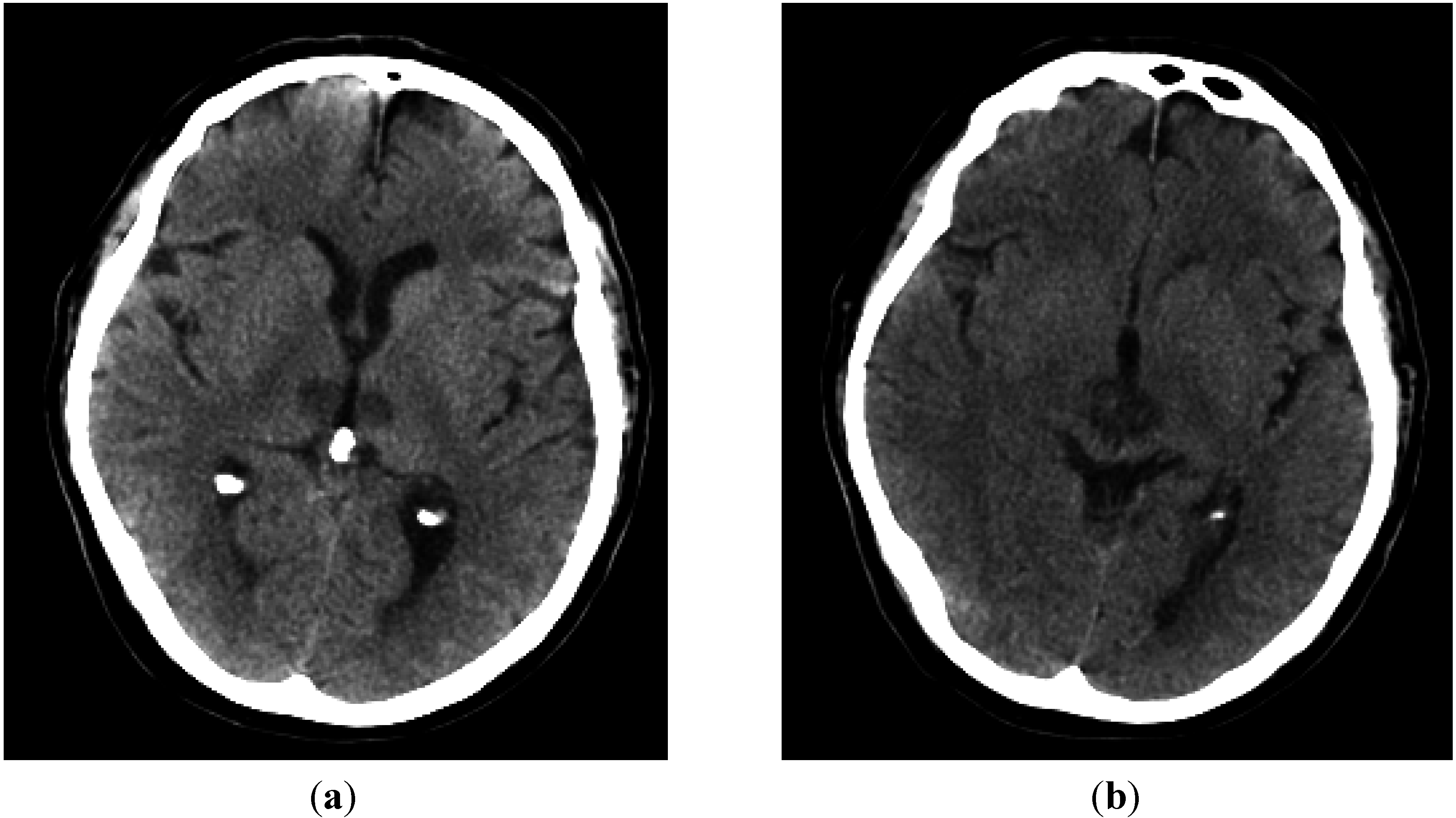
2. Case Report

3. Discussion

| Differentials | Acute/Subacute Presentations |
|---|---|
| Vascular–Arterial | Stroke-Infarct-tip of basilar artery syndrome, PoA occlusion |
| Vascular–Venous | Cerebral Vein Thrombosis especially Great vein of Galen, Straight Sinus thrombosis |
| Infections–Viral | Flavi virus infections e.g., Japanese Encephalitis (90% of patients have bithalamic involvement), West Nile Virus encephalitis |
| Infections–others | Tuberculous Meningio-encephalitis, Fungal Infection, Malaria, Toxoplasmosis |
| Demyelination | Acute Demyelinating Encephalomyelitis, Multiple Sclerosis |
| Spongiform encephalopathy | Variant Creutzfeldt-Jakob disease (vCJD), CJD |
| Thiamine deficiency | Wernicke’s encephalopathy |
| Hypoxic injury newborn | Profound Hypoxia of the Newborn |
| Non-Acute Presentations | |
| Vascular | Chronic Hypertensive encephalopathy leading to lacunar infarcts/microbleeds |
| Tumours | Astrocytoma, Glioblastoma Multiforme, Germinoma, lymphomas (1% of Central nervous System tumours) |
| Congenital Metabolic Syndrome | Leigh Syndrome (mitrochondropathy), Gangliosidoses (Lysosomal disorders), Krabbe’s disease, Wilson’s Disease |
| Cerebrovascular Ferrocalcinosis | Fahr’s Disease |
4. Conclusions
Author Contributions
Conflicts of Interest
References
- Percheron, G. The anatomy of the arterial supply of the human thalamus and its use for the interpretation of the thalamic vascular pathology. J. Neurol. 1973, 205, 1–13. [Google Scholar] [CrossRef]
- Amin, O.M.; Shwani, S.S.; Zangana, H.M.; Hussein, E.M.H.; Ameen, N. Bilateral infarction of paramedian thalami: A report of two cases of artery of Percheron occlusion and review of the literature. BMJ Case Rep. 2011, 2011. [Google Scholar] [CrossRef]
- Kostanian, V.; Cramer, S.C. Artery of Percheron Thrombolysis. AJNR Am. J. Neuroradiol. 2007, 28, 870–871. [Google Scholar] [PubMed]
- Lazzaro, N.A.; Wright, B.; Castillo, M.; Fischbein, N.J.; Glastonbury, C.M.; Hildenbrand, P.G.; Wiggins, R.H.; Quigley, E.P.; Osborn, A.G. Artery of Percheron infarction—Imaging patterns and clinical spectrum. Am. J. Neuroradiol. 2010, 31, 1283–1289. [Google Scholar] [CrossRef] [PubMed]
- Intercollegiate Stroke Working Party. National Clinical Guideline for Stroke, 4th ed.; The Royal College of Physicians: London, UK, 2012. [Google Scholar]
- Ben Slamia, L.; Jemaa, H.B.; Benammou, S.; Tlili-Graiess, K. Occlusion of the artery of percheron: Clinical and neuroimaging correlation. J. Neuroradiol. 2008, 35, 244–245. [Google Scholar]
- Linn, J.; Danek, A.; Hoffmann, L.A.; Seelos, K.C.; Brückmann, H. Differential Diagnosis of Bilateral Thalamic Lesions. Clin. Neuroradiol. 2007, 17, 3–22. [Google Scholar] [CrossRef]
- Agarwal, N.; Tolia, A.; Hansberry, D.R.; Duffis, E.J.; Barrese, J.C.; Gandhi, C.D.; Prestigiacomo, C.J. Current differential diagnoses and treatment options of vascular occlusions presenting as bilateral thalamic infarcts: A review of the literature. J. NeuroIntervent. Surg. 2013, 5, 419–425. [Google Scholar] [CrossRef]
- Arauz, A.H.; Patino-Rodriguez, M.; Vargas-Gonzalez, J.C.; Arguelles-Morales, N.; Silos, H.; Ruiz-Franco, A.; Ochoa, M.A. Clinical Spectrum of artery of Percheron infarct: Clinical-Radiological correlations. J. Stroke Cerebrovasc. Dis. 2014, 23, 1083–1088. [Google Scholar] [CrossRef] [PubMed]
- Reilly, M.; Connolly, S.; Stack, J.; Martin, E.A.; Hutchinson, M. Bilateral paramedian thalamic infarction a distinct but poorly recognized syndrome. Q. J. Med. 1992, 82, 63–70. [Google Scholar] [PubMed]
- Waterston, J.A.; Stark, R.J.; Gilligan, B.S. Paramedian thalamic and mid brain infarction: The mesencephalothalamic syndrome. Clin. Exp. Neurol. 1987, 24, 45–53. [Google Scholar] [PubMed]
- López-Serna, R.; González-Carmona, P.; López-Martínez, M. Bilateral thalamic stroke due to occlusion of the artery of Percheron in a patient with patent foramen ovale: A case report. J. Med. Case Rep. 2009, 3, 7392. [Google Scholar] [CrossRef]
- Raphaeli, G.; Liberman, A.; Gomori, J.M.; Steiner, I. Acute bilateral paramedian thalamic infarcts after occlusion of the artery of Percheron. Neurology 2006, 66. [Google Scholar] [CrossRef] [PubMed]
- Cassourret, G.; Bertrand, P.; Sbardella, F.; Bordes, J.; Maurin, O.; Boret, H. Ischaemic stroke of the Artery of Percheron with Normal Initial MRI: A case report. Case Rep. Med. 2010, 2010. [Google Scholar] [CrossRef]
© 2015 by the authors; licensee MDPI, Basel, Switzerland. This article is an open access article distributed under the terms and conditions of the Creative Commons Attribution license (http://creativecommons.org/licenses/by/4.0/).
Share and Cite
Almamun, M.; Suman, A.; Arshad, S.; Kumar, S.J.S. A Case of Midbrain and Thalamic Infarction Involving Artery of Percheron. J. Clin. Med. 2015, 4, 369-374. https://doi.org/10.3390/jcm4030369
Almamun M, Suman A, Arshad S, Kumar SJS. A Case of Midbrain and Thalamic Infarction Involving Artery of Percheron. Journal of Clinical Medicine. 2015; 4(3):369-374. https://doi.org/10.3390/jcm4030369
Chicago/Turabian StyleAlmamun, Muhammad, Appu Suman, Syed Arshad, and Sonni Jayathirthachar Sanjeev Kumar. 2015. "A Case of Midbrain and Thalamic Infarction Involving Artery of Percheron" Journal of Clinical Medicine 4, no. 3: 369-374. https://doi.org/10.3390/jcm4030369
APA StyleAlmamun, M., Suman, A., Arshad, S., & Kumar, S. J. S. (2015). A Case of Midbrain and Thalamic Infarction Involving Artery of Percheron. Journal of Clinical Medicine, 4(3), 369-374. https://doi.org/10.3390/jcm4030369