Diameters of Arteries Supplying Horseshoe Kidneys and the Level They Branch off Their Parental Vessels: A CT-Angiographic Study

 , ,
, ,
Abstract
:1. Background
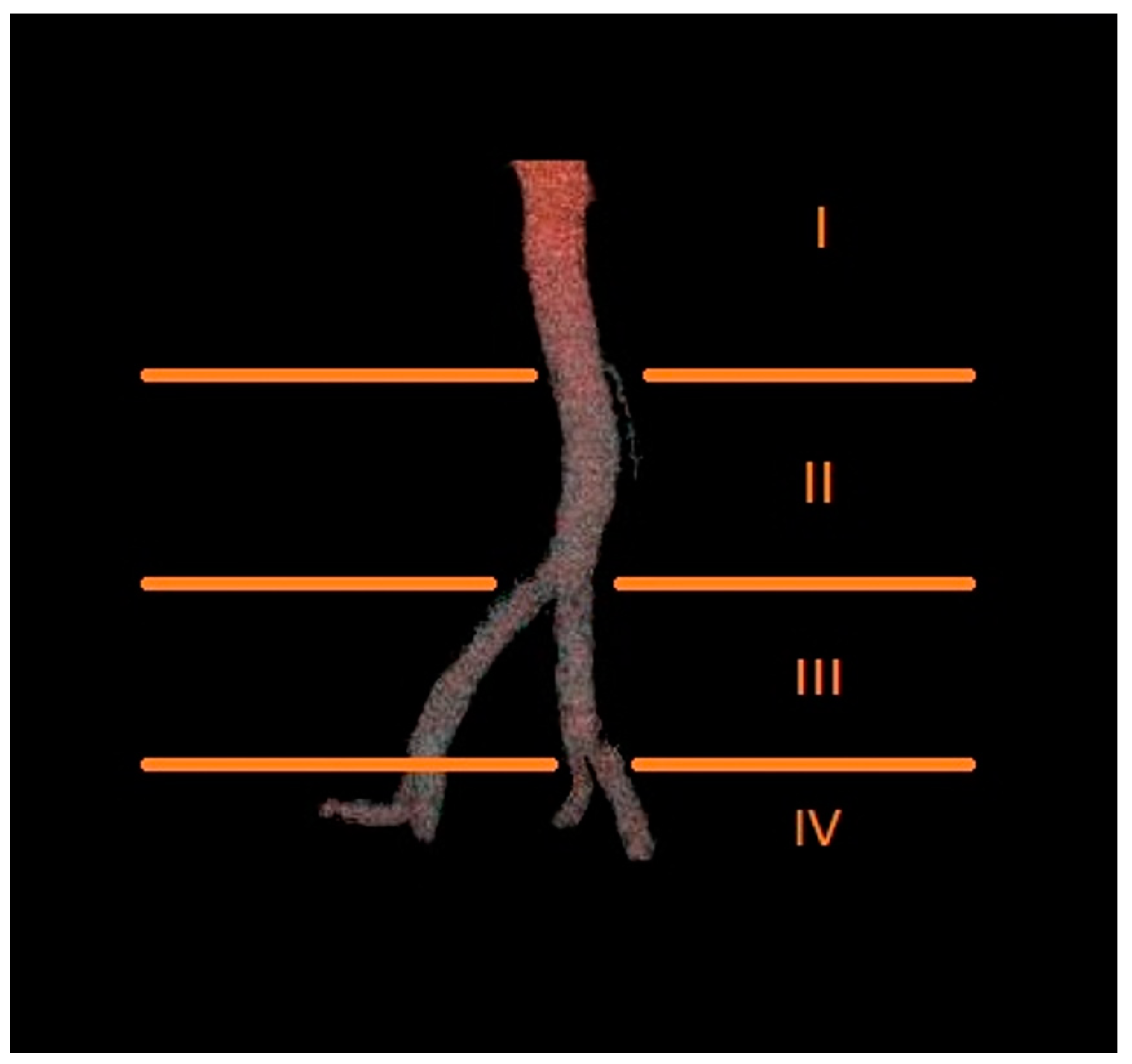
2. Methods
Statistical Analysis
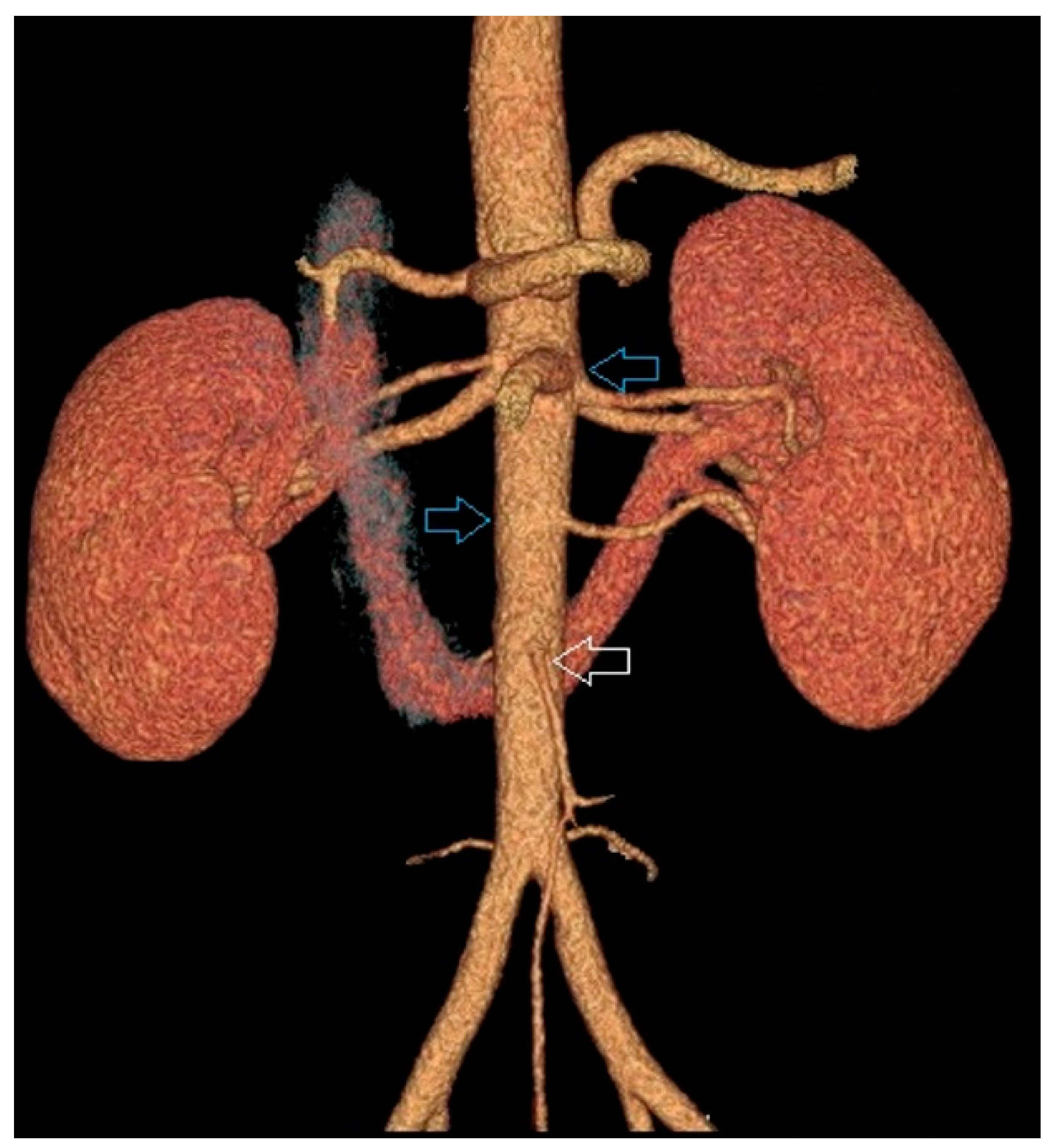
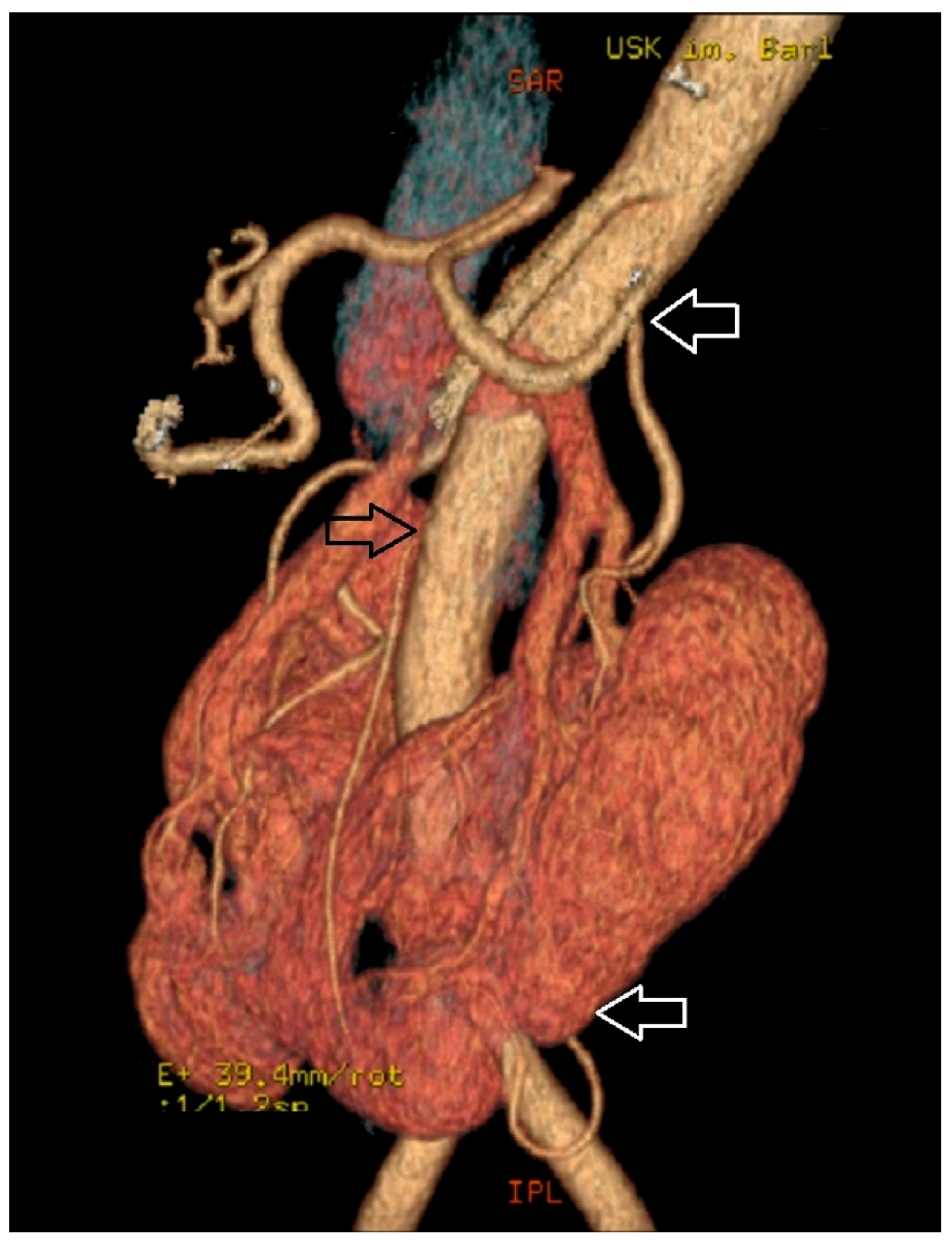
3. Results
4. Discussion
5. Conclusions
Author Contributions
Funding
Acknowledgments
Conflicts of Interest
References
- Taghavi, K.; Kirkpatrick, J.; Mirjalili, S.A. The horseshoe kidney: Surgical anatomy and embryology. J. Pediatr. Urol. 2016, 12, 275–280. [Google Scholar] [CrossRef] [PubMed]
- Ichikawa, T.; Tanno, K.; Okochi, T.; Koizumi, J.; Mori, N.; Yamada, Y.; Joishi, D.; Enkhbaatar, N.; Terachi, T.; Nishibe, T.; et al. Evaluation of renal artery anomalies associated with horseshoe kidney using CT angiography. Tokai J. Exp. Clin. Med. 2015, 40, 16–21. [Google Scholar]
- Natsis, K.; Piagkou, M.; Skotsimara, A.; Protogerou, V.; Tsitouridis, I.; Skandalakis, P. Horseshoe kidney: A review of anatomy and pathology. Surg. Radiol. Anat. 2014, 36, 517–526. [Google Scholar] [CrossRef] [PubMed]
- Ichikawa, T.; Kawada, S.; Koizumi, J.; Endo, J.; Iino, M.; Terachi, T.; Usui, Y.; Nishibe, T.; Dardik, A.; Imai, Y. Major venous anomalies are frequently associated with horseshoe kidneys. Circ. J. 2011, 75, 2872–2877. [Google Scholar] [CrossRef] [PubMed]
- Majos, M.; Polguj, M.; Szemraj-Rogucka, Z.; Arazińska, A.; Stefańczyk, L. The level of origin of renal arteries in horseshoe kidney vs. in separated kidneys: CT-based study. Surg. Radiol. Anat. 2018, 24. [Google Scholar] [CrossRef]
- Mhaske, S.M.; Patil, B.; Patwardhan, S.K.; Gopalakrishnan, G.; Shelke, U.R.; Pamecha, Y.G. Outcome following renal autotransplantation in renal artery stenosis. Urol. Ann. 2019, 11, 46–52. [Google Scholar] [CrossRef]
- Studzińska, D.; Rudel, B.; Polok, K.; Lewandowski, K.; Studziński, K.; Gajdosz, A.; Oo, A.; Szczeklik, M.; Zaczek, M.; Zaniewski, M.; et al. Infra-renal vs supra-renal abdominal aortic aneurysms: Comparison of associated aneurysms and renal artery stenosis. Ann. Vasc. Surg. 2019. [Google Scholar] [CrossRef]
- Mastrocostas, K.; Chingkoe, C.M.; Pace, K.T.; Barfett, J.J.; Kirpalani, A.; Mnatzakanian, G.N.; Vlachou, P.A.; Colak, E. Computed tomography identified factors that preclude living kidney donation. Can. Urol. Assoc. J. 2018, 12, 276–279. [Google Scholar] [CrossRef]
- Yun, S.; Woo, H.D.; Doo, S.W.; Kwon, S.H.; Noh, H.; Song, D. Transplantation of a horseshoe kidney found during harvest operation of a cadaveric donor: A case report. J. Korean Med. Sci. 2014, 29, 1166–1169. [Google Scholar] [CrossRef]
- Kin, K.; Takano, H.; Nakagawa, T.; Shirakawa, Y. Hybrid repair of an abdominal aortic aneurysm: Debranching with endovascular aneurysm repair in a patient with horseshoe kidney. Ann. Vasc. Dis. 2017, 10, 41–43. [Google Scholar] [CrossRef]
- Ding, J.; Huang, Y.; Gu, S.; Chen, Y.; Peng, J.; Bai, Q.; Ye, M.; Qi, J. flexible ureteroscopic management of horseshoe kidney renal calculi. Int. Braz. J. Urol. 2015, 41, 683–689. [Google Scholar] [CrossRef]
- Sharma, K.; Babrowski, T.; Milner, R. A novel chimney approach for management of horseshoe kidney during EVAR. EJVES Short Rep. 2016, 33, 16–19. [Google Scholar] [CrossRef] [PubMed]
- De Monti, M.; Ghilardi, G.; Caverni, L.; Ceriani, L.; Soldi, S.; Massaro, F.; Buchbut, R.; Gobatti, D.; Scorza, R. Multidetector helical angio CT oblique reconstructions orthogonal to internal carotid artery for preoperative evaluation of stenosis. A prospective study of comparison with color Doppler US, digital subtraction angiography and intraoperative data. Minerva Cardioangiol. 2003, 51, 373–385. [Google Scholar] [PubMed]
- Addis, K.A.; Hopper, K.D.; Iyriboz, T.A.; Liu, Y.; Wise, S.W.; Kasales, C.J.; Blebea, J.S.; Mauger, D.T. CT angiography: In vitro comparison of five reconstruction methods. Am. J. Roentgenol. 2001, 177, 1171–1176. [Google Scholar]
- Gulas, E.; Wysiadecki, G.; Cecot, T.; Majos, A.; Stefańczyk, L.; Topol, M.; Polguj, M. Accessory (multiple) renal arteries—Differences in frequency according to population, visualizing techniques and stage of morphological development. Vascular 2016, 24, 531–537. [Google Scholar] [CrossRef] [PubMed]
- Scavuzzo, A.; Santana Rios, Z.; Diaz-Gomez, C.; Varguez Gonzalez, B.; Osornio-Sanchez, V.; Bravo-Castro, E.; Linden-Castro, E.; Martinez-Cervera, P.; Jimenez-Rios, M.A. Renal cell carcinoma in a pregnant woman with horseshoe kidney. Urol. Case Rep. 2017, 13, 58–60. [Google Scholar] [CrossRef] [PubMed]
- Ohtake, S.; Kawahara, T.; Noguchi, G.; Nakaigawa, N.; Chiba, K.; Uemura, H.; Yao, M.; Makiyama, K. Renal cell carcinoma in a horseshoe kidney treated with laparoscopic partial nephrectomy. Case Rep. Oncol. Med. 2018, 2018, 7135180. [Google Scholar] [CrossRef]
- Bleve, C.; Bucci, V.; Conighi, M.L.; Battaglino, F.; Costa, L.; Fasoli, L.; Zolpi, E.; Chiarenza, S.F. Horseshoe kidney and uretero-pelvic-junction obstruction in a pediatric patient. Laparoscopic vascular hitch: A valid alternative to dismembered pyeloplasty? Pediatr. Med. Chir. 2017, 39, 178. [Google Scholar] [CrossRef] [PubMed]
- Eisendrath, D.N.; Phifer, F.M.; Culver, H.B. Horseshoe kidney. Ann. Surg. 1925, 82, 735–764. [Google Scholar] [CrossRef]
- Graves, F.T. The arterial anatomy of the congenitally abnormal kidney. Br. J. Surg. 1969, 56, 533–541. [Google Scholar] [CrossRef]
- Iwanaga, J.; Saga, T.; Tabira, Y.; Watanabe, K.; Yamaki, K. Contrast imaging study of the horseshoe kidney for transplantation. Surg. Radiol. Anat. 2015, 37, 1267–1271. [Google Scholar] [CrossRef]
- Crawford, E.S.; Coselli, J.S.; Safi, H.J.; Martin, T.D.; Pool, J.L. The impact of renal fusion and ectopia on aortic surgery. J. Vasc. Surg. 1988, 8, 375–383. [Google Scholar] [CrossRef]
- Kaplan, D.B.; Kwon, C.C.; Marin, M.L.; Hollier, L.H. Endovascular repair of abdominal aortic aneurysms in patients with congenital renal vascular anomalies. J. Vasc. Surg. 1999, 30, 407–416. [Google Scholar] [CrossRef]
- Ruppert, V.; Umscheid, T.; Rieger, J.; Schmedt, C.G.; Mussack, T.; Steckmeier, B.; Stelter, W.J. Endovascular aneurysm repair: Treatment of choice for abdominal aortic aneurysm coincident with horseshoe kidney? Three case reports and review of literature. J. Vasc. Surg. 2004, 40, 367–370. [Google Scholar] [CrossRef] [PubMed]


| N | Mean (mm) | Median (mm) | Min (mm) | Max (mm) | SD (mm) | |
|---|---|---|---|---|---|---|
| Arteries supplying horseshoe kidneys | 398 | 4.30 | 4.20 | 0.60 | 8.50 | 1.54 |
| Arteries supplying separated kidneys | 598 | 5.52 | 5.70 | 1.00 | 9.80 | 1.61 |
| Arteries Supplying Horseshoe Kidneys | Arteries Supplying Separated Kidneys | |||||||||||
|---|---|---|---|---|---|---|---|---|---|---|---|---|
| Level of Origin of Renal Artery | N | Mean (mm) | Median (mm) | Min (mm) | Max (mm) | SD (mm) | N | Mean (mm) | Median (mm) | Min (mm) | Max (mm) | SD (mm) |
| I | 230 | 4.54 | 4.60 | 0.60 | 8.50 | 1.64 | 596 | 5.53 | 5.70 | 1.00 | 9.80 | 1.61 |
| II | 108 | 4.28 | 4.20 | 1.40 | 7.40 | 1.35 | 2 | 4.45 | 4.45 | 2.80 | 6.10 | 2.33 |
| III | 57 | 3.41 | 3.30 | 1.10 | 6.30 | 1.11 | 0 | 0 | 0 | 0 | 0 | 0 |
| IV | 3 | 3.43 | 3.20 | 2.80 | 4.30 | 0.78 | 0 | 0 | 0 | 0 | 0 | 0 |
| Arteries Supplying Horseshoe Kidneys | Arteries Supplying Separated Kidneys | |||||||
|---|---|---|---|---|---|---|---|---|
| Level of Origin of Renal Artery | <4.0 mm | 4.0–5.7 mm | 5.7–7.4 mm | >7.4 mm | <4.0 mm | 4.0–5.7 mm | 5.7–7.4 mm | >7.4 mm |
| I | 82 (35.7%) | 95 (41,3%) | 38 (16.5%) | 15 (6.5%) | 94 (15.8%) | 201 (33.7%) | 234 (39.3%) | 67 (11.2%) |
| II | 44 (40,7%) | 48 (44,4%) | 15 (13.9%) | 1 (1%) | 1 | 0 | 1 | 0 |
| III | 42 (73.7%) | 12 (21.1%) | 3 (5.3%) | 0 | 0 | 0 | 0 | 0 |
| IV | 2 | 1 | 0 | 0 | 0 | 0 | 0 | 0 |
| Arteries Supplying Horseshoe Kidneys | Arteries Supplying Separated Kidneys | |||||
|---|---|---|---|---|---|---|
| Level of Origin of Renal Artery | Whole Group | ♀ | ♂ | Whole Group | ♀ | ♂ |
| I | 191 (83.0%) | 71 (86.6%) | 120 (81.1%) | 546 (91.8%) | 261 (92.6%) | 285 (91.1%) |
| II | 89 (82.4%) | 36 (83.7%) | 53 (81,5%) | 1 | 0 | 1 |
| III | 39 (68.4%) | 12 (70.6%) | 28 (70.0%) | 0 | 0 | 0 |
| IV | 2 | 0 | 2 | 0 | 0 | 0 |
© 2019 by the authors. Licensee MDPI, Basel, Switzerland. This article is an open access article distributed under the terms and conditions of the Creative Commons Attribution (CC BY) license (http://creativecommons.org/licenses/by/4.0/).
Share and Cite
Majos, M.; Majos, A.; Polguj, M.; Szymczyk, K.; Chrostowski, J.; Stefańczyk, L. Diameters of Arteries Supplying Horseshoe Kidneys and the Level They Branch off Their Parental Vessels: A CT-Angiographic Study. J. Clin. Med. 2019, 8, 464. https://doi.org/10.3390/jcm8040464
Majos M, Majos A, Polguj M, Szymczyk K, Chrostowski J, Stefańczyk L. Diameters of Arteries Supplying Horseshoe Kidneys and the Level They Branch off Their Parental Vessels: A CT-Angiographic Study. Journal of Clinical Medicine. 2019; 8(4):464. https://doi.org/10.3390/jcm8040464
Chicago/Turabian StyleMajos, Marcin, Agata Majos, Michał Polguj, Konrad Szymczyk, Jakub Chrostowski, and Ludomir Stefańczyk. 2019. "Diameters of Arteries Supplying Horseshoe Kidneys and the Level They Branch off Their Parental Vessels: A CT-Angiographic Study" Journal of Clinical Medicine 8, no. 4: 464. https://doi.org/10.3390/jcm8040464
APA StyleMajos, M., Majos, A., Polguj, M., Szymczyk, K., Chrostowski, J., & Stefańczyk, L. (2019). Diameters of Arteries Supplying Horseshoe Kidneys and the Level They Branch off Their Parental Vessels: A CT-Angiographic Study. Journal of Clinical Medicine, 8(4), 464. https://doi.org/10.3390/jcm8040464