A Systematic Review of the Clinical Value and Applications of Three-Dimensional Printing in Renal Surgery


Abstract
:1. Introduction
2. Methods
2.1. Search Strategy
2.2. Inclusion Criteria and Data Extraction
3. Results
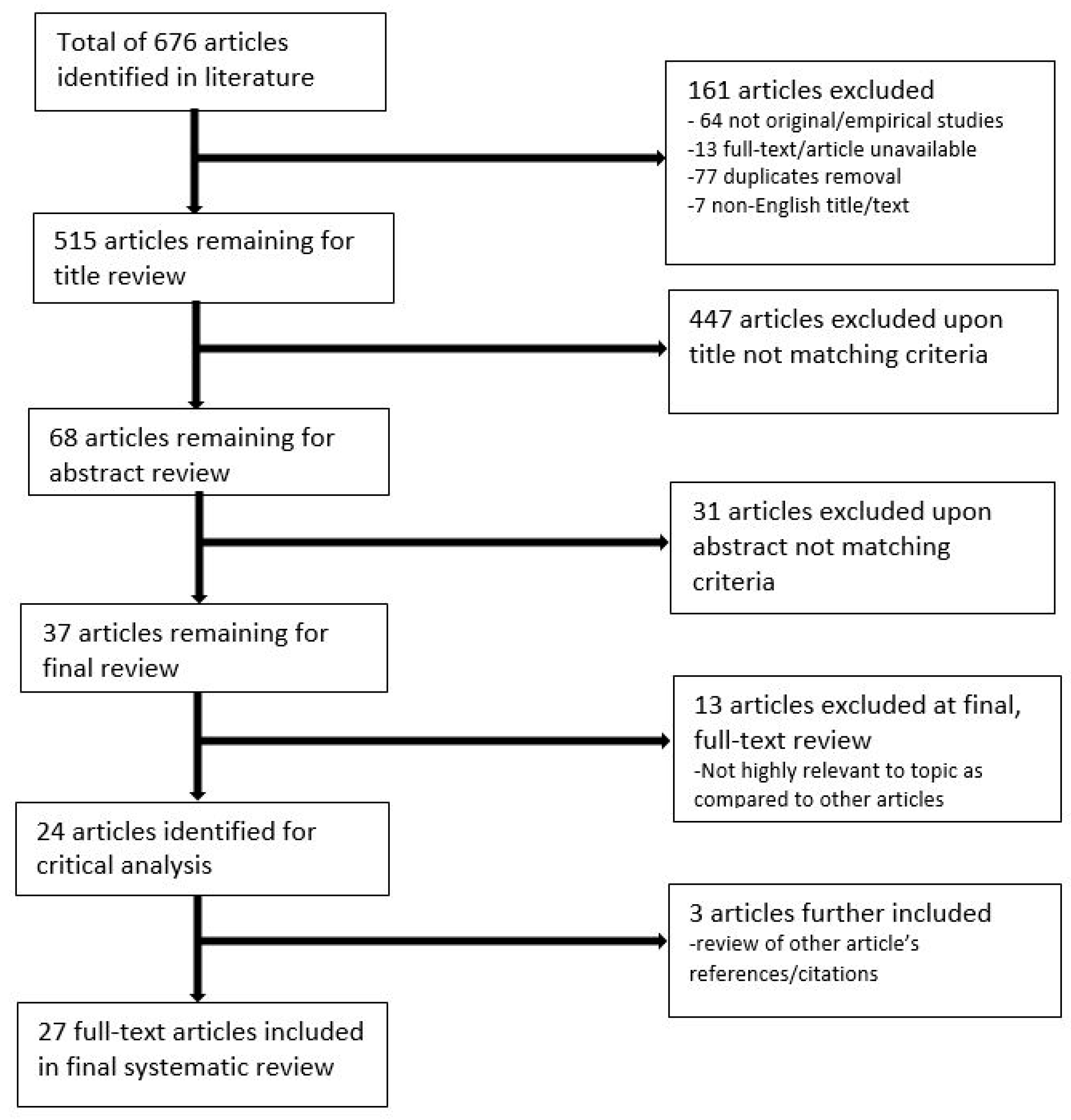
3.1. Literature Search Outcomes
3.2. Study Characteristics
3.3. Quantitative Analysis of 3D-Printed Kidney Models
3.4. Qualitative Analysis of 3D-Printed Kidney Models
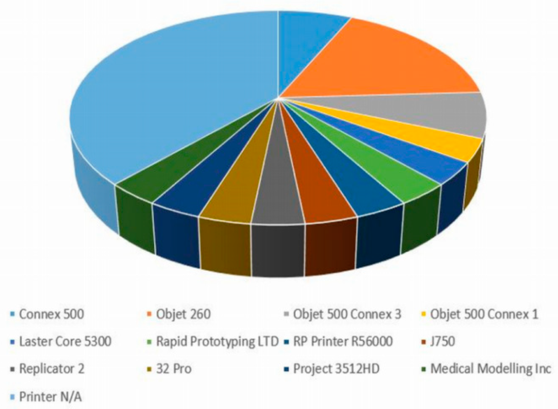
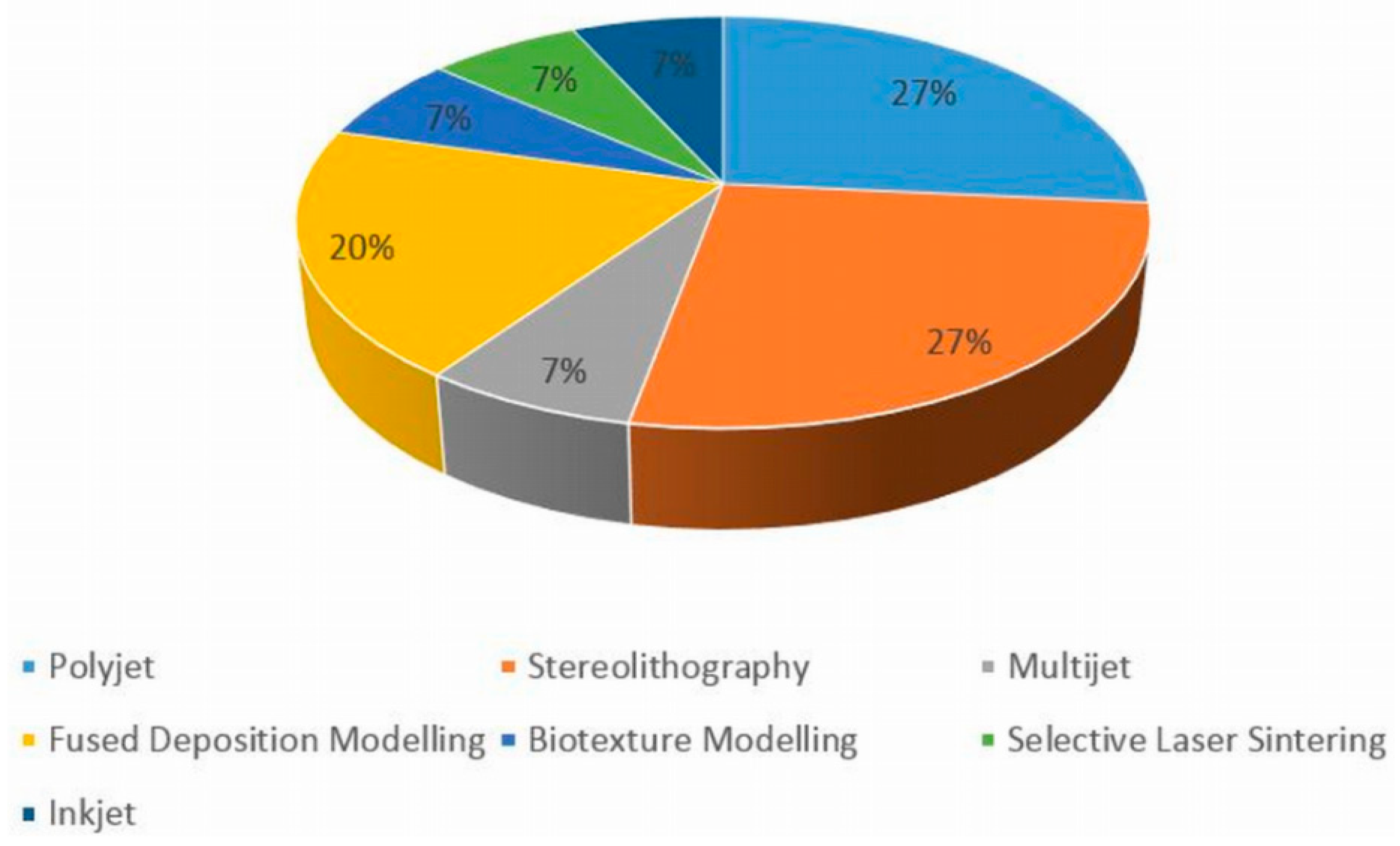
3.5. 3D-Printing Technologies, Materials and Software Tools
3.6. Manufacturing Cost and Time Duration
3.7. Study Purpose
4. Discussion
4.1. Key Findings
4.2. Participant Types
4.3. Clinical Value and Applications in Pre-Surgical Planning
4.4. 3D-Printing Technologies and Materials
4.5. Limitations
5. Conclusion and Future Recommendations
Author Contributions
Acknowledgments
Conflicts of Interest
References
- Ventola, C.L. Medical applications for 3D printing: Current and projected uses. Pharm. Ther. 2014, 39, 704–711. [Google Scholar]
- Mitsouras, D.; Liacouras, P.; Imanzadeh, A.; Giannopoulos, A.A.; Cai, T.; Kumamaru, K.K.; George, E.; Wake, N.; Caterson, E.J.; Pomahac, B.; et al. Medical 3D printing for the radiologist. Radiographics 2015, 35, 1965–1988. [Google Scholar] [CrossRef] [PubMed]
- Soliman, Y.; Feibus, A.; Baum, N. 3D printing and its urologic applications. Rev. Urol. 2015, 17, 20–24. [Google Scholar] [PubMed]
- Chepelev, L.; Hodgdon, T.; Gupta, A.; Wang, A.; Torres, C.; Krishna, S.; Akyuz, E.; Mitsouras, D.; Sheikh, A. Medical 3D printing for vascular interventions and surgical oncology: A primer for the 2016 radiological society of North America (RSNA) hands-on course in 3D printing. 3D Print. Med. 2016, 2, 2–17. [Google Scholar] [CrossRef] [PubMed]
- Wake, N.; Rude, T.; Kang, S.K.; Stifelman, M.D.; Borin, J.F.; Sodickson, D.K.; Huang, W.C.; Chandarana, H. 3D Printed renal cancer models derived from MRI data: Application in pre-surgical planning. Abdom. Radiol. 2017, 42, 1501–1509. [Google Scholar] [CrossRef] [PubMed]
- Bernhard, J.C.; Isotani, S.; Matsugasumi, T.; Duddalwar, V.; Hung, A.J.; Suer, E.; Baco, E.; Satkunasivam, R.; Djaladat, H.; Metcalfe, C.; et al. Personalized 3D printed model of kidney and tumour anatomy: A useful tool for patient education. World. J. Urol. 2016, 34, 337–345. [Google Scholar] [CrossRef]
- Maddox, M.M.; Feibus, A.; Liu, J.; Wang, J.; Thomas, R.; Silberstein, J.L. 3D-printed soft-tissue physical models of renal malignancies for individualized surgical simulation: A feasibility study. J. Robot. Surg. 2018, 12, 27–33. [Google Scholar] [CrossRef]
- Libby, R.S.; Silberstein, J.L. Physical model of clear-cell renal carcinoma with inferior vena cava extension created from a 3-dimensional printer to aid in surgical resection: A case report. Clin. Genitourin. Cancer 2017, 15, 867–869. [Google Scholar] [CrossRef]
- Glybochko, P.V.; Rapoport, L.M.; Alyaev, Y.G.; Sirota, E.S.; Bezrukov, E.A.; Fiev, D.N.; Byadretdinov, I.S.; Bukatov, M.D.; Letunovskiy, A.V.; Korolev, D.O. Multiple application of three-dimensional soft kidney models with localized kidney cancer: A pilot study. Urol. J. 2018, 85, 99–105. [Google Scholar] [CrossRef]
- Zhang, Y.; Ge, H.W.; Li, N.C.; Yu, C.F.; Guo, H.F.; Jin, S.H.; Liu, J.S.; Na, Y.Q. Evaluation of three-dimensional printing for laparoscopic partial nephrectomy of renal tumors: A preliminary report. World J. Urol. 2016, 34, 533–537. [Google Scholar] [CrossRef]
- Yang, T.; Lin, S.; Tan, T.; Yang, J.; Pan, J.; Hu, C.; Li, J.; Zou, Y. Impact of 3D printing technology on comprehension of surgical anatomy of retroperitoneal tumour. World. J. Surg. 2018, 42, 2339–2343. [Google Scholar] [CrossRef] [PubMed]
- Marconi, S.; Pugliese, L.; Botti, M.; Peri, A.; Cavazzi, E.; Latteri, S.; Auricchio, F.; Pietrabissa, A. Value of 3D printing for the comprehension of surgical anatomy. Surg. Endosc. 2017, 31, 4102–4110. [Google Scholar] [CrossRef] [PubMed]
- Porpiglia, F.; Bertolo, R.; Checcucci, E.; Amparore, D.; Autorino, R.; Dasgupta, P.; Wiklund, P.; Tewari, A.; Liatsikos, E.; Fiori, C. Development and validation of 3D printed virtual models for robot-assisted radical prostatectomy and partial nephrectomy: Urologists’ and patients’ perception. World J. Urol. 2017, 36, 201–207. [Google Scholar] [CrossRef] [PubMed]
- Wake, N.; Rosenkrantz, A.B.; Huang, R.; Park, K.U.; Wysock, J.S.; Taneja, S.S.; Huang, W.C.; Sodickson, D.K.; Chandarana, H. Patient-specific 3D printed and augmented reality kidney and prostate cancer models: Impact on patient education. 3D Print. Med. 2019, 5, 4–9. [Google Scholar] [CrossRef] [PubMed]
- Golab, A.; Slojewski, M.; Brykczynski, M.; Lukowiak, M.; Boehlke, M.; Matias, D.; Smektala, T. Three-dimensional printing as an interdisciplinary communication tool: Preparing for removal of a giant renal tumor and atrium neoplastic mass. Heart. Surg. Forum. 2016, 19, E185–E186. [Google Scholar] [CrossRef] [PubMed]
- Golab, A.; Smektala, T.; Kaczmarek, K.; Stamirowski, R.; Hrab, M.; Slojewski, M. Laparoscopic partial nephrectomy supported by training involving personalized silicone replica poured in three-dimensional printed casting mold. J. Laparoendosc Adv. Surg. Tech. 2017, 27, 420–422. [Google Scholar] [CrossRef]
- Adams, F.; Qiu, T.; Mark, A.; Fritz, B.; Kramer, L.; Schlager, D.; Wetterauer, U.; Miernik, A.; Fischer, P. Soft 3D-printed phantom of the human kidney with collecting system. Ann. Biomed. Eng. 2017, 45, 963–972. [Google Scholar] [CrossRef] [PubMed]
- Atalay, H.A.; Canat, H.L.; Ulker, V.; Alkan, I.; Ozkuvanci, U.; Altunrende, F. Impact of personalized three-dimensional (3D) printed pelvicalyceal system models on patient information in percutaneous nephrolithotripsy surgery: A pilot study. Int. Braz. J. Urol. 2017, 43, 470–475. [Google Scholar] [CrossRef]
- Dwivedi, D.K.; Chatzinoff, Y.; Zhang, Y.; Yuan, Q.; Fulkerson, M.; Chopra, R.; Brugarolas, J.; Cadeddu, J.A.; Kapur, P.; Pedrosa, I. Development of a patient-specific tumor mold using magnetic resonance imaging and 3-dimensional printing technology for targeted tissue procurement and radiomics analysis of renal masses. Urology 2018, 14, 112–209. [Google Scholar] [CrossRef]
- Knoedler, M.; Feibus, A.H.; Lange, A.; Maddox, M.M.; Ledet, E.; Thomas, R.; Silberstein, J.L. Individualized physical 3-dimensional kidney tumor models constructed from 3-dimensional printers result in improved trainee anatomic understanding. Urology 2015, 85, 1257–1262. [Google Scholar] [CrossRef]
- Silberstein, J.L.; Maddox, M.M.; Dorsey, P.; Feibus, A.; Thomas, R.; Lee, B.R. Physical models of renal malignancies using standard cross-sectional imaging and 3-dimensional printers: A pilot study. Urology 2014, 84, 268–272. [Google Scholar] [CrossRef] [PubMed]
- Smektala, T.; Goląb, A.; Królikowski, M.; Slojewski, M. Low cost silicone renal replicas for surgical training-technical note. Arch. Esp. Urol. 2016, 69, 434–436. [Google Scholar] [PubMed]
- Kusaka, M.; Sugimoto, M.; Fukami, N.; Sasaki, H.; Takenaka, M.; Anraku, T.; Ito, T.; Kenmochi, T.; Shiroki, R.; Hoshinaga, K. Initial experience with a tailor-made simulation and navigation program using a 3-D printer model of kidney transplantation surgery. Transplant Proc. 2015, 47, 596–599. [Google Scholar] [CrossRef] [PubMed]
- Lee, H.; Nguyen, N.H.; Hwang, S.; Lee, H.J.; Hong, S.K.; Byun, S.S. Personalized 3D kidney model produced by rapid prototyping method and its usefulness in clinical applications. Int. Braz. J. Urol. 2018, 44, 952–957. [Google Scholar] [CrossRef] [PubMed]
- Alyaev, Y.G.; Sirota, E.S.; Bezrukov, E.A.; Fiev, D.N.; Bukatov, M.D.; Letunovskii, A.V.; Byadretdinov, I.S. Application of 3D soft print models of the kidney for treatment of patients with localized cancer of the kidney (a pilot study). Urologiia 2017, 6, 12–19. [Google Scholar]
- Monda, S.M.; Weese, J.R.; Anderson, B.G.; Vetter, J.M.; Venkatesh, R.; Du, K.; Andriole, G.L.; Figenshau, R.S. Development and validity of a silicone renal tumor model for robotic partial nephrectomy training. Urology 2018, 114, 114–120. [Google Scholar] [CrossRef] [PubMed]
- Chandak, P.; Byrne, N.; Coleman, A.; Karunanithy, N.; Carmichael, J.; Marks, S.D.; Stojanovic, J.; Kessaris, N.; Mamode, N. Patient-specific 3D printing: A novel technique for complex pediatric renal transplantation. Ann. Surg. 2019, 269, e18–e23. [Google Scholar] [CrossRef] [PubMed]
- Von Rundstedt, F.C.; Scovell, J.M.; Agrawal, S.; Zaneveld, J.; Link, R.E. Utility of patient-specific silicone renal models for planning and rehearsal of complex tumour resections prior to robot-assisted laparoscopic partial nephrectomy. Robot. Laparosc. 2016, 119, 598–604. [Google Scholar] [CrossRef] [PubMed]
- Woliner-van der Weg, W.; Deden, L.N.; Meeuwis, A.P.W.; Koenrades, M.; Peeters, L.H.; Kuipers, H.; Laanstra, G.J.; Gotthardt, M.; Slump, C.H.; Visser, E.P. A 3D-printed anatomical pancreas and kidney phantom for optimizing SPECT/CT reconstruction settings in beta cell imaging using 111In-exendin. EJNMMI Phys. 2016, 3. [Google Scholar] [CrossRef]
- Liu, D.; Sun, Z.; Chaichana, T.; Ducke, W.; Fan, Z. Patient-specific 3D printed models of renal tumours using home-made 3D printer in comparison with commercial 3D printer. J. Med. Imaging Health Inf. 2018, 8, 303–308. [Google Scholar] [CrossRef]
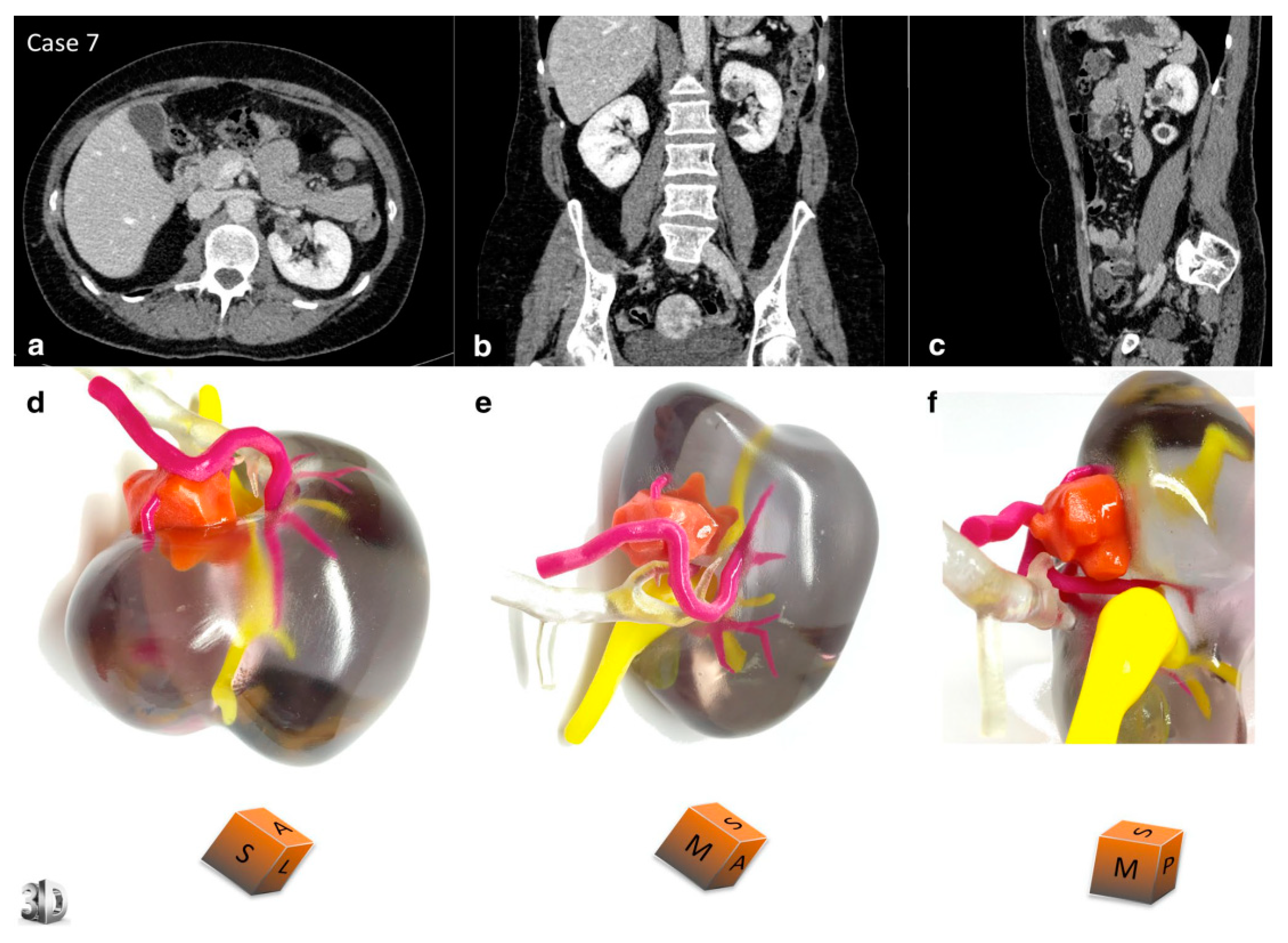
- Komai, Y.; Suglmoto, M.; Gotohda, N.; Matsubara, N.; Kobayashl, T.; Sakal, Y.; Shlga, Y.; Salto, N. Patient-specific 3-dimensional printed kidney designed for 4D surgical navigation: A novel aid to facilitate minimally invasive off-clamp partial nephrectomy in complex tumor cases. Technol. Eng. 2016, 91, 226–233. [Google Scholar] [CrossRef] [PubMed]
- Moher, D.; Shamseer, L.; Clarke, M.; Ghersi, D.; Liberati, A.; Petticrew, M.; Shekelle, P.; Stewart, L.A. Preferred reporting items for a systematic review and meta-analysis protocols (PRISMA-P) 2015 statement. Syst. Rev. 2015, 4, 1. [Google Scholar] [CrossRef] [PubMed]
- Sun, Z.; Liu, D. A systematic review of clinical value of three-dimensional printing in renal disease. Quant. Imaging Med. Surg. 2018, 8, 311–325. [Google Scholar] [CrossRef] [PubMed] [Green Version]
- Sun, Z.; Lau, I.; Wong, Y.H.; Yeong, C.H. Personalized three-dimensional printed models in congenital heart disease. J. Clin. Med. 2019, 8, 522. [Google Scholar] [CrossRef] [PubMed]
- Lau, I.; Sun, Z. Three-dimensional printing in congenital heart disease: A systematic review. J. Med. Radiat. Sci. 2018, 65, 226–236. [Google Scholar] [CrossRef]
- Sun, Z. 3D printing in medicine: Current applications and future perspectives. Quant. Imaging Med. Surg. 2018, 8, 1069–1077. [Google Scholar] [CrossRef] [PubMed]


| First Author and Publication Date | Study Sample Size and Participants | Study Purpose | Technology/Software Used for Segmentation and Post-Processing/Time Duration | Imaging Modality Used for 3D Printing | 3D Printing Technology/Material/Costs/Time Duration | Key Findings |
|---|---|---|---|---|---|---|
| Wake et al. 2017 [5] | 10 renal mass cases collected. 3 urologists used as participants for qualitative questionnaire. | Assess ability of patient-specific 3D-printed kidney models with tumours to enhance pre-surgical planning for complex cases of RCC. | Mimics 16.0 (Mimics, Materialise, Leuven, BE) software. 7 h time duration. | MRI | Flexible, transparent material (HeartPrint Flex, Materialise, Leuven, BE) used with cyan and magenta rigid material combinations (VeroCyan and VeroMagenta, Stratasys, Eden, Prairie). Stereolithography technology using Connex 500 (Stratasys, USA) 3D printer. USD 1000 per kidney model. 10 h taken per model. | High degree of verisimilitude and good correlation between tumour size measurements of 2D data and 3D model. 3D models impacted clinician pre-surgical planning decisions, most specifically in the trans-peritoneal or retroperitoneal approach and clamping. 3D models could be valuable for pre-surgical planning of complex cases and reduce occurrence of intra-operative complications. |
| Bernhard et al. 2016 [6] | 7 patients with primary renal tumours of sizes 2.5–7.2 cm. | Investigate ability of 3D-printed models of kidneys with renal tumours to facilitate patients’ understanding and education of their condition. | Image recognition algorithm Synapse 3D (Fujifilm, Tokyo, Japan. Time duration N/A. | CT | Combination of opaque magenta, opaque yellow, and transparent photopolymer materials. Objet 500 Connex 3 (Stratasys, Eden Prairie, MN, USA) 3D printer. USD 560 per kidney model. | Significant improvement in patient’s understanding of their planned surgical procedure, and kidney physiology and anatomy after 3D model presentation (37.6% improvement, p < 0.05). Patient satisfaction with the usefulness of the models and their experience was on average 9.4/10. |
| Maddox et al. 2018 [7] | 6 patients with 7 enhancing tumours (1 bilateral) ranging from 2.8–5.5 cm. | Investigate the feasibility of using 3D-printed kidney models for pre-operative simulation of renal tumour resection and application into simulation labs. | 3D Systems (Rock Hill, SC). Time duration N/A. | CT | Photopolymer, flexible resin material (translucent for parenchyma and red for tumour). Multi-jet 3D printer (brand N/A). Time-duration and cost N/A. | 3D models were able to closely resemble the feel and texture of real kidneys and thus assisted in the education of junior urologists and their anatomical understanding. Considerably lower estimated blood loss in patients with pre-surgical simulation using the 3D models prior to surgery (p = 0.01). |
| Libby et al. 2017 [8] | 1 case of a 76-year-old woman with right mass extending into adrenal gland and inferior vena cava. | Assess ability of 3D-printed kidney models to guide surgical treatment of complex cases of renal disease. | Software and time duration N/A. | MRI | Deep red colour for thrombus and pink colour for parenchyma and vasculature. 3D printer, time duration and cost N/A. | 3D-printed model was able to improve knowledge of the patient and patient’s family on their condition, required treatment, and general kidney anatomy. 3D-printed kidney model improved visualisation of tumour thrombus and its relationship to surrounding critical anatomy, promoting surgeon confidence in the interventional technique. |
| Glybochko et al. 2018 [9] | 5 patients with renal tumours. | Assess usefulness and clinical applications of soft, 3D-printed kidney models for localised surgical treatment planning of renal disease. | Amira Version 5.4.4 (license ASTND.44644) software; Meshmixer (Autodesk, Inc., San Rafael, CA, USA); Blender (Blender Foundation, Amsterdam, the Netherlands, open-source software). Time duration N/A. | CT | Red, blue, and yellow polylactic acid plastic material Fused Deposition Modelling technology (Cura, open-source software). 3D printer brand N/A. Printing duration of 10–20 h per kidney model and total model completion duration of 2–4 days. USD 150–450 per mode. | 3D-printed model analysis resulted in a change in initial surgical approach and access method (3/5 surgeons changed their approach to partial/radical nephrectomy surgery and 4 surgeons changed their initial decision for transperitoneal/retroperitoneal access). 3D-printed models demonstrated anatomical structures and tumour location superiorly, and thus are useful tools for pre-operative training and trainee education. |
| Zhang et al. 2016 [10] | 10 patients with solitary renal tumours clinically indicated for laparoscopic partial nephrectomy. | Investigate the value of 3D-printed models for laparoscopic partial nephrectomy planning, surgical training, and patient education on their condition and management. | Medical Imaging ToolKit (MITK) and 3DMed. Time duration N/A. | CT | Thermoplastic plastic material with manual colouring of vasculature, parenchyma, collecting system, and tumour performed. LaserCore5300 (Longyuan Rapid Prototyping LTD., Beijing, China) 3D printer. USD 150 per model. 3–4 day duration per model. | The overall usefulness of the models, impact on pre-surgical planning and training, and verisimilitude to real kidneys were rated as 7.8, 6.0, and 7.3 out of 10, respectively. Patients were satisfied with the usefulness of the models (9 or over out of 10 in all questions). |
| Yang et al. 2018 [11] | 1 case of a kidney with a retroperitoneal tumour. 30 participants for model evaluation (10 students, 10 surgeons, 10 residents). | Investigate usefulness of 3D-printed kidney models in enhancing understanding of retroperitoneal tumour anatomy and surgical procedure. | Mimics v.14.01 (Materialize Corp, Leuven, Belgium) software. Time duration N/A. | CT | Stereolithography RP printer RS6000 (Shanghai Union 3D Technology Corp., Shanghai, China). Time duration and cost N/A. | Junior surgeon participant success of anatomical recognition and identifying correct vasculature was improved at 83.33 on 3D-printed model compared to 73.33 on 3D imaging (out of 100). 3D-printed model demonstrated little advantage over 3D imaging for the surgeon participants. |
| Marconi et al. 2017 [12] | 15 patients scheduled for laparoscopic splenectomy, nephrectomy and/or pancreatectomy (2 patients with renal tumours). 30 participants for model evaluation (10 medical students, 10 general surgeons, 10 radiologists). | Assess if 3D-printed models can provide a superior anatomical representation to standard 2D and 3D visualisations for pre-surgical planning of renal disease and surgical training. | Visualisation software Paraview (http://www.paraview.org). Time duration N/A. | CT | Technology and printer N/A. USD 150–200 per model. 20–30 h taken per process. | The highest percentage of correct answers in identifying anatomical structures was achieved for the 3D-printed model group (53.9%), compared to 3D reconstructions (53.4%) and 2D images (45.5%). There was less time spent by participants evaluating anatomy on the 3D models (60.67 ± 25.5 s) compared to 3D reconstructions (70.8 ± 28.18 s) and 2D images (127.04 ± 35.91 s). |
| Porpiglia et al. 2017 [13] | 18 patients (8 undergoing robot-assisted radical prostatectomy, 10 undergoing minimally invasive partial nephrectomy). | Clinical value of 3D-printed models for pre-surgical planning of robot-assisted prostate cancer and nephron-sparing surgeries, training, and education of both urologists and patients with the condition. | Software M3DICS. Time duration N/A. | CT (renal tumour cases) MRI (prostate cancer cases) | Selective laser sintering with thermoplastic polymer technology was used for the kidney models, and PolyJet technology was chosen for the prostate models. Printer brand, time duration and cost N/A. | Patient participants rated the 3D-printed models on average 10/10 for their educational benefits and ability to improve comprehension of the disease. The usefulness of the 3D-printed kidney models for pre-operative planning and comprehending surgical complexity was rated 8/10 by participants. |
| Wake et al. 2019 [14] | 200 patients with confirmed prostate cancer OR RCC to undergo surgical treatment (151 prostate cancer, 49 kidney cancer). | Investigate the value of using augmented reality or 3D-printed kidney and prostate models for patient education on their condition, as well as pre-surgical planning for renal and prostate cancer. | Mimics 20.0 (Materialise, Leuven, Belgium). Time duration N/A. | MRI and CT | Multi-coloured 3D printing (J750, Stratasys, Eden Prairie, MN). Time duration and cost N/A. | Patients described having a better understanding of their disease, tumour characteristics, location, and surgical procedure/plan utilising the 3D-printed models compared to augmented reality models and volumetric imaging. |
| Golab et al. 2016 [15] | 1 patient with a giant renal tumour and neoplastic mass reaching right atrium and venous system. | Assess implementation of 3D-printed models into pre-surgical planning for a rare, complex surgery requiring interdisciplinary communication. | Software and time duration N/A. | CT | Fused Deposition Modelling technology (printer brand N/A). Euro 100/USD 123 per model. 22 h time duration per model. | 3D-printed models improved interdisciplinary communication between physicians of different specialisations, facilitating treatment decisions regarding renal disease. 3D-printed models aided pre-surgical planning and visualisation of anatomy, reducing surgery duration and improving surgical safety. |
| Golab et al. 2017 [16] | 3 patients with renal tumours eligible for partial nephrectomy surgery. | Evaluate the clinical value of 3D-printed kidney models for training purposes in the context of laparoscopic partial nephrectomy surgery. | 3D Slicer (Surgical Planning Lab, Harvard University, MA, USA) program; TinkerCAD (Autodesk, San Rafal, CA, USA) software. Time duration N/A. | CT | Polylactic acid filament material Elite Double 8 (Zhermack Spa, Italy) silicone material for parenchyma Replicator 2 (MakerBot Industries, LLC) 3D printer 120 USD per kidney model Kidney and tumour form printing duration of 4 h 15 min and 55 min, respectively. Total time duration of 7–8 h per kidney model | The accuracy of the models and silicone flexible material closely mimicked real kidney tissue and enabled easy cutting of the model for surgical simulation. The models improved trainee recognition of renal structures and tumour anatomy and hence the surgical process. Implementation of patient-specific 3D-printed kidney models may assist in shortening laparoscopic surgery times and intra-operative renal ischemia. |
| Adams et al. 2017 [17] | 3D-printed kidney models based on 3 kidney cadavers of persons over 18 years. 10 kidney phantoms with 3D-printed collecting systems. | Assess the ability of 3D-printed kidney models to simulate endoscopic urological procedures, and thus assist in pre-operative planning. | InVesalius 3.0.0 (Centro de Technologia da Informacao Renato Archer, Brazil). Models scaled down 80% in all dimensions for printing. Time duration N/A. | CT | Collecting system was printed using an engineered wax material UV curable photopolymer (VeroClear) material used for outer moulds 3Z pro (Solidscape, NH, USA) 3D printer used for collecting system; Objet 260 (Connex, Stratasys, Israel) 3D printer for outer moulds. 25 h taken for 3D printing of engineered wax material and 4.3 h for 3D printing of VeroClear material. Total of 2 days taken for model to become useable phantom. Cost N/A. | The three 3D-printed models accurately resembled morphological details of the renal collecting system and anatomy (0.6 mm distance error for phantoms). 3D-printed collecting systems were able to be easily visualised endoscopically, promoting possibilities for implementation into endoscopic training. |
| Atalay et al. 2017 [18] | 5 patients with unilateral staghorn renal stones and indication of percutaneous nephrolithotomy. | Assess if personalised 3D-printed kidney models can improve patient understanding of their condition prior to undergoing pelvicalyceal surgery. | Mimics 16.0 (Materialise, Belgium). Time duration N/A. | CT | Fused deposition modelling technology, using a polymer filament (Stratasys Inc.). Printer model N/A. 100 USD per model. 2 h taken per model. | 3D-printed models were able to assist pre-surgical planning for collecting system access in complex renal stone-removal cases. Physician-patient interaction and communication was improved after the presentation of customised 3D-printed kidney models (patients rated a 50% improvement in conversation). Patient understanding of basic kidney anatomy and the location of their renal calculus and planned surgical approach improved by 50% (p < 0.05). |
| Dwivedi et al. 2018 [19] | 6 patients with renal tumours > 2.5 cm and eGFR <0.30 mL/min/1.73 m2. | Assess usefulness of patient-specific 3D-printed kidney moulds for radiomics and radiogenomic analyses. | 3D slicer (http://www.slicer.org); CAD software SolidWorks (Dassault Systemes, Velizy-Villacoublay, France). Time duration N/A. | MRI | Project 3512HD (3D Systems, Rock Hall, SC) 3D printer. USD 160.7 ± 111.1 per mould (range USD 20.9–350.7). 1 patient had an entire kidney and large tumour printed for USD 1000. 12–14 h taken per mould. | Study is the first report of patient-specific 3D-printed renal models to correlate MRI imaging features with kidney tumour histopathology. MRI patient-specific 3D moulds of renal tumours are able to facilitate tissue-based analyses for radiomics and radiogenomic studies. |
| Knoedler et al. 2015 [20] | 6 cases of kidneys with renal tumours. | Investigate the effects of 3D-printed renal models with enhancing masses on junior medics’ understandings, localisations, and characterisations of renal tumours. | Software and time duration N/A. | CT | Translucent plastic resin material. Stereolithography (SLA) (3D printer brand N/A). Cost and time duration N/A. | 3D-printed models improved trainee nephrectomy accuracy rating significantly compared to CT images (p < 0.01). Implementation of 3D-printed kidney models can improve junior medical understanding and characterisation of renal pathology. |
| Silberstein et al. 2014 [21] | 5 cases of kidneys with suspicious renal tumours. | Disseminate impact of personalised, 3- printed kidney models with enhancing lesions on education of trainees, patients, and surgeons for characterisation and management of RCC. | Software and time duration N/A. | CT | Translucent resin for parenchyma and red translucent resin for lesions and proximal ureter. Stereolithography printing technology (Medical Modeling Inc., Golden, CO.). Printer brand N/A. Time duration and cost N/A. | Medical trainees showed improvement in their understanding of tumour characteristics and anatomical relationships. Patients and patients’ families suggested an improvement in their comprehension of their condition and the location and size of the tumour, alongside the planned surgery. |
| Smektala et al. 2016 [22] | 5 patients undergoing lapraroscopic kidney tumorectomy for RCC. | Investigate feasibility of implementing low-cost, customised silicone kidney replicas for pre-operative planning and simulation of complex nephron-sparing surgeries. | 94 min time duration per kidney model. Software N/A. | CT | Silicone material. 14 h and 30 min per kidney model. USD 7.4–14.4 per kidney model. Printing technology/printer brand N/A. | 3D printing of the models was simple to perform and inexpensive, and thus may be feasible to implement into practice. Silicone material of the 3D-printed models enabled them to have malleable properties that closely resembled kidney tissue, making them useful in surgical simulation for complex cases. |
| Kusaka et al. 2015 [23] | 1 case of a donor graft kidney model and 1 case of a donor kidney and recipient pelvic cavity. | The feasibility and vale of 3D-printed kidney graft models and pelvic cavity replicas for pre-operative and intra-operative simulation in kidney transplantation. | OsiriX (Pixemeo, Geneva, Switzerland) image processing software. Magics (Materialise, Leuven, Belgium) softwar program. Time duration N/A. | CT | Parenchyma printed with VeroClear and TangoPlus material, with VeroMagenta/TangoPlus for the renal artery, VeroCyan/TangoPlus for the renal vein, and VeroMagenta/VeroCyan/TangoPlus blend for urinary tract anatomical structures. TangoPlus for skin layer, and TangoPlus/VeroWhite Plus for pelvic bones, bladder, muscles, and vessels of the pelvic cavity replica. Inkjet printing technology using Connex 500 (Stratasys Ltd., MN, USA) 3D printer. Time duration and cost N/A. | Understanding of spatial relationships amongst vital structures and anatomy was improved using the 3D-printed life-size kidney models due to their accurate replication of anatomy. 3D printing is able to overcome the limitations of viewing 3D anatomy on a 2D monitor and can provide superior depth visualisation. Implementation of 3D-printed kidney models into planning for transplantation surgery may be able to reduce intra-operative complications. |
| Lee et al. 2018 [24] | 10 patients with kidney tumours on the list for robot-assisted partial nephrectomy surgery. 1 surgeon, 1 urologist, 1 resident, and 20 medical students to evaluate models. | Investigate if stereoscopic, 3D-printed customised kidney models can provide superior representations of anatomical structures and be implemented for training of medical students for partial nephrectomy surgery. | Compact View III v.1.03. (Optimum Solution, Korea) software; Blender v.2.76 (Blender Foundation, Amsterdam, NL). Time duration N/A. | CT | Photopolymer material (transparent for renal parenchyma and red for tumour). Objet 260 Connex 3 3D printer (Stratasys, Eden Prairie, MN, USA). USD 650 per model. | Urologist and surgeon group rated the clinical value of the 3D-printed models highly, suggesting they enhance anatomical understanding, facilitate pre-surgical planning, and intraoperative tumour identification (9.9/10, 8.2/10 and 8.4/10, respectively). With the assistance of the 3D-printed models, the student group answered 70% of questions on anatomy, tumour location and morphology correct, compared to 47.3% with only the CT imaging and without the models (p < 0.001). |
| Alyaev et al. 2017 [25] | 5 patients with localised RCC. | Investigate usefulness of soft 3D-printed kidney models for treatment and pre-operative planning for patients with localised kidney cancer. | Software and time duration N/A. | CT | Technology N/A. | There was an improvement in intra-operative efficiency utilising the 3D-printed kidney models in pre-operative planning (mean operative time was reduced). Accurate nature of the soft-material, patient-specific 3D-printed kidney models suggests the implementation of 3D-printed organs for surgical training and planning may be of promising use in the future. |
| Monda et al. 2018 [26] | 1 case of a kidney with a tumour 4 cm in diameter. 24 participants (4 medical students, 14 residents, 3 attending surgeons, and 3 endourology fellows) to perform 2 mock surgeries on 2 different days. | Investigate the usefulness of 3D-printed silicone kidney models as an educational resource for inexperienced surgeons. | Invesalius open-source segmentation software (Centro de Tecnologia de Informacao, Amarais, Brazil); Blender (Blender Foundation, Amsterdam, The Netherlands) open-source, 3D-modelling software. Time duration N/A. | MRI | Dragonskin 20 silicone and Slacker silicone deadener (Smooth-On, Inc., Macungie, PA) material. Objet Eden260VS 3D printer (Stratasys, Eden Prairie, MN). 15 min taken per kidney model. 125 g of silicone material used at a cost of USD 3.90 per model. | 3D-printed models were useful in improving new technical skills of trainees (mean of 93.8/100) and existing technical skills of trainees (mean of 85.7/100). Silicone material was useful in giving the models tear-resistibility for pre-operative mock surgery to be carried out (needle-driving accuracy mean of 78.3/100 and cutting accuracy mean of 78/100). |
| Chandak et al. 2019 [27] | 3 children < 20 kg referred for renal transplantation. | Assess feasibility of implementing 3D-printed, patient-specific kidney models into pre-surgical planning and practitioner-patient communication for complex cases of pediatric renal transplantation surgery. | Mimics Medical v18.0 software (Materialise NV, Leuven, Belgium); CAD software 3-Matic Medical v.10.0 (Materialise NV, Leuven, Belgium). 4–6 h duration. | CT and MRI | Acrylic polymer material (Objet-Stratasys, Rehovot, Israel). PolyJet technology using Objet500 Connex 1 (Objet-Stratasys, Rehovot, Israel) 3D printer. Cost and time duration N/A. | The kidney models were useful in simulating complex cases such as cases where there was a large size difference between the donor kidney and recipient abdomen, or vascular abnormalities. 3D-printed models improved patient communication, enabling donors to visualise their own and their recipient’s anatomy. 3D-printed models can be incorporated into pre-surgical planning by enabling surgeons to perform a mock transplantation prior to the patient’s transplantation surgery, thereby reducing intra-operative risks. |
| Von Rundstedt et al. 2016 [28] | 10 patients with complex renal tumours. | Investigate if 3D-printed, personalized kidney models with soft-tissue-like properties are able to be used as simulations for robot-assisted partial nephrectomy surgery. | 3D Slicer (https://www.slicer.org) editing software; Lazarus 3D (Houston, TX, USA) software. Time duration N/A. | CT and MRI | Mixes of silicone rubber and silicone oil as a thinner (70% by volume rubber and 30% by volume thinner). 3D printer model, time duration and cost N/A. | No discrepancy in surgical resection times between 3D phantoms and real kidneys (6:58 vs. 8:22 min, respectively, p = 0.162). 3D-printed kidney models that mimic soft-tissue properties are valuable in their depiction of tumour location, depth, and morphology, suggesting that they may be implemented for pre-surgical rehearsal. |
| Woliner-van der Weg et al. 2016 [29] | 1 case of a kidney and pancreas 3D printed from an 87-kg male patient. | Investigate accuracy and feasibility of 3D-printed phantoms of pancreas and kidneys for use in optimisation of SPECT/CT reconstruction protocols in beta cell imaging using 111In-exendin. | Mimics v.14.0 (Materialise HQ, Leuven, Belgium); Meshlab v.1.3.2 (open-source software); SolidWorks v.2012 (Dassault Systemes SolidWorks Corp, Waltham, MA, USA). Time duration N/A. | MRI (T2) | Transparent plastic VeroClear RGD810 material. PolyJet technology using Object Eden250 (Stratasys, Eden Prairie, MN, USA) 3D printer. Time duration and cost N/A. | 3D phantom images had similarity to clinical images and showed similar artefacts, with corrections required for pancreas visualisation. The 3D phantoms enabled quantification of pancreatic 111In-exendin uptake and selection of the most suitable protocol, alongside the potential for education of clinical 111In-exendin image interpretation. |
| Liu et al. 2018 [30] | 2 patients with renal tumours. | Compare the accuracy and associated cost difference of 3D-printed, diseased kidney models printed from a homemade printer vs. a commercial 3D printer. | Analyze12.0 (AnalyzeDirect, Inc., Lexana, KS, USA). 1.5 h time duration. | CT | FLX930 TangoPlus commercial material vs. filament polylactide material for homemade printed models. PolyJet technology using Objet 260 (Stratasys, EDEN 260VS) commercial printer and a homemade 3D printer. 3.5 h taken commercially compared to 4–6 h using homemade printer (per kidney). USD 200 commercially compared to USD 1 homemade. | Good correlation between 2D and 3D images, and 3D-printed models in terms of tumour diameter measurements for both commercially and homemade 3D models (differences less than 0.1 mm). The homemade printer is just as accurate as the commercial printer in its replication of renal anatomical spatial relationships and tumour dimensions, at a much lower cost. |
| Komai et al. 2016 [31] | 10 patients with renal tumours. | Assess 4D navigation experience in minimally invasive off-clap partial nephrectomy procedures utilising 3D-printed kidney models. | CAD software ZedView (LEXI Co. Ltd, Tokyo, Japan) and Freeform (Geomagic, Rock Hill, SC). Time duration N/A. | CT | Acrylic resin material. Biotexture modelling technology using Objet Connex500 (Stratasys, Eden Prairie, MN, USA) 3D printer. USD 450–680 per kidney model. 4–9 h printing duration and 3–9 day duration for total completion of model. | 3D-printed models assisted surgeons in visualising the overall kidney anatomical makeup and tumour size and depth, thus facilitating the minimally invasive partial nephrectomy procedure. 3D printed models enhanced patient education on their condition and the planned surgical approach. |
© 2019 by the authors. Licensee MDPI, Basel, Switzerland. This article is an open access article distributed under the terms and conditions of the Creative Commons Attribution (CC BY) license (http://creativecommons.org/licenses/by/4.0/).
Share and Cite
Lupulescu, C.; Sun, Z. A Systematic Review of the Clinical Value and Applications of Three-Dimensional Printing in Renal Surgery. J. Clin. Med. 2019, 8, 990. https://doi.org/10.3390/jcm8070990
Lupulescu C, Sun Z. A Systematic Review of the Clinical Value and Applications of Three-Dimensional Printing in Renal Surgery. Journal of Clinical Medicine. 2019; 8(7):990. https://doi.org/10.3390/jcm8070990
Chicago/Turabian StyleLupulescu, Catalina, and Zhonghua Sun. 2019. "A Systematic Review of the Clinical Value and Applications of Three-Dimensional Printing in Renal Surgery" Journal of Clinical Medicine 8, no. 7: 990. https://doi.org/10.3390/jcm8070990