Diffusional Kurtosis Imaging of White Matter Degeneration in Glaucoma

 , , , and
Abstract
:1. Introduction
2. Methods
2.1. Patient Population
2.2. Visual Field Examination and Patient Groups
2.3. Magnetic Resonance Imaging
2.4. Image Preprocessing and Model Fitting
2.5. Statistical Analysis
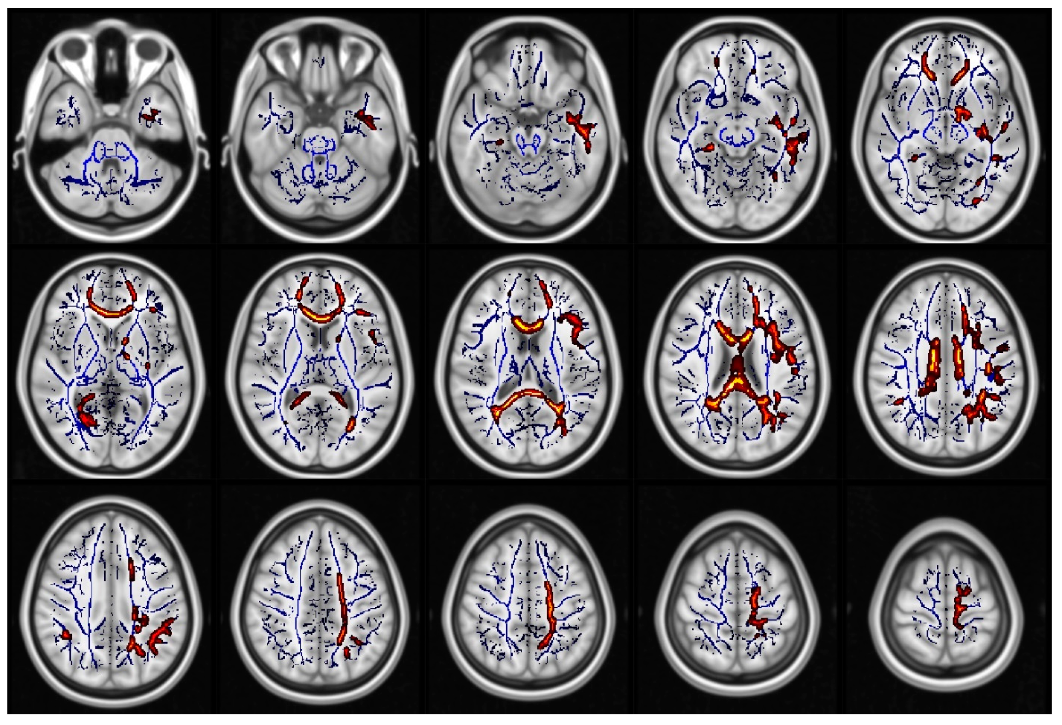
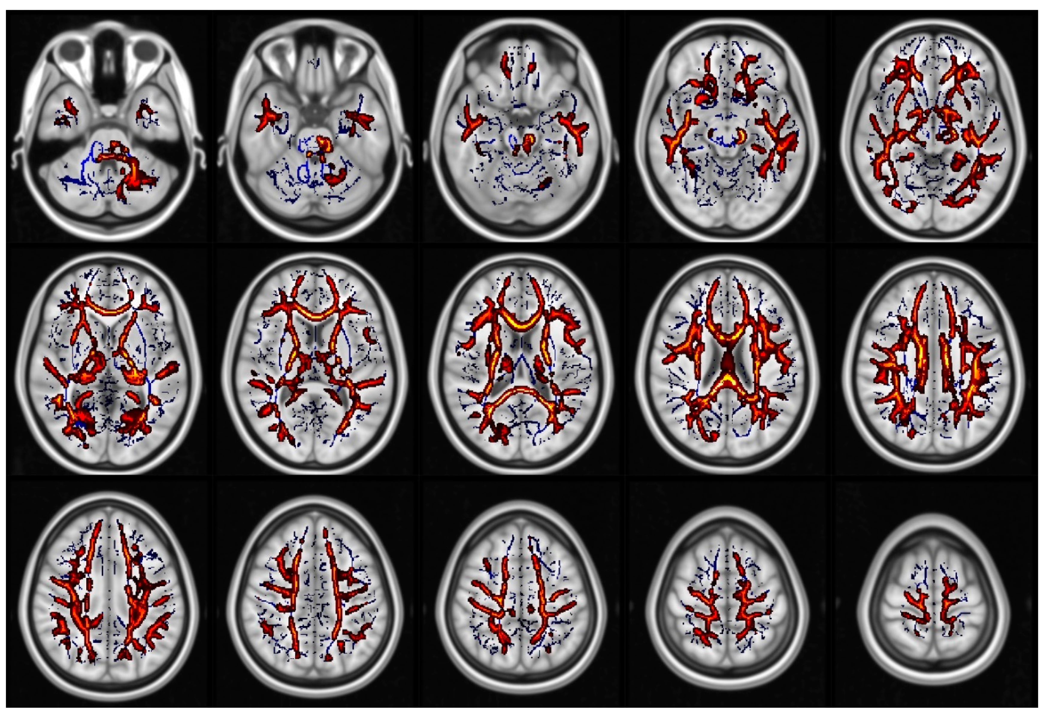
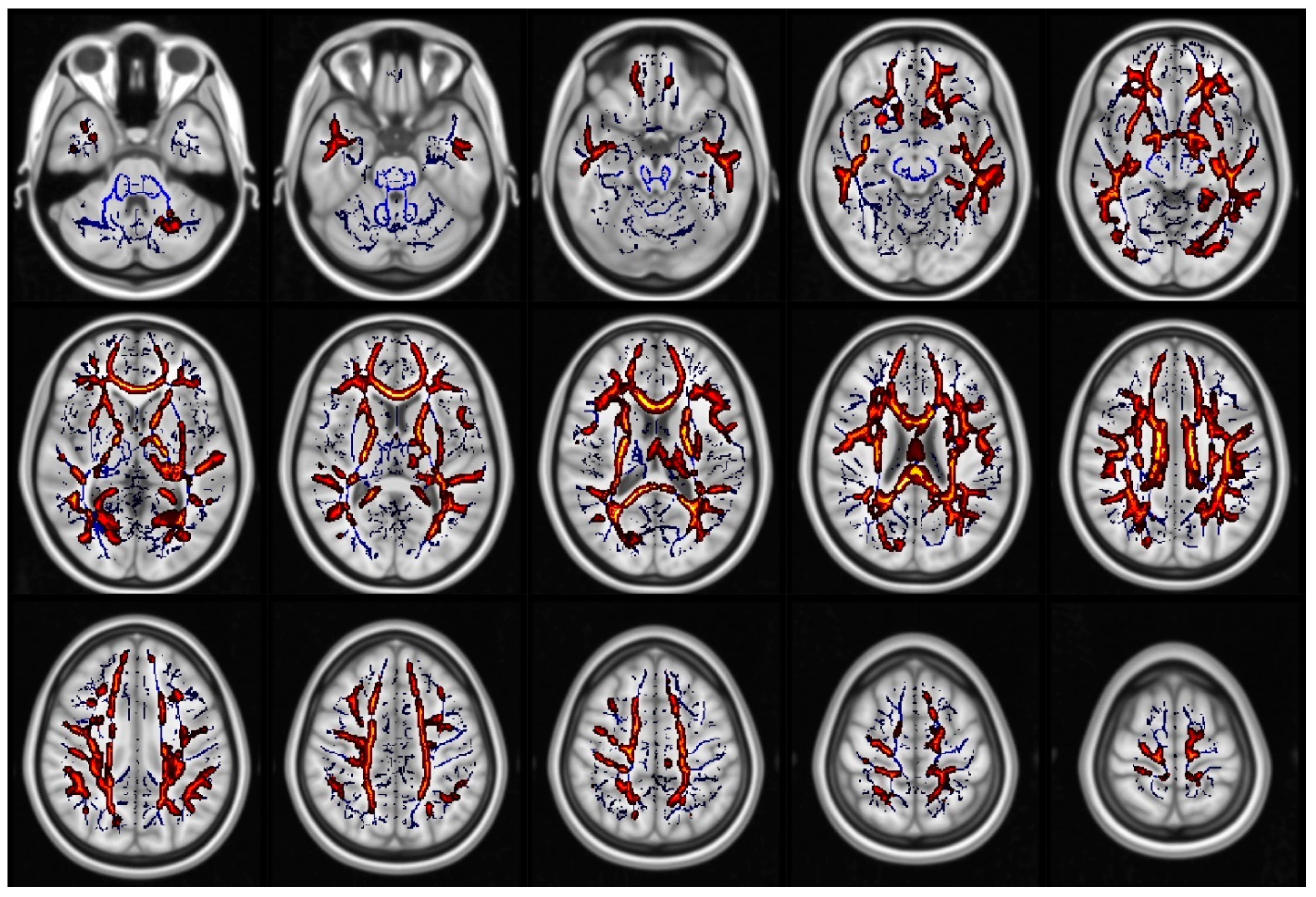
3. Results
4. Discussion
Supplementary Materials
Author Contributions
Funding
Conflicts of Interest
References
- Tham, Y.C.; Li, X.; Wong, T.Y.; Quigley, H.A.; Aung, T.; Cheng, C.-Y. Global Prevalence of Glaucoma and Projections of Glaucoma Burden through 2040. Ophthalmology 2014, 121, 2081–2090. [Google Scholar] [CrossRef] [PubMed]
- Martucci, A.; Toschi, N.; Cesareo, M.; Giannini, C.; Pocobelli, G.; Garaci, F.; Mancino, R.; Nucci, C. Spectral Domain Optical Coherence Tomography Assessment of Macular and Optic Nerve Alterations in Patients with Glaucoma and Correlation with Visual Field Index. J. Ophthalmol. 2018, 2018, 1–9. [Google Scholar] [CrossRef] [PubMed] [Green Version]
- European Glaucoma Society Terminology and Guidelines for Glaucoma, 4th Edition—Chapter 2: Classification and terminologySupported by the EGS Foundation. Br. J. Ophthalmol. 2017, 101, 73–127. [CrossRef] [PubMed] [Green Version]
- Nucci, C.; Martucci, A.; Cesareo, M.; Garaci, F.; Morrone, L.A.; Russo, R.; Corasaniti, M.T.; Bagetta, G.; Mancino, R. Links among glaucoma, neurodegenerative, and vascular diseases of the central nervous system. Prog. Brain Res. 2015, 221, 49–65. [Google Scholar] [PubMed]
- Sotimehin, A.E.; Ramulu, P.Y. Measuring Disability in Glaucoma. J. Glaucoma 2018, 27, 939–949. [Google Scholar] [CrossRef] [PubMed]
- Ramulu, P. Glaucoma and disability: Which tasks are affected, and at what stage of disease? Curr. Opin. Ophthalmol. 2009, 20, 92–98. [Google Scholar] [CrossRef] [Green Version]
- Ehrlich, J.R.; Moroi, S.E. Glaucoma, Cognitive Decline, and Healthy Aging. JAMA Ophthalmol. 2017, 135, 740–741. [Google Scholar] [CrossRef]
- Diniz-Filho, A.; Delano-Wood, L.; Daga, F.B.; Cronemberger, S.; Medeiros, F.A. Association between Neurocognitive Decline and Visual Field Variability in Glaucoma. JAMA Ophthalmol. 2017, 135, 734–739. [Google Scholar] [CrossRef] [Green Version]
- Honjo, M.; Numaga, J.; Hara, T.; Asaoka, R. The association between structure-function relationships and cognitive impairment in elderly glaucoma patients. Sci. Rep. 2017, 7, 1–9. [Google Scholar] [CrossRef] [Green Version]
- Jonas, J.B.; Bin Wei, W.; Zhu, L.P.; Xu, L.; Wang, Y.X. Cognitive Function and Ophthalmological Diseases: The Beijing Eye Study. Sci. Rep. 2018, 8, 4816. [Google Scholar] [CrossRef]
- Mancino, R.; Cesareo, M.; Martucci, A.; Di Carlo, E.; Ciuffoletti, E.; Giannini, C.; Morrone, L.A.; Nucci, C.; Garaci, F.; Raffaele, M. Neurodegenerative Process Linking the Eye and the Brain. Curr. Med. Chem. 2019, 26, 3754–3763. [Google Scholar] [CrossRef] [PubMed]
- Nucci, C.; Martucci, A.; Cesareo, M.; Mancino, R.; Russo, R.; Bagetta, G.; Cerulli, L.; Garaci, F.G. Brain involvement in glaucoma: Advanced neuroimaging for understanding and monitoring a new target for therapy. Curr. Opin. Pharmacol. 2013, 13, 128–133. [Google Scholar] [CrossRef] [PubMed]
- Nucci, C.; Russo, R.; Martucci, A.; Giannini, C.; Garaci, F.; Floris, R.; Bagetta, G.; Morrone, L.A.; Carlo, N.; Rossella, R.; et al. New strategies for neuroprotection in glaucoma, a disease that affects the central nervous system. Eur. J. Pharmacol. 2016, 787, 119–126. [Google Scholar] [CrossRef] [PubMed] [Green Version]
- Frezzotti, P.; Giorgio, A.; Motolese, I.; De Leucio, A.; Iester, M.; Motolese, E.; Federico, A.; De Stefano, N. Structural and Functional Brain Changes beyond Visual System in Patients with Advanced Glaucoma. PLoS ONE 2014, 9, e105931. [Google Scholar] [CrossRef] [PubMed] [Green Version]
- Boucard, C.C.; Hanekamp, S.; Ćurčić-Blake, B.; Ida, M.; Yoshida, M.; Cornelissen, F.W. Neurodegeneration beyond the primary visual pathways in a population with a high incidence of normal-pressure glaucoma. Ophthalmic Physiol. Opt. 2016, 36, 344–353. [Google Scholar] [CrossRef]
- Chen, W.W.; Wang, N.-L.; Cai, S.; Fang, Z.; Yu, M.; Wu, Q.; Tang, L.; Guo, B.; Feng, Y.; Jonas, J.B.; et al. Structural Brain Abnormalities in Patients with Primary Open-Angle Glaucoma: A Study with 3T MR Imaging. Investig. Opthalmol. Vis. Sci. 2013, 54, 545–554. [Google Scholar] [CrossRef]
- Wang, Y.; Wang, X.; Zhou, J.; Qiu, J.; Yan, T.; Xie, Y.; Li, L.; Lu, W. Brain morphological alterations of cerebral cortex and subcortical nuclei in high-tension glaucoma brain and its associations with intraocular pressure. Neuroradiology 2019, 62, 495–502. [Google Scholar] [CrossRef]
- Giorgio, A.; Zhang, J.; Costantino, F.; De Stefano, N.; Frezzotti, P. Altered Large-Scale Brain Functional Connectivity in Ocular Hypertension. Front. Mol. Neurosci. 2020, 14. [Google Scholar] [CrossRef] [Green Version]
- Minosse, S.; Garaci, F.; Martucci, A.; Lanzafame, S.; Di Giuliano, F.; Picchi, E.; Cesareo, M.; Mancino, R.; Guerrisi, M.; Pistolese, C.A.; et al. Primary Open Angle Glaucoma Is Associated with Functional Brain Network Reorganization. Front. Neurol. 2019, 10. [Google Scholar] [CrossRef]
- Frezzotti, P.; Giorgio, A.; Toto, F.; De Leucio, A.; De Stefano, N. Early changes of brain connectivity in primary open angle glaucoma. Hum. Brain Mapp. 2016, 37, 4581–4596. [Google Scholar] [CrossRef]
- Giorgio, A.; Zhang, J.; Costantino, F.; De Stefano, N.; Frezzotti, P. Diffuse brain damage in normal tension glaucoma. Hum. Brain Mapp. 2017, 39, 532–541. [Google Scholar] [CrossRef] [PubMed] [Green Version]
- Jensen, J.H.; Helpern, J.A.; Ramani, A.; Lu, H.; Kaczynski, K. Diffusional kurtosis imaging: The quantification of non-gaussian water diffusion by means of magnetic resonance imaging. Magn. Reson. Med. 2005, 53, 1432–1440. [Google Scholar] [CrossRef]
- Jensen, J.H.; Helpern, J.A. MRI quantification of non-Gaussian water diffusion by kurtosis analysis. NMR Biomed. 2010, 23, 698–710. [Google Scholar] [CrossRef] [PubMed]
- Fieremans, E.; Jensen, J.H.; Helpern, J.A. White matter characterization with diffusional kurtosis imaging. NeuroImage 2011, 58, 177–188. [Google Scholar] [CrossRef] [Green Version]
- Lanzafame, S.; Giannelli, M.; Garaci, F.; Floris, R.; Duggento, A.; Guerrisi, M.; Toschi, N. Differences in Gaussian diffusion tensor imaging and non-Gaussian diffusion kurtosis imaging model-based estimates of diffusion tensor invariants in the human brain. Med. Phys. 2016, 43, 2464–2475. [Google Scholar] [CrossRef]
- Xu, Z.; Sun, J.; Zhang, X.; Feng, Y.; Pan, A.; Gao, M.; Zhao, H. Microstructural visual pathway abnormalities in patients with primary glaucoma: 3 T diffusion kurtosis imaging study. Clin. Radiol. 2018, 73, 591.e9–591.e15. [Google Scholar] [CrossRef] [PubMed]
- Li, T.; Qu, X.; Chen, W.; Wang, Q.; Wang, H.; Wang, Y.; Huang, C.; Zhang, X.; Wang, N.; Xian, J. Altered information flow and microstructure abnormalities of visual cortex in normal-tension glaucoma: Evidence from resting-state fMRI and DKI. Brain Res. 2020, 1741, 146874. [Google Scholar] [CrossRef]
- Hodapp, E.; Parrish, R.; Anderson, D.R. Clinical Decisions in Glaucoma; Mosby Incorporated.: St. Louis, MO, USA, 1993. [Google Scholar]
- Leemans, A.; Jeurissen, B.; Sijbers, J.; Jones, D.K. ExploreDTI: A graphical toolbox for processing, analyzing, and visualizing diffusion MR data. Int. Soc. Magn. Reson. Med. 2009, 17. [Google Scholar]
- Smith, S.M.; Jenkinson, M.; Johansen-Berg, H.; Rueckert, D.; Nichols, T.E.; Mackay, C.; E Watkins, K.; Ciccarelli, O.; Cader, M.Z.; Matthews, P.M.; et al. Tract-based spatial statistics: Voxelwise analysis of multi-subject diffusion data. NeuroImage 2006, 31, 1487–1505. [Google Scholar] [CrossRef] [PubMed]
- Jenkinson, M.; Beckmann, C.F.; Behrens, T.E.J.; Woolrich, M.W.; Smith, S.M. Review FSL. NeuroImage 2012, 62, 782–790. [Google Scholar] [CrossRef] [Green Version]
- Smith, S.M.; Nichols, T.E. Threshold-free cluster enhancement: Addressing problems of smoothing, threshold dependence and localisation in cluster inference. NeuroImage 2009, 44, 83–98. [Google Scholar] [CrossRef] [PubMed] [Green Version]
- Taoka, T.; Fujioka, M.; Kashiwagi, Y.; Obata, A.; Rokugawa, T.; Hori, M.; Masutani, Y.; Aoki, S.; Naganawa, S.; Abe, K. Time Course of Diffusion Kurtosis in Cerebral Infarctions of Transient Middle Cerebral Artery Occlusion Rat Model. J. Stroke Cerebrovasc. Dis. 2016, 25, 610–617. [Google Scholar] [CrossRef] [PubMed]
- Weber, R.A.; Hui, E.S.; Jensen, J.H.; Nie, X.; Falangola, M.F.; Helpern, J.A.; Adkins, D.L. Diffusional kurtosis and diffusion tensor imaging reveal different time-sensitive stroke-induced microstructural changes. Stroke 2015, 46, 545–550. [Google Scholar] [CrossRef] [PubMed] [Green Version]
- Delouche, A.; Attyé, A.; Heck, O.; Grand, S.; Kastler, A.; Lamalle, L.; Renard, F.; Krainik, A. Diffusion MRI: Pitfalls, literature review and future directions of research in mild traumatic brain injury. Eur. J. Radiol. 2016, 85, 25–30. [Google Scholar] [CrossRef] [PubMed]
- Stokum, J.A.; Sours, C.; Zhuo, J.; Kane, R.; Shanmuganathan, K.; Gullapalli, R.P. A longitudinal evaluation of diffusion kurtosis imaging in patients with mild traumatic brain injury. Brain Inj. 2014, 29, 47–57. [Google Scholar] [CrossRef] [PubMed]
- Raab, P.; Hattingen, E.; Franz, K.; Zanella, F.E.; Lanfermann, H. Cerebral Gliomas: Diffusional Kurtosis Imaging Analysis of Microstructural Differences. Radiology 2010, 254, 876–881. [Google Scholar] [CrossRef]
- Van Cauter, S.; Veraart, J.; Sijbers, J.; Peeters, R.R.; Himmelreich, U.; De Keyzer, F.; Van Gool, S.W.; Van Calenbergh, F.; De Vleeschouwer, S.; Van Hecke, W.; et al. Gliomas: Diffusion Kurtosis MR Imaging in Grading. Radiology 2012, 263, 492–501. [Google Scholar] [CrossRef] [Green Version]
- Giannelli, M.; Toschi, N.; Passamonti, L.; Mascalchi, M.; Diciotti, S.; Tessa, C. Diffusion kurtosis and diffusion-tensor MR imaging in Parkinson disease. Radiology 2012, 265, 645–646. [Google Scholar] [CrossRef]
- Struyfs, H.; Van Hecke, W.; Veraart, J.; Sijbers, J.; Slaets, S.; De Belder, M.; Wuyts, L.; Peters, B.; Sleegers, K.; Robberecht, C.; et al. Diffusion Kurtosis Imaging: A Possible MRI Biomarker for AD Diagnosis? J. Alzheimer’s Dis. 2015, 48, 937–948. [Google Scholar] [CrossRef] [Green Version]
- Bester, M.; Jensen, J.H.; Babb, J.; Tabesh, A.; Miles, L.; Herbert, J.; I Grossman, R.; Inglese, M. Non-Gaussian diffusion MRI of gray matter is associated with cognitive impairment in multiple sclerosis. Mult. Scler. J. 2014, 21, 935–944. [Google Scholar] [CrossRef] [Green Version]
- Bonilha, L.; Lee, C.Y.; Jensen, J.H.; Tabesh, A.; Spampinato, M.V.; Edwards, J.C.; Breedlove, J.; Helpern, J.A. Altered microstructure in temporal lobe epilepsy: A diffusional kurtosis imaging study. Am. J. Neuroradiol. 2014, 36, 719–724. [Google Scholar] [CrossRef] [Green Version]
- Tummala, S.; Palomares, J.; Kang, D.W.; Park, B.; Woo, M.A.; Harper, R.M.; Kumar, R. Global and Regional Brain Non-Gaussian Diffusion Changes in Newly Diagnosed Patients with Obstructive Sleep Apnea. Sleep 2016, 39, 51–57. [Google Scholar] [CrossRef] [PubMed]
- Herbet, G.; Zemmoura, I.; Duffau, H. Functional Anatomy of the Inferior Longitudinal Fasciculus: From Historical Reports to Current Hypotheses. Front. Neuroanat. 2018, 12. [Google Scholar] [CrossRef] [PubMed] [Green Version]
- Garaci, F.; Picchi, E.; Di Giuliano, F.; Lanzafame, S.; Minosse, S.; Manenti, G.; Pistolese, C.A.; Sarmati, L.; Teti, E.; Andreoni, M.; et al. Cerebral Multishell Diffusion Imaging Parameters are Associated with Blood Biomarkers of Disease Severity in HIV Infection. J. Neuroimaging 2019, 29, 771–778. [Google Scholar] [CrossRef] [PubMed]
- Kandel, E.R. Intermediate-level visual processing and visula primitives. In Principles of Neural Science, 5th ed.; McGraw-Hill: New York, NY, USA, 2013. [Google Scholar]
- Kandel, E. High-level visual processing: Cognitive influences. In Principles of Neural Science, 5th ed.; McGraw-Hill: New York, NY, USA, 2013. [Google Scholar]
- Goodale, M.A.; Milner, A.D. Separate visual pathways for perception and action. Trends Neurosci. 1992, 15, 20–25. [Google Scholar] [CrossRef]
- Jiang, F.; Zeng, F.-F.; Yu, C.; Ye, Y.-Q.; Zeng, X.-J. Altered whole-brain gray matter volume in primary angle closure glaucoma patients. NeuroReport 2018, 29, 1405–1412. [Google Scholar] [CrossRef]
- Wang, J.; Li, T.; Wang, N.; Xian, J.; He, H. Graph theoretical analysis reveals the reorganization of the brain network pattern in primary open angle glaucoma patients. Eur. Radiol. 2016, 26, 3957–3967. [Google Scholar] [CrossRef]
- Tyler, L.K.; Chiu, S.; Zhuang, J.; Randall, B.; Devereux, B.J.; Wright, P.; Clarke, A.; Taylor, K.I. Objects and categories: Feature statistics and object processing in the ventral stream. J. Cogn. Neurosci. 2013, 25, 1723–1735. [Google Scholar] [CrossRef] [Green Version]
- Kanwisher, N.; Yovel, G. The fusiform face area: A cortical region specialized for the perception of faces. Philos. Trans. R. Soc. B Biol. Sci. 2006, 361, 2109–2128. [Google Scholar] [CrossRef] [Green Version]
- Iidaka, T. Role of the fusiform gyrus and superior temporal sulcus in face perception and recognition: An empirical review. Jpn. Psychol. Res. 2013, 56, 33–45. [Google Scholar] [CrossRef]
- Ma, Y.; Han, S. Functional dissociation of the left and right fusiform gyrus in self-face recognition. Hum. Brain Mapp. 2011, 33, 2255–2267. [Google Scholar] [CrossRef] [PubMed]
- Barton, J.J.S.; Press, D.Z.; Keenan, J.P.; O’Connor, M. Lesions of the fusiform face area impair perception of facial configuration in prosopagnosia. Neurology 2002, 58, 71–78. [Google Scholar] [CrossRef] [PubMed]
- Glen, F.C.; Crabb, D.P.; Smith, N.D.; Burton, R.; Garway-Heath, D.F. Do patients with glaucoma have difficulty recognizing faces? Investig. Ophthalmol. Vis. Sci. 2012, 53, 3629–3637. [Google Scholar] [CrossRef] [PubMed] [Green Version]
- McCandliss, B.D.; Cohen, L.; Dehaene, S. The visual word form area: Expertise for reading in the fusiform gyrus. Trends Cogn. Sci. 2003, 7, 293–299. [Google Scholar] [CrossRef] [Green Version]
- Dehaene, S.; Le Clec’H, G.; Poline, J.-B.; Le Bihan, D.; Cohen, L. The visual word form area: A prelexical representation of visual words in the fusiform gyrus. NeuroReport 2002, 13, 321–325. [Google Scholar] [CrossRef] [PubMed]
- Dehaene, S.; Cohen, L. The unique role of the visual word form area in reading. Trends Cogn. Sci. 2011, 15, 254–262. [Google Scholar] [CrossRef]
- Ramulu, P.Y.; West, S.K.; Muñoz, B.; Jampel, H.D.; Friedman, D.S. Glaucoma and Reading Speed. Arch. Ophthalmol. 2009, 127, 82. [Google Scholar] [CrossRef]
- Kandel, E. Visual processing and action. In Principles of Neural Science, 5th ed.; McGraw-Hill: New York, NY, USA, 2013. [Google Scholar]
- Gallivan, J.P.; Goodale, M.A. The dorsal “action” pathway. Handb. Clin. Neurol. 2018, 151, 449–466. [Google Scholar]
- Murphy, M.C.; Conner, I.P.; Teng, C.Y.; Lawrence, J.D.; Safiullah, Z.; Wang, B.; Bilonick, R.A.; Kim, S.G.; Wollstein, G.; Schuman, J.S.; et al. Retinal Structures and Visual Cortex Activity are Impaired Prior to Clinical Vision Loss in Glaucoma. Sci. Rep. 2016. [Google Scholar] [CrossRef]
- Berlucchi, G. Visual interhemispheric communication and callosal connections of the occipital lobes. Cortex 2014, 56, 1–13. [Google Scholar] [CrossRef]
- Tamura, I.; Kitagawa, M.; Otsuki, M.; Kikuchi, S.; Tashiro, K.; Dubois, B. Pure Topographical Disorientation Following a Right Forceps Major of the Splenium Lesion: A Case Study. Neurocase 2007, 13, 178–184. [Google Scholar] [CrossRef] [PubMed]
- Friedman, D.S.; Freeman, E.; Munoz, B.; Jampel, H.D.; West, S.K. Glaucoma and Mobility Performance. Ophthalmology 2007, 114, 2232–2237.e1. [Google Scholar] [CrossRef] [PubMed]
- Welniarz, Q.; Dusart, I.; Roze, E. The corticospinal tract: Evolution, development, and human disorders. Dev. Neurobiol. 2016, 77, 810–829. [Google Scholar] [CrossRef] [PubMed]
- Trivedi, V.; Bang, J.W.; Parra, C.; Colbert, M.K.; O’Connell, C.; Arshad, A.; Faiq, M.A.; Conner, I.P.; Redfern, M.S.; Wollstein, G.; et al. Widespread brain reorganization perturbs visuomotor coordination in early glaucoma. Sci. Rep. 2019, 9, 14168. [Google Scholar] [CrossRef]
- Filippi, C.G.; Cauley, K. Lesions of the Corpus Callosum and Other Commissural Fibers: Diffusion Tensor Studies. Semin. Ultrasound CT MRI 2014, 35, 445–458. [Google Scholar] [CrossRef] [PubMed]
- Levine, D.N.; Lee, J.M.; Fisher, C.M. The visual variant of Alzheimer’s disease: A clinicopathologic case study. Neurology 1993, 43, 305. [Google Scholar] [CrossRef]
- Bokde, A.L.W.; Pietrini, P.; Ibáñez, V.; Furey, M.L.; Alexander, G.E.; Graff-Radford, N.R.; Rapoport, S.I.; Schapiro, M.B.; Horwitz, B. The effect of brain atrophy on cerebral hypometabolism in the visual variant of Alzheimer disease. Arch. Neurol. 2001, 58, 480–486. [Google Scholar] [CrossRef]
- Lee, A.G.; O Martin, C. Neuro-ophthalmic findings in the visual variant of Alzheimer’s disease. Ophthalmology 2004, 111, 376–380. [Google Scholar] [CrossRef]
- Yoshida, T.; Shiga, K.; Yoshikawa, K.; Yamada, K.; Nakagawa, M. White Matter Loss in the Splenium of the Corpus callosum in a Case of Posterior Cortical Atrophy: A Diffusion Tensor Imaging Study. Eur. Neurol. 2004, 52, 77–81. [Google Scholar] [CrossRef]
- Mancino, R.; Martucci, A.; Cesareo, M.; Giannini, C.; Corasaniti, M.T.; Bagetta, G.; Nucci, C. Glaucoma and Alzheimer Disease: One Age-Related Neurodegenerative Disease of the Brain. Curr. Neuropharmacol. 2018, 16, 971–977. [Google Scholar] [CrossRef]
- Inyushin, M.; Zayas-Santiago, A.; Rojas, L.; Kucheryavykh, Y.; Kucheryavykh, L. Platelet-generated amyloid beta peptides in Alzheimer’s disease and glaucoma. Histol. Histopathol. 2019, 34, 843–856. [Google Scholar] [PubMed]
- Xu, X.; Zou, J.; Geng, W.; Wang, A. Association between glaucoma and the risk of Alzheimer’s disease: A systematic review of observational studies. Acta Ophthalmol. 2019, 97, 665–671. [Google Scholar] [CrossRef]
- Mitsuda, M.; Suzuki, Y.; Kunimatsu, A.; Kasahara, A.; Watanabe, Y.; Ino, K.; Yano, K.; Ohtomo, K. Feasibility of diffusion tensor imaging at 1.5T using multi-band echo planar acquisition. Magn. Reson. Med. Sci. 2017, 16, 169–175. [Google Scholar] [CrossRef] [PubMed]
- Lee, H.-L.; Zhou, X.A.; Li, Z.; Chuang, K.-H. Optimizing diffusion MRI acquisition efficiency of rodent brain using simultaneous multislice EPI. NMR Biomed. 2020. Online ahead of print. [Google Scholar] [CrossRef] [PubMed]
- Veraart, J.; Poot, D.; Van Hecke, W.; Blockx, I.; Van Der Linden, A.M.; Verhoye, M.; Sijbers, J. More accurate estimation of diffusion tensor parameters using diffusion kurtosis imaging. Magn. Reson. Med. 2010, 65, 138–145. [Google Scholar] [CrossRef] [PubMed]


| POAG | Healthy Controls | |
|---|---|---|
| Population | n = 23 | n = 15 |
| Gender | 15 F, 8 M | 6 F, 9 M |
| Average age | 61.2 ± 6.9 | 60.2 ± 9.8 |
| Stages | I: 4, II: 6, III: 6, IV: 5, V: 2 |
© 2020 by the authors. Licensee MDPI, Basel, Switzerland. This article is an open access article distributed under the terms and conditions of the Creative Commons Attribution (CC BY) license (http://creativecommons.org/licenses/by/4.0/).
Share and Cite
Nucci, C.; Garaci, F.; Altobelli, S.; Di Ciò, F.; Martucci, A.; Aiello, F.; Lanzafame, S.; Di Giuliano, F.; Picchi, E.; Minosse, S.; et al. Diffusional Kurtosis Imaging of White Matter Degeneration in Glaucoma. J. Clin. Med. 2020, 9, 3122. https://doi.org/10.3390/jcm9103122
Nucci C, Garaci F, Altobelli S, Di Ciò F, Martucci A, Aiello F, Lanzafame S, Di Giuliano F, Picchi E, Minosse S, et al. Diffusional Kurtosis Imaging of White Matter Degeneration in Glaucoma. Journal of Clinical Medicine. 2020; 9(10):3122. https://doi.org/10.3390/jcm9103122
Chicago/Turabian StyleNucci, Carlo, Francesco Garaci, Simone Altobelli, Francesco Di Ciò, Alessio Martucci, Francesco Aiello, Simona Lanzafame, Francesca Di Giuliano, Eliseo Picchi, Silvia Minosse, and et al. 2020. "Diffusional Kurtosis Imaging of White Matter Degeneration in Glaucoma" Journal of Clinical Medicine 9, no. 10: 3122. https://doi.org/10.3390/jcm9103122
APA StyleNucci, C., Garaci, F., Altobelli, S., Di Ciò, F., Martucci, A., Aiello, F., Lanzafame, S., Di Giuliano, F., Picchi, E., Minosse, S., Cesareo, M., Guerrisi, M. G., Floris, R., Passamonti, L., & Toschi, N. (2020). Diffusional Kurtosis Imaging of White Matter Degeneration in Glaucoma. Journal of Clinical Medicine, 9(10), 3122. https://doi.org/10.3390/jcm9103122