Volumetric Carotid Flow Characteristics in Doppler Ultrasonography in Healthy Population Over 65 Years Old

Abstract
:1. Introduction
2. Subjects and Methods
3. Statistics
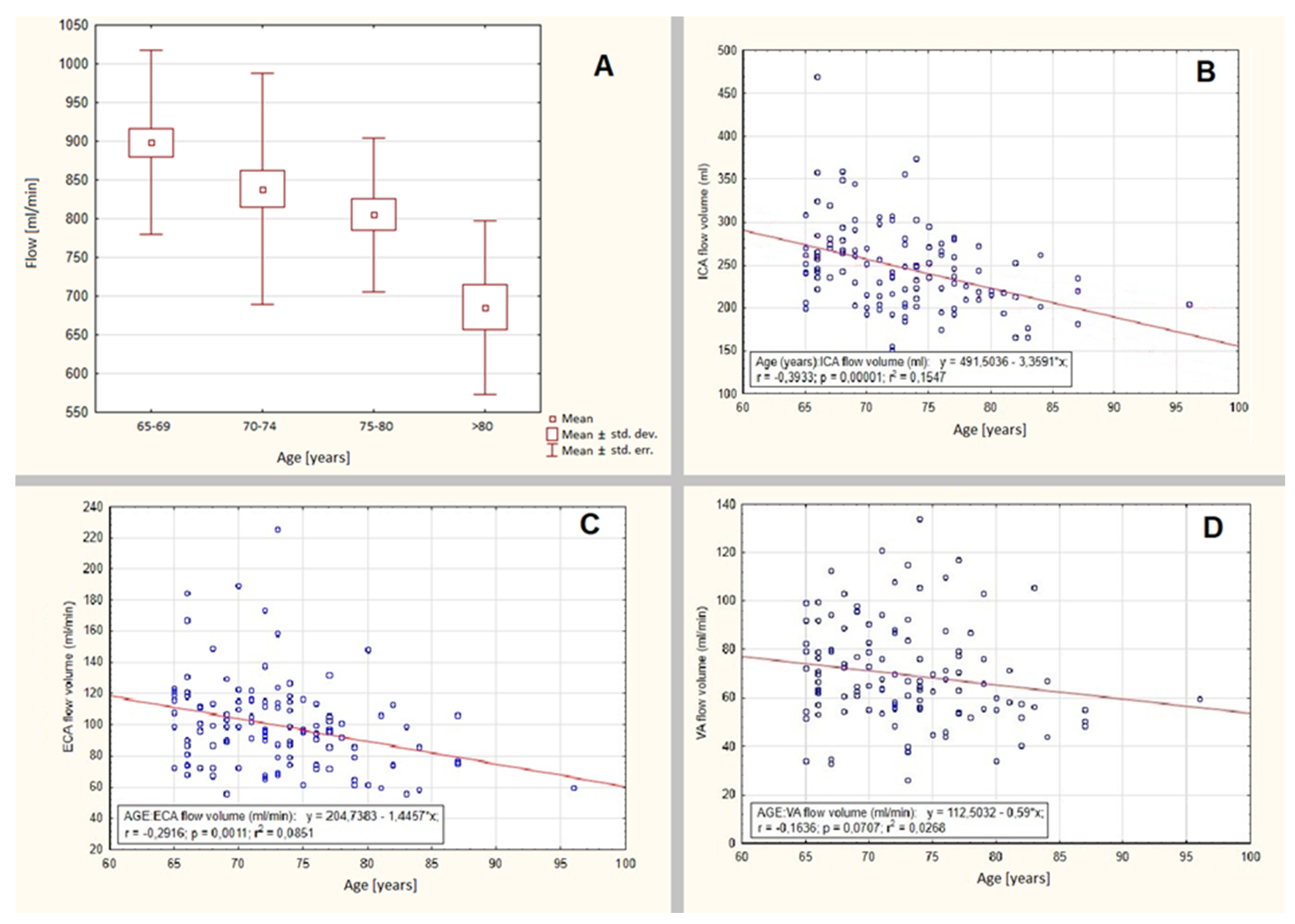
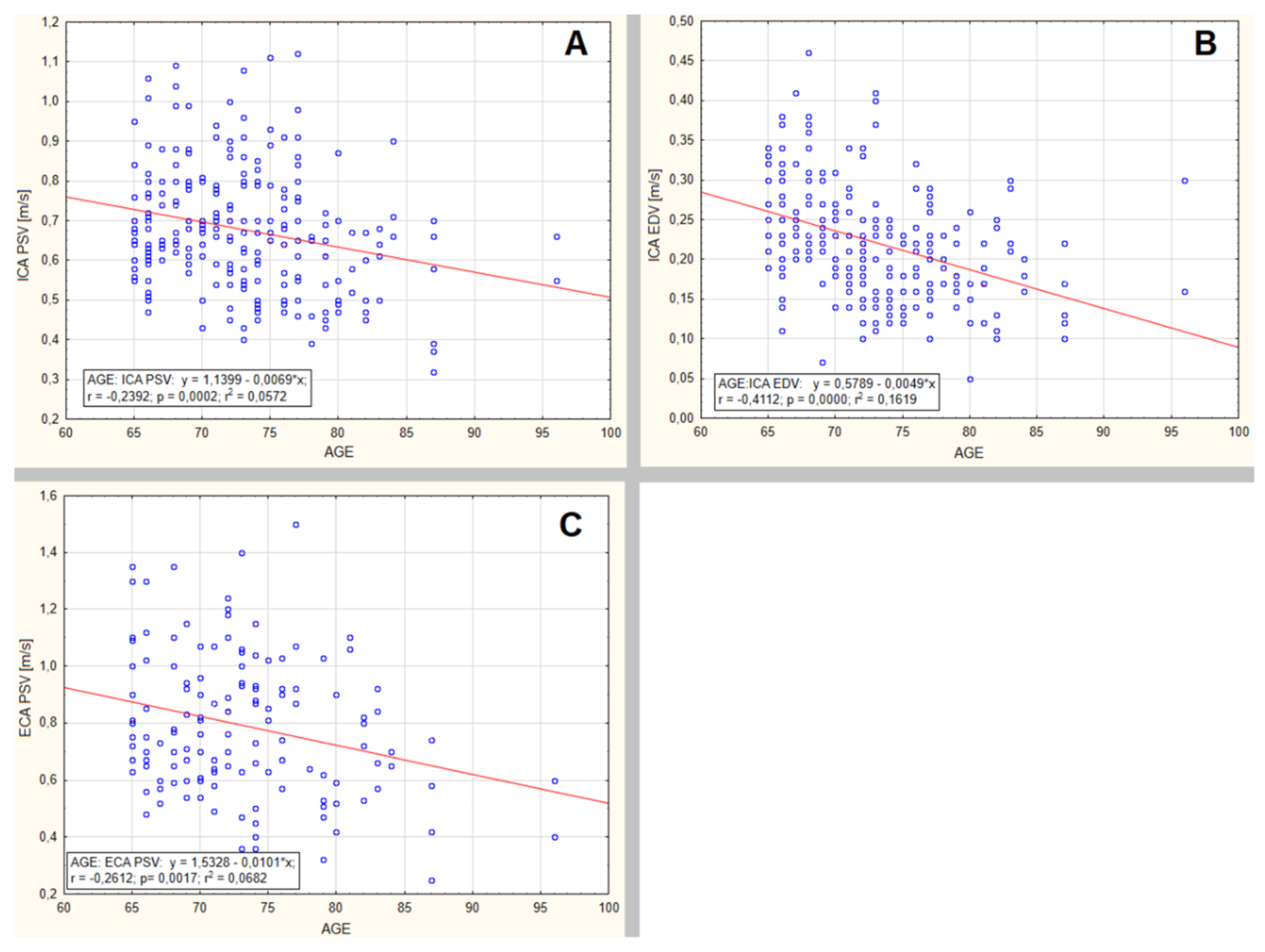
4. Results
5. Discussion
6. Conclusions
Author Contributions
Funding
Conflicts of Interest
References
- Sigvant, B.; Wiberg-Hedman, K.; Bergqvist, D.; Rolandsson, O.; Andersson, B.; Persson, E.; Wahlberg, E. A population-based study of peripheral arterial disease prevalence with special focus on critical limb ischemia and sex differences. J. Vasc. Surg. 2007, 45, 1185–1191. [Google Scholar] [CrossRef] [PubMed] [Green Version]
- Sigvant, B.; Lundin, F.; Wahlberg, E. The Risk of Disease Progression in Peripheral Arterial Disease is Higher than Expected: A Meta-Analysis of Mortality and Disease Progression in Peripheral Arterial Disease. Eur. J. Vasc. Endovasc. Surg. 2016, 51, 395–403. [Google Scholar] [CrossRef] [PubMed] [Green Version]
- WHO Cardiovascular Diseases Fact Sheets. Available online: https://www.who.int/en/news-room/fact-sheets/detail/cardiovascular-diseases-(cvds) (accessed on 28 May 2019).
- WHO Top 10 Causes of Death Fact Sheets. Available online: https://www.who.int/news-room/fact-sheets/detail/the-top-10-causes-of-death (accessed on 28 May 2019).
- WHO: The Atlas of Heart Disease and Stroke: Global Burden of Stroke. Available online: http://www.who.int/cardiovascular_diseases/resources/atlas/en (accessed on 28 May 2019).
- Roger, V.L.; Go, A.S.; Lloyd-Jones, N.M.; Benjamin, E.J.; Berry, J.D.; Borden, W.B.; Bravata, D.M.; Dai, S.; Ford, E.S.; Fox, C.S.; et al. Heart Disease and Stroke Statistics—2012 Update. Circulation 2012, 125, 2–220. [Google Scholar]
- Aboyans, V.; Ricco, J.B.; Bartelink, M.L.; Björck, M.; Brodmann, M.; Cohnert, T.; Collet, J.P.; Czerny, M.; De Carlo, M.; Debus, S. Editor’s Choice - 2017 ESC Guidelines on the Diagnosis and Treatment of Peripheral Arterial Diseases, in collaboration with the European Society for Vascular Surgery (ESVS). Eur. J. Vasc. Endovasc. Surg. 2018, 55, 305–368. [Google Scholar] [CrossRef] [PubMed]
- Kalayci, T.O.; Sonmezgoz, F.; Apaydin, M.; Inci, M.F.; Erdogan, H.; Oyar, O.; Çakir, H.; Kestelli, M. Effects of carotid artery stenosis and plaque localization on the incidence of cerebral infarct and diameter of vertebral artery: A duplex ultrasonography and MRI evaluation. Int. J. Clin. Exp. Med. 2016, 9, 22393–22397. [Google Scholar]
- Scheel, P.; Ruge, C.; Petruch, U.R.; Schoning, M. Color Duplex Measurement of Cerebral Blood Flow Volume in Healthy Adults. Stroke 2000, 31, 147–150. [Google Scholar] [CrossRef] [PubMed] [Green Version]
- Schöning, M.; Walter, J.; Scheel, P. Estimation of cerebral blood flow through color duplex sonography of the carotid and vertebral arteries in healthy adults. Stroke 1994, 25, 17–22. [Google Scholar] [CrossRef] [Green Version]
- Schöning, M.; Scheel, P. Color Duplex Measurement of Cerebral Blood Flow Volume: Intra- and Interobserver Reproducibility and Habituation to Serial Measurements in Normal Subjects. J. Cereb. Blood Flow Metab. 1996, 16, 523–531. [Google Scholar] [CrossRef] [Green Version]
- Matthew, E.; Andreason, P.; Carson, R.E.; Herscovitch, P.; Pettigrew, K.; Cohen, R.; King, C.; Johanson, C.-E.; Paul, S.M. Reproducibility of Resting Cerebral Blood Flow Measurements with H215O Positron Emission Tomography in Humans. Br. J. Pharmacol. 1993, 13, 748–754. [Google Scholar] [CrossRef] [Green Version]
- Wardlaw, J.M.; Chappell, F.M.; Stevenson, M.; De Nigris, E.; Thomas, S.; Gillard, J.; Berry, E.; Young, G.; Rothwell, P.; Roditi, G.; et al. Accurate, practical and cost-effective assessment of carotid stenosis in the UK. Health Technol. Assess. 2006, 10. [Google Scholar] [CrossRef]
- Gupta, A.; Kesavabhotla, K.; Baradaran, H.; Kamel, H.; Pandya, A.; Giambrone, A.E.; Wright, D.N.; Pain, K.; Mtui, E.E.; Suri, J.S.; et al. Plaque echolucency and stroke risk in asymptomatic carotid stenosis: A systematic review and meta-analysis. Stroke 2014, 46, 91–97. [Google Scholar] [CrossRef] [PubMed] [Green Version]
- Kakkos, S.K.; Nicolaides, A.; Charalambous, I.; Thomas, D.; Giannopoulos, A.; Naylor, A.R.; Geroulakos, G.; Abbott, A.L. Predictors and clinical significance of progression or regression of asymptomatic carotid stenosis. J. Vasc. Surg. 2014, 59, 956–967. [Google Scholar] [CrossRef] [PubMed] [Green Version]
- Markus, H.S.; King, A.; Shipley, M.; Topakian, R.; Cullinane, M.; Reihill, S.; Bornstein, N.M.; Schaafsma, A. Asymptomatic embolisation for prediction of stroke in the Asymptomatic Carotid Emboli Study (ACES): A prospective observational study. Lancet Neurol. 2010, 9, 663–671. [Google Scholar] [CrossRef] [Green Version]
- King, A.; Serena, J.; Bornstein, N.M.; Markus, H.S. Does impaired cerebrovascular reactivity predict stroke risk in asymptomatic carotid stenosis? A prospective substudy of the asymptomatic carotid emboli study. Stroke 2011, 42, 1550–1555. [Google Scholar] [CrossRef] [PubMed] [Green Version]
- Oktar, S.O.; Yücel, C.; Karaosmanoglu, D.; Akkan, K.; Ozdemir, H.; Tokgöz, N.; Tali, T. Blood-flow volume quantification in internal carotid and vertebral arteries: Comparison of 3 different ultrasound techniques with phase-contrast MR imaging. AJNR Am. J. Neuroradiol. 2006, 27, 363–369. [Google Scholar] [PubMed]
- Yazici, B.; Erdogmus, B.; Tugay, A. Cerebral blood flow measurements of the extracranial carotid and vertebral arteries with Doppler ultrasonography in healthy adults. Diagn. Interv. Radiol. 2005, 11, 195–198. [Google Scholar] [PubMed]
- Schöning, M.; Hartig, B. Age Dependence of Total Cerebral Blood Flow Volume from Childhood to Adulthood. Br. J. Pharmacol. 1996, 16, 827–833. [Google Scholar] [CrossRef]
- Uematsu, S.; Yang, A.; Preziosi, T.J.; Kouba, R.; Toung, T.J. Measurement of carotid blood flow in man and its clinical application. Stroke 1983, 14, 256–266. [Google Scholar] [CrossRef] [Green Version]
- Buijs, P.C.; Krabbe-Hartkamp, M.J.; Bakker, C.J.; De Lange, E.; Ramos, L.M.; Breteler, M.; Mali, W.P. Effect of age on cerebral blood flow: Measurement with ungated two-dimensional phase-contrast MR angiography in 250 adults. Radiology 1998, 209, 667–674. [Google Scholar] [CrossRef]
- Kashimada, A.; Machida, K.; Honda, N.; Mamiya, T.; Takahashi, T.; Kamano, T.; Osada, H. Measurement of cerebral blood flow with two-dimensional cine phase-contrast mR imaging: Evaluation of normal subjects and patients with vertigo. Radiat. Med. 1995, 13, 95–102. [Google Scholar]
- Scheel, P.; Ruge, C.; Schöning, M. Flow velocity and flow volume measurements in the extracranial carotid and vertebral arteries in healthy adults: Reference data and the effects of age. Ultrasound Med. Biol. 2000, 26, 1261–1266. [Google Scholar] [CrossRef]
- Schebesch, K.-M.; Simka, S.; Woertgen, C.; Brawanski, A.; Rothoerl, R.D. Normal values of volume flow in the internal carotid artery measured by a new angle-independant Doppler technique for evaluating cerebral perfusion. Acta Neurochir. 2004, 146, 983–986. [Google Scholar] [CrossRef] [PubMed]
- Albayrak, R.; Degirmenci, B.; Acar, M.; Haktanir, A.; Colbay, M.; Yaman, M.; Degirmenci, B.; Haktanir, A. Doppler sonography evaluation of flow velocity and volume of the extracranial internal carotid and vertebral arteries in healthy adults. J. Clin. Ultrasound 2006, 35, 27–33. [Google Scholar] [CrossRef] [PubMed]
- Dekaban, A.S.; Sadowsky, D. Changes in brain weights during the span of human life: Relation of brain weights to body heights and body weights. Ann. Neurol. 1978, 4, 345–356. [Google Scholar] [CrossRef]
- Dobbing, J.; Sands, J. Quantitative growth and development of human brain. Arch. Dis. Child. 1973, 48, 757–767. [Google Scholar] [CrossRef] [Green Version]
- Ho, K.C.; Roessmann, U.; Straumfjord, J.V.; Monroe, G. Analysis of brain weight. I. Adult brain weight in relation to sex, race, and age. Arch. Pathol. Lab. Med. 1980, 104, 635–639. [Google Scholar]
- Udomphorn, Y.; Armstead, W.M.; Vavilala, M.S. Cerebral Blood Flow and Autoregulation After Pediatric Traumatic Brain Injury. Pediatr. Neurol. 2008, 38, 225–234. [Google Scholar] [CrossRef] [Green Version]
- Vavilala, M.S.; A Lee, L.; Lam, A.M. Cerebral blood flow and vascular physiology. Anesthesiol. Clin. N. Am. 2002, 20, 247–264. [Google Scholar] [CrossRef]
- Peters, R. Ageing and the brain. Postgrad. Med. J. 2006, 82, 84–88. [Google Scholar] [CrossRef]
- Kuo, H.-K.; A Lipsitz, L. Cerebral White Matter Changes and Geriatric Syndromes: Is There a Link? J. Gerontol. A Biol. Sci. Med Sci. 2004, 59, 818–826. [Google Scholar] [CrossRef] [PubMed]
- Marstrand, J.R.; Garde, E.; Rostrup, E.; Ring, P.; Rosenbaum, S.; Mortensen, E.L.; Larsson, H. Cerebral perfusion and cerebrovascular reactivity are reduced in white matter hyperintensities. Stroke 2002, 33, 972–976. [Google Scholar] [CrossRef] [PubMed] [Green Version]
- Moody, D.M.; Thore, C.R.; Anstrom, J.A.; Challa, V.R.; Langefeld, C.D.; Brown, W.R. Quantification of Afferent Vessels Shows Reduced Brain Vascular Density in Subjects with Leukoaraiosis. Radiology 2004, 233, 883–890. [Google Scholar] [CrossRef] [PubMed]
- Taki, Y.; Goto, R.; Evans, A.; Zijdenbos, A.; Neelin, P.; Lerch, J.; Sato, K.; Ono, S.; Kinomura, S.; Nakagawa, M.; et al. Voxel-based morphometry of human brain with age and cerebrovascular risk factors. Neurobiol. Aging 2004, 25, 455–463. [Google Scholar] [CrossRef] [PubMed]
- Bendick, P.J.; Glover, J.L. Vertebrobasilar insufficiency: Evaluation by quantitative duplex flow measurements. A preliminary report. J. Vasc. Surg. 1987, 5, 594–600. [Google Scholar] [CrossRef] [Green Version]
- Blanco, P. Volumetric blood flow measurement using Doppler ultrasound: Concerns about the technique. J. Ultrasound 2015, 18, 201–204. [Google Scholar] [CrossRef] [Green Version]
- Gill, R.W. Measurement of blood flow by ultrasound: Accuracy and sources of error. Ultrasound Med. Biol. 1985, 11, 625–641. [Google Scholar] [CrossRef]
- Li, S.; Hoskins, P.R.; Anderson, T.; McDicken, W.N. Measurement of mean velocity during pulsatile flow using time-averaged maximum frequency of Doppler ultrasound waveforms. Ultrasound Med. Biol. 1993, 19, 105–113. [Google Scholar] [CrossRef]
- Tegler, L.; Gillquist, J.; Anderberg, B.; Lundström, B.; Johansson, H. Thyroid blood flow rate in man. Electromagnetic flowmetry during operation in euthyroid normal gland, nontoxic goiter, and hyperthyroidism. J. Endocrinol. Investig. 1981, 4, 335–341. [Google Scholar] [CrossRef]


| Age Group | Number of Patients | Average Age (Years) | Number of Females | Average Age of Females (Years) | Number of Males | Average Age of Males (Years) | Age Median in Females (Years) | Age Median in Males (Years) | Age Standard Deviation in Females (Years) | Age Standard Deviation in Males (Years) |
|---|---|---|---|---|---|---|---|---|---|---|
| 65–69 years | 42 | 66.9 | 21 | 67 | 21 | 66.8 | 67 | 66 | 1.5 | 1.4 |
| 70–74 years | 41 | 72.2 | 16 | 72.1 | 25 | 72.3 | 72 | 72 | 1.2 | 1.5 |
| 75–79 years | 24 | 76.8 | 11 | 76.8 | 13 | 76.8 | 77 | 77 | 1.0 | 1.6 |
| ≥80 years | 16 | 83.7 | 13 | 83.5 | 3 | 84.7 | 82 | 87 | 4.21 | 4.0 |
| <65 years | 56 | 50.3 | 34 | 51.2 | 22 | 49 | 54 | 45 | 10.6 | 10.1 |
| Inclusion Criteria–Study Group | |
| 1 | Age ≥ 65 years |
| 2 | Informed consent before the examination |
| 3 | No hemodynamically significant carotid atherosclerotic lesions, causing blood flow disturbances (ICA stenosis < 30%) |
| 4 | No exclusion criteria |
| Exclusion Criteria–Study Group | |
| 1 | Age < 65 years |
| 2 | No informed consent given before the examination |
| 3 | Internal Carotid Artery stenosis > 30% |
| 4 | Stenosis of Common Carotid, External Carotid or Vertebral Artery |
| 5 | Concomitant diseases: uncontrolled hypertension, ischemic heart disease, heart insufficiency, positive history of heart infraction, positive history of stent implantation to coronary or any other arteries, cardiac arrhythmia, tachycardia, bradycardia, congenital vascular or heart failure, positive history of vascular interventions, presence of endocrine diseases: thyroid goiter, hyper-, hypothyroidism diabetes, adrenal diseases, positive history of thyroid surgery, smoking, alcohol use. |
| 6 | Positive history of ischemic stroke, TIA symptoms or other neurological symptoms. |
| Group-Age | 65–69 | 70–74 | 75–80 | >80 |
|---|---|---|---|---|
| Mean (mL/min) | 898.5 | 838.5 | 805.1 | 685.7 |
| Std. err (mL/min) | 18.4 | 23.3 | 20.3 | 29.0 |
| Std. dev (mL/min) | 119.1 | 148.9 | 99.3 | 112.3 |
| Proposed reference value (mL/min) | 898.5 ± 119.1 | 838.5 ± 148.9 | 805.1 ± 99.3 | 685.7 ± 112.3 |
| ICA volume (mL/min) | 273.8 ± 60.5 | 237.9 ± 54.3 | 240.1 ± 47.3 | 203.3 ± 42.7 |
| VA volume (mL/min) | 71.8 ± 32.3 | 70.3 ± 28.2 | 60.5 ± 25 | 57.3 ± 18.5 |
| ECA volume (mL/min) | 103.6 ± 32.9 | 104.2 ± 32.7 | 91.5 ± 23 | 81 ± 35 |
| ICA PSV (m/s) | 0.72 ± 0.14 | 0.67 ± 0.15 | 0.68 ± 0.17 | 0.59 ± 0.14 |
| ICA EDV (m/s) | 0.26 ± 0.07 | 0.21 ± 0.06 | 0.20 ± 0.05 | 0.18 ± 0.06 |
| VA PSV (m/s) | 0.45 ± 0.11 | 0.45 ± 0.14 | 0.44 ± 0.12 | 0.41 ± 0.11 |
| VA EDV (m/s) | 0.13 ± 0.06 | 0.12 ± 0.05 | 0.13 ± 0.04 | 0.13 ± 0.04 |
| ECA PSV (m/s) | 0.8 ± 0.24 | 0.81 ± 0.25 | 0.78 ± 0.26 | 0.67 ± 0.21 |
| ECA EDV (m/s) | 0.1 ± 0.05 | 0.13 ± 0.07 | 0.13 ± 0.05 | 0.12 ± 0.05 |
© 2020 by the authors. Licensee MDPI, Basel, Switzerland. This article is an open access article distributed under the terms and conditions of the Creative Commons Attribution (CC BY) license (http://creativecommons.org/licenses/by/4.0/).
Share and Cite
Kaszczewski, P.; Elwertowski, M.; Leszczynski, J.; Ostrowski, T.; Galazka, Z. Volumetric Carotid Flow Characteristics in Doppler Ultrasonography in Healthy Population Over 65 Years Old. J. Clin. Med. 2020, 9, 1375. https://doi.org/10.3390/jcm9051375
Kaszczewski P, Elwertowski M, Leszczynski J, Ostrowski T, Galazka Z. Volumetric Carotid Flow Characteristics in Doppler Ultrasonography in Healthy Population Over 65 Years Old. Journal of Clinical Medicine. 2020; 9(5):1375. https://doi.org/10.3390/jcm9051375
Chicago/Turabian StyleKaszczewski, Piotr, Michal Elwertowski, Jerzy Leszczynski, Tomasz Ostrowski, and Zbigniew Galazka. 2020. "Volumetric Carotid Flow Characteristics in Doppler Ultrasonography in Healthy Population Over 65 Years Old" Journal of Clinical Medicine 9, no. 5: 1375. https://doi.org/10.3390/jcm9051375