Article

Influence of Temperature and Screw Pressing on the Quality of Cassava Leaf Fractions

by
Haimanot Hailegiorgis Ayele
*,
Sajid Latif
and
Joachim Müller
Tropics and Subtropics Group, Institute of Agricultural Engineering, University of Hohenheim, 70599 Stuttgart, Germany
*
Author to whom correspondence should be addressed.
Agriculture 2022, 12(1), 42; https://doi.org/10.3390/agriculture12010042
Submission received: 7 December 2021 / Accepted: 28 December 2021 / Published: 31 December 2021

Abstract

:
In this study, the development of a mild processing method for cassava leaves to remove cyanogenic compounds with minimum nutritional loss is evaluated. Fresh leaves were reduced in size using a mixer at temperatures of 25 (room temperature), 55, 80, and 100 °C for 1 min before screw pressing to separate the juice and press cake fractions. Cyanide content in the fresh leaves was reduced by 60% at 100 °C and by 57% in the juice sample processed at 25 °C. The press cake cyanide content was low (210 ppm) in both the control and the sample that was processed at 55 °C. An increase in the temperature for processing cassava leaves to 100 °C resulted in a loss of 5–13% of the CP and 7–18% of the vitamin C content. The press-cake fraction had high beta-carotene, lutein, and chlorophyll a and b content, and low values were registered for the juice fraction. Processing fresh cassava leaves at 25 and 55 °C resulted in fractions with high beta-carotene and lutein content. The protein quality of press cake was better than that of juice for feed. Short thermal shredding with pressing resulted in minimal loss of nutrients and a significant reduction of cyanide in the leaves.

1. Introduction

Cassava (Manihot esculenta Crantz) is an essential staple crop in tropical and subtropical areas [1]. The crop was introduced to Ethiopia in the 1960s and regarded as a food crop after 1984, where it is of strategic importance for combating food insecurity caused by drought [2]. It is mainly grown in the southern region of the country as a food security crop, and the roots are primarily used [3]. In Ethiopia, due to cultural bias and fear of toxicity, the leaves are not used for human consumption; instead, they are left in the field as green manure [2]. Cassava leaves are commonly considered as a byproduct of cassava root harvest and used for human and animal consumption in different parts of the world [4]. Depending on the age, variety, and growth conditions of cassava plants, the leaves contain a relatively high amount of protein, vitamins, minerals, and phytochemicals that are of nutritional and biochemical importance to humans and animals [5,6]. However, the consumption of the leaves in these areas is also limited due to the high level of antinutrients and toxic compounds, such as cyanogenic glucosides [4]. These compounds reduce nutrient absorption and might even lead to other adverse effects [7].
Recently developed cassava leaf processing and protein isolation methods have focused on cyanogen removal, reducing the levels of antinutrient compounds, and reducing nutrient loss [8,9,10]. On a household level, cassava leaf processing is usually performed by pounding and boiling the leaves in water for long time [11]. This process facilitates the rapid removal of cyanogen but also reduces the content of proteins, vitamins, and sulfur-containing amino acids that are necessary to detoxify ingested cyanide. The loss of methionine is particularly unfavorable because it is necessary for the conversion of cyanide to thiocyanate in the body [8]. Cyanide content of cassava leaves can be decreased by more than 98% through solid-state fermentation [12], 81% by combination of blanching, dry heating and wet heating [13], and 93% via chemical treatment with NaHCO3 [10]. The loss of ascorbic acid and protein content can reach 38–75 g 100 g−1DM for different cassava varieties and leaf processing methods [10,14,15]. Other milder cassava leaf processing methods such as pounding and sun or shade drying of leaves can reduce the cyanogen content but result in dull-colored products as well as a reduced water-soluble vitamin, protein, and methionine contents [8]. Leaf processing and fractioning have also been performed by chemical, thermal, and mechanical actions using screw pressing [16,17]. Processing of leaves by screw pressing is commonly used to separate the liquid fraction from the fiber and to concentrate protein [18,19]. However, the challenge remains to find a suitable processing method to produce cassava leaves with high nutrition, low cyanide content, and low fiber content for human consumption [20].
Cassava leaves also have the potential to be used as a major protein source in animal feed for ruminants and monogastric animals, but the high cyanide and fiber contents limit such use [21]. The development of a mild processing method to remove cyanogens and preserve the nutritional content of cassava leaves will play a significant role in its wider use [8]. Therefore, in this study, a method for cassava leaf processing involving short-term heat application followed by size reduction and pressing was assessed. The two fractions obtained during processing with different temperatures were evaluated as food (juice) and feed (press cake).

2. Materials and Methods
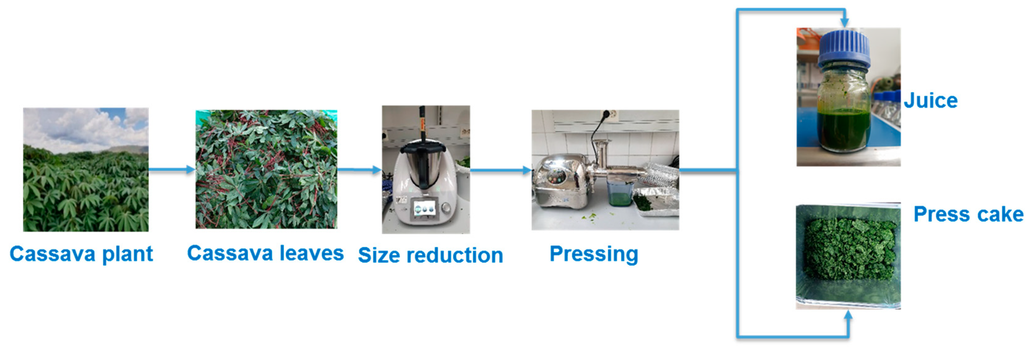
2.1. Plant Material

Cassava (Manihot esculenta Crantz) cultivar Chichu grown at Hawassa Agricultural Research Center, Ethiopia, (6°48′54.05″ N, 38°16′55.58″ E, 1862 m.a.s.l) was used for the experiment. Cassava stem cuttings were planted in the 2018/2019 growing season with a spacing of 1 m × 1 m. The plants were cultivated under rainfed conditions with an annual mean temperature of 22.08 °C and mean annual rainfall of 887.4 mm. Cassava leaves with the petiole attached were harvested by hand from 100 plants within the same plot at the age of one year. All leaf samples, ranging from the first fully expanded leaf to the 15th leaf, were collected on the same day. The leaves were cleaned, packed in polyethylene zip bags, and placed in a box with ice for cooling. The leaves were transported within 24 h to the laboratory at the University of Hohenheim for processing.

2.2. Treatments

After removing the petiole, the size of the leaves was reduced using a food processor with a chopping and heating function (Thermomix TM5, Vorwerk, Cloyes, France). Chopping was conducted for 1 min at a speed of 3100 rpm and at four different temperatures—25 (room temperature), 55, 80, and 100 °C—i.e., temperature was set as the variable factor in the experiment. The temperatures were set based on previous research recommendations for cassava leaves processing [10,21,22,23]. Untreated leaves were taken as a control. Treated leaves and control were kept at room temperature for 30 min before mechanical extraction with a screw press. Screw pressing was done using a commercial lab-scale twin gear screw stainless-steel press (AG- 8500S, Angel Juicers, Queensland, Australia). The press was equipped with a coarse size screen (hole size 1 mm). The processing was conducted at room temperature at a screw speed of 82 rpm. The double screw press was fed continuously to collect the juice and press cake separately (Figure 1). After measuring moisture and cyanide content, the samples were freeze-dried, packed, and stored at −20 °C for further analysis. Treatments were replicated twice.

2.3. Sample Analysis

2.3.1. Antinutritional Factors

The total cyanide content in cassava leaves and fractions was analyzed using the picrate paper kit method [24,25]. Picrate paper was prepared by dipping 0.3 mm thick filter paper into a 2.5% (w/v) picrate solution (Sigma-Aldrich, St. Louis, MO, USA) followed by drying in a fume hood. The dried papers were cut into a 3 cm × 1 cm rectangle and attached to the plastic strip (size 5 cm × 1 cm, 1 mm thickness). Linamarase was isolated according to the method described by Yeoh et al. [26] involving the extraction of enzymes followed by subsequent purification using gel filtration chromatography. A sample of 0.05 g, 1 mL of 0.1 M Na-phosphate buffer, and 100 µL linamarase were placed in a vile and the strip carrying a picrate paper was placed inside the vial, which was closed immediately with a screw cap. The sample and solutions in the vial were gently mixed and left at 30 °C for 24 h. Then, the picrate paper was removed and soaked in 5 mL distilled water for 30 min. A picrate paper suspended in a vial without a sample was used as a blank. The standard curve for cyanide content was prepared from a series of linamarin (Sigma-Aldrich) concentrations (0.2–2.4 µM). The picrate papers from the blank and the standard were treated the same way as the picrate papers of the samples. The absorbance of the solutions was measured at 510 nm.
Phytate in the samples was analyzed according to the method described by Latta and Eskin [27]. Sample extraction was done by placing 0.5 g of the dried sample in a 10 mL of 3.5% HC1 solution. The solution was stirred for 1 h and centrifuged for 10 min at 3000× g. The aliquot was removed from the supernatant, filled into a 2 mL tube, and centrifuged again at 10,000× g for 10 min. Wade reagent was prepared by mixing 30 mg of FeC13∙6H2O and 300 mg of sulfosalicylic acid in 100 mL of distilled water. Standard phytate solution was prepared by dissolving 2632 mg sodium phytate (Sigma-P8810, Merck KGaA, Darmstadt, Germany) in 1 mL of distilled water (2632 mg mL−1). Distilled water (9 mL) was added to the solution (dilution of 1:10). A standard curve was prepared with a range of 0.0–1.0 mL and absorbance was measured at 500 nm using a UV-spectrophotometer (DR6000, Hach Lange, Düsseldorf, Germany).
The total phenolic content (TPC) of the samples was determined using the Folin–Ciocalteu reagent method [28]. A freeze-dried sample of 0.5 g was diluted in 5 mL of 80% methanol and placed in a 60 °C water bath for 20 min. The solution was centrifuged at 13,500 rpm for 10 min (Z 326 K, Hermle Labortechnik GmbH, Wehingen, Germany). The supernatant was transferred, and the residue was mixed again with 3 mL of 80% methanol and centrifuged. The supernatant was combined with the previously extracted solution and the volume was adjusted to 10 mL with 80% methanol. The extracted solution was kept at 4 °C until analysis (max. 48 h). To avoid a deviation of values from the standard curve, the sample was further diluted with 80% methanol (press cake (1:20), leaves (1:20), and juice (1:40)). The sample (150 µL) was mixed with 150 µL of 0.25 N Folin–Ciocalteu reagent and 2400 µL of HPLC water and incubated for 3 min before adding a further 300 µL of 1 N sodium carbonate solution. The sample and standard were incubated for 2 h at room temperature in the dark using 80% methanol as a blank. The absorbance of the standards and the samples at 725 nm were measured using a UV spectrophotometer. The standard calibration curve was prepared by measuring the absorbance of dilutions of a gallic acid stock solution ranging in concentration from 0.005 to 0.1 mg mL−1.

2.3.2. Micronutrients

The method described by Valente et al. [29] was used to measure the vitamin C content of the fractions, with minor modifications. A sample of 1 g was transferred into a 50 mL Falcon tube, and a 15 mL extraction solution (10% perchloric acid and 1% metaphosphoric acid in ultrapure water) was added to stabilize the ascorbic acid and precipitate proteins. The solution was homogenized for 1 min with a vortex and centrifuged at 20,000 rcf for 15 min at 4 °C. A supernatant of 8 mL was transferred into a 12 mL tube and centrifuged again under the same condition. A total 3 mL of the supernatant from each sample was transferred into a 10 mL volumetric flask and filled up with a mobile phase (20 mM ammonium dihydrogen phosphate, pH 3.5, containing 0.015% (w/v) of metaphosphoric acid). The samples were filtrated into HPLC vials with 0.45 µm nylon filter membranes. The separation and quantification were performed using a HPLC system (Shimadzu Co., Kyoto, Japan) equipped with a column of 250 mm × 4.6 mm Luna 5u C18(2) 100A (Phenomenex, Torrance, CA, USA). The quantification of the ascorbic acid components was performed at 254 nm. To calculate the recovery rate, samples were spiked with the main standard.
β-Carotene, lutein, and chlorophyll a and b levels were characterized using HPLC (Agilent 1200, Agilent Technologies, Waldbronn, Germany) according to the method of Lee et al. [30], with some modifications. Mixed analytical standards consisting of β-carotene, lutein, and chlorophyll a and b were prepared in acetone at concentrations from 0 to 100 ppm. Extraction was performed by adding 1 g of the sample in 30 mL of acetone, which was then placed in an ultrasonic bath at 35–40 °C for 90 min. The mixture was filtered through a syringe filter (PTFE, 0.45 µm) and evaluated by HPLC analysis. To separate pigments, a C30 column (stability 100, 5 µm, dimensions 250 mm × 4.6 mm) with guard column (stability 100 C30, 5 µm, 5 mm × 4.6 mm, Dr. Maisch, Ammerbuch-Entringen, Germany) was used. The column temperature was maintained at 30 °C. The mobile consisted of solvent A (75% methanol) and B (100% ethyl acetate). The gradient at a flow rate of 1.0 mL min−1 was set as follows: 0–15 min, 30–90% B; 15–20 min, 90–30% B, followed by a constant 30% B until the end of the running time of 25 min. A 20 µL injection volume was used each time. The peak area of a photodiode array detector was used at 450 nm to calculate the amount of each pigment.

2.3.3. Moisture, Ash, and Crude Protein Content

Cassava leaves, press cake, and juice fraction moisture content was measured by drying the samples in an oven at 105 °C for 12 h [31]. The ash content was measured by placing the oven-dried samples in a muffle furnace, as described in AOAC [31] official method 923.03. Freeze-dried leaf, press cake, and juice crude protein (CP) contents were measured using the Kjeldahl method and a Kjeldahl analysis system (Vapodest 500, C. Gerhardt GmbH & Co. KG, Königswinter, Germany). A conversion factor of 6.25 was used to calculate the amount of CP content from the total nitrogen content.

2.3.4. Acid Detergent Fiber, Acid Detergent Lignin, and Neutral Detergent Fiber

The acid detergent fiber (ADF), acid detergent lignin (ADL), and neutral detergent fiber (NDF) contents of fresh leaf, press cake, and juice were measured according to the method described by AOAC [31] official method 973.18 using an automated fiber analysis system (FibreBag Analysis System FBS6, Gerhardt GmbH & Co. KG, Königswinter, Germany).

2.3.5. Protein Fractioning

The CP of cassava leaves, juice, and press cake was partitioned following the procedures described by Licitra et al. [32]. Samples were analyzed in duplicate, and repetitions were performed in cases where the variation coefficient was greater than 5%. The N concentrations were determined using the Kjeldahl procedure, and all N concentrations were converted to CP using a conversion factor of 6.25. The nonprotein nitrogen (NPN) concentration of the samples was determined using the tungstic acid method [32]. A sample of 0.5 g was weighed into a 100 mL Erlenmeyer flask; then, 50 mL distilled water and 8 mL of 0.3 M sodium tungstate solution were added. The solution was mixed for 30 min and continuously stirred, and the solution pH was reduced to 2.0 using a sulfuric acid solution (0.5 M). The flask was covered and kept at room temperature overnight. Soluble true protein concentrations were determined using a borate–phosphate buffer (pH 6.7–6.8). In total, 50 mL of buffer and 1 mL of freshly prepared sodium azide were added to 0.5 g of the sample in 100 mL Erlenmeyer flasks. The flasks were covered for 3 h before filtration.
NPN and soluble true protein filtration of the suspensions were followed by washing both the residue and filter paper (Whatman paper N° 54, GE Healthcare Life Sciences, Darmstadt, Germany) with 250 mL of cold distilled water. The washed filter paper with residue was dried at 38 °C for 1 h. The N value for the residue and filter paper was analyzed. The NPN and soluble true protein concentration of the samples was calculated by subtracting the N concentration in the residual material from the total N concentration in the sample.
The neutral detergent-insoluble CP (NDICP) was determined following the procedure of NDF analysis without using sodium sulfite. The sample (0.5 g) was boiled in 100 mL of neutral detergent solution for 1 h. A 25 µL aliquot of alpha-amylase (Ankom Technology, NY, USA) was added 1 and 30 min after the solution started boiling. The solution was filtered through a filter paper (Whatman paper N° 54, GE Healthcare Life Sciences, Darmstadt, Germany). The acid detergent-insoluble CP (ADICP) was determined in the same way as the NDICP, except that the neutral detergent solution was substituted with an acid detergent solution and alpha-amylase was not used. The residue with filter paper was washed with 250 mL hot distilled water (80 °C). Then, it was rinsed twice with 5 mL acetone and dried at 38 °C for 1 h. The filter paper with residue was then analyzed for N. The concentrations of different CP fractions were then calculated according to Sniffen et al. [33]. These have been described as fractions NPN (A), soluble true protein (B1), insoluble true protein (B2), protein that is insoluble in neutral detergent but soluble in acid detergent (B3), and protein that is insoluble in acid detergent (C).

2.4. Statistical Analysis

One-way analysis of variance (ANOVA) was conducted to determine the effect of the size reduction of leaves subject to different processing temperatures on the nutritional and antinutritional content of cassava leaf, press cake, and juice. The significant difference of sample means was identified using Tukey’s test at a significance level of p ≤ 0.05. All analyses were performed using SAS statistical software (version 9.2, SAS Institute Inc., Cary, NC, USA) and two independent replicates.

3. Results and Discussion
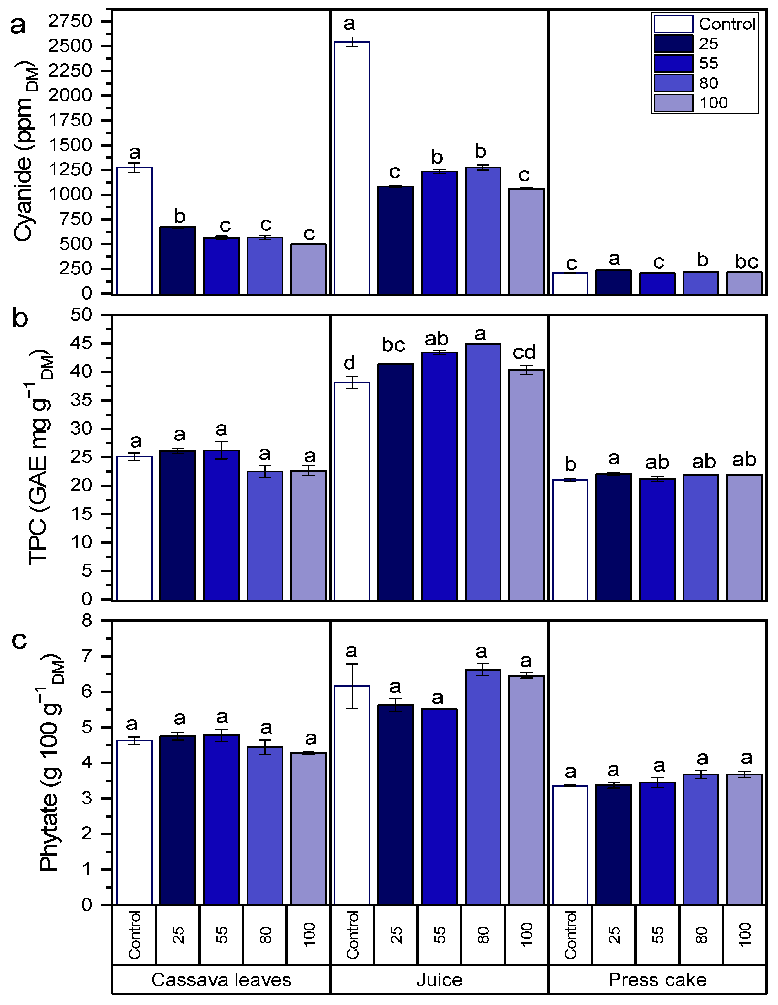
3.1. Antinutrients

A general comparison of the antinutrient contents across the fractions—namely, leaves, juice, and press cake—showed that mechanical extraction by screw pressing leads to unequal partitioning of the original content, with higher contents in the juice and lower contents in the press cake. Observing the controls after pressing revealed that the original cyanide content of 1275 ppm in the leaves increased to 2543 ppm in the juice but decreased to 211 ppm in the press cake. This means that pressing alone was effective for obtaining a press cake with a low toxicity level; however, for the juice, the need for detoxification was increased. The difference between control and chopping at 25 °C showed the effect of chopping alone, i.e., without additional heating. Regarding cyanide content, it decreased to 671 ppm in the leaves, which corresponds to a 47% reduction. In the juice, the reduction was even more pronounced at 57%. The reduction in cyanide content caused by the disruption of leaf tissue and juice through grinding was caused by the action of endogenous linamarase on glucosides [21]. The positive impact of size reduction on lowering cyanide content in the present experiment is similar to what was reported by Ravindran et al. [34]. Applying heat during chopping further reduced the cyanide content in the leaves but to a smaller amount and without significant differences between the temperatures. Already at a moderate temperature of 55 °C, the reduction was 56%. In the juice, heating at 55 and 80 °C during chopping slightly weakened the detoxification effect, and cyanide was reduced by 50%. In the press cake, chopping with and without heating yielded negligible differences (Figure 2a). The significant reduction of cyanide in leaves processed at 55 °C can be explained by the stability of linamarase enzyme being optimum at a temperature of 55 °C [21]. The amount of cyanide in all the fractions in the current study was higher than what was stated as a safe level (10 ppm) by the FAO/WHO [35]. The remaining cyanide content in the leaves and press cake fractions can be reduced further by drying [36] or using membrane filtration or coagulation for the juice fraction [37]. It was observed by Bradbury and Denton [8] that processing methods with longer heat application time can reduce up to 99% of the total cyanogens in cassava leaves but at the expense of high nutritional loss.
After pressing, the initial TPC of 25 GAE mg g−1DM in the fresh cassava leaves was concentrated in the juice to 38 GAE mg g−1DM while it was lowered to 21 GAE mg g−1DM in the press cake fraction. The impact of size reduction on the TPC of the fresh leaves was not significant, whereas it tends to slightly increase the TPC in the juice and press cake from 38 to 41 GAE mg g−1DM and 21 to 22 GAE mg g−1DM, respectively. Increasing the processing temperature to 55 and 80 °C tends to increase the TPC of the juice fraction. The slight increase in TPC of the juice and press cake fraction after heat application can be explained by the increase of free-radical scavenging activities or the inactivation of several enzymes. The same result was observed on blanched Carica papaya L. leaf by Raja et al. [38]. In the juice fraction, TPC was increased to some extent when processing temperatures were increased and then decreased, similarly to tea leaf drying at different temperatures [39]. Leaf fractions processed without heat application and size reduction showed a significantly low value of TPC (Figure 2b). As previously reported, leaf processing can help dephosphorylate phytate to release minerals and facilitate their absorption [40]. Even though the impact of size reduction and application of temperature did not show a significant difference in the phytate content in all cassava leaf fractions, higher concentrations were observed in all juice fractions after pressing (Figure 2c). Phytate is relatively heat-stable during processing. To minimize phytate in cassava leaf, longer heat application or fermentation is needed after pressing, as suggested by Montagnac et al. [40].

3.2. Micronutrients (Vitamin C, Beta-Carotene, Lutein, Chlorophyll a, and Chlorophyll b)

Pressing of the fresh leaves with vitamin C content of 1425 mg 100 g−1DM resulted in a juice fraction with high (4077 mg 100 g−1DM) and a press cake with low (327 mg 100 g−1DM) levels of vitamin C. Size reduction of the leaves leads to a loss of 3%, 18%, and 13% of vitamin C in the leaves, juice, and press cake fractions, respectively. The increase of processing temperature to 100 °C led to a reduction in the vitamin C content of cassava leaves by 73%, while for the juice fraction, the reduction was only 13% (Figure 3a). The highest loss of vitamin C was recorded for the juice extracted after size reduction without any heat application (18%). The reason for the loss is the sensitivity of vitamin C to light, high temperatures, and exposure to oxygen [10]. In leafy vegetables, vitamin C content is considered to be high, which was the case in cassava leaves and juice fractions compared with the values reported for other vegetables [41,42]. The loss of vitamin C in the current study is small compared with what was reported for other cassava leaf processing methods (7–60%) [10,42].
The beta-carotene and lutein contents of cassava leaves after pressing were higher in the press cake than the juice fraction. Grinding of fresh cassava leaves showed a significant increase in beta-carotene of 18%. Pressing after size reduction resulted in low content of beta-carotene and lutein in the press cake fraction (Figure 3b,c). The processing temperature increase to 55 °C resulted in a slightly higher amount of lutein in fresh leaves (70.4 mg 100 g−1DM). In the juice fraction, the highest loss of beta-carotene and lutein was recorded when the temperature was above 80 °C (Figure 3b,c). The contents of the important, nutritional, plant-derived carotenoids, namely beta-carotene (hydrocarbon carotene) and lutein (oxygenated xanthophyll), was high in cassava leaves [43]. Lutein is a major component of the human retina and is considered beneficial for eye health through reducing macular degeneration [44,45]. The higher amount of chlorophyll a and b in samples after processing was mainly caused by the occurrence of cell disruption during size reduction and pressing, generating a more intense bright green color on the surface [46]. The change or loss of lutein and β-carotene due to an increase in temperature might differ based on time of exposure and crop type. Increasing the processing temperature to above 55 °C reduced the beta-carotene and lutein content of cassava leaves, similarly to what has been observed for red pepper and green pepper treated at higher temperatures [46].
Pressing of cassava leaves resulted in higher amounts of chlorophyll a and b in the press cake fraction, while the size reduction of fresh cassava leaves led to a 24% increase in chlorophyll a and b. By contrast, pressing after size reduction resulted in a 9% loss of chlorophyll a and 5% loss of chlorophyll b in the press cake fraction. In the juice fraction, the highest losses of chlorophyll a (24.3%) and chlorophyll b (12%) were recorded at a temperature of 80 °C (Figure 3d,e). In the current study, the content of chlorophyll a is more than three times that of chlorophyll b, which is similar to what was reported by Sánchez et al. [46] for different vegetables. The chlorophyll content presented a negative correlation with vitamin C, which indicates that the highest antioxidant levels might be found when the plant presents low chlorophyll levels [41]. In all fractions, the effects of temperature increase are more pronounced on chlorophyll a than on chlorophyll b. This can be explained by the greater stability of chlorophyll b to increases in temperature [46].

3.3. Macronutrient (Ash, Crude Protein, Acid Detergent Fiber, Acid Detergent Lignin, and Neutral Detergent Fiber)

The ash content was not significantly affected by a size reduction in the leaves and fractions. The ash content was higher in the press cake fraction than in the juice fraction. Ash content increased significantly when the processing temperatures of fresh cassava leaves (9.6–10.4 g 100 g−1DM) and juice (12.4–13.3 g 100 g−1DM) were increased. The ash content of the leaves in this study lies within the range reported by Ravindran [21] but is higher compared with other findings [47,48]. These variations are attributed to the differences in varietal, age, and processing methods in the experiments [9,49]. A positive relationship of ash content with the processing temperature of cassava leaves and juice fraction was observed in this study, and a similar trend was seen in the case of tea leaves dried at different temperatures [39]. The lower ash content in the press cake is similar to that reported by Latif et al. [19] for pressed frozen cassava leaves.
Cassava leaf pressing led to higher CP content in the press cake, which is similar to what was reported by Latif et al. [19] for mechanical pressing of frozen cassava leaves (Table 1). Tenorio et al. [50] also found a higher CP content in the press cake fraction in their study of sugar beet leaf pressing. Mechanical pressing of leaves usually results in higher protein content in the press cake, as most proteins are retained in the fiber structure [51,52]. The CP content of cassava leaves and press cake was unaffected by size reduction, which is similar to what was reported by Achidi et al. [53] and Ravindran et al. [34] but a positive effect was seen in the juice fraction. A temperature increase to 100 °C for processing of cassava leaves resulted in a loss of 5–13% CP content. The juice fraction processed at 80 °C had the lowest CP (25.8 g 100 g−1DM) content among the samples. In the current study, there was less CP loss compared with what was reported in other studies [14,54].
The leaves processed at 25 °C showed a significant increase in ADL and NDF content in the press cake, whereas the ADL and ADF content of the juice fraction increased at 55 °C. The NDF content in the juice fraction increased significantly as the temperature increased during processing, while the press cake showed an opposite trend. The NDF is normally associated with cell-wall-bound protein nitrogen, which also includes the indigestible nitrogen found in the acid-detergent residue [32]. The NDF values in the current study (23.0–25.7 g 100 g−1DM) are higher than what was recorded previously by Ayele et al. [17] (19 g 100 g−1DM) and less than those reported by Paengkoum et al. [55] (47.5 g 100 g−1DM). The reason for this discrepancy might be caused by the difference in age and variety of plants used for the study. After processing, the NDF content in the press cake increased, which is similar to what was reported for sun-dried and ensiled cassava leaves [17]. Overall, among the three factions, the ADF and ADL contents were higher in the press cake fraction (Table 1). Increasing the processing temperature resulted in increased ADF in all fractions, which is caused by the production of artifact lignin via the nonenzymatic browning reaction [32]. A similar result was observed for the ADF value of cassava leaves by Paengkoum et al. [55].

3.4. Protein Fractions

SP (true protein) and NPN were very low in the press cake fraction. NDICP was high in the press cake, whereas NDICP and ADICP were very low in the juice fraction. A low amount of ADICP in all three fractions indicates the high protein quality of the fractions [56]. CP contains both SP and NPN compounds, including amides, amine peptides, nucleic acids, free amino acids, ammonia, and nitrate [57,58]. Levels of SP, soluble in buffer at rumen pH, were very low in the press cake fraction. This result is attributed to the separation of the juice, which showed a higher value for SP after pressing. Contents of A, B1, and C were higher in the juice and leaf fraction whereas B2 and B3 were higher in the press cake (Table 2). NPN (A) (trichloroacetic (TCA) acid-soluble N) also showed a similar trend. The NPN values of the three fractions were similar to those of cassava leaf meal but lower than those reported for alfalfa hay (10 to 20%) [59]. To estimate feed degradation rates in the rumen, classification of B is subdivided into B1 (rapidly degraded in the rumen), B2 (fermented in the rumen and, depending on the relative rates of digestion and passage, some may escape to the lower gut), and B3 (associated with the cell wall and slowly degradable in the rumen) [33]. The higher amount of B2 in the press cake fraction is an indication of high feed quality for ruminants [60]. The values in this study are in a similar range to those reported by Tham et al. [60] for cassava leaf meal. However, the values are much higher than those of most roughage feeds [61] and lower compared with alfalfa leaves that are also used as a forage [61]. Levels of C, the unavailable or bound protein that cannot be degraded by ruminal bacteria and does not provide amino acids postruminally, were very low in all three fractions.

4. Conclusions

The present study advocates the possibility of using simple processing techniques to minimize nutritional loss and produce cassava leaf products that are low in cyanide. We found that the impact of size reduction on the antinutritional content of the different fractions was higher than that of temperature change. In this study, we observed that short-term temperature increases do not lead to very significant nutritional losses but few losses were observed in the leaves and juice fraction when the processing temperature was 80 and 100 °C Pressing of size-reduced samples alone can result in significant cyanide detoxification of the juice fraction. Even though the nutritional content of the resulting juice fraction was high, the antinutritional factors must be further reduced before it can be considered suitable as food. The press cake cyanide content was low (210 ppm) in both the control and the sample that was processed at 55 °C as compared to the fresh leaves (1275 ppm). The low cyanide content and protein quality of the press cake support its use as a viable animal feed source. Further studies investigating the time of exposure of the leaves at higher temperatures with screw pressing should be conducted. The results of the current study will help in establishing methods for exploiting cassava leaves as food and feed in Ethiopia in the long run.

Author Contributions

Conceptualization, H.H.A. and S.L.; methodology, H.H.A.; software, H.H.A.; formal analysis, H.H.A.; investigation, H.H.A.; resources, J.M. and S.L.; data curation, H.H.A.; writing—original draft preparation, H.H.A.; writing—review and editing, H.H.A., J.M. and S.L.; supervision, J.M. and S.L.; project administration, S.L.; funding acquisition, J.M. and S.L. All authors have read and agreed to the published version of the manuscript.

Funding

This publication is an output of a Ph.D. scholarship from the University of Hohenheim in the framework of the project “German-Ethiopian SDG Graduate School: Climate Change Effects on Food Security (CLIFOOD)” between the University of Hohenheim (Germany) and the Hawassa University (Ethiopia), supported by the DAAD and with funds from the Federal Ministry for Economic Cooperation and Development (BMZ).): 57316245.

Data Availability Statement

The data presented in this study are available on request from the corresponding author. The data are not publicly available due to privacy.

Acknowledgments

The authors are appreciative of all the help from the lab team and colleagues in the Institute of Agricultural Engineering, Tropics and Subtropics Group, University of Hohenheim, Germany.

Conflicts of Interest

The authors declare no conflict of interest. The funders had no role in the design of the study; in the collection, analyses, or interpretation of data; in the writing of the manuscript, or in the decision to publish the results.

References

  1. Ding, Z.; Zhang, Y.; Xiao, Y.; Liu, F.; Wang, M.; Zhu, X.; Liu, P.; Sun, Q.; Wang, W.; Peng, M.; et al. Transcriptome response of cassava leaves under natural shade. Sci. Rep. 2016, 6, 31673. [Google Scholar] [CrossRef] [PubMed] [Green Version]
  2. Parmar, A.; Fikre, A.; Sturm, B.; Hensel, O. Post-harvest management and associated food losses and by-products of cassava in southern Ethiopia. Food Secur. 2018, 10, 419–435. [Google Scholar] [CrossRef]
  3. Daba, M. Nutritional and Hydrogen Cyanide Compositions and Consumers Preference in Cassava Varieties Grown in East Hararghe Zone of Oromia, Ethiopia. Int. J. Sci. Technol. Res. 2019, 8, 6. [Google Scholar]
  4. Latif, S.; Müller, J. Potential of cassava leaves in human nutrition: A review. Trends Food Sci. Technol. 2015, 44, 147–158. [Google Scholar] [CrossRef]
  5. Jamila, S.S.; Bujangb, A. Nutrient and antinutrient composition of different variety of cassava (Manihot esculenta Crantz) leaves. J. Teknol. 2016, 78, 59–63. [Google Scholar] [CrossRef] [Green Version]
  6. Chaiareekitwat, S.; Latif, S.; Mahayothee, B.; Khuwijitjaru, P.; Nagle, M.; Amawan, S.; Müller, J. Protein composition, chlorophyll, carotenoids, and cyanide content of cassava leaves (Manihot esculenta Crantz) as influenced by cultivar, plant age, and leaf position. Food Chem. 2022, 372, 131173. [Google Scholar] [CrossRef]
  7. Ogbuji, C.A.; David-Chukwu, N.P. Phytochemical, Antinutrient and Mineral Compositions of Leaf Extracts of Some Cassava Varieties. J. Environ. Sci. Toxicol. Food Technol. 2016, 10, 4. [Google Scholar] [CrossRef]
  8. Bradbury, J.H.; Denton, I.C. Mild method for removal of cyanogens from cassava leaves with retention of vitamins and protein. Food Chem. 2014, 158, 417–420. [Google Scholar] [CrossRef]
  9. Popoola, J.O.; Egwari, L.O.; Bilewu, Y.; Omonigbehin, E.; Ogunlana, O.O.; Daramola, F. Proximate analysis and SDS-PAGE protein profiling of cassava leaves: Utilization as leafy vegetable in Nigeria. MOJ Ecol. Environ. Sci. 2019, 4, 5. [Google Scholar] [CrossRef]
  10. Latif, S.; Zimmermann, S.; Barati, Z. Detoxification of Cassava Leaves by Thermal, Sodium Bicarbonate, Enzymatic, and Ultrasonic Treatments. J. Food Sci. 2019, 84, 1986–1991. [Google Scholar] [CrossRef]
  11. Poonsri, T.; Jafarzadeh, S.; Ariffin, F.; Abidin, S.Z.; Barati, Z.; Latif, S.; Müller, J. Improving nutrition, physicochemical and antioxidant properties of rice noodles with fiber and protein-rich fractions derived from cassava leaves. J. Food Nutr. Res. 2019, 7, 325–332. [Google Scholar] [CrossRef]
  12. Hawashi, M.; Aparamarta, H.; Widjaja, T.; Gunawan, S. Optimization of Solid State Fermentation Conditions for Cyanide Content Reduction in Cassava Leaves using Response Surface Methodology. Int. J. Technol. 2019, 10, 291–319. [Google Scholar] [CrossRef] [Green Version]
  13. Leguizamón, A.J.; Rompato, K.M.; Hoyos, R.E.; Audisio, M.C. Nutritional evaluation of three varieties of cassava leaves (Manihot esculenta Crantz) grown in Formosa, Argentina. J. Food Compos. Anal. 2021, 101, 103986. [Google Scholar] [CrossRef]
  14. Diasolua Ngudi, D.; Kuo, Y.-H.; Lambein, F. Amino acid profiles and protein quality of cooked cassava leaves or ‘saka-saka’. J. Sci. Food Agric. 2003, 83, 529–534. [Google Scholar] [CrossRef]
  15. Hidayat, A.; Zuraida, N.; Hanarida, I. The Cyanogenic Potential of Roots and Leaves of Ninety Nine Cassava Cultivars. Indones. J. Agric. Sci. 2002, 3, 25–32. [Google Scholar] [CrossRef] [Green Version]
  16. Colas, D.; Doumeng, C.; Pontalier, P.Y.; Rigal, L. Green crop fractionation by twin-screw extrusion: Influence of the screw profile on alfalfa (Medicago sativa) dehydration and protein extraction. Chem. Eng. Process. Process Intensif. 2013, 72, 1–9. [Google Scholar] [CrossRef]
  17. Ayele, H.H.; Latif, S.; Bruins, M.E.; Müller, J. Partitioning of Proteins and Anti-Nutrients in Cassava (Manihot esculenta Crantz) Leaf Processing Fractions after Mechanical Extraction and Ultrafiltration. Foods 2021, 10, 1714. [Google Scholar] [CrossRef] [PubMed]
  18. Tamayo Tenorio, A.; Gieteling, J.; de Jong, G.A.H.; Boom, R.M.; van der Goot, A.J. Recovery of protein from green leaves: Overview of crucial steps for utilization. Food Chem. 2016, 203, 402–408. [Google Scholar] [CrossRef] [PubMed]
  19. Latif, S.; Romuli, S.; Barati, Z.; Müller, J. CFD assisted investigation of mechanical juice extraction from cassava leaves and characterization of the products. Food Sci. Nutr. 2020, 8, 3089–3098. [Google Scholar] [CrossRef] [Green Version]
  20. Hawashi, M.; Sitania, C.; Caesy, C.; Aparamarta, H.W.; Widjaja, T.; Gunawan, S. Kinetic data of extraction of cyanide during the soaking process of cassava leaves. Data Brief. 2019, 25, 104279. [Google Scholar] [CrossRef]
  21. Ravindran, V. Cassava leaves as animal feed: Potential and limitations. J. Sci. Food Agric. 1993, 61, 141–150. [Google Scholar] [CrossRef]
  22. Rizki, I.L.N.; Yulianto, M.E.; Hartati, I.; Paramita, V.; Abidin, Z.; Nisa’, Q.A.y.K.; Waspada, I. Optimization of simultaneous enzymatic inactivation and extraction of linamarin from cassava leaf by UV-assisted photobioextraction. AIP Conf. Proc. 2018, 2026, 020016. [Google Scholar] [CrossRef]
  23. EL, G.; TPR, S.; DS, F.; MM, M. Cassava derivatives in the preparation of unconventional gluten-free snacks. Int. Food Res. J. 2019, 26, 801–809. [Google Scholar]
  24. Bradbury, M.G.; Egan, S.V.; Bradbury, J.H. Picrate paper kits for determination of total cyanogens in cassava roots and all forms of cyanogens in cassava products. J. Sci. Food Agric. 1999, 79, 593–601. [Google Scholar] [CrossRef]
  25. Egan, S.V.; Yeoh, H.H.; Bradbury, J.H. Simple picrate paper kit for determination of the cyanogenic potential of cassava flour. J. Sci. Food Agric. 1998, 76, 39–48. [Google Scholar] [CrossRef]
  26. Yeoh, H.-H.; Bradbury, J.H.; Egan, S.V. A simple and rapid method for isolating cassava leaf linamarase suitable for cassava cyanide determination. J. Sci. Food Agric. 1997, 75, 258–262. [Google Scholar] [CrossRef]
  27. Latta, M.; Eskin, M. A simple and rapid colorimetric method for phytate determination. J. Agric. Food Chem. 1980, 28, 1313–1315. [Google Scholar] [CrossRef]
  28. Thaipong, K.; Boonprakob, U.; Crosby, K.; Cisneros-Zevallos, L.; Hawkins Byrne, D. Comparison of ABTS, DPPH, FRAP, and ORAC assays for estimating antioxidant activity from guava fruit extracts. J. Food Compos. Anal. 2006, 19, 669–675. [Google Scholar] [CrossRef]
  29. Valente, A.; Albuquerque, T.G.; Sanches-Silva, A.; Costa, H.S. Ascorbic acid content in exotic fruits: A contribution to produce quality data for food composition databases. Food Res. Int. 2011, 44, 2237–2242. [Google Scholar] [CrossRef]
  30. Lee, J.; Hwang, Y.-S.; Kang, I.-K.; Choung, M.-G. Metabolic association of lipophilic pigments in the organs of soybean sprouts. Food Sci. Biotechnol. 2015, 24, 859–865. [Google Scholar] [CrossRef]
  31. AOAC. Official Methods of Analysis of AOAC; Association of Official Analytical Chemists: Arlington, VA, USA, 2005. [Google Scholar]
  32. Licitra, G.; Hernandez, T.M.; Van Soest, P.J. Standardization of procedures for nitrogen fractionation of ruminant feeds. Anim. Feed Sci. Technol. 1996, 57, 347–358. [Google Scholar] [CrossRef]
  33. Sniffen, C.J.; O’Connor, J.D.; Van Soest, P.J.; Fox, D.G.; Russell, J.B. A net carbohydrate and protein system for evaluating cattle diets: II. Carbohydrate and protein availability. J. Anim. Sci. 1992, 70, 3562–3577. [Google Scholar] [CrossRef]
  34. Ravindran, V.; Kornegay, E.; Rajaguru, A. Influence of processing methods and storage time on the cyanide potential of cassava leaf meal. Anim. Feed Sci. Technol. 1987, 17, 227–234. [Google Scholar] [CrossRef]
  35. Joint, F.; WHO Expert Committee on Food Additives; World Health Organization. Safety Evaluation of Certain Food Additives and Contaminants: Prepared by the Seventy Fourth Meeting of the Joint FAO/WHO Expert Committee on Food Additives (JECFA); World Health Organization: Geneva, Switzerland, 2012. [Google Scholar]
  36. Padmaja, G. Cyanide detoxification in cassava for food and feed uses. Crit. Rev. Food Sci. Nutr. 1995, 35, 299–339. [Google Scholar] [CrossRef]
  37. Castellanos, R.; Altamirano, S.; Moretti, R. Nutritional characteristics of cassava (Manihot esculenta Crantz) leaf protein concentrates obtained by ultrafiltration and acidic thermocoagulation. Plant Foods Hum. Nutr. 1994, 45, 357–363. [Google Scholar] [CrossRef]
  38. Raja, K.S.; Taip, F.S.; Azmi, M.M.Z.; Shishir, M.R.I. Effect of pre-treatment and different drying methods on the physicochemical properties of Carica papaya L. leaf powder. J. Saudi Soc. Agric. Sci. 2019, 18, 150–156. [Google Scholar] [CrossRef]
  39. Ismanto, S.D. The Effect of Drying Temperature to Chemical Components of Surian Herbal Tea Leaves (Toona sureni, (Blume) Merr.). Res. J. Pharm. Biol. Chem. Sci. 2017, 8, 229–238. [Google Scholar]
  40. Montagnac, J.A.; Davis, C.R.; Tanumihardjo, S.A. Processing Techniques to Reduce Toxicity and Antinutrients of Cassava for Use as a Staple Food. Compr. Rev. Food Sci. Food Saf. 2009, 8, 17–27. [Google Scholar] [CrossRef]
  41. Santos, M.A.I.; Fraguas, R.M.; Braga, M.A.; Marques, T.R.; Duarte, M.H.; dos Santos, C.M.; Freire, J.M.; Correcirc, A.D. Antioxidants and chlorophyll in cassava leaves at three plant ages. Afr. J. Agric. Res. 2013, 8, 3724–3730. [Google Scholar]
  42. Karri, V.R.; Nalluri, N. Cassava: Meeting the global protein need. Plant Sci. Today 2016, 3, 304–311. [Google Scholar] [CrossRef] [Green Version]
  43. Mou, B. Genetic variation of beta-carotene and lutein contents in lettuce. J. Am. Soc. Hortic. Sci. 2005, 130, 870–876. [Google Scholar] [CrossRef] [Green Version]
  44. Ma, L.; Lin, X.-M. Effects of lutein and zeaxanthin on aspects of eye health. J. Sci. Food Agric. 2010, 90, 2–12. [Google Scholar] [CrossRef]
  45. Achir, N.; Randrianatoandro, V.A.; Bohuon, P.; Laffargue, A.; Avallone, S. Kinetic study of β-carotene and lutein degradation in oils during heat treatment. Eur. J. Lipid Sci. Technol. 2010, 112, 349–361. [Google Scholar] [CrossRef]
  46. Sánchez, C.; Baranda, A.B.; Martínez de Marañón, I. The effect of High Pressure and High Temperature processing on carotenoids and chlorophylls content in some vegetables. Food Chem. 2014, 163, 37–45. [Google Scholar] [CrossRef]
  47. Oresegun, A.; Fagbenro, O.A.; Ilona, P.; Bernard, E.; Yildiz, F. Nutritional and anti-nutritional composition of cassava leaf protein concentrate from six cassava varieties for use in aqua feed. Cogent Food Agric. 2016, 2. [Google Scholar] [CrossRef]
  48. Salvador, E.; Steenkamp, V.; McCrindle, C.M.E. Production, consumption and nutritional value of cassava (Manihot esculenta, Crantz) in Mozambique: An overview. J. Agric. Biotechnol. Sustain. Dev. 2014, 6, 29–38. [Google Scholar] [CrossRef] [Green Version]
  49. Ujong, A.; Baridia, D. Effect of Processing on the Chemical and Anti-Nutritional Properties of Cassava Leaves (Sweet and Bitter Varieties). ARC J. Nutr. Growth 2020, 6, 6–12. [Google Scholar] [CrossRef]
  50. Santamaría-Fernández, M.; Lübeck, M. Production of leaf protein concentrates in green biorefineries as alternative feed for monogastric animals. Anim. Feed Sci. Technol. 2020, 268, 114605. [Google Scholar] [CrossRef]
  51. Tenorio, A.T. Sugar Beet Leaves for Functional Ingredients. Ph.D. Dissertation, Wageningen University, Wageningen, The Netherlands, 2017. [Google Scholar]
  52. Achidi, A.U.; Ajayi, O.A.; Maziya-Dixon, B.; Bokanga, M. The Effect of Processing on the Nutrient Content of Cassava (Manihot esculenta Crantz) Leaves. J. Food Process. Preserv. 2008, 32, 486–502. [Google Scholar] [CrossRef]
  53. Lyimo, M.; Nyagwegwe, S.; Mnkeni, A. Investigations on the effect of traditional food processing, preservation and storage methods on vegetable nutrients: A case study in Tanzania. Plant Foods Hum. Nutr. 1991, 41, 53–57. [Google Scholar] [CrossRef] [PubMed]
  54. Paengkoum, P.; Thongpea, S.; Paengkoum, S. Utilization of concentrate supplements containing varying levels of cassava leaf pellet by growing goats fed a basal diet of pangola hay. Indian J. Anim. Res. 2017, 51, 1091–1096. [Google Scholar] [CrossRef] [Green Version]
  55. Jayanegara, A.; Dewi, S.; Laylli, N.; Laconi, E.; Nahrowi, N.; Ridla, M. Determination of cell wall protein from selected feedstuffs and its relationship with ruminal protein digestibility in vitro. Media Peternak. 2016, 39, 134–140. [Google Scholar] [CrossRef] [Green Version]
  56. Schwab, C.G.; Tylutki, T.; Ordway, R.; Sheaffer, C.; Stern, M.D. Characterization of proteins in feeds. J. Dairy Sci. 2003, 86, E88–E103. [Google Scholar] [CrossRef] [Green Version]
  57. Islam, S.; Haque, M.; Hossain, S. Non-protein nitrogen (NPN) test protocol for raw materials of feed. Ijppr. Hum. 2016, 6, 129–140. [Google Scholar]
  58. Stanton, T.L. Urea and NPN for Cattle and Sheep. Ph.D. Dissertation, Colorado State University. Libraries, Fort Collins, CO, USA, 1981. [Google Scholar]
  59. Das, L.K.; Kundu, S.S.; Kumar, D.; Datt, C. Fractionation of carbohydrate and protein content of some forage feeds of ruminants for nutritive evaluation. Vet. World 2015, 8, 197–202. [Google Scholar] [CrossRef] [Green Version]
  60. Tham, H.T.; Man, N.V.; Preston, T.R. Estimates of protein fractions of various heat-treated feeds in ruminant production. Livest. Res. Rural Dev. 2008, 20. Available online: http://www.lrrd.org/lrrd20/supplement/tham2.htm (accessed on 25 March 2021).
  61. Hakl, J.; Fuksa, P.; Konečná, J.; Šantrůček, J. Differences in the crude protein fractions of lucerne leaves and stems under different stand structures. Grass Forage Sci. 2016, 71, 413–423. [Google Scholar] [CrossRef]
Figure 1. Cassava leaf processing at different temperatures.
Agriculture 12 00042 g001
Figure 2. Influence of pressing at different temperatures on the (a) cyanide, (b) total phenolic content (TPC), and (c) phytate contents of cassava leaves and fractions. Control represents the sample without heat application and size reduction. Bars with the same letter are not significantly different from other samples within the same fraction.
Agriculture 12 00042 g002
Figure 3. Impact of temperature and processing method on (a) vitamin C, (b) beta-carotene, (c) lutein, (d) chlorophyll a, and (e) chlorophyll b content of cassava leaves and fractions. Control represents the sample without heat application and size reduction. Bars with the same letter are not significantly different from other samples within the same fraction.
Agriculture 12 00042 g003
Table 1. Nutritional content of cassava leaf, press cake, and juice processed at different temperatures.
FractionTemperature (°C)DMAsh CPADF ADL NDF
(%)(g 100 g−1DM)(g 100 g−1DM) (g 100 g−1DM)(g 100 g−1DM)(g 100 g−1DM)
LeavesControl29.1 ± 0.1 a9.7 ± 0.1 b31.0 ± 0.5 a23.8 ± 0.4 a7.5 ± 0.1 a25.7 ± 0.2 a
2527.7 ± 0.2 b9.6 ± 0.1 b30.5 ± 0.4 a b22.6 ± 0.9 a7.0 ± 0.1 a23.0 ± 0.5 a
5527.9 ± 0.1 b9.9 ± 0.0 b29.9 ± 0.3 a b c21.8 ± 1.0 a7.0 ± 0.1 a23.7 ± 0.5 a
8028.3 ± 0.4 a b10.4 ± 0.2 a29.4 ± 0.3 b c24.3 ± 0.3 a7.6 ± 0.3 a25.1 ± 0.6 a
10028.4 ± 0.4 a b10.4 ± 0.0 a28.9 ± 0.2 c22.4 ± 0.6 a7.6 ± 0.1 a25.2 ± 1.3 a
JuiceControl13.6 ± 0.0 a12.4 ± 0.1 b27.7 ± 0.2 b3.2 ± 0.2 a b1.1 ± 0.0 a3.8 ± 0.3 c
2513.1 ± 0.3 a12.4 ± 0.3 b29.6 ± 0.7 a3.1 ± 0.1 a b1.1 ± 0.0 a5.2 ± 0.9 b c
5512.9 ± 0.1 a12.7 ± 0.3 a b26.6 ± 0.5 b c2.9 ± 0.1 b0.9 ± 0.0 b8.6 ± 1.9 a b
8013.1 ± 0.3 a13.1 ± 0.0 a b25.8 ± 1.1 c3.1 ± 0.0 a b1.1 ± 0.0 a11.5 ± 0.6 a
10013.6 ± 0.1 a13.3 ± 0.0 a27.8 ± 0.0 b3.5 ± 0.1 a1.1 ± 0.0 a12.2 ± 0.0 a
Press cakeControl52.5 ± 0.1 b8.6 ± 0.3 a31.0 ± 0.4 a24.9 ± 0.4 a b10.4 ± 0.6 a28.7 ± 0.3 a b
2554.8 ± 0.2 a8.5 ± 0.0 a31.2 ± 0.0 a26.6 ± 0.2 a9.0 ± 0.1 a31.9 ± 1.0 a
5556.1 ± 0.8 a8.6 ± 0.1 a31.2 ± 0.2 a25.1 ± 1.0 a b9.5 ± 0.0 a29.2 ± 1.1 a b
8055.1 ± 0.3 a9.1 ± 0.1 a31.0 ± 0.1 a23.9 ± 0.1 b9.7 ± 0.3 a29.4 ± 0.9 a b
10054.5 ± 0.6 a9.1 ± 0.1 a29.5 ± 0.1 b25.1 ± 0.6 a b9.4 ± 0.4 a28.2 ± 0.5 b
Note: Control represents the sample without heat application and size reduction. CP—crude protein, ADF—acid detergent fiber, ADL—acid detergent lignin, NDF—neutral detergent fiber. All the results are expressed on a dry matter basis; values in columns followed by the same superscript letters are not significantly different.
Table 2. Primary protein and fractions of cassava leaf, press cake, and juice.
FractionCP (g 100 g−1DM)SP (g 100 g−1DM)NPN (g 100 g−1DM)NDICP (g 100 g−1DM)ADICP (g 100 g−1DM)A (g 100 g−1 CP)B1 (g 100 g−1 CP)B2 (g 100 g−1 CP)B3 (g 100 g−1 CP)C (g 100 g−1 CP)
Leaves31.0 ± 0.5 a8.9 ± 0.3 a7.4 ± 0.1 a2.1 ± 0.0 a1.3 ± 0.0 a24.0 ± 0.2 a4.7 ± 0.8 a64.6 ± 0.6 b2.5 ± 0.2 b0.7 ± 0.0 a
Juice27.7 ± 0.2 b9.9 ± 0.5 a8.3 ± 0.6 a0.3 ± 0.0 b0.1 ± 0.0 b30.0 ± 2.5 a5.8 ± 0.5 a63.3 ± 2.4 b0.6 ± 0.0 c0.1 ± 0.0 b
Press cake31.0 ± 0.4 a2.3 ± 0.1 b1.5 ± 0.1 b2.6 ± 0.3 a1.3 ± 0.1 a5.0 ± 0.4 b2.4 ± 0.1 b84.2 ± 1.3 a4.4 ± 0.6 a0.7 ± 0.1 a
CP—crude protein, SP—soluble protein, NPN—nonprotein nitrogen, NDICP—neutral-detergent-insoluble CP, ADICP—acid-detergent-insoluble CP, A—nonprotein nitrogen, B1—soluble true protein, B2—insoluble true protein, B3—protein insoluble in neutral detergent but soluble in acid detergent, C—protein insoluble in acid detergent. Values in columns followed by the same superscript letters are not significantly different.
Publisher’s Note: MDPI stays neutral with regard to jurisdictional claims in published maps and institutional affiliations.

