Article

Disruption of the Expression of the Cinnamoyl–CoA Reductase (CCR) Gene OsCCR18 Causes Male Sterility in Rice (Oryza sativa L. japonica)

1
The Key Laboratory for Quality Improvement of Agricultural Products of Zhejiang Province, College of Advanced Agricultural Sciences, Zhejiang A&F University, Hangzhou 311300, China
2
Institute of Horticulture, Zhejiang Academy of Agricultural Sciences, Hangzhou 310021, China
*
Author to whom correspondence should be addressed.
These authors contributed equally to this work.
Agriculture 2022, 12(10), 1685; https://doi.org/10.3390/agriculture12101685
Submission received: 25 August 2022 / Revised: 1 October 2022 / Accepted: 9 October 2022 / Published: 13 October 2022
(This article belongs to the Special Issue Genetics, Genomics and Breeding of Rice)

Abstract

:
The biological process of anther development is very complex. It remains largely unclear how the cinnamoyl–CoA reductase (CCR) encoding genes function in the regulation of anther development in plants. Here, we establish that the CCR family gene OsCCR18 is essential for maintaining male fertility in rice. The OsCCR18 transcripts were greatly abundant in the panicles at the S4 and S5 developmental stages in rice. The subcellular localization of OsCCR18 proteins was in the nucleus of the rice. The knockout of the OsCCR18 gene resulted in a severely abnormal degradation of the tapetum as well as the abnormal development of granular Ubisch bodies, leading to the inability to form normal pollen in the mutants. Compared with the wild–type (WT) rice, the osccr18 mutants had no visible pollen grains and had entirely male sterility. Furthermore, several anther development–related genes, including OsPDA1, OsDTD, OsC6, OsACOS12, OsTDR, OsWDA1, OsDPW, OsCYP703A3, and OsNOP, were significantly lower expressed in the panicles at the stages from S5 to S8 in the osccr18 mutants than in the WT plants. Additionally, hundreds of genes involved in phenylpropanoid biosynthesis, fatty acid synthesis and metabolism exhibited distinct expression patterns between the WT and mutants, which may be crucial for controlling anther development in rice. These findings add a new regulatory role to CCR family gene–mediated male fertility in rice.

1. Introduction

It has been estimated that the production of major grains—including rice, corn, and wheat—will double to meet growing human demands over the next twenty years [1]. As one of the staple crop foods, rice provides a calorie intake for more than 65% of Chinese people [2]; the yield has been extensively elevated during the past six decades in China [3]. As widely accepted, the utilization of heterosis is one of the most important ways to improve the rice yield [4]. The adoption of male sterile lines could greatly facilitate the cultivation of high-yielding rice varieties by hybridization [4]. Under the same growth conditions, hybrid rice could give about a 25% higher yield compared with inbred varieties [5].
The development of male gametes is vital for plant sexual reproduction, which is a complex biological process from the formation of anther primordia to the maturation and release of pollen [6]. In the process of pollen growth and development, the programmed cell death of the tapetum provides essential nutrients for the development of microspores [7,8,9]; the formation of the pollen outer wall, which consists of a rigid substance called sporopollenin, plays a crucial role in division and fertility [10]. Accordingly, male sterile rice are plants that are defective in one or more of these developmental stages, resulting in the inability to produce normal fertile pollen.
Several functional genes have been reported to be involved in the regulation of anther development. The OsPDA1/OsABCG15 gene encodes a rice ABC transporter and functions in anther and pollen exine development [11,12,13]. The lipid transfer protein encoding gene OsC6 participates in the process of post–meiotic anther development in rice [14]. Several genes, including DTD, DTM1, DCM1, DTC1 and AMD1, function in controlling the programmed cell death of the rice tapetum [15,16,17,18,19]. In addition, the WDA1, DPW2, CYP704B2, and CYP703A3 genes play important regulatory roles in the synthesis of sporopollenin, the major component of the pollen outer wall in rice [20,21,22,23].
The phenylpropanoid derivatives that are the intermediate metabolites of the lignin synthesis pathway are essential raw materials for the synthesis of sporopollenin in vascular plants [24]. As known, the biosynthetic pathway of lignin is divided into two branches: the general phenylpropanoid pathway, from phenylalanine to hydroxycinnamoyl–CoAs [25]; and the monolignol pathway, from hydroxycinnamoyl–CoAs to monolignols [26]. Cinnamoyl–CoA reductase (CCR) is the first enzyme of the monolignol pathway and catalyzes the conversion of p–coumaroyl–, feruloyl–, and sinapoyl–CoAs to p–coumaraldehyde, coniferaldehyde, and sinapaldehyde, respectively [26]. The CCR family genes are widespread in plants. In Arabidopsis thaliana and Oryza sativa, there are 11 and 26 CCR genes that perform different functions, respectively [25,27]. In A. thaliana, the downregulation of the AtCCR1 gene expression severely affects the assembly of the secondary wall of fibers and ducts [28] and results in a 50% decrease in the lignin content [29]. In rice, OsCCR1 was involved in plant defense against the infection of Xanthomonas oryza and blast Magnaporthe grisea [27,30]. However, whether the CCR genes are involved in the regulation of male fertility in rice remains unclear.
In this study, we first established the regulatory role of OsCCR18 in controlling male fertility in rice by regulating the expression of several key genes related to the development of the tapetum, anther exine, and pollen exine. This research is of significance in further understanding the molecular mechanisms of male sterility in rice.

2. Materials and Methods

2.1. Plant Materials

Oryza sativa L. japonica cv. Nipponbare was used as the wild–type (WT) and for the genetic transformation. The osccr18 mutants were developed by adopting the clustered regularly interspaced short palindromic repeats (CRISPR)/Cas9 gene editing system with the Nipponbare background and used in the following experiments.

2.2. Generation of Transgenic Rice Plants

The coding sequence (CDS) of the OsCCR18 gene (LOC_Os08g17500) was retrieved from the China Rice Data Center (https://www.ricedata.cn/gene/ (accessed on 5 November 2019)). Based on the amino acid sequence of OsCCR18, two specific CRISPR target sites mapping on the second and third exons, TGCCAAGAACGCGCATCTCA and GGCGCCGTGTACATGGGCGG, were designed by CRISPR-PLANT (https://www.genome.arizona.edu/crispr/ (accessed on 5 November 2019)). The OsCCR18 knockout vector was constructed using the method as described previously [31]. The constructed CRISPR vector was introduced into Agrobacterium tumefaciens (EHA105) and then transformed into a Nipponbare callus to generate the transgenic lines [31]. To screen out the osccr18 mutants, the genomic DNA sequence region spanning the gRNA target site was amplified using specific primers (OsCCR18–EDIT–FP, ATATGGCTGGCTGACACACC and OsCCR18–EDIT–RP, ATCGCTAGCTCACGGCAT) and was further sequenced to detect the targeted mutations.

2.3. Histological Analysis of Anther Morphology

Based on the length of the spikelet, the panicles of WT and osccr18 mutants at different developmental stages were separately collected [6] and immediately immersed and fixed with an FAA solution that contained 50% ethanol, 5% glacial acetic acid, 10% formaldehyde, and 35% ultrapure water. The tissues were dehydrated with 50%, 75%, 85%, 95%, and 100% ethanol aqueous solutions and were then sequentially treated with anhydrous ethanol–ylene (3:1), anhydrous ethanol–xylene (1:1), anhydrous ethanol–xylene (1:3), and 100% xylene. After that, the samples were immersed in 100% xylene, placed in an oven to gradually add paraffin until saturation at 42 °C, and then soaked in pure paraffin liquid for 48 h at 60 °C. The treated samples were sliced with a microtome; the slices were then fully oven–dried at 42 °C and stained with safranin and fast green. The stained sections were observed and photographed under an optical microscope.

2.4. Quantitative Real–Time PCR (qRT–PCR)

The total RNA was extracted from different rice tissues and organs using a TRIzol reagent (HLINGENE, Shanghai, China). The RNA was then used to synthesize the cDNA using a Hifair® II 1st Strand cDNA Synthesis Kit (YEASEN, Shanghai, China). Quantitative real–time PCR (qRT–PCR) was performed with a BIO–RAD CFX96 Real–Time PCR System using Hieff® qPCR SYBR Green Master Mix (YEASEN, Shanghai, China) under the conditions of 95 °C for 5 min, followed by 40 cycles of 95 °C for 15 s, 60 °C for 30 s, and 72 °C for 30 s. For each experiment, three biological replicates and three technical replicates were performed. The Ubiquitin gene was used as the internal control for normalization and the 2−ΔΔCT method was employed to evaluate the relative expression level of the corresponding genes. The primers used for the qRT–PCR are listed in Table S1.

2.5. Subcellular Localization of OsCCR18

The CDS of the OsCCR18 gene was amplified from the rice full–length cDNA library using the primers listed in Table S1. The PCR product was cloned into pCAMBIA1301GFP between the XbaI and SalI restriction sites to generate the OsCCR18GFP construct using Novozan’s ClonExpress II One Step Cloning Kit. The recombinant plasmid was then separately introduced into the rice protoplasts and tobacco leaves employing the PEG transformation method and the Agrobacterium–mediated injection method, respectively [32]. The samples were photographed using a fluorescence confocal microscope (FV3000, OLYMPUS, Tokyo, Japan).

2.6. Library Construction and Transcriptome Sequencing

The inflorescences at the S9 stage were separately harvested from the WT and the osccr18 mutants. The total RNA was extracted using a TRIzol reagent (HLINGENE, Shanghai, China) and used as the input for the RNA sample preparations. Briefly, mRNAs were purified from the total RNA using poly–T oligo–attached magnetic beads. The first and second strand cDNA were synthesized using M–MuLV Reverse Transcriptase and DNA Polymerase I, respectively. Adaptors with a hairpin loop structure were ligated to prepare for hybridization after the adenylation of the 3′ ends of the DNA fragments. The library fragments were then purified with an AMPure XP system (Beckman Coulter, Beverly, MA, USA) to facilitate the collection of the cDNA fragments with lengths of 370–420 bp. The library quality was assessed using an Agilent Bioanalyzer 2100 system (Agilent, Palo Alto, CA, USA). Finally, the clustering of the index–coded samples was conducted in a cBot Cluster Generation System using a TruSeq PE Cluster Kit (Illumia, San Diego, CA, USA). The library preparations were then sequenced on an Illumina Novaseq Platform (Illumia, San Diego, CA, USA). The experiments were performed with three biological replicates for the WT and osccr18 mutants.

2.7. Sequence Mapping and Enrichment Analysis

After removing the low-quality reads as well as the reads with adapters or containing N, the remaining clean reads were mapped onto the Oryza sativa L. japonica cv. Nipponbare reference genome (MSU v7.0) using HISAT2 v2.0.2 [33] with default parameters. The featureCounts program (v1.5.0–p3) [34] was used to count the read numbers mapped onto each gene. The gene expression level was calculated using the expected number of fragments per kilobase of the transcript sequence per million base pairs sequenced (FPKM) method. The differential expression analysis of the two groups was performed using the DESeq2 R package (1.20.0) [35] with adjusted p–values using the Benjamini and Hochberg approaches. Genes with a corrected p–value < 0.05 and an absolute fold change ≥ 2.0 were considered to be differentially expressed. Gene Ontology (GO) and Kyoto Encyclopedia of Genes and Genomes (KEGG) enrichment analyses of the differentially expressed genes were conducted using the clusterProfiler R package, in which the gene length bias was corrected [36]. The GO terms and KEGG pathways with a corrected p–value < 0.05 were considered to be significantly enriched.

3. Results

3.1. OsCCR18 Exhibits Tissue– and Developmental Stage–Specific Expression Patterns in Rice

To explore the potential biological function of the OsCCR18 gene in rice, we first investigated its temporal and spatial expression patterns in different tissues and developmental stages. The OsCCR18 transcripts were relatively abundant in the leaves at the seedling stage, but less so in the stem at the seedling stage and in the glume at the flowering stage (Figure 1A). Furthermore, the expression level of the OsCCR18 gene was examined along the anther developmental stages. OsCCR18 was significantly highly expressed at the S4 and S5 stages, but markedly lower expressed at the stages from S1 to S3 and from S6 to S8 in the rice (Figure 1B). These observations strongly support that the expression of OsCCR18 is related to anther development in rice.

3.2. OsCCR18 Is Located in the Nucleus

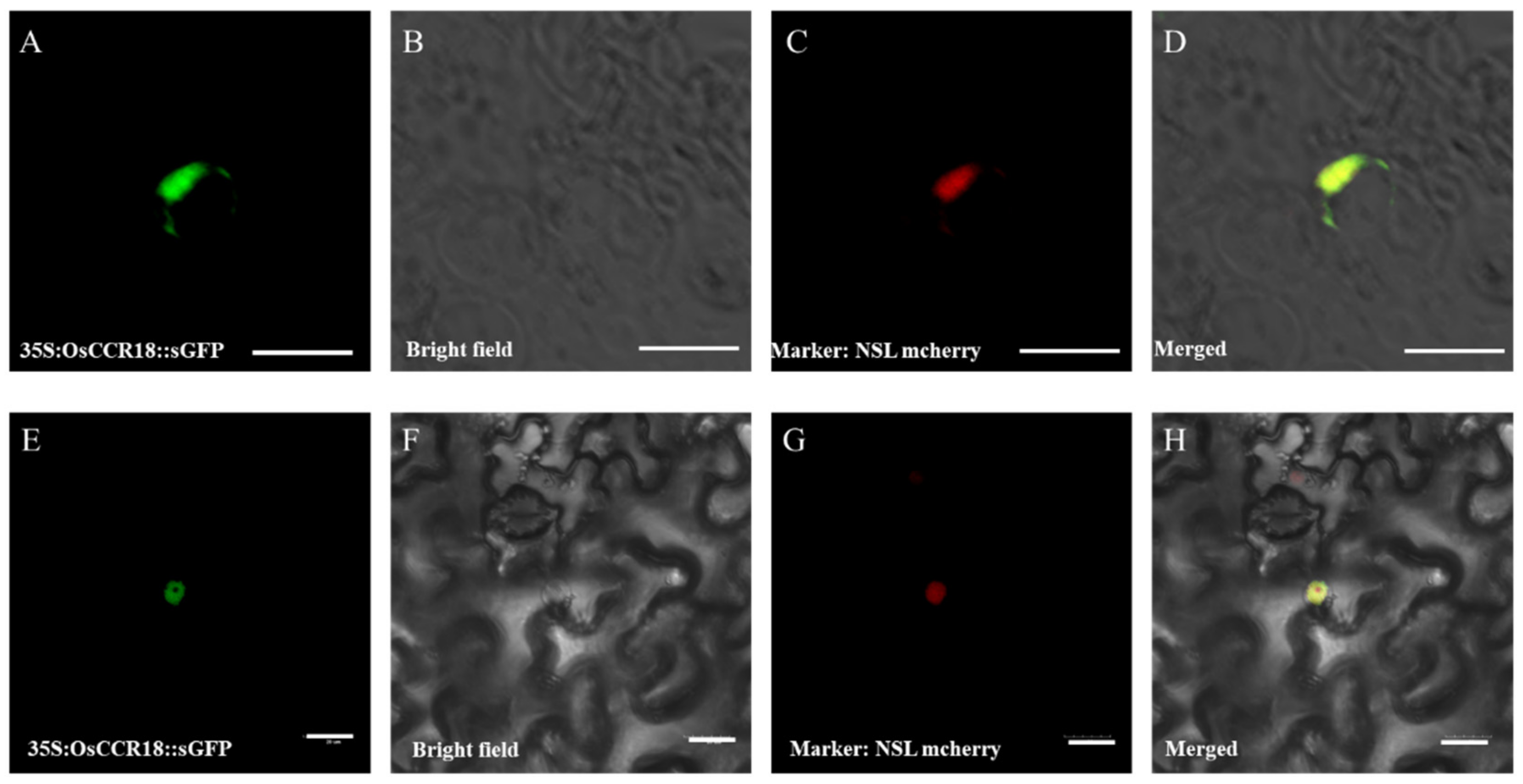
After separately co–transforming the OsCCR18GFP plasmid and the nuclear localization marker NSL mCherry into the rice protoplasts and tobacco leaves, fluorescence was detected in the nucleus of both (Figure 2), indicative of the nucleus localization of OsCCR18 proteins in rice.

3.3. Knockout of OsCCR18 Results in Complete Male Sterility

To investigate the regulatory role of the OsCCR18 gene during anther development, two osccr18 knockout lines were created by CRISPR/Cas9. In the osccr18–1 line, there was a “T” insertion, followed by a 376 bp deletion; the osccr18–2 line had a 373 bp deletion (Figure S1). These mutations caused a severe frameshift of the CDS of the OsCCR18 gene, resulting in significant changes in the protein structure where the NADPH–binding and catalytic activity motifs were lacking in the osccr18 mutants (Figure S1). The OsCCR18 expression was remarkably repressed in the leaves and anthers of the two mutants compared with the WT (Figure S1).
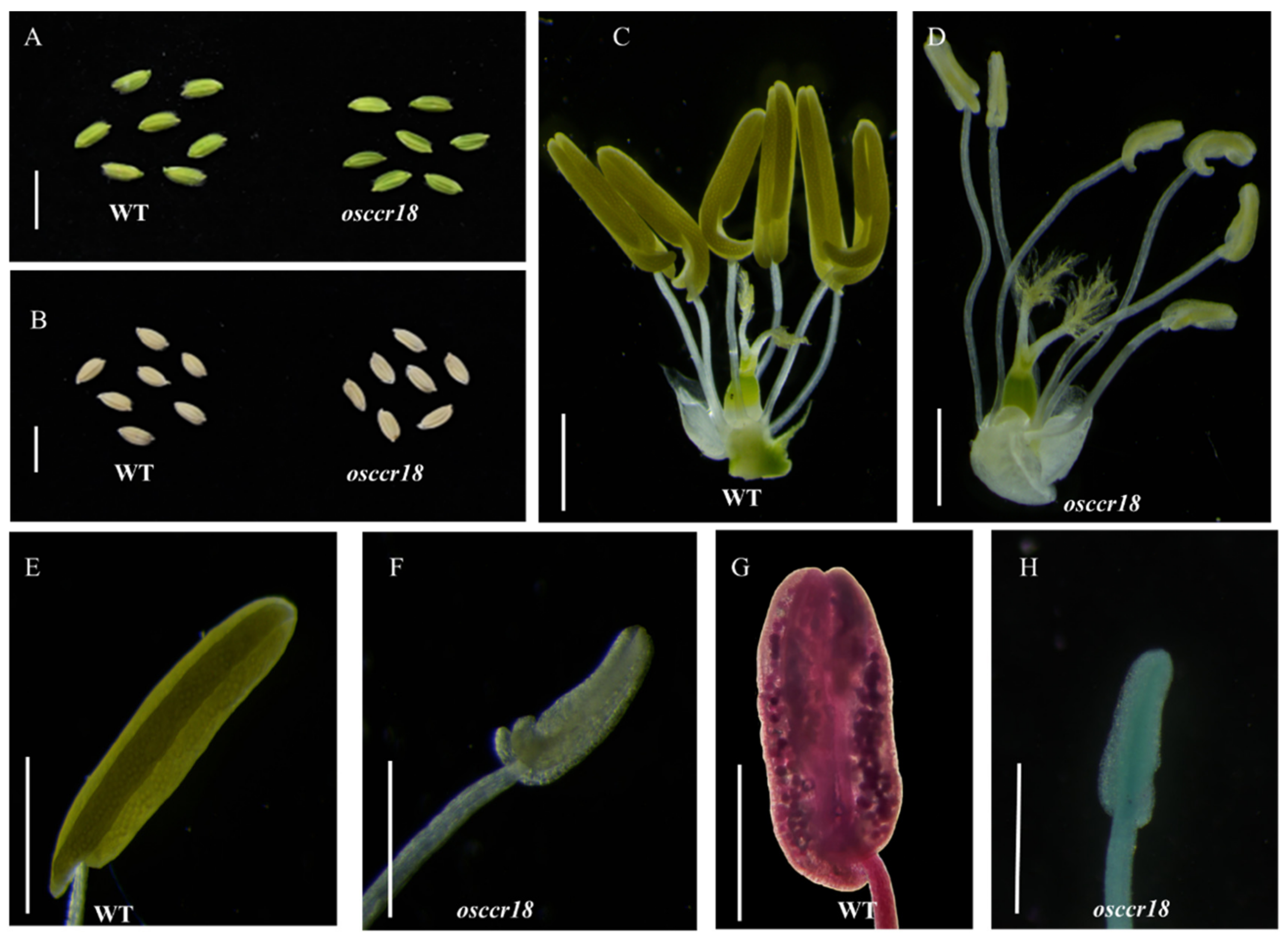
Under normal cultivation conditions, the osccr18 mutants exhibited no significant difference to the WT plants with regard to the plant growth and development as well as the spikelet morphology (Figure 3A,B). However, the anthers of the mutants were much more transparent, whiter, and smaller than the WT, suggestive of a severe defection in the anther development of the osccr18 lines (Figure 3C–F). The pistil morphology had no obvious difference between the WT and the mutants. Alexander staining showed that the mutant lines did not produce any visible pollen grains whereas the WT plants did (Figure 3G,H). These observations indicated that the loss of function of the OsCCR18 gene caused complete male sterility in the rice.

3.4. Abnormal Tapetum Degradation Contributes to the Male Sterility of the Osccr18 Mutants

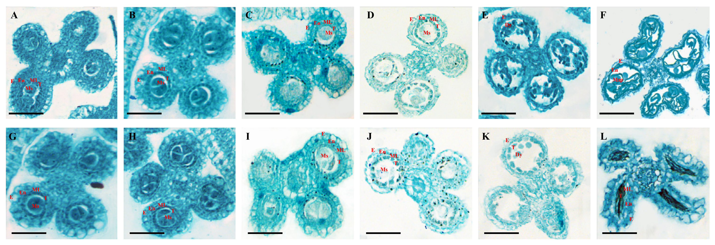
To clarify the cellular defects during the anther development in osccr18 mutants, we observed and compared the differences in the anther cross-sections of the WT and the mutants at each developmental stage. As known, the anther development of rice is divided into twelve stages, from the stamen primordia to the maturation and release of pollen [37]. At stages S6 and S7, there was no significant difference between the WT and the mutants; both could form microspores through normal meiosis (Figure 4A,B,G,H). However, at the S8 stage, the intermediate cells of the WT plants began to significantly degenerate (Figure 4C) whereas no obvious degeneration occurred in the osccr18 mutants (Figure 4I). At the S10 stage, we observed densely arranged granular Ubisch bodies on the lumen surface of the WT anthers (Figure 4E) but not in the mutants (Figure 4K), suggesting that the knockout of the expression of the OsCCR18 gene strongly affected the development of granular Ubisch bodies, further leading to abnormal pollen development. Moreover, at the S11 stage, the outer wall of the pollen was sickle-shaped and the tapetum cells were almost degraded and collapsed in the WT (Figure 4F) whereas the microspores of the mutants collapsed in the anther lumen in a non–regular manner (Figure 4L). These observations indicated that the abnormal degradation of the tapetum caused by the downregulation of the OsCCR18 expression played a crucial role in controlling male fertility in the rice.

3.5. OsCCR18 Regulates the Expression of anther Development–Related Genes

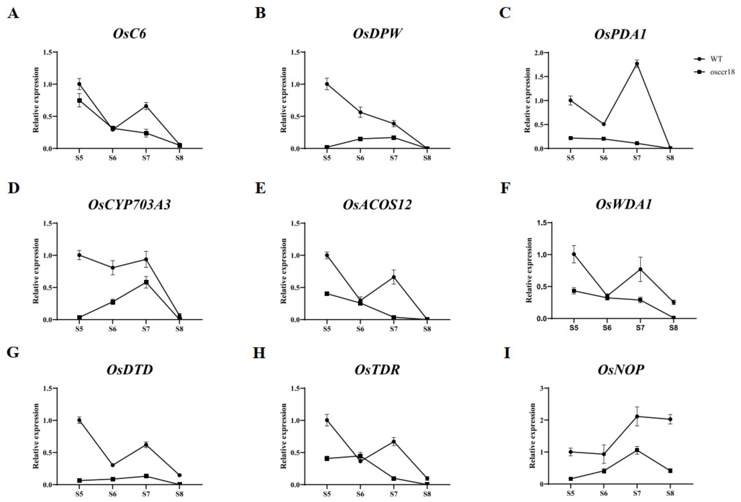
Did the male sterility of the osccr18 mutants result from the changes in the expression of several key genes involved in the regulation of anther development in rice? To verify this, we examined the expression patterns of the genes related to lipid metabolism and transport in anthers (OsC6 and OsDPW) and anther cuticle and sporopollenin precursors (OsPDA1, OsCYP703A3, OsACOS12, and OsWDA1) as well as two tapetum programed cell death (PCD)–related genes (OsDTD and OsTDR) and one gene involved in male gamete development (OsNOP) at the late developmental stages (from S5 to S8) of the anthers in the WT and the osccr18 mutants.
Most of the examined genes were expressed at remarkably lower levels at the S6 to S8 stages than at the S5 stage in the WT plants except for the OsPDA1 and OsNOP genes, whose transcript abundances were the highest at the S7 stage (Figure 5). However, the expression level of all of nine examined genes was severely downregulated at each of the four developmental stages from S5 to S8 in the osccr18 mutants compared with WT (Figure 5). Therefore, the significant downregulation of the expression of the genes related to anther development, especially those involved in the regulation of tapetum PCD (OsDTD and OsTDR), was likely to have strong effects on pollen formation in the osccr18 mutants.

3.6. Genes Involved in Fatty Acid Metabolism Are Differentially Expressed in anther at the S9 Stage between osccr18 and WT Plants

To further explore the molecular mechanism of OsCCR18 in controlling male fertility in rice, we compared the transcriptomes in the anthers at the S9 stage between the WT and the osccr18 mutants by RNA-Seq. A total of 41.43–48.85 million clean reads were generated for each library and separately mapped onto the Nipponbare reference genome. Differentially expressed genes (DEGs) were identified in the osccr18 mutants accordingly.
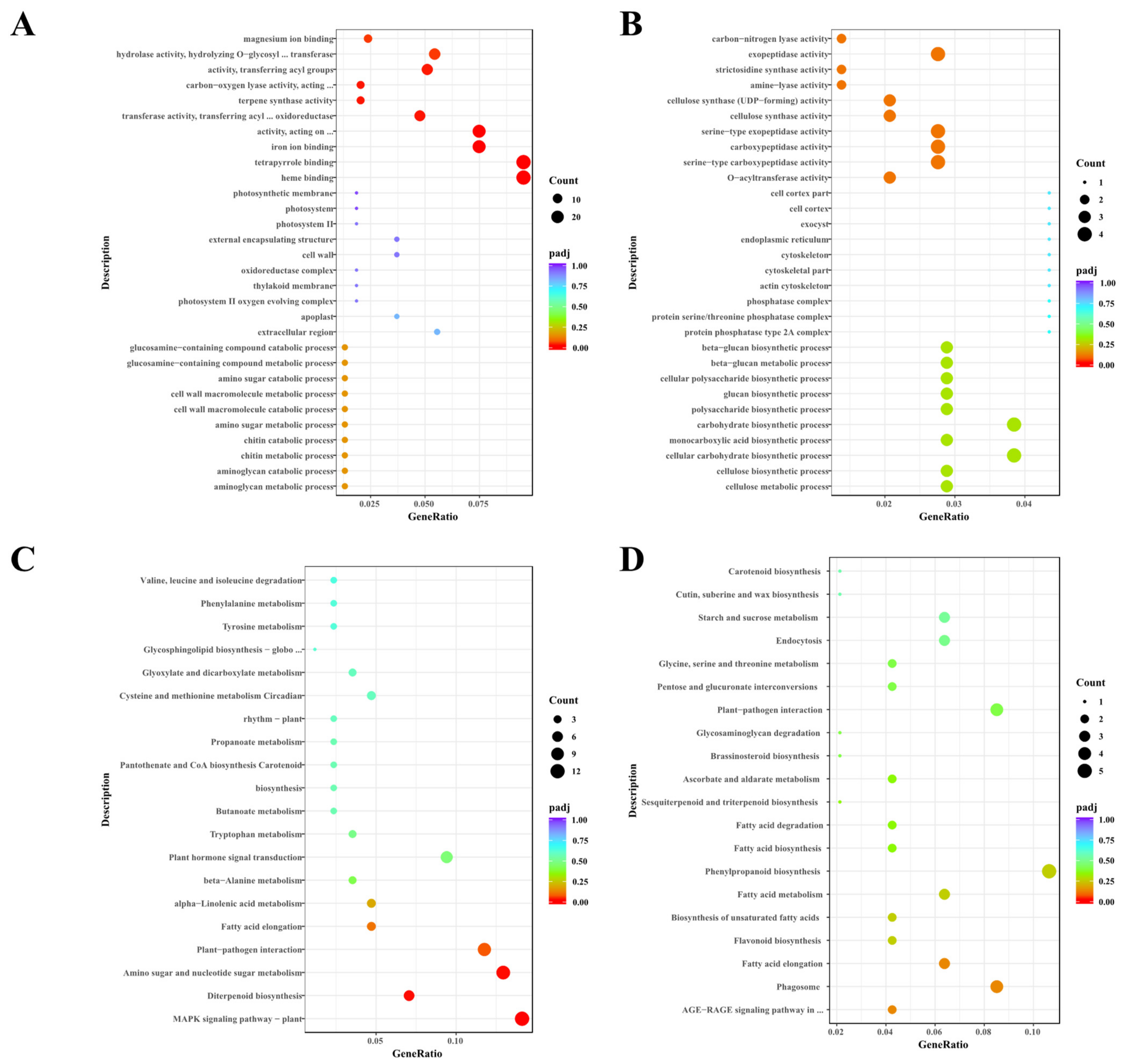
In total, 574 up– and 330 downregulated genes were characterized in the anthers at the S9 stage in the osccr18 mutants compared with the WT (Figure 6, Table S2). Notably, a total of 26 genes that exhibited differential expression level between the osccr18 mutants and the WT were previously reported to be related to male sterility in rice (Table S3). A GO enrichment analysis revealed that the upregulated DEGs primarily functioned in “tetrapyrrole binding” and “heme binding” (Figure 7A) whereas the downregulated DEGs mainly participated in a carbohydrate biosynthetic process (Figure 7B). The KEGG database was searched to examine the pathway enrichment of the DEGs in the osccr18 mutants. The upregulated DEGs were abundantly related to the “MAPK signaling pathway-plant”, “amino sugar and nucleotide sugar metabolism”, and “plant-pathogen interaction” pathways (Figure 7C) whereas the downregulated DEGs were greatly enriched in the “phenylpropanoid biosynthesis” and “fatty acid biosynthesis and metabolism” pathways, including “fatty acid elongation”, “fatty acid metabolism”, “biosynthesis of unsaturated fatty acids”, and “fatty acid degradation” (Figure 7D). Therefore, the OsCCR18 gene should be partially involved in modulating the carbohydrate and fatty acid metabolisms, which is crucial for controlling anther development in rice.

4. Discussion

In hybrid rice breeding, male sterile lines are usually adopted to facilitate the efficiency of heterosis utilization. Accordingly, the identification of male sterility–related genes is fundamental for uncovering its underneath molecular mechanism and further usage in rice breeding. As known, wax, cutin, and sporopollenin are indispensable components of the outer wall of the pollen and anthers in rice [24,38,39,40]. Cinnamoyl–CoA reductase (CCR) family genes are involved in the process of sporopollenin synthesis by regulating the PCD of tapetum cells [24]. An abnormal PCD occurring in the tapetum would cause incomplete pollen development or even fail to produce pollen in rice [24]. In this study, we created two rice male sterile lines by a loss of function of the OsCCR18 gene that belongs to the CCR family [41]. All CCR family proteins contain NADPH-binding and catalytic motifs [42]. In the two mutants, the OsCCR18 gene was completely knocked out in its function; both the NADPH–binding and catalytic motifs were deleted by CRISPR and the PCD of the tapetum cells in the anthers of the osccr18 mutants was blocked, resulting in an abnormal formation of the outer wall of the pollen. These findings may deepen our understanding of the function of the CCR genes as well as male fertility in rice.
To answer how OsCCR18 affects the development of the anther tapetum and pollen exine leading to male sterility in rice, we compared the expression level of nine genes related to anther development in the WT and mutants using qRT–PCR and found that all of the nine genes examined were significantly downregulated at stages from S5 to S8 in the osccr18 mutants (Figure 5). Notably, with one exception (OsDPW), all the tested genes—OsC6, OsACOS12, and OsPDA1 in particular—were expressed at a lower level at the S9 stage in the osccr18 mutants compared with the WT (Table S3). Additionally, the transcripts of OsGSL5, OsDTC1, and OsCYP704B2 were markedly abundant at the S9 stage in the WT than in the osccr18 mutants (Table S3). OsGSL5 (glucan synthase-like 5) encodes a rice callose synthase and a lack of OsGSL5 would result in developmental defects in the callose wall of microspores, further leading to serious male sterility in rice [43]. These observations indicated that a knockout of the OsCCR18 function would strongly affect the processes of the formation of the anther epidermis, pollen exine [11,12,13,14], pollen outer wall [20,21,22,23], and PCD of the tapetum cells [15,16,17,18,43] in rice. However, whether OsCCR18 could directly regulate the expression of the corresponding genes needs to be further experimentally validated.
Lipids play important roles in the development of the anther cuticle and the synthesis of the pollen exine and sporopollenin [44,45]. Abnormal lipid metabolism often causes male sterility in plants [46]. In this study, compared with the WT, hundreds of DEGs that were mainly enriched in the pathways of phenylpropanoid and fatty acid biosynthesis and metabolism were identified in the osccr18 mutants (Figure 7), which should have been responsible for the male sterility of the mutants. In maize, based on RNA-Seq data, Wan et al. identified 125 novel putative male fertility–related genes that participated in the process of lipid metabolism [46]. Moreover, several maize genes involved in lipid metabolism, including ZmMs33, ZmABCG26, and ZmFAR1, are crucial for anther development and have shown a good application future in developing male sterile lines [47,48]. Interestingly, the GDSL lipase OsRMS2, which plays diverse roles, determines male fertility by modulating the lipid homeostasis in the anthers of rice [49]. Similarly, in maize, the GDSL lipase ZmMs30 is a determinant for pollen exine formation and anther cuticle development by affecting the lipid metabolisms [50], indicative of the conserved roles of GDSLs in male fertility in plants [49,50]. However, the glucose-6-phosphate/phosphate translocator 1 encoding genes OsGPT1 and AtGPT1 might function distinctly in controlling anther development and male fertility in rice and Arabidopsis [51,52].

5. Conclusions

In this study, we identified a novel CCR family gene OsCCR18, which plays an important role in the regulation of anther development in rice. The OsCCR18 gene apparently exhibited developmental and tissue–specific expression patterns, whose transcripts were highly abundant in the leaves at the seedling stage and in the panicles at the S4–S5 flowering stage. The subcellular localization of the OsCCR18 proteins was in the nucleus of the rice. When OsCCR18 was dysfunctional, the osccr18 mutants were completely male-sterile, mainly because of the abnormal degradation of the tapetum and abnormal development of granular Ubisch bodies. Several anther development–related genes (OsPDA1, OsDTD, OsC6, OsACOS12, OsTDR, OsWDA1, OsDPW, OsCYP703A3, and OsNOP) were strongly downregulated in the panicles at the stages from S5 to S8 in the osccr18 mutants. Furthermore, hundreds of genes that were mainly involved in phenylpropanoid biosynthesis and fatty acid synthesis and metabolism were differentially expressed in the osccr18 mutants and the WT plants. Notably, 26 differentially expressed genes were previously considered to be male sterility-related. Overall, the knockout of the OsCCR18 gene primarily affected the development of the anther epidermis and the pollen outer wall, eventually leading to male sterility in the rice. This study provides a new genetic resource for the utilization of heterosis in rice breeding.

Supplementary Materials

The following supporting information can be downloaded at: https://www.mdpi.com/article/10.3390/agriculture12101685/s1, Supplementary Figure S1. Mutational analysis of OsCCR18 gene by CRISPR/Cas9. (A) Schematic diagram of the specific target sites of the OsCCR18 gene. Boxes represent exons (black) and untranslated regions (white); black lines represent introns; the vertical black lines indicate the two target sites, gRNA1 and gRNA2. The up– and downstream primers are indicated by arrows. (B) The editing profile of two osccr18 knockout lines. (C–E) Predicted protein structure of OsCCR18 and two mutated proteins using SWISS–MODEL. (F,G) Expression analysis of OsCCR18 gene in the leaves (F) and anthers (G) of WT and osccr18 mutants by qRT–PCR. For each experiment, three biological replicates and three technical replicates were performed. ** indicates significant difference between two samples by Student’s t–test (p > 0.001). Supplementary Table S1. Primers used for qRT–PCR analysis and vector construction. Supplementary Table S2. Differentially expressed genes in panicles at the S9 stage between osccr18 mutants and WT plants. Supplementary Table S3. List of differentially expressed genes between osccr18 mutants and WT plants that were previously reported to be male fertility-related in rice.

Author Contributions

Conceptualization, Q.L.; formal analysis, X.P., X.J., J.W., M.H., Y.W., M.W. and H.D.; data curation, X.P., X.J. and Q.L.; writing—original draft preparation, X.P., X.J. and Q.L.; writing—review and editing, X.P. and Q.L.; supervision, Q.L.; funding acquisition, M.W. and Q.L. All authors have read and agreed to the published version of the manuscript.

Funding

The Key Project of Zhejiang Provincial Natural Science Foundation of China (LZ19B070001), the National Natural Science Foundation of China (31972959, 32101658), and the Zhejiang Provincial Natural Science Foundation of China (LQ21C130001).

Data Availability Statement

The datasets supporting the conclusions of this article are included within the article and its Supplementary files.

Conflicts of Interest

The authors declare no conflict of interest.

References

  1. Zhao, Y.; Li, Z.; Liu, G.; Jiang, Y.; Maurer, H.P.; Würschum, T.; Mock, H.P.; Matros, A.; Ebmeyer, E.; Schachschneider, R.; et al. Genome-based establishment of a high-yielding heterotic pattern for hybrid wheat breeding. Proc. Natl. Acad. Sci. USA 2015, 112, 15624–15629. [Google Scholar] [CrossRef] [PubMed] [Green Version]
  2. Zhang, X.; Wang, D.; Fang, F.; Zhen, Y.; Liao, X. Food safety and rice production in China. Res. Agric. Mod. 2005, 26, 85–88. [Google Scholar]
  3. Xu, L.; Yuan, S.; Man, J. Changes in rice yield and yield stability in China during the past six decades. J. Sci. Food Agric. 2020, 100, 3560–3569. [Google Scholar] [CrossRef] [PubMed]
  4. Bai, S.; Yu, H.; Wang, B.; Li, J. Retrospective and perspective of rice breeding in China. J. Genet. Genom. 2018, 45, 603–612. [Google Scholar] [CrossRef]
  5. Tu, J.; Zhang, G.; Datta, K.; Xu, C.; He, Y.; Zhang, Q.; Khush, G.S.; Datta, S.K. Field performance of transgenic elite commercial hybrid rice expressing bacillus thuringiensis delta-endotoxin. Nat. Biotechnol. 2000, 18, 1101–1104. [Google Scholar] [CrossRef]
  6. Zhang, D.; Luo, X.; Zhu, L. Cytological analysis and genetic control of rice anther development. J. Genet. Genom. 2011, 38, 379–390. [Google Scholar] [CrossRef]
  7. Nonomura, K.I.; Miyoshi, K.; Eiguchi, M.; Suzuki, T.; Miyao, A.; Hirochika, H.; Kurata, N. The MSP1 gene is necessary to restrict the number of cells entering into male and female sporogenesis and to initiate anther wall formation in rice. Plant Cell 2003, 15, 1728–1739. [Google Scholar] [CrossRef] [Green Version]
  8. Li, N.; Zhang, D.S.; Liu, H.S.; Yin, C.S.; Li, X.X.; Liang, W.Q.; Yuan, Z.; Xu, B.; Chu, H.W.; Wang, J.; et al. The rice tapetum degeneration retardation gene is required for tapetum degradation and anther development. Plant Cell 2006, 18, 2999–3014. [Google Scholar] [CrossRef] [Green Version]
  9. Huang, M.D.; Hsing, Y.I.C.; Huang, A.H.C. Transcriptomes of the anther sporophyte: Availability and uses. Plant Cell Physiol. 2011, 52, 1459–1466. [Google Scholar] [CrossRef] [Green Version]
  10. Guilford, W.J.; Schneider, D.M.; Labovitz, J.; Opella, S.J. High resolution solid state 13C NMR spectroscopy of sporopollenins from different plant taxa. Plant Physiol. 1988, 86, 134–136. [Google Scholar] [CrossRef] [Green Version]
  11. Niu, B.X.; He, F.R.; He, M.; Ren, D.; Chen, L.T.; Liu, Y.G. The ATP-binding cassette transporter OsABCG15 is required for anther development and pollen fertility in rice. J. Integr. Plant Biol. 2013, 55, 710–720. [Google Scholar] [CrossRef]
  12. Qin, P.; Tu, B.; Wang, Y.; Deng, L.; Quilichini, T.D.; Li, T.; Wang, H.; Ma, B.; Li, S. ABCG15 encodes an ABC transporter protein, and is essential for post-meiotic anther and pollen exine development in rice. Plant Cell Physiol. 2013, 54, 138–154. [Google Scholar] [CrossRef] [Green Version]
  13. Zhu, L.; Shi, J.; Zhao, G.; Zhang, D.; Liang, W. Post-meiotic deficient anther1 (PDA1) encodes an ABC transporter required for the development of anther cuticle and pollen exine in rice. J. Plant Biol. 2013, 56, 59–68. [Google Scholar] [CrossRef]
  14. Zhang, D.; Liang, W.; Yin, C.; Zong, J.; Gu, F.; Zhang, D. OsC6, encoding a lipid transfer protein, is required for postmeiotic anther development in rice. Plant Physiol. 2010, 154, 149–162. [Google Scholar] [CrossRef] [Green Version]
  15. Yi, J.; Kim, S.R.; Lee, D.Y.; Moon, S.; Lee, Y.S.; Jung, K.H.; Hwang, I.; An, G. The rice gene DEFECTIVE TAPETUM AND MEIOCYTES 1 (DTM1) is required for early tapetum development and meiosis. Plant J. 2012, 70, 256–270. [Google Scholar] [CrossRef]
  16. Ji, C.; Li, H.; Chen, L.; Xie, M.; Wang, F.; Chen, Y.; Liu, Y.G. A novel rice bHLH transcription factor, DTD, acts coordinately with TDR in controlling tapetum function and pollen development. Mol. Plant 2013, 6, 1715–1718. [Google Scholar] [CrossRef] [Green Version]
  17. Yi, J.; Moon, S.; Lee, Y.S.; Zhu, L.; Liang, W.; Zhang, D.; Jung, K.H.; An, G. Defective Tapetum Cell Death 1 (DTC1) regulates ROS levels by binding to metallothionein during tapetum degeneration. Plant Physiol. 2016, 170, 1611–1623. [Google Scholar] [CrossRef] [Green Version]
  18. Zhang, C.; Shen, Y.; Tang, D.; Shi, W.; Zhang, D.; Du, G.; Zhou, Y.; Liang, G.; Li, Y.; Cheng, Z. The zinc finger protein DCM1 is required for male meiotic cytokinesis by preserving callose in rice. PLoS Genet. 2018, 14, e1007769. [Google Scholar] [CrossRef] [Green Version]
  19. Zou, T.; Xiong, P.; Zhou, F.; Zhou, D.; Chen, H.; Li, G.; Peng, K.; Zheng, K.; Han, Y.; Zhang, K.; et al. Grass-specific ABERRANT MICROSPORE DEVELOPMENT 1 is required for maintaining pollen fertility in rice. Plant J. 2022. [Google Scholar] [CrossRef]
  20. Jung, K.H.; Han, M.J.; Lee, D.Y.; Lee, Y.S.; Schreiber, L.; Franke, R.; Faust, A.; Yephremov, A.; Saedler, H.; Kim, Y.W.; et al. Wax-deficient anther1 is involved in cuticle and wax production in rice anther walls and is required for pollen development. Plant Cell 2006, 18, 3015–3032. [Google Scholar] [CrossRef] [Green Version]
  21. Li, H.; Pinot, F.; Sauveplane, V.; Werck-Reichhart, D.; Diehl, P.; Schreiber, L.; Franke, R.; Zhang, P.; Chen, L.; Gao, Y.; et al. Cytochrome P450 family member CYP704B2 catalyzes the ω-hydroxylation of fatty acids and is required for anther cutin biosynthesis and pollen exine formation in rice. Plant Cell 2010, 22, 173–190. [Google Scholar] [CrossRef]
  22. Xu, D.; Shi, J.; Rautengarten, C.; Yang, L.; Qian, X.; Uzair, M.; Zhu, L.; Luo, Q.; An, G.; Waßmann, F.; et al. Defective Pollen Wall 2 (DPW2) encodes an acyl transferase required for rice pollen development. Plant Physiol. 2017, 173, 240–255. [Google Scholar] [CrossRef] [Green Version]
  23. Yang, Z.; Zhang, Y.; Sun, L.; Zhang, P.; Liu, L.; Yu, P.; Xuan, D.; Xiang, X.; Wu, W.; Cao, L.; et al. Identification of cyp703a3-3 and analysis of regulatory role of CYP703A3 in rice anther cuticle and pollen exine development. Gene 2018, 649, 63–73. [Google Scholar] [CrossRef]
  24. Xue, J.S.; Zhang, B.; Zhan, H.; Lv, Y.L.; Jia, X.L.; Wang, T.; Yang, N.Y.; Lou, Y.X.; Zhang, Z.B.; Hu, W.J.; et al. Phenylpropanoid derivatives are essential components of sporopollenin in vascular plants. Mol. Plant 2020, 13, 1644–1653. [Google Scholar] [CrossRef]
  25. Costa, M.A.; Collins, R.E.; Anterola, A.M.; Cochrane, F.C.; Davin, L.B.; Lewis, N.G. An in silico assessment of gene function and organization of the phenylpropanoid pathway metabolic networks in Arabidopsis thaliana and limitations thereof. Phytochemistry 2003, 64, 1097–1112. [Google Scholar] [CrossRef]
  26. Li, L.; Cheng, X.; Lu, S.; Nakatsubo, T.; Umezawa, T.; Chiang, V.L. Clarification of cinnamoyl co-enzyme A reductase catalysis in monolignol biosynthesis of aspen. Plant Cell Physiol. 2005, 46, 1073–1082. [Google Scholar] [CrossRef]
  27. Kawasaki, T.; Koita, H.; Nakatsubo, T.; Hasegawa, K.; Wakabayashi, K.; Takahashi, H.; Umemura, K.; Umezawa, T.; Shimamoto, K. Cinnamoyl-CoA reductase, a key enzyme in lignin biosynthesis, is an effector of small GTPase Rac in defense signaling in rice. Proc. Natl. Acad. Sci. USA 2006, 103, 230–235. [Google Scholar] [CrossRef] [PubMed] [Green Version]
  28. Ruel, K.; Berrio-Sierra, J.; Derikvand, M.M.; Pollet, B.; Thévenin, J.; Lapierre, C.; Jouanin, L.; Joseleau, J.P. Impact of CCR1 silencing on the assembly of lignified secondary walls in Arabidopsis thaliana. New Phytol. 2009, 184, 99–113. [Google Scholar] [CrossRef]
  29. Goujon, T.; Ferret, V.; Mila, I.; Pollet, B.; Ruel, K.; Burlat, V.; Joseleau, J.P.; Barrière, Y.; Lapierre, C.; Jouanin, L. Down-regulation of the AtCCR1 gene in Arabidopsis thaliana: Effects on phenotype, lignins and cell wall degradability. Planta 2003, 217, 218–228. [Google Scholar] [CrossRef]
  30. Kawasaki, T.; Henmi, K.; Ono, E.; Hatakeyama, S.; Iwano, M.; Satoh, H.; Shimamoto, K. The small GTP-binding protein Rac is a regulator of cell death in plants. Proc. Natl. Acad. Sci. USA 1999, 96, 10922–10926. [Google Scholar] [CrossRef] [PubMed] [Green Version]
  31. Zhao, J.; Liu, X.; Wang, M.; Xie, L.; Wu, Z.; Yu, J.; Wang, Y.; Zhang, Z.; Jia, Y.; Liu, Q. The miR528-D3 module regulates plant height in rice by modulating the gibberellin and abscisic acid metabolisms. Rice 2022, 15, 27. [Google Scholar] [CrossRef] [PubMed]
  32. Trinidad, J.L.; Longkumer, T.; Kohli, A. Rice protoplast isolation and transfection for transient gene expression analysis. Methods Mol. Biol. 2021, 2238, 313–324. [Google Scholar] [PubMed]
  33. Kim, D.; Paggi, J.M.; Park, C.; Bennett, C.; Salzberg, S.L. Graph-based genome alignment and genotyping with HISAT2 and HISAT-genotype. Nat. Biotechnol. 2019, 37, 907–915. [Google Scholar] [CrossRef]
  34. Liao, Y.; Smyth, G.K.; Shi, W. featureCounts: An efficient general purpose program for assigning sequence reads to genomic features. Bioinformatics 2014, 30, 923–930. [Google Scholar] [CrossRef] [Green Version]
  35. Love, M.I.; Huber, W.; Anders, S. Moderated estimation of fold change and dispersion for RNA-seq data with DESeq2. Genome Biol. 2014, 15, 550. [Google Scholar] [CrossRef] [Green Version]
  36. Yu, G.; Wang, L.G.; Han, Y.; He, Q.Y. clusterProfiler: An R package for comparing biological themes among gene clusters. OMICS J. Integr. Biol. 2012, 16, 284–287. [Google Scholar] [CrossRef]
  37. Zhang, D.; Wilson, Z.A. Stamen specification and anther development in rice. Chin. Sci. Bull. 2009, 54, 2342–2353. [Google Scholar] [CrossRef]
  38. Samuels, L.; Kunst, L.; Jetter, R. Sealing plant surfaces: Cuticular wax formation by epidermal cells. Annu. Rev. Plant Biol. 2008, 59, 683–707. [Google Scholar] [CrossRef] [Green Version]
  39. Li, H.; Zhang, D. Biosynthesis of anther cuticle and pollen exine in rice. Plant Signal Behav. 2010, 5, 1121–1123. [Google Scholar] [CrossRef] [Green Version]
  40. Shi, J.; Cui, M.; Yang, L.; Kim, Y.J.; Zhang, D. Genetic and biochemical mechanisms of pollen wall development. Trends Plant Sci. 2015, 20, 741–753. [Google Scholar] [CrossRef]
  41. Park, H.L.; Bhoo, S.H.; Kwon, M.; Lee, S.W.; Cho, M.H. Biochemical and expression analyses of the rice cinnamoyl-CoA reductase gene family. Front. Plant Sci. 2017, 8, 2099. [Google Scholar] [CrossRef] [PubMed]
  42. Lacombe, E.; Hawkins, S.; Van Doorsselaere, J.; Piquemal, J.; Goffner, D.; Poeydomenge, O.; Boudet, A.M.; Grima-Pettenati, J. Cinnamoyl CoA reductase, the first committed enzyme of the lignin branch biosynthetic pathway: Cloning, expression and phylogenetic relationships. Plant J. 1997, 11, 429–441. [Google Scholar] [CrossRef] [PubMed]
  43. Shi, X.; Sun, X.; Zhang, Z.; Feng, D.; Zhang, Q.; Han, L.; Wu, J.; Lu, T. GLUCAN SYNTHASE-LIKE 5 (GSL5) plays an essential role in male fertility by regulating callose metabolism during microsporogenesis in rice. Plant Cell Physiol. 2015, 56, 497–509. [Google Scholar] [CrossRef] [Green Version]
  44. Ischebeck, T. Lipids in pollen—They are different. Biochim. Biophys. Acta 2016, 1861, 1315–1328. [Google Scholar] [CrossRef]
  45. Zhang, D.; Shi, J.; Yang, X. Role of lipid metabolism in plant pollen exine development. Subcell Biochem. 2016, 86, 315–337. [Google Scholar]
  46. Wan, X.; Wu, S.; Li, Z.; An, X.; Tian, Y. Lipid metabolism: Critical roles in male fertility and other aspects of reproductive development in plants. Mol. Plant 2020, 13, 955–983. [Google Scholar] [CrossRef]
  47. Zhu, T.; Li, Z.; An, X.; Long, Y.; Xue, X.; Xie, K.; Ma, B.; Zhang, D.; Guan, Y.; Niu, C.; et al. Normal structure and function of endothecium chloroplasts maintained by ZmMs33-mediated lipid biosynthesis in tapetal cells are critical for anther development in maize. Mol. Plant 2020, 13, 1624–1643. [Google Scholar] [CrossRef]
  48. Jiang, Y.; Li, Z.; Liu, X.; Zhu, T.; Xie, K.; Hou, Q.; Yan, T.; Niu, C.; Zhang, S.; Yang, M.; et al. ZmFAR1 and ZmABCG26 regulated by microrna are essential for lipid metabolism in maize anther. Int. J. Mol. Sci. 2021, 22, 7916. [Google Scholar] [CrossRef]
  49. Zhao, J.; Long, T.; Wang, Y.; Tong, X.; Tang, J.; Li, J.; Wang, H.; Tang, L.; Li, Z.; Shu, Y.; et al. RMS2 encoding a GDSL lipase mediates lipid homeostasis in anthers to determine rice male fertility. Plant Physiol. 2020, 182, 2047–2064. [Google Scholar] [CrossRef] [Green Version]
  50. An, X.; Dong, Z.; Tian, Y.; Xie, K.; Wu, S.; Zhu, T.; Zhang, D.; Zhou, Y.; Niu, C.; Ma, B.; et al. ZmMs30 encoding a novel gdsl lipase is essential for male fertility and valuable for hybrid breeding in maize. Mol. Plant 2019, 12, 343–359. [Google Scholar] [CrossRef] [Green Version]
  51. Qu, A.; Xu, Y.; Yu, X.; Si, Q.; Xu, X.; Liu, C.; Yang, L.; Zheng, Y.; Zhang, M.; Zhang, S.; et al. Sporophytic control of anther development and male fertility by glucose-6-phosphate/phosphate translocator 1 (OsGPT1) in rice. J. Genet. Genom. 2021, 48, 695–705. [Google Scholar] [CrossRef] [PubMed]
  52. Niewiadomski, P.; Knappe, S.; Geimer, S.; Fischer, K.; Schulz, B.; Unte, U.S.; Rosso, M.G.; Ache, P.; Flügge, U.I.; Schneider, A. The Arabidopsis plastidic glucose 6-phosphate/phosphate translocator GPT1 is essential for pollen maturation and embryo sac development. Plant Cell 2005, 17, 760–775. [Google Scholar] [CrossRef] [PubMed]
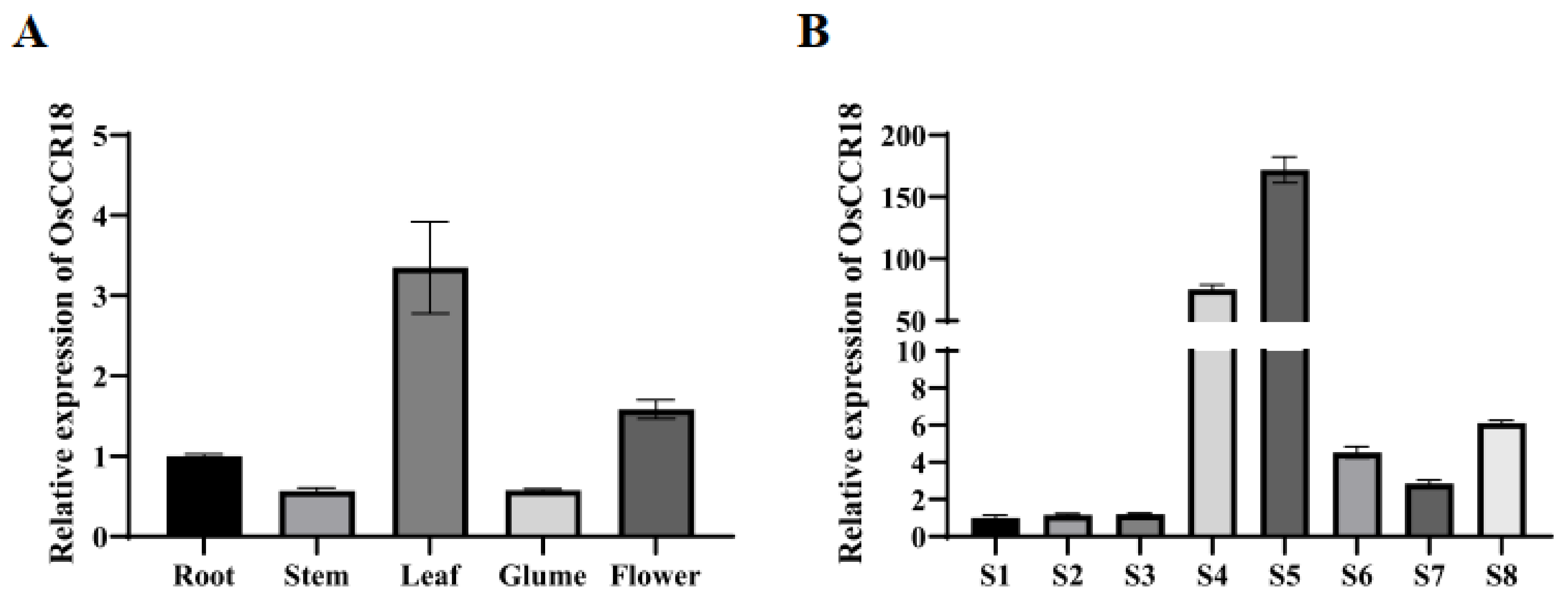
Figure 1. Analysis of the OsCCR18 gene expression pattern in rice. (A) Expression pattern analysis of OsCCR18 in roots, stems, and leaves at the seedling stage and in the glume and flower organ at the flowering stage by qRT–PCR. (B) Expression pattern analysis of OsCCR18 in anther at different developmental stages by qRT–PCR. Data are shown as the means ± SD (n = 3).
Agriculture 12 01685 g001
Figure 2. Subcellular localization analysis of OsCCR18. (AD) Transient expression of 35S:OsCCR18::sGFP fusion vector in rice protoplast. (EH) Transient expression of 35S:OsCCR18::sGFP fusion vector in a tobacco leaf. In the experiment, the 35S:OsCCR18::sGFP plasmid was co–transformed with the nuclear localization marker NSL mCherry. Bar = 100 µm.
Agriculture 12 01685 g002
Figure 3. Phenotypic analysis of WT and osccr18 mutants. (A,B) Comparison of the spikelet morphology of WT and mutants at the grain filling (A) and maturity stages (B). (CF) Comparison of the anther morphology of WT and mutants at the flowering stage. (G,H) Alexander staining of pollen of WT and mutants at the flowering stage.
Agriculture 12 01685 g003
Figure 4. Paraffin sections of anthers of WT and osccr18 mutants at different developmental stages. (AF) Paraffin sections of WT anthers at the developmental stages from S6 to S11. (GL) Paraffin sections of osccr18 anthers at the developmental stages from S6 to S11. E: epidermis; En: endothecium; ML: middle layer; Mp: mature pollen; Ms: microsporocyte; Msp: microspores; T: tapetum; Tds: tetrads. Bar = 30 μm.
Agriculture 12 01685 g004
Figure 5. Expression analysis of the tapetum programmed cell death (PCD)related genes in WT and osccr18 mutants. The experiments were performed with three biological replicates and three technical replicates. Data are shown as the means ± SD (n = 3).
Agriculture 12 01685 g005
Figure 6. Analysis of differentially expressed genes (DEGs) at the S9 stage during anther development in osccr18 mutants and WT. (A) The volcano plot of DEGs. The red and green dots represent the up– and downregulated DEGs in osccr18 mutants compared with WT. (B) Clustering heat map of DEGs. The analysis was performed using the normalized expression level of DEGs with three biological replicates of osccr18 and WT samples.
Agriculture 12 01685 g006
Figure 7. GO and KEGG enrichment analyses of differentially expressed genes (DEGs) at the S9 stage during anther development in osccr18 mutants and WT. (A,B) GO enrichment analysis of up– and downregulated DEGs. (C,D) KEGG pathway enrichment analyses of up– and downregulated DEGs. In both the GO and KEGG analyses, the top 20 most represented categories were shown.
Agriculture 12 01685 g007
Publisher’s Note: MDPI stays neutral with regard to jurisdictional claims in published maps and institutional affiliations.