Article

Nectar Secretion, Morphology, Anatomy and Ultrastructure of Floral Nectary in Selected Rubus idaeus L. Varieties

by
Mikołaj Kostryco
and
Mirosława Chwil
*
Department of Botany and Plant Physiology, University of Life Sciences in Lublin, Akademicka 15, 20-950 Lublin, Poland
*
Author to whom correspondence should be addressed.
Agriculture 2022, 12(7), 1017; https://doi.org/10.3390/agriculture12071017
Submission received: 7 June 2022 / Revised: 7 July 2022 / Accepted: 7 July 2022 / Published: 13 July 2022
(This article belongs to the Section Farm Animal Production)

Abstract

:
The distinctive features of floral nectaries facilitate identification of ecological and phylogenetic links between related taxa. The structure and functioning of nectaries determine the relationships between plants, pollinators, and the environment. The aim of the study was to determine and compare the micromorphology of the epidermis in the floral nectaries of six Rubus idaeus cultivars belonging to biennial (‘Glen Ample’, ‘Laszka’, ‘Radziejowa’) and repeated fruiting (‘Pokusa’, ‘Polana’, ‘Polka’) groups. Another objective was to characterize the cuticle ornamentation and stomatal morphology, the anatomy of the nectary epidermis, parenchyma, and sub-nectary parenchyma in the initial nectar secretion phase, as well as the ultrastructure of the nectary epidermis and parenchyma cells in the initial and full nectar secretion phases. The study was carried out using light, fluorescence, scanning and transmission-electron microscopy techniques. Semi-thin and ultrathin sections were used for the microscopic analyses. The cuticular ornamentation and stomatal morphology may be helpful elements in the identification of relatedness between Rubus species. The interaction of the extensive system of endoplasmic reticulum membranes, mitochondria, and Golgi apparatus indicates high metabolic activity, and the fusion of transport vesicles with the membrane suggests granulocrine nectar secretion. The results bring new data to the biology of plants.

1. Introduction

Plants of the genus Rubus are highly valued for their wide range of consumer, medicinal, cosmetic, decorative, and beekeeping uses [1,2]. Rubus is one of the most numerous and phylogenetically most diverse genera with a complex taxonomy resulting from hybridization and facultative apomixes [1,2,3,4]. It belongs to the tribe Rubeae, subfamily Rosoideae, family Rosaceae, order Rosales, and clade Fabids [1,2,3,5,6,7]. The estimated number of species of the genus Rubus ranges from 600 to 800 [8]. As suggested by Lechowicz et al. [9], depending on the classification, there may be even 1000 species representing 12 subgenera [10,11,12]. Rubus idaeus belongs to the subgenus Ideobatus [13]. In Poland, this species occurs in its natural state but there are also numerous varieties grown in amateur and commercial cultivation [14,15,16].
The distinctive traits of floral nectaries facilitate the identification of ecological and phylogenetic links between related taxa. The structure and functioning of nectaries in combination with the composition of secreted nectar provides comprehensive knowledge of the relationships between plants, pollinators, and the environment [17]. Nectary micromorphology, anatomy, and ultrastructure as well as nectary secretion are important elements of plant reproduction, as they ensure visits of insect pollinators foraging for the offered reward. Considering the decline in pollinator abundance, which poses a serious threat to agriculture and food production, studies of the structure and development phases of floral nectaries as well as nectar secretion are extremely important [18]. The morpho-anatomical and ultrastructural traits of nectaries are of key importance for elucidation of the mechanisms of the dynamics of production and characteristics of nectar during anthesis [19]. The CRABS CLAW gene is the main regulator of nectary development in Rosids. STY genes engaged in the auxin biosynthetic pathway play a key role in the formation of nectaries in Eudicots [18]. The nectary is independent of the floral organ specification homeotic genes ABC [17]. Nectaries in the Rosaceae family represent the receptacular type. They are composed of three layers: nectary epidermis, nectary parenchyma, and subnectary parenchyma [20,21]. The structure of floral nectaries in the Rubus genus has not been studied. Various types of nectaries have been distinguished in various species from many genera of the Pomoideae subfamily: automorphic (Crataegus, Pyrus, and Chaenomeles), epimorphic (Cydonia), and transitional (Malus) [22,23,24]. Ornamental apple cultivars from the Maloideae subfamily (Malus baccata var. jackii, M. sieboldi var. arborescens, M. floribunda ‘Van Houtte’. etc.) produce epimorphic, transitional, or automorphic nectaries [25]. Other authors have described receptaculo-ovarial nectaries in the flowers of Cydonia and Pyrus [21,26,27]. In turn, Farkas [28] has distinguished xeromorphic, mesomorphic, and hygromorphic nectaries. Floral nectar has various functions, e.g., it attracts bees, thus facilitating pollination. Extra-floral nectar additionally protects plants against herbivores, as shown by Chatt et al. [29]. Secreted nectar mediates mutualistic plant–animal relationships aimed at the improvement of the condition and reproductive success of plants [30,31,32,33].
Prenectar is transported from vascular tissue elements via the apoplastic pathway, and proper nectar is secreted through various mechanisms [34]. The composition of nectar depends on the plant species and environmental conditions. Nectar contains water (30–90%), sugars (from several to 70%), nitrogenous substances, organic acids, dyes, essential oils, vitamins, and mineral salts [35].
Although there are many reports on floral nectaries in various species from many taxonomic families, including the Rosaceae family, researchers have not explored the micromorphology, anatomy, and ultrastructure of floral nectaries in Rubus plants. This issue is of fundamental importance for molecular biology and knowledge of nectar secretion at the cellular level. There are also no data on nectar abundance and nectar transport processes at the cellular level in common raspberry varieties cultivated in commercial production in Poland, i.e., ‘Glen Ample’, ‘Laszka’, ‘Radziejowa’, ‘Pokusa’, ‘Polana’, and ‘Polka’. The present study is a continuation of previous research [16,36].
The aim of the study was to determine and compare (i) the micromortphology, (ii) anatomy, and (iii) ultrastructure of floral nectaries, as well as (iv) nectar secretion at the cellular level in six R. idaeus cultivars, i.e., three biennial fruiting cultivars ‘Glen Ample’, ‘Laszka’, and ‘Radziejowa’ and three repeated fruiting cultivars ‘Pokusa’, ‘Polana’, and ‘Polka’. Additionally, the paper presents the micromorphological characteristics of selected floral elements in the studied cultivars.

2. Materials and Methods

2.1. Plant Material

The study, carried out in 2016–2018, involved six Rubus ideaus cultivars, i.e., three from the biennial fruiting group: ‘Glen Ample’, ‘Laszka’, and ‘Radziejowa’ and three from the repeated fruiting group: ‘Pokusa’, ‘Polana’, and ‘Polka’. Raspberry flowering was observed on a plantation located in Blinów II village in the Lublin Province, Kraśnik County, Szastarka commune in south-eastern Poland (50°51′59″ N 22°23′09″ E). Buds in the bud-burst stage and flowers on the first and last day of flowering were collected in the full flowering stage.

2.2. Scope of the Study

The structure of the nectaries was analyzed at different cellular levels in two stages of development: the initial secretion phase (bud-burst stage) and the full nectar secretion stage (anthesis stage). The comparative micromorphological studies were carried out in the full nectar secretion phase, and the anatomical analyses were performed in the initial nectar secretion phase. In turn, the ultrastructure was analyzed in both phases. The micromorphology of selected floral elements was observed in the initial nectar secretion phase. The structure of floral nectaries was subjected to micromorphological, anatomical, and ultrastructural studies conducted with the use of fluorescence (FM), light (LM), scanning (SEM), and electron transmission (TEM) microscopy techniques.

2.3. Microscopic Studies

For the comparative microscopic studies, fragments of nectaries and floral elements (pedicels, sepals, corolla petals, pistils) were collected from fresh flowers and fixed in 4% glutaraldehyde in a 0.1 M phosphate buffer at pH 7.0 for 6 h at room temperature. Afterwards, a 0.01 M phosphate buffer pH 7.0 was used at 4 °C for 48 h [37,38].
Light microscopy (LM). In order to prepare semi-thin microscopic slides for observation in a bright-field light microscope and ultrathin preparations for the comparative studies of the ultrastructure of nectary cells in a transmission electron microscope, the fixed material was washed twice in the phosphate buffer and was contrasted in a 1.5% osmium tetroxide solution for 1.5 h [37,38].
After rinsing, the nectary samples were dehydrated for 15 min in an ethyl alcohol series with the following concentrations: 15, 30, 50, 70, 90, 96, and 99.8% (twice in anhydrous ethanol). The dehydrated sections were embedded in Spurr Low Viscosity resin and polymerized at 60 °C for 48 h [39].
For the observations of the nectary anatomy (initial nectar secretion phase) under a light microscope, longitudinal semi-thin (0.8–1 µm thick) sections were made from fragments of resin-embedded nectaries. A glass knife and a Reichert Ultracut S microtome were used to cut the nectaries. Fragments of the nectaries were stained with 1% toluidine blue and 1% azure II (1:1) at 60 °C for 5 min. Next, the preparations were rinsed with distilled water and 5% ethyl alcohol, dried, and sealed in Eukitt [40]. The comparative anatomical studies of the nectaries in the initial secretion stage were focused on epidermis cells, nectary parenchyma, and subnectary parenchyma.
The location of starch grains in plastids and the presence of other polysaccharides (cellulose and pectins) were determined by application of the periodic acid-Schiff (PAS) reaction [41,42]. Semi-thin sections were treated with 1% periodic acid for 20 min and rinsed with water (5 min). Next, the Schiff’s reagent was applied for 45 min. After rinsing with running water, the sections were dried at 60 °C and sealed in Eukitt. The anatomy of the nectary cells was observed using a Nikon Eclipse 90i bright-field microscope.
Fluorescence microscopy (FM). Semi-thin sections of the fixed material were placed in a drop of fluorochrome (0.01% auramine O) [42]. The preparations were sealed in a 50% glycerin solution. The observations were carried out using a Nikon Eclipse 90i bright-field microscope (Nikon, Tokyo, Japan) equipped with filters: FITC (excitation light 465–495 nm) and barrier (wavelength 515–555 nm).
Scanning electron microscopy (SEM). Fixed fragments of nectaries (5 × 5 mm squares) taken (i) from the central part of the nectary, (ii) the marginal part at the filament base, and (iii) the basal part near the pistil ovaries were drained in an acetone series: 15; 30; 50; 70; 90, and 99.5%, (anhydrous acetone was used twice) [43]. Subsequently, they were critical-point dried in liquid CO2 in an Emitech K850 dryer (Emitech, Ashford, UK) and gold sputtered using an Emitech K550X sputter coater (Emitech, Corato, Italy). The observations of the nectary epidermis surface and photographic documentation were made using a Tescan Vega II LMU scanning electron microscope (SEM) (Brno, Czech Republic).
Transmission electron microscopy (TEM). Ultrathin sections (70 nm thick; longitudinal section) were cut from the resin-embedded nectaries (middle part). The sections were treated with an 8% solution of uranyl acetate in acetic acid for 40 min [39]. After rinsing with distilled water twice for 10 min, the sections were contrasted with Reynolds reagent for 15 min [44]. Next, they were rinsed with water and dried. The observations of the ultrastructure of epidermis and nectary parenchyma cells were carried out in the initial and full nectar secretion phases, and the photographic documentation was made using a transmission electron microscope (TEM) (FEI, Tecnai G2 spirit Biotwin, New York, NY, USA).

2.4. Microscopic and Morphometric Measurements

The morphometric measurements consisted in assessment of the perianth morphology and the micromorphology, anatomy, and ultrastructure of R. idaeus nectaries. The size (length and width) of sepals and corolla petals was measured in the full flowering stage. At the beginning of nectar secretion, the following micromorphological traits of nectary epidermis were compared: (i) the length (ii) width, and (iii) surface of stomata, (iv) the number of stomata per 1 mm2 area, (v) the length and (vi) width of the aperture between the cuticle ledges, (vii) the length and (viii) width of the stomatal complex, (ix) width of band-forming striae, (x) width of cuticle bands, (xi) length of cuticle bands, (xii) width of striae (in the interband region) connecting cuticle bands, and (xiii) distance between cuticle bands (Figure 1).
The anatomical studies consisted in comparison of (xiv) the height and (xv) width of epidermis cells, (xvi) the thickness of the parenchyma layer (xvii), the number of nectary parenchyma cell layers, (xviii) the diameter of nectary parenchyma cells, (xix) the thickness of the subnectary parenchyma layer, (xx) number of subnectary parenchyma layers, and (xxi) the thickness of the nectary. The analyses of the ultrastructure of nectary epidermis cells in the initial phase of nectar secretion consisted in comparison of (xxii) the height of protuberances of the outer cell wall, (xxiii) the cuticle thickness, (xxiv) and the thickness of the outer, (xxv) periclinal inner, and (xvi) anticlinal cell walls. These measurements were performed in sixteen replications. They were made using the Nikon NIS-Elements version 3.0 Advance Research computer program for microscopic image analysis.

2.5. Statistical Analysis

One-way analysis of variance was carried out for the parameters of cuticle ornamentation, the size and number of stomata, the stomatal complex diameter, and the cell wall thickness. A two-way analysis of variance (ANOVA) was performed for the analyzed traits of the perianth in the six R. idaeus varieties. Tukey’s post-hoc comparison tests were also performed at the significance level of α = 0.05. The calculations were made using SAS 9.2 and Statistica 6.0 statistical software. The parameters of the cuticular ornamentation, the size and number of stomata, and the diameter of the stomatal complex were used in the hierarchical cluster analysis to distinguish homogeneous subsets indicating the similarity between the studied R. idaeus varieties. The number of clusters was determined on the basis of the scree criterion with standardization of the original data to avoid misclassifications associated with the different variable units. Ward’s minimal variance classification algorithm with the Euclidean distance as a measure of similarity was used in the dendrograms.

3. Results

The observations show that the lifespan of the raspberry flowers was 2–4 days in the biennial fruiting cultivars (‘Glen Ample’, ‘Laszka’, and ‘Radziejowa) and 2–5 days in the repeated fruiting cultivars (‘Pokusa’, ‘Polka’, and ‘Polana’). The Rubus idaeus flowers were located on pedicels bearing hairs and prickles, except for the ‘Glen Ample’ cultivar (Figure 2A). The perianth in the biennial fruiting cultivars had shorter sepals in comparison with the repeated fruiting cultivars, with a length ranging from 8.6 mm in ‘Radziejowa’ to 9.2 mm in ‘Glen Ample’ and from 11.9 mm in ‘Polka’ to 12.3 mm in ‘Polana’, respectively. The width of the sepals was similar in both groups of cultivars, i.e., from 4.7 mm in ‘Polka’ to 5.4 mm in ‘Radziejowa’. A similar relationship was found in the size of corolla petals. Their length ranged from 6.5 mm in ‘Radziejowa’ to 7.7 mm in ‘Glen Ample’ in the former group of cultivars and from 7.4 mm in ‘Polka’ to 8.5 mm in ‘Polana’ in the latter group. The width of the petals in both groups was in the range of 3.3 mm in ‘Radziejowa’ to 3.7 mm in ‘Glen Ample’. In general, there were no significant differences within and between the cultivars in the consecutive study years in terms of the analyzed traits of the perianth in the six R. idaeus cultivars: the length and width of sepals and the length and width of corolla petals. An exception was the significantly greater length of sepals in the repeated fruiting cultivars than in the biennial fruiting cultivars (Table 1).

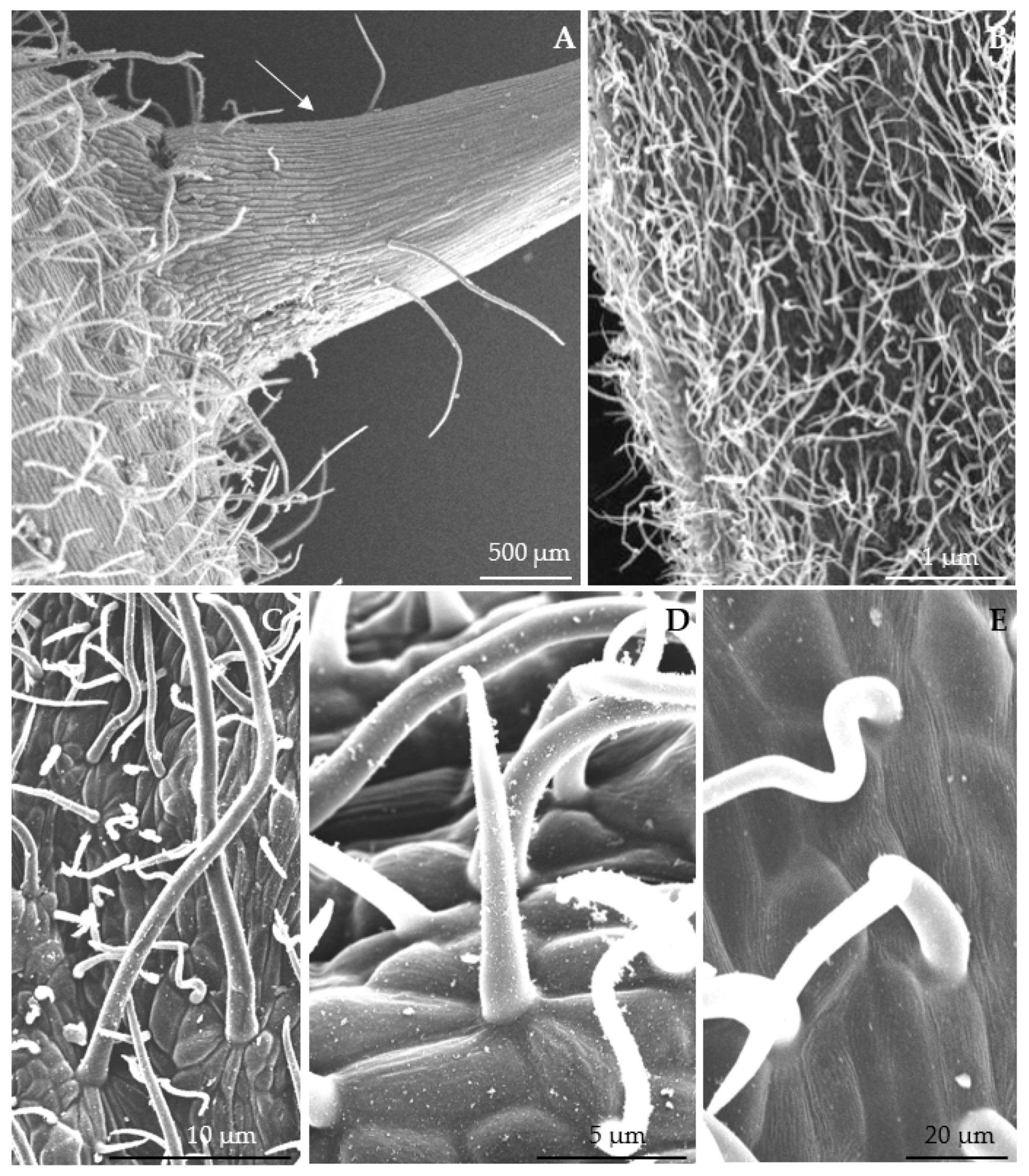
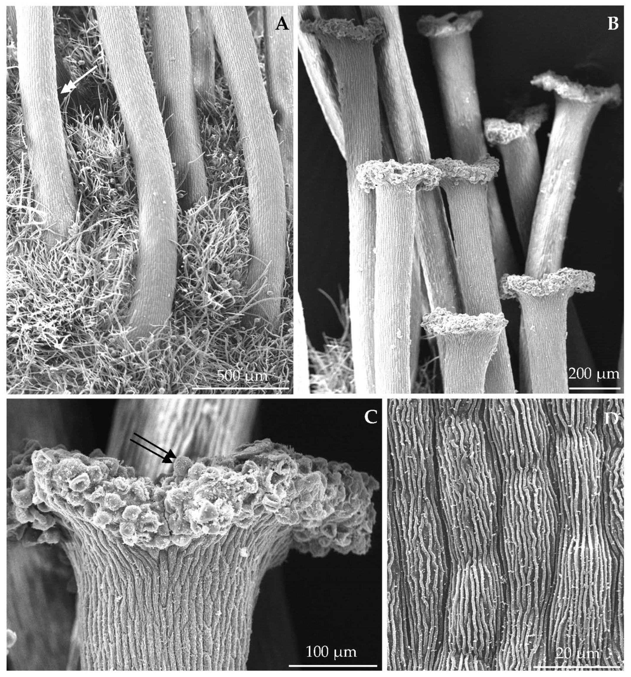
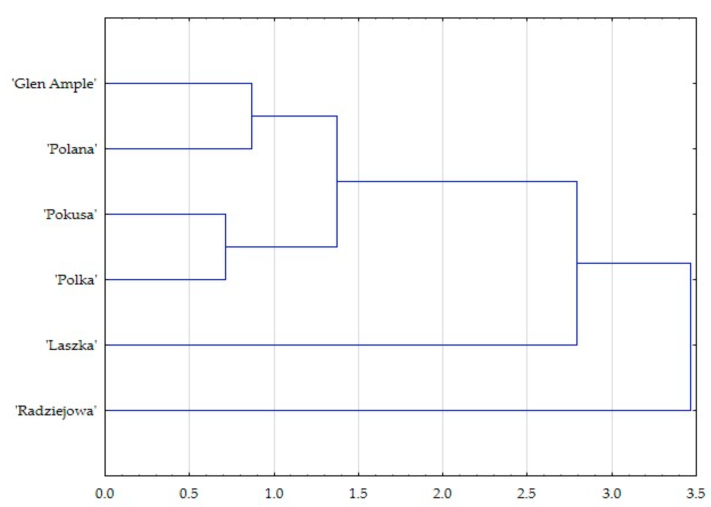
3.1. Micromorphological Traits of Some Flower Elements

The abaxial face of the sepals had an epidermis with striated cuticle ornamentation and was covered by dense hairs. The non-glandular trichomes were unicellular and had various lengths (Figure 2B–E). The epidermal cells of the adaxial face in the petals were tetra-, penta-, and hexagonal in the outline. In the central part, the outer periclinal wall of the epidermis cells was slightly convex, forming a conical protuberance (Figure 3A,B). The surface of these cells exhibited striated cuticle ornamentation. The striae in the central apical part of the cells were undulating and densely arranged. At some distance from the protuberance, the striae were arranged radially towards adjacent cells, forming a looser array in which they usually exhibited parallel arrangement and sometimes overlapped (Figure 3C,D).
In turn, similar to the adaxial surface of the sepals, the surface of the ovary epidermis cells was covered by dense hairs. The unicellular non-glandular trichomes were short–medium length, and strongly elongated. Their dense arrangement masked the surface of the epidermis cells (Figure 4A). The cuticle ornamentation on the surface of style epidermis cells was composed of straight striae, sometimes slightly undulating or with delicate overlapping curves. Some of them extended onto adjacent cells towards the longer axis of the epidermis cells. This ornamentation was visible from the base of the style to the basal part of the stigma (Figure 4A–D). The pistil stigmata were flat and slightly dilated beyond the margin of the style. Their surface was covered by papillae (Figure 4B,C).

3.2. Floral Nectaries

The floral nectaries in the analyzed R. idaeus L. cultivars were located on the adaxial surface of the receptacle between the basal parts of the ovaries to the filament bases (Figure 5A–D). In the initial nectar secretion phase, nectar drops of various sizes were visible on the nectary surface (Figure 5B). In the full nectar secretion phase, the nectary had a shape of a circular disc with a slightly raised margin and an irregular outline at the filament base. On the opposite side, the surface of the nectary epidermis cells near the ovary was covered by elongated and tapering different-length unicellular non-glandular trichomes (Figure 5D). The nectary was light green in the initial secretion phase, bit the color changed into green-yellow in the full secretion phase. The surface of the nectaries was flat. The floral nectaries in R. idaeus represent the receptactular type.

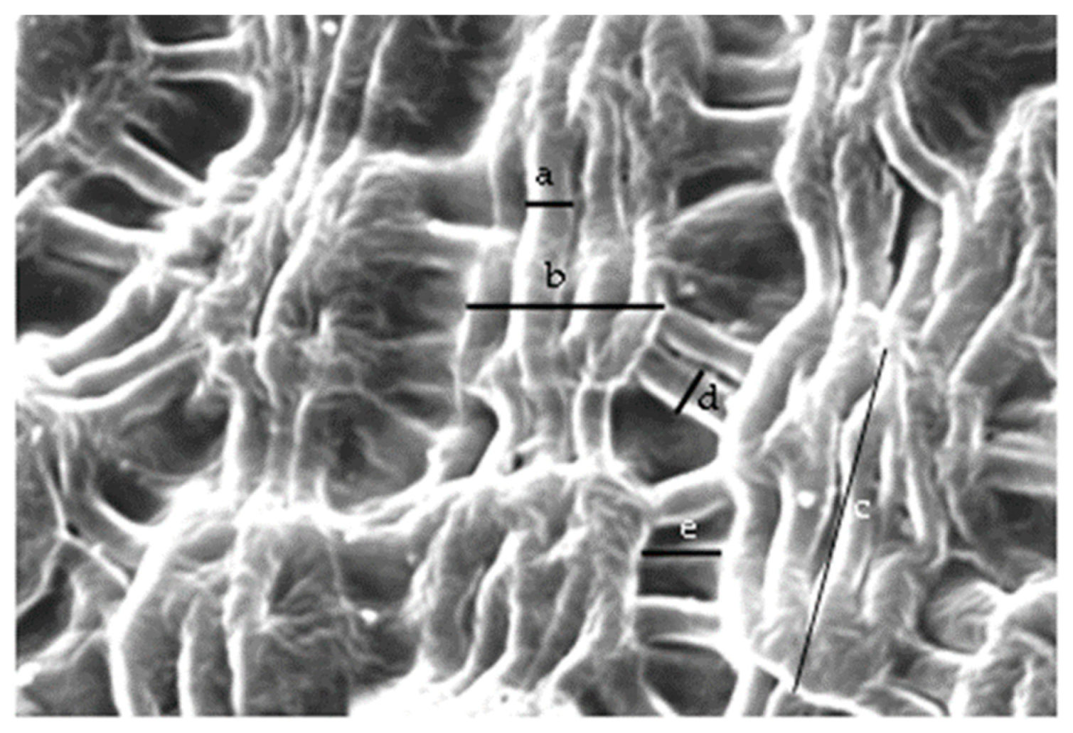
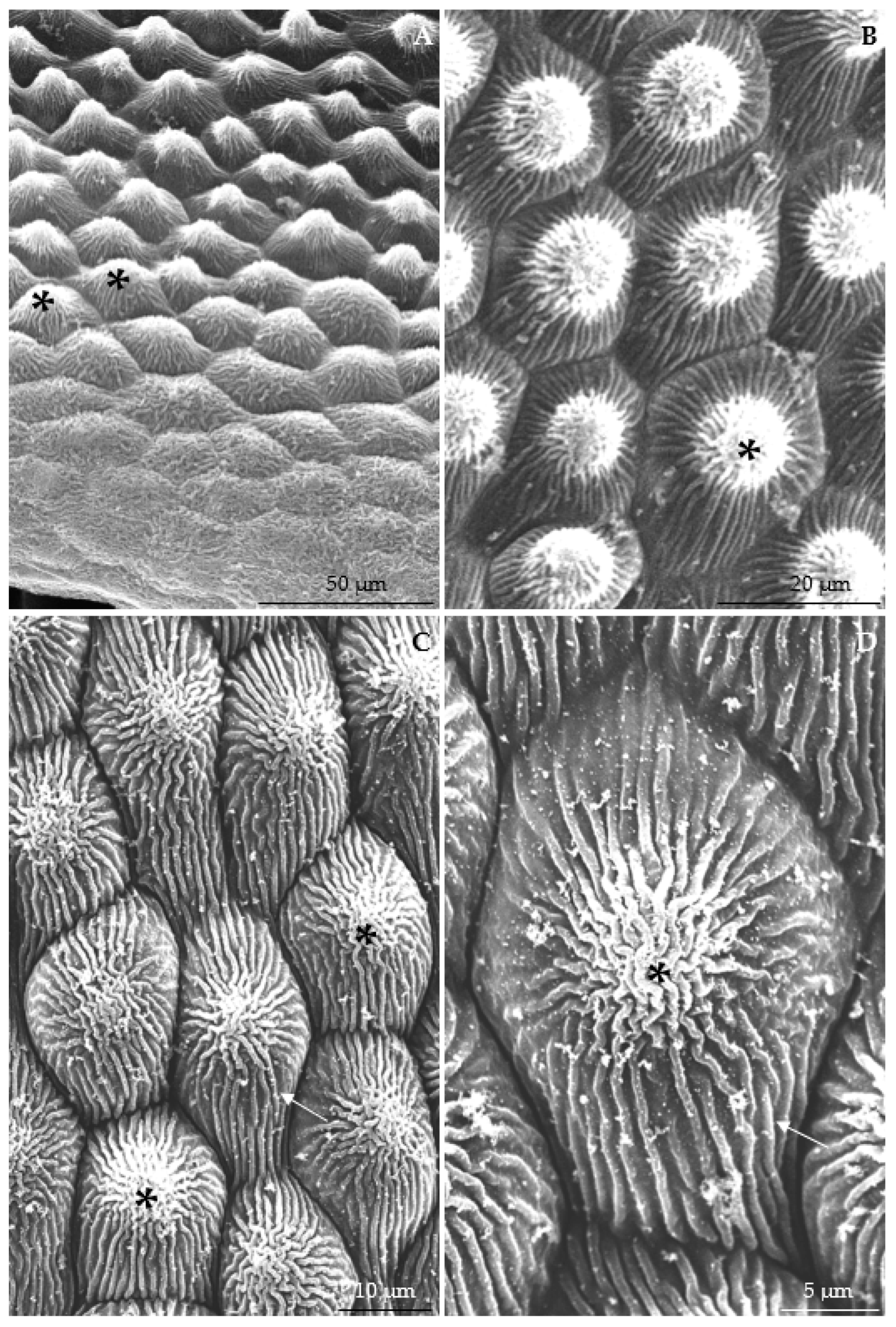
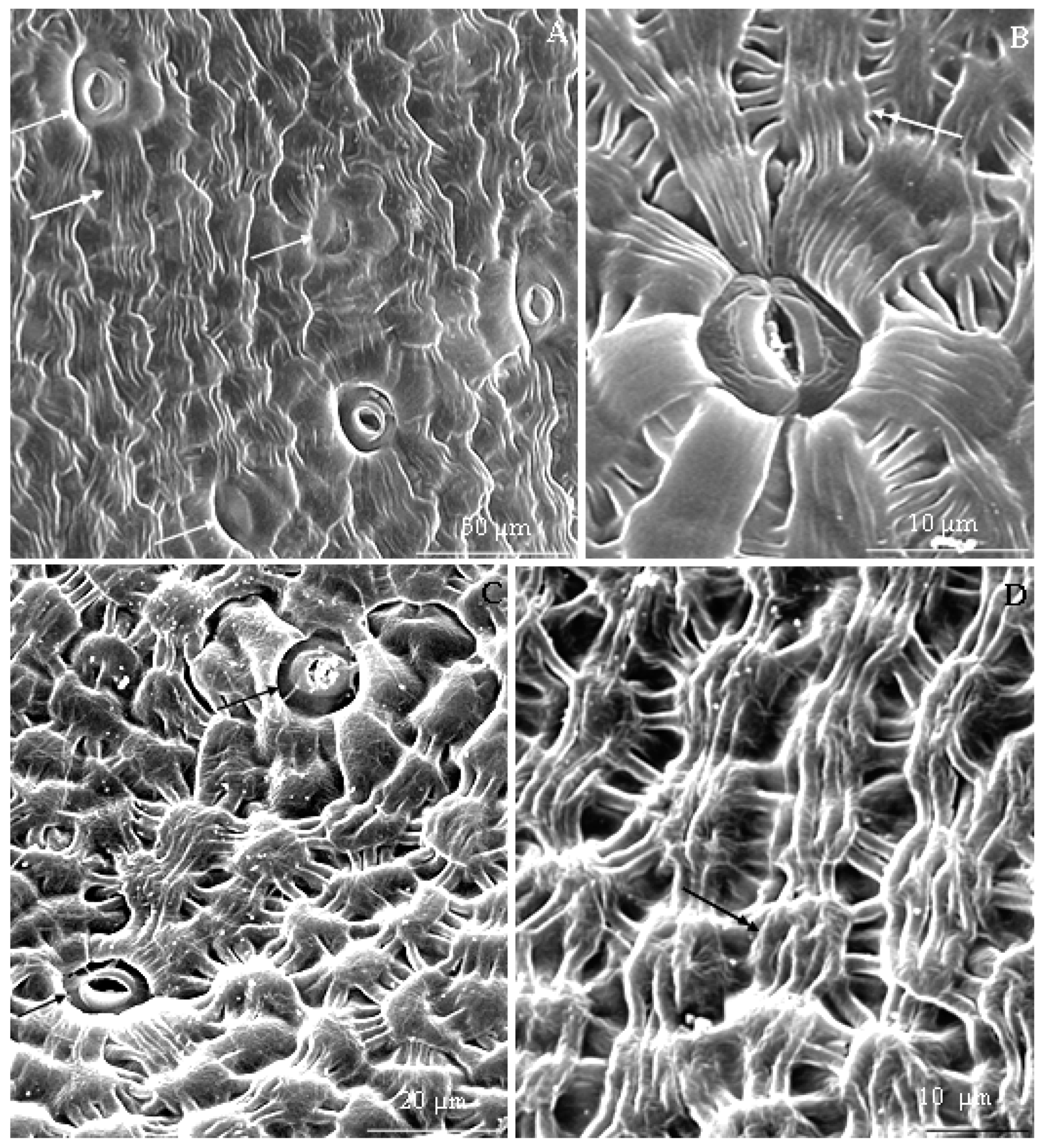
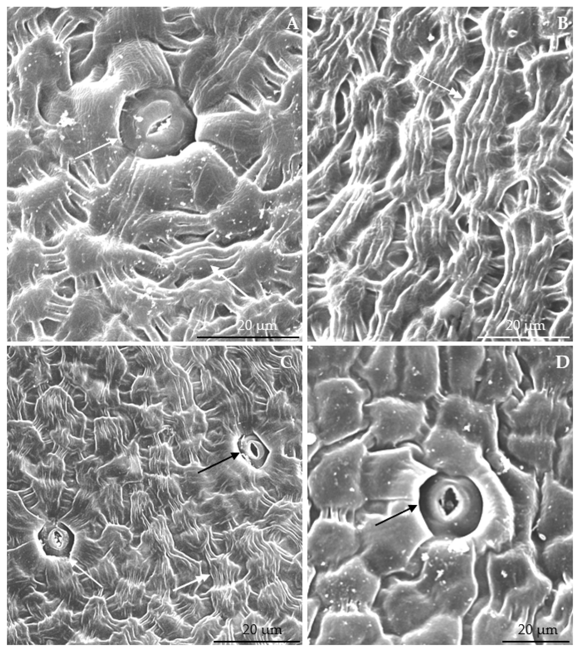
3.2.1. Micromorphology

Cuticle ornamentation. The surface of nectary epidermis cells in the six R. idaeus cultivars exhibited striated cuticle ornamentation (Figure 6A,C, Figure 7A,C and Figure 8A,C). It varied depending on the type of the cells. A smooth cuticle covered the surface of the stomatal cells. These cells were located at the same level or slightly below other epidermis cells (Figure 6C, Figure 7C and Figure 8A,D). The stomatal complexes surrounding the stomata were composed of 4–7 radially arranged cells. Their surface was covered by striated cuticle ornamentation. The striae were arranged side by side forming a parallel pattern extending from the stomata to cells adjacent to the stomatal complex. The striae on the surface of the stomatal complex cells adjacent to the stomatal cells were densely arranged, whereas a much looser arrangement was visible on the opposite side (Figure 6B,C, Figure 7A and Figure 8A,C).
The surface of other epidermis cells exhibited characteristic striated cuticle ornamentation composed of almost parallel striae, sometimes with a slight arcuate curve. These striae on a single epidermis cell formed a band located along its longer axis; it sometimes extended onto the surface of cells adjacent to the gentle indentation on the anticlinal cell wall. In this area, some striae retained continuity, while others overlapped or intertwined (Figure 6A,D, Figure 7A,B,D and Figure 8B,C). The width of the striae forming the band in the analyzed cultivars ranged from 1.64 μm in ‘Radziejowa’ to 1.92 μm in ‘Polana’. The length and width of the cuticle bands in the analyzed cultivars were in the range from 9.45 μm in ‘Pokusa’ to 12.51 μm in ‘Polana’ and from 7.82 μm in ‘Pokusa’ to 10.31 μm in ‘Polana’, respectively. Between the adjacent cells, there were sparse striae arranged towards their shorter axis and perpendicular to the cell layers or sometimes slightly slanting. The width of these striae was in the range from 1.41 μm in ‘Radziejowa’ to 2.08 μm in ‘Polana’. The distance between the bands of adjacent nectary epidermis cells ranged from 1.63 μm in ‘Radziejowa’ to 4.81 μm ‘Polana’. The statistical analysis of the selected traits of cuticle ornamentation on the surface of nectar epidermis cells in the full nectar secretion phase in the raspberry cultivars showed that the band width in ‘Polana’ was significantly higher than that in ‘Pokusa’ and ‘Radziejowa’, and the distance between cuticle bands in ‘Radziejowa’, ‘Pokusa’, and ‘Polka’ was lower than in the other cultivars. There were no statistically confirmed differences between the cultivars in the width of the band-forming striae, the length of the cuticle bands, and the width of striae connecting the cuticle bands (Table 2).
Stomata. The nectar in the raspberry flowers was released onto the nectary surface through stomata arranged evenly on the entire epidermis. The outline of the stomata was elongated. Various development stages, i.e., open, half-open, and closed stomata, were observed in the full nectar secretion phase (Figure 6A,C, Figure 7A,C and Figure 8A,C,D). The width of the aperture between the cuticular ledges in the open stomata of the biennial fruiting cultivars and the repeated fruiting cultivars ranged from 1.93 μm in ‘Glen Ample’ to 2.78 µm in Laszka’ and from 1.92 μm in ‘Polana’ to 3.29 µm in ‘Pokusa’, respectively. Its length ranged from 3.26 μm in ‘Radziejowa’ to 7.65 µm in ‘Laszka’ and 4.12 ‘Pokusa’—6.25 μm ‘Polana’ (Table 3).
The size of the stomata varied; their length and width in the biennial fruiting cultivars ranged from 11.91 µm in ‘Radziejowa’ to 14.96 µm in ‘Laszka’ and 8.74 ‘Radziejowa’ –14,96 ‘Laszka’. In the group of the repeated fruiting cultivars, the respective sizes ranged from 10.82 μm in ‘Pokusa’ to 13.41 µm in ‘Polana’ and from 8.32 μm in ‘Pokusa’ to 11.25 µm in ‘Polana’. The surface area of the stomata was in the range of 70.84 µm2 in ‘Radziejowa’ to 130.28 µm2 in ‘Laszka’ in the biennial fruiting cultivars, and of 87.24 µm2 in ‘Pokusa’ to 101,32 µm2 in ‘Polana’ in the repeated fruiting cultivars. The number of stomata per 1 mm2 of epidermis in the full nectar secretion phase ranged from 70 in ‘Laszka’ to 107 in ‘Radziejowa’ in the biennial fruiting group and from 48.21 in ‘Polka’ to 66.48 in ‘Polana’ in the group of the repeated fruiting cultivars. The analyzed stomata were classified as the anomocytic type. The diameter of the stomatal complex in the biennial and repeated fruiting cultivars ranged from 26.53 in ‘Glen Ample’ to 38.78 in ‘Laszka’ and from 24.36 in ‘Polana’ to 34.12 in ‘Polka’. The statistical analysis showed a significantly smaller surface area of the stomata and a higher value of this parameter in ‘Laszka’ in comparison with the other cultivars. In turn, ‘Pokusa’ had a clearly greater width of the aperture between the cuticular ledges than ‘Glen Ample’, ‘Radziejowa’, and ‘Polana’. In turn, ‘Polana’ and ‘Laszka’ were characterized by a longer aperture between the cuticular ledges than the other cultivars. In ‘Radziejowa’, there were significantly higher numbers of stomata than in the other cultivars. In general, ‘Radziejowa’ and ‘Laszka’ has a significantly lower minimum and maximum diameter of the stomatal complex than ‘Glen Ample’, ‘Pokusa’, ‘Polana’, and ‘Polka’. There were no significant differences between the cultivars in the length and width of stomata (Table 3).
The hierarchical agglomeration analysis of clusters yielded two clusters. One of the clusters comprises the ‘Radziejowa’ cultivar, which differs from the other varieties, as indicated by the analyzed parameters (cuticle ornamentation, size and number of stomata, and diameter of the stomatal complex). In this variety, the surface area of stomata is much smaller, the number of stomata per 1 mm2 is much higher than in the other varieties. In the other cluster, the greatest similarity was found between ‘Pokusa’ and ‘Polka’ and between ‘Glen Ample’ and ‘Polana’. The ‘Laszka’ variety assigned to the second cluster shows greater similariy to ‘Glen Ample’, ‘Pokusa’, ‘Polana’, and ‘Polka’ than to ‘Radziejowa’ (Figure 9).

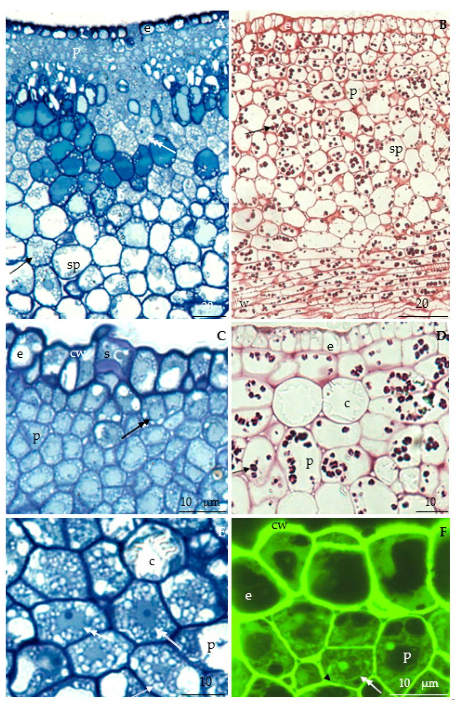
3.2.2. Anatomical Traits of Nectaries

The floral nectaries in the analyzed R. idaeus cultivars were composed of a single-layer epidermis, a layer of nectary parenchyma cells, and several layers of subnectary parenchyma cells (Figure 10A,B).
Nectary epidermis cells. The height and width of the cells in the initial secretion stage ranged from 9.24 µm in ‘Polana’ to 12.24 µm in ’Polana’ and from 9.61 µm in ‘Polana’ to 13.98 µm in ‘Glen Ample’, respectively (Table 4). The outer wall of the nectary epidermis cells was intensely stained and slightly convex (Figure 10A,C). A varied degree of vacuolization was observed in these cells (Figure 10A–F). There were numerous amyloplasts in the cytoplasm (Figure 10A,C), which were intensely stained in the PAS reaction (Figure 10B,D). These cells and nectary parenchyma cells showed green fluorescence (Figure 10F).
Nectary parenchyma cells. The thickness of the nectary parenchyma composed of 7–9 layers of cells was in the range from 90 μm in ‘Laszka’ to 174 µm in ‘Glen Ample’ and from 82 μm in ‘Polka’ to 116 µm in ‘Polana’ in the biennial and repeated fruiting cultivars, respectively. The diameter of these cells in the former and latter group ranged from 13 μm in ‘Laszka’ to 20 µm in ‘Glen Ample’ and from 9 μm in ‘Polana’ to 14 µm in ‘Pokusa’, respectively. The statistical analysis of the selected traits of the nectary in the raspberry cultivars showed the thickness of the nectary parenchyma layer in ‘Pokusa’ and ‘Polana’ was significantly lower than in ‘Glen Ample’ and ‘Radziejowa’ and higher than in ‘Laszka’ and ‘Polka’. ‘Glen Ample’ and ‘Radziejowa’ had a larger diameter of nectary parenchyma cells than the other cultivars (Table 4). In the initial secretion phase, the thin-walled cells of the nectary parenchyma viewed in the longitudinal section adhered tightly to each other. These cells were clearly smaller than the epidermis and subnectary parenchyma cells. The outlines of the parenchyma cells at different developmental stages were 4–6-angular. Among fully developed cells, there were sometimes small cells with a large centrally located nucleus and a protoplast resembling meristematic tissue (Figure 10A,B). The nectary parenchyma cells were characterized by intensely stained cytoplasm, a centrally located nucleus, and numerous plastids (Figure 10C,E). Greater numbers of amyloplasts were observed in the nectary parenchyma than epidermis cells (Figure 10B–D). Calcium oxalate crystals were observed in nectary parenchyma cells (Figure 10D,E).
Subnectary parenchyma cells. The subnectary parenchyma in the six cultivars was composed of 5–8 cell layers. Its thickness ranged from 173 μm in ‘Radziejowa’ to 199 µm in ‘Glen Ample’ in the biennial fruiting group and from 145 µm in ‘Polana’ to 176 µm in ‘Polka’ in the repeated fruiting group. The diameter of the cells was in the range of 27 μm in ‘Laszka’ to 35 µm in ‘Radziejowa’ in the former group, and of 19 μm in ‘Pokusa’ to 27 µm in ‘Polana’ in the latter group. Moreover, the thickness of the subnectary parenchyma layer in ‘Polana’ was significantly lower than that in ‘Glen Ample’. In turn, the number of subnectary parenchyma layers in ‘Pokusa’ was higher than that in ‘Radziejowa’. The diameter of the subnectary parenchyma cells in the latter cultivar was greater than in ‘Pokusa’ and ‘Polka’ (Table 4). The thin-walled cells of the subnectary parenchyma exhibited a parietal cytoplasm band and a large centrally located vacuole. Intercellular spaces were visible between these cells. Amyloplasts were visible in the subnectary parenchyma. Vascular xylem and phloem elements supplying the nectary with substances necessary for metabolic processes and nectar secretion were located near the subnectary parenchyma (Figure 10B).

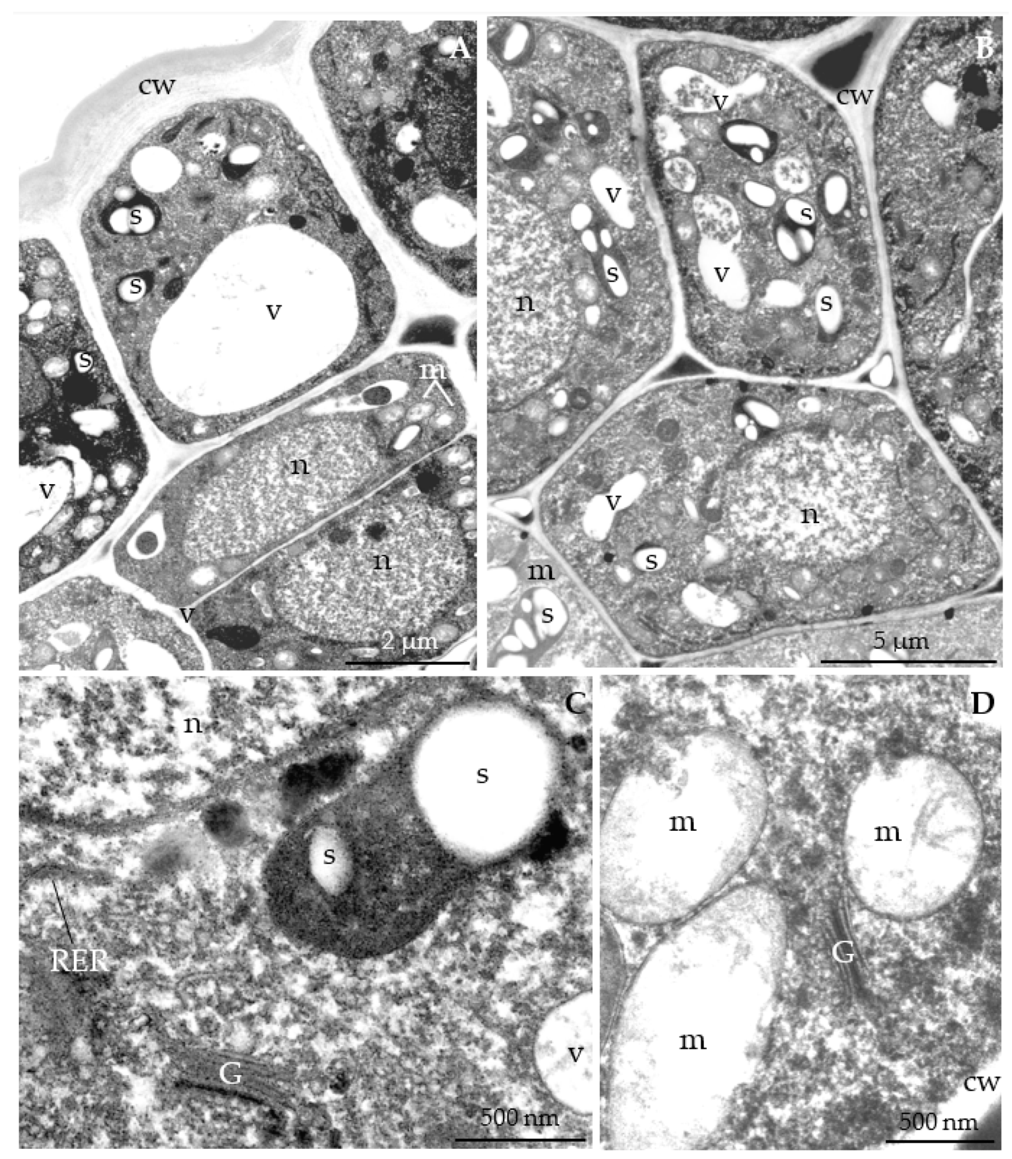
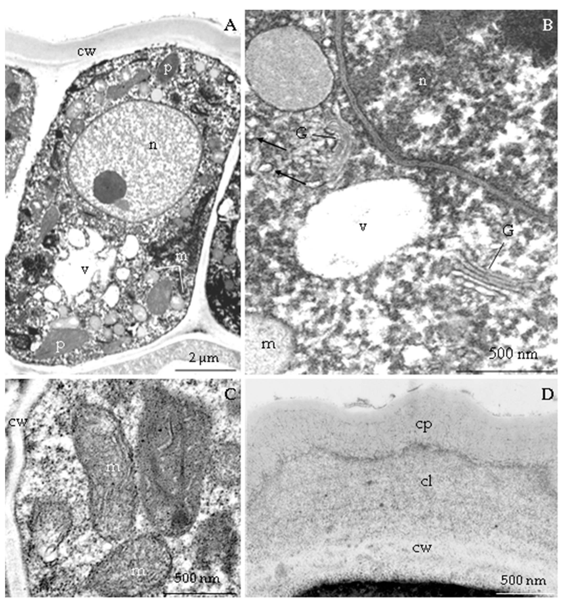
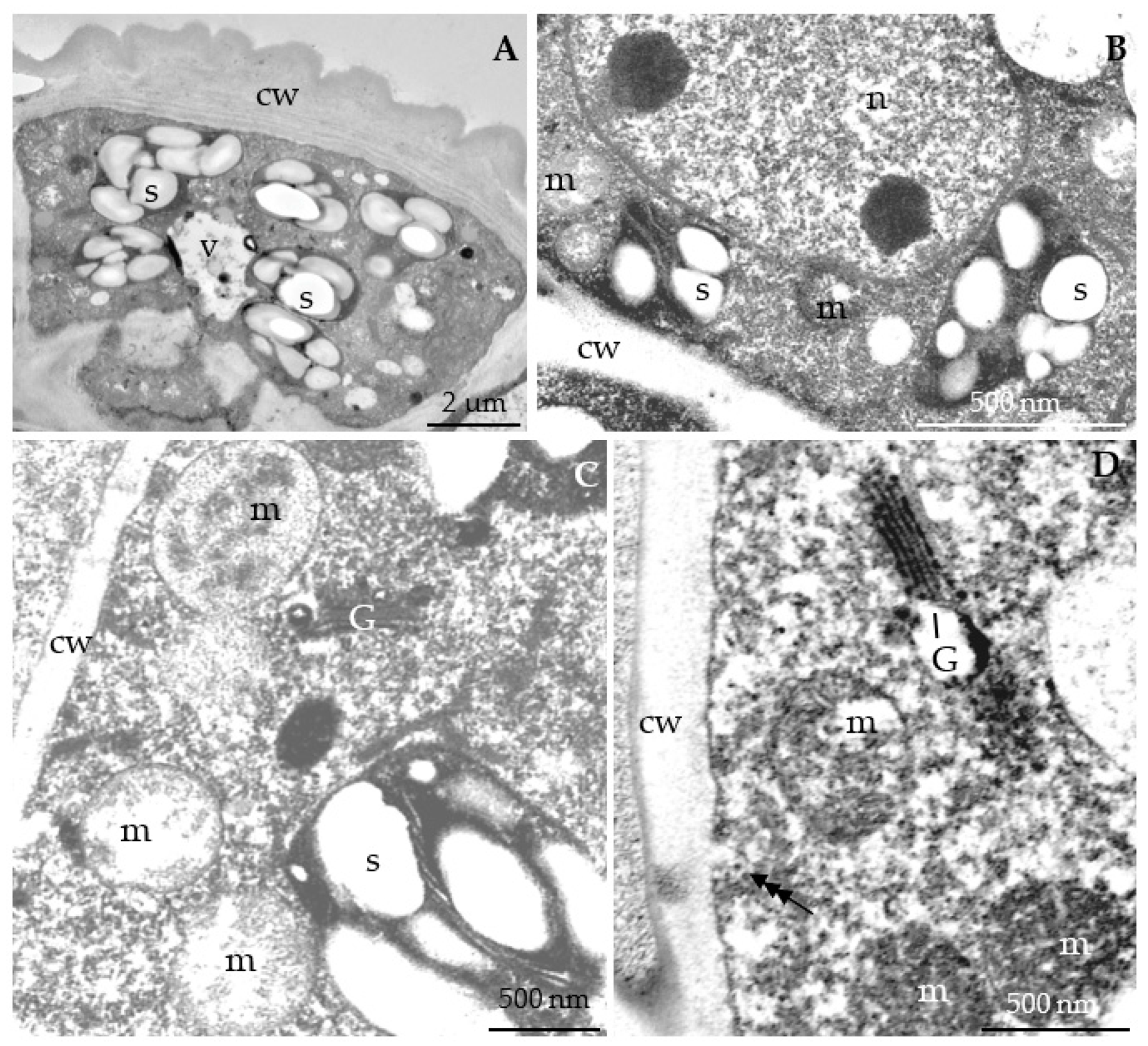
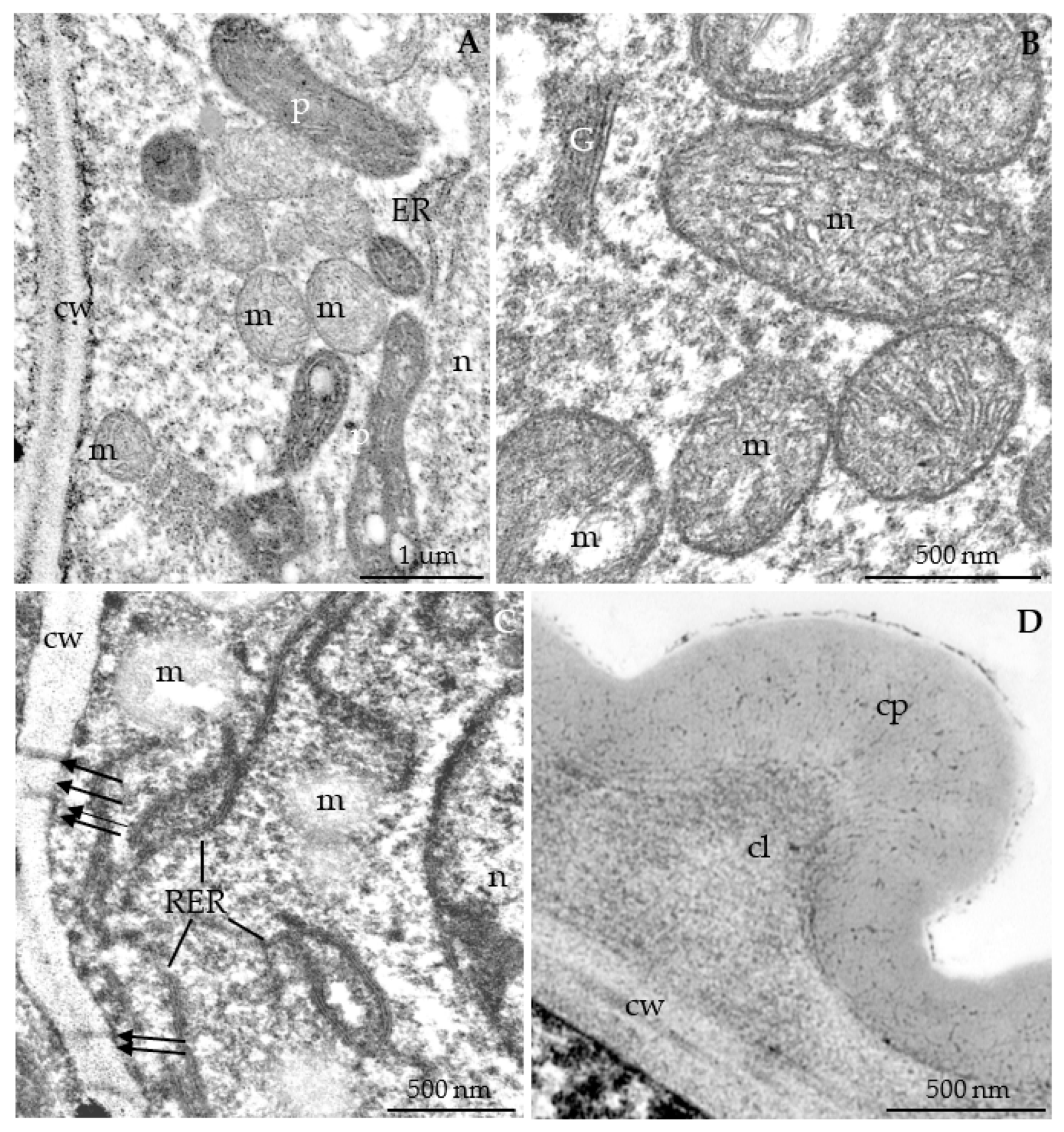
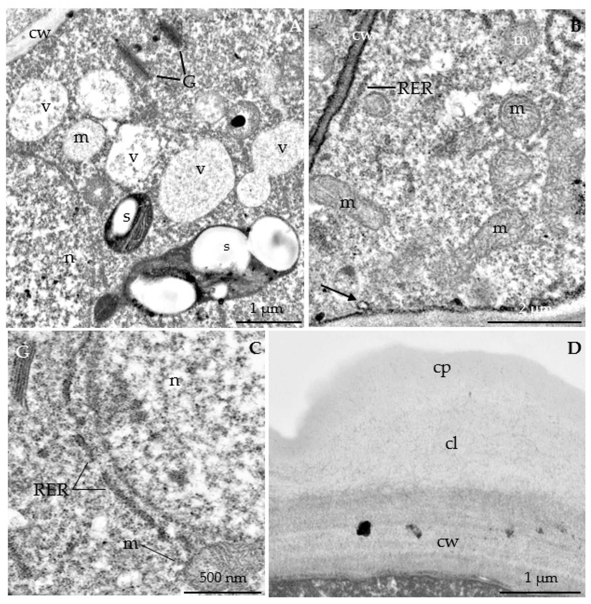
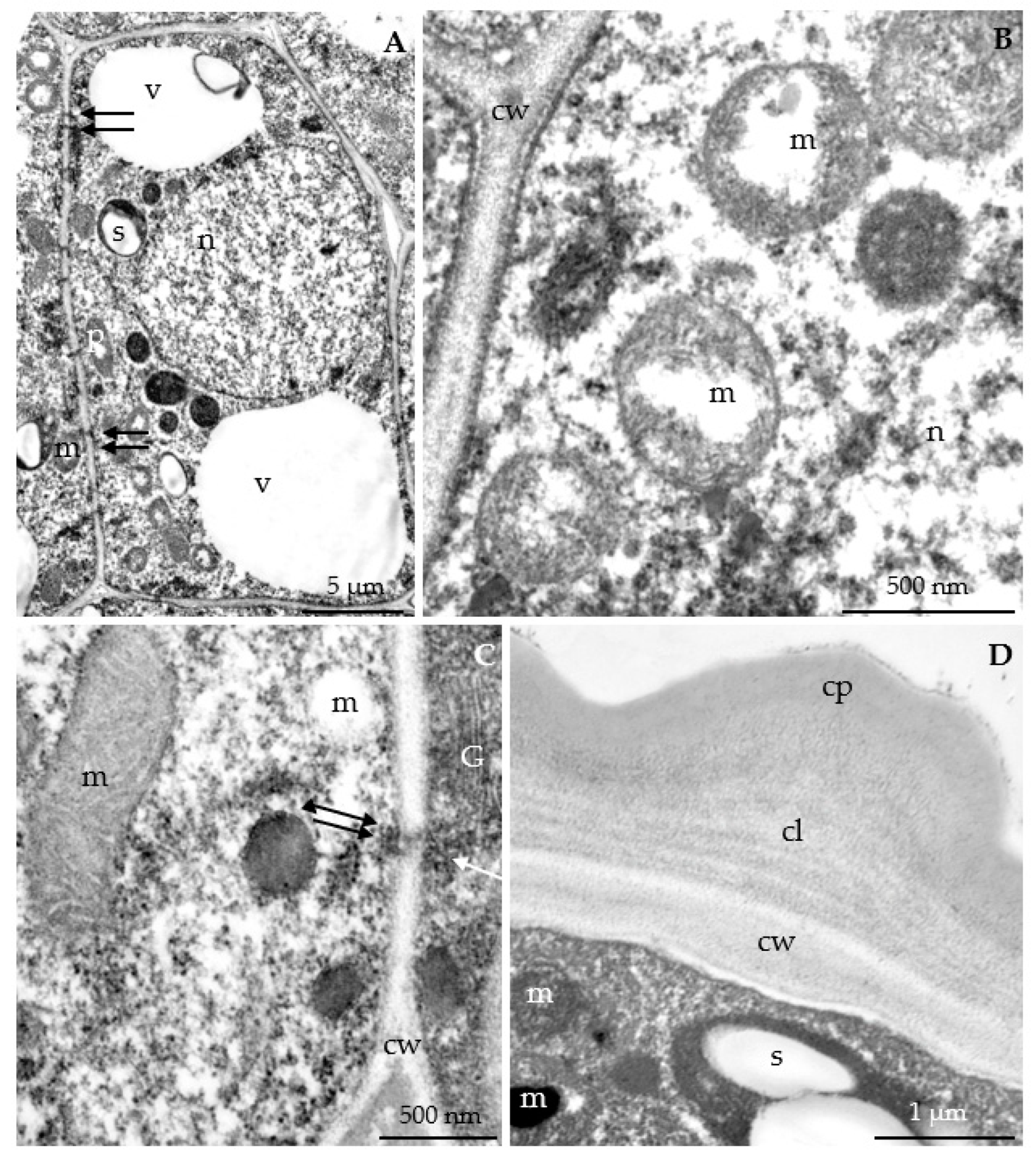
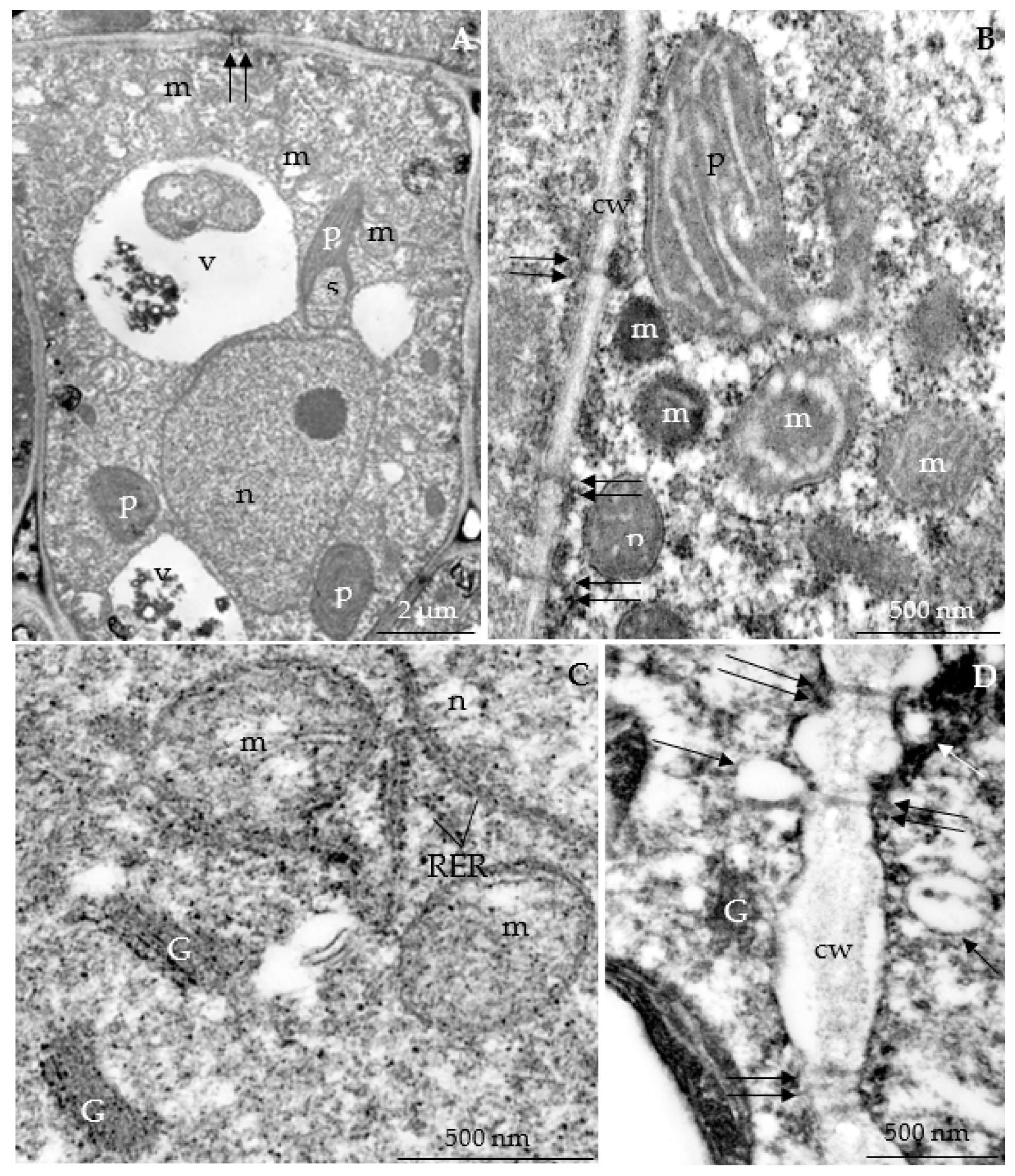
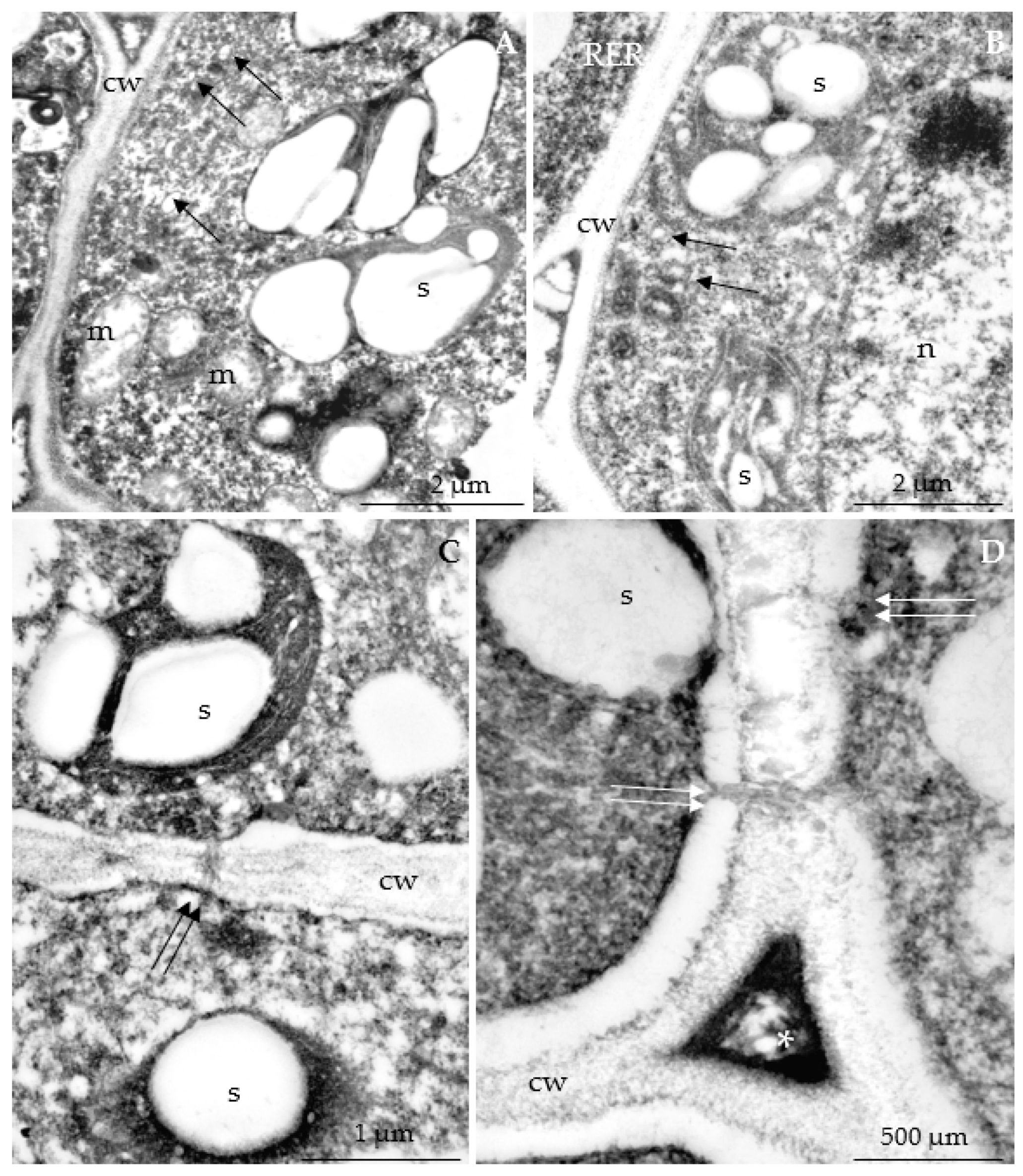
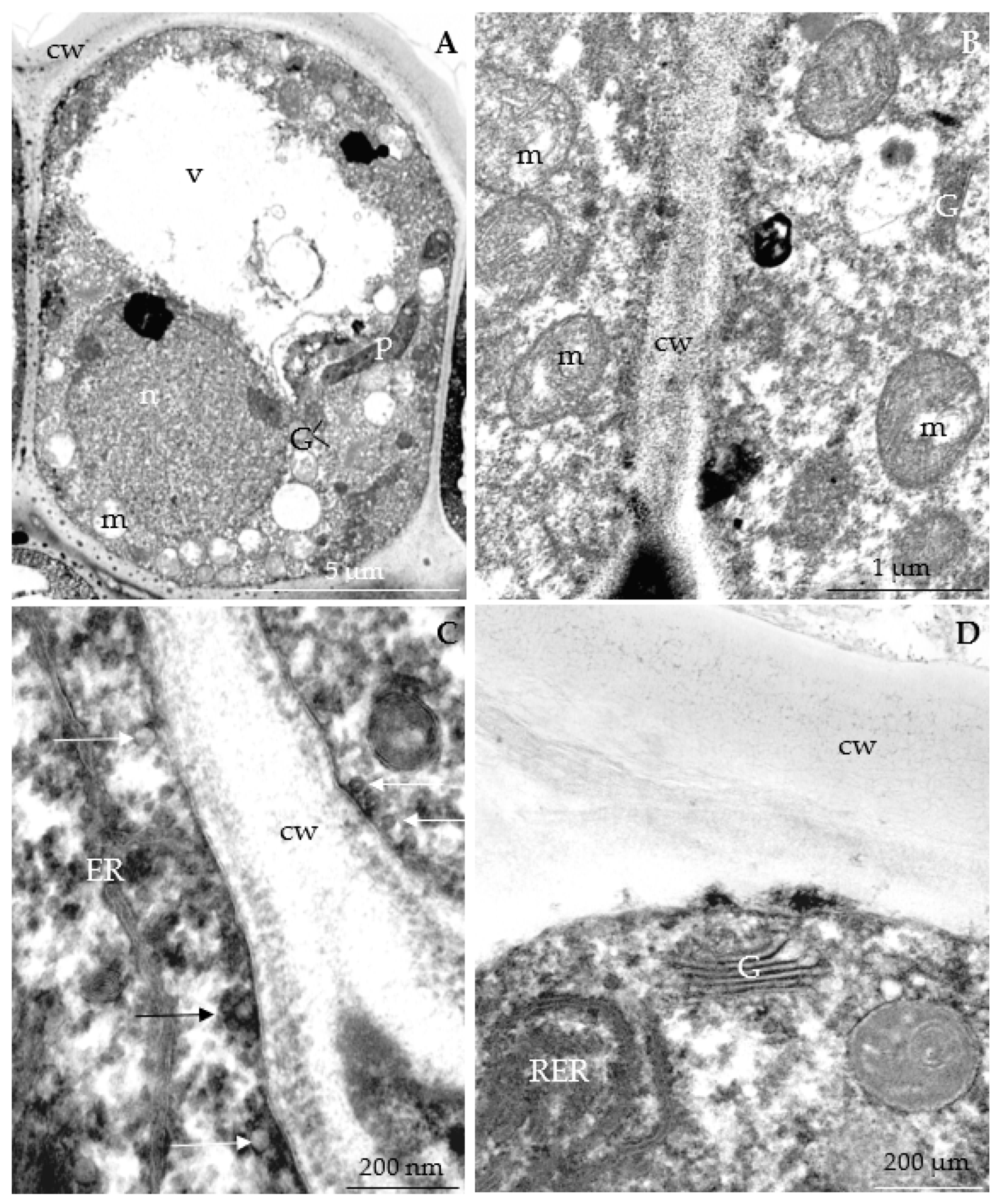
3.2.3. Nectary Ultrastructure

Nectary epidermis cells. The comparative ultrastructural analysis of longitudinal nectary cell sections in the initial and full secretion phases showed that, the nectary epidermis cells had much thicker outer cell walls (1.37–2.7 µm) than the anticlinal (0.33–0.58 µm) and periclinal (0.38–0.63 µm) inner walls (Figure 11A, Figure 12A, Figure 15A, Figure 19A and Figure 22A). The thickness of the anticlinal and periclinal inner epidermis cell walls constituted 18–44% and 16–47% of the outer cell wall thickness, respectively (Table 5). The outer wall of the nectary epidermis cells formed distinct protuberances with different heights and outlines located perpendicular to the cell surface (Figure 13A, Figure 14D, Figure 15A, Figure 16D and Figure 17A). The surface of these protuberances was covered by an evenly distributed continuous cuticle with a thickness ranging from 650 nm in ‘Glen Ample’ to 850 nm in ‘Pokusa’. The cuticle was composed of lamellar and reticulate layers as well as cellulose microfibrils (Figure 12D, Figure 14D, Figure 16D, Figure 17A and Figure 18D). In the epidermal cell walls plasmodesmata were visible (Figure 20D and Figure 21D). The statistical analysis showed that ‘Polana’ was characterized by a significantly greater thickness of the outer cell wall than the other cultivars. Concurrently, the thickness of the anticlinal cell wall in ‘Radziejowa’ and ‘Polana’ was significantly lower than that in ‘Glen Ample’ and ‘Polka’. The thickness of the inner periclinal cell wall in ‘Polka’ was clearly greater than that in ‘Glen Ample’ and ‘Laszka’. There were no statistically confirmed differences between the cultivars in the thickness of the lamellar layer of the cuticle (Table 5).
The inner periclinal walls were tightly adjacent to the nectary parenchyma cells. Numerous plasmodesmata, possibly involved in the symplastic transport of prenectar to nectary cells, were present in the cell walls (Figure 14C, Figure 15D, Figure 18B, Figure 19D, Figure 20B and Figure 21C,D). There were a few small vacuoles or one or two differently outlined larger vacuoles. Their area expanded during nectar release. Multilamellar bodies, flocculent material, or vesicular structures with varying diameters were observed in the cell sap (Figure 12A, Figure 13A, Figure 15A, Figure 20A and Figure 22A). The common traits of the protoplast and organelles of epidermis and nectary parenchyma cells are discussed in the subsection on nectary parenchyma cells.
Nectary parenchyma cells. The nectary parenchyma and epidermis cells in the initial stage of secretion had a centrally located spherical nucleus with a visible electron-dense karyolymph and one or two clearly visible spherical osmophilic nucleoli with dense chromatin stroma (Figure 12A, Figure 13B, Figure 15C, Figure 17A, Figure 19B, Figure 20A and Figure 22A). During the secretion process, the cell nucleus was characterized by an irregular outline and parietal location.
A well-developed endoplasmic reticulum, sometimes constituting an extension of the nuclear membrane, was located near the cell nucleus. The perinuclear endoplasmic reticulum formed oblong tubules (Figure 16C and Figure 20C). An extensive network of cortical endoplasmic reticulum membranes was located near other organelles and cell walls. The rough endoplasmic reticulum was located near mitochondria, plastids, and plasmalemma. The reticulum membrane system had elongated tubules with a loop-shaped (Figure 14C) or circular arrangement (Figure 14C and Figure 22D).
The nectary epidermis and parenchyma cells contained numerous plastids, which were arranged especially densely around the cell nucleus and close to the cell walls (Figure 11B, Figure 13A,B, Figure 15B, Figure 17B and Figure 18A). In the initial phase of secretion, the amyloplasts contained from 1 to 7 starch grains. They varied from small and medium-sized to large grains, which sometimes filled the amyloplast completely (Figure 11B, Figure 13B, Figure 15B, Figure 17C, Figure 19C and Figure 21A,C). In the full secretion phase, the amyloplasts were visible but contained substantially lower numbers of starch grains, as this carbohydrate was hydrolyzed during nectar secretion. The plastids usually exhibited pleomorphic shapes: spherical, lenticular, or elongated. Sometimes they were narrow in the central part or dilated at one of the poles and tapering at the other. The stroma contained a loose thylakoid system, one or two small starch grains, and sometimes osmophilic plastoglobules (Figure 12A, Figure 14A, Figure 20A,D and Figure 22A).
Numerous mitochondria were located near the plastids, nucleus, and walls. The close contact between these organelles provided energy necessary for metabolic processes during nectar secretion. Some mitochondria had well-developed cristae. A transparent matrix and a reduced number of cristae were observed in the subsequent phase of secretion. The shape of the mitochondrion varied from spherical to lenticular and sometimes elongated. The mitochondria were arranged in clusters near other organelles in the central part of the protoplast or serially near the plasmalemma, tonoplast, and nuclear membrane (Figure 12C, Figure 14A, Figure 17A, Figure 18C, Figure 20C and Figure 22B). These organelles interacted with the Golgi apparatus present nearby (Figure 11D, Figure 13C,D, Figure 14B and Figure 20C).
Golgi apparatus composed of 4–8 dictyosome cisternae were located close to the nucleus, plastids, and plasmalemma (Figure 11C,D, Figure 12B, Figure 13C,D, Figure 14B, Figure 16A,C, Figure 17D, Figure 19D, Figure 20C and Figure 22D). Numerous transport vesicles were visible at the poles of the cisternae and in the entire protoplast (Figure 12B, Figure 15D, Figure 16B, Figure 17C,D, Figure 18B, Figure 19C,D, Figure 21B and Figure 22C). They were mostly concentrated near the plasmalemma. Some of them fused with the membrane and released nectar into the periplasmic space (Figure 13C, Figure 17C and Figure 19C,D).

4. Discussion

4.1. Nectary Micromorphology

Cuticular ornamentation. The present observations showed cuticular striation on the surface of nectary epidermis cells in the six R. idaeus cultivars. Such ornamentation was also reported in other members of the Rosaceae family, e.g., various species of the genera Cotoneaster, Crataegus, Malus, Prunus, Sorbus, and Chaenomeles [20,45,46,47,48]. Literature data indicate that cuticular ornamentation is a characteristic trait of various cultivars or a group of cultivars [49]. The striae on the surface of stomatal complex cells in the groups of raspberry cultivars analyzed in the present study exhibited parallel arrangement between the stomatal cells and those adjacent to the stomatal complex. In turn, the striae on the other epidermal cells formed a single band along the longer cell axis, which sometimes extended onto the surface of neighboring cells. The inter-band region of the adjacent cells was linked by striae arranged loosely towards the shorter axis of the cells.
In the present study, the thickness of the band-forming striae and those visible in the inter-band area varied in the range of 1.6–1.9 µm and 1.4–2.1 µm, respectively, and the distance between the bands was 1.6–4.8 µm. As reported by Orosz-Kovács et al. [50], the narrow grooves present between thin striae on the entire nectary surface serve as microcapillaries distributing secreted nectar in the inter-band region, and the distribution of this secretion is greatly facilitated by the hydrophobic nature of the cuticle. The ability of the cuticle to repel water molecules is associated with the presence of polymerized fatty acids, which facilitate the flowing drops of the liquid to clean the surface of this layer [51,52,53]. Concurrently, cuticular striae protect nectar against drying and, through limitation of evaporation, extend the duration of nectar exposure to insects, thus substantially enhancing the flower pollination efficiency [20,27,50,54]. As demonstrated by Koteyeva [55], the cuticle layer in specialized secretory tissues, mainly in nectaries, is characterized by a high degree of diffusion-based penetration. Cuticular striae with different heights and arrangements probably determine the absorption and reflectance of ultraviolet radiation [56]. As shown by Schulte et al. [57], in addition to the color of flowers in the visible spectrum (VIS, 400–700 nm) and in UV (280–400 nm), the glossy cuticle ornamentation is an additional visual signaling system for pollinators. Specific carbohydrates and waxes present in the cuticle reflect UV radiation, which is especially important for the secretory tissue [58]. The cuticle reduces the penetration of unfavorable chemicals into tissues, constituting a mechanical barrier against biotic and abiotic factors. The cuticle ornamentation and the location of stomata relative to epidermis cells indicate the plant ecotype [50].
Stomata. The nectary epidermis in the analyzed R. idaeus cultivars exhibited the presence of anomocytic stomata. The number of stomata per 1 mm2 of the nectary epidermis in the biennial and repeated fruiting raspberry cultivars (from 70 in ‘Laszka’ to 107 in ‘Radziejowa’ and from 48 in ‘Polka’ to 67 in ‘Polana’, respectively) was lower than in species of the genera Crataegus (175–308), Prunus (146–230), and Polemonium (134–156) [45,59,60,61]. In comparison with the value of the parameter in Cucurbita pepo (83–152) [61,62], the number of stomata per 1 mm2 of the nectary epidermis was similar or comparable in the former cultivar group and clearly lower in the latter group. Chwil et al. [20] reported the presence of 45 and 71 stomata in the epidermis of Prunus laurocerasus ‘Schipkaensis’ and ‘Zabeliana’, respectively. Nectar precursors generated through metabolic processes overcome various barriers, e.g., the cell membrane, cell wall, and hydrophobic cuticle. Nectar is secreted onto the nectary surface through the stomata and cuticle pores, cracks, or detached fragments of the layer [52]. Nectar release through the stomata proceeds passively as a result of the concentration gradient and the action of capillary forces [63]. Abedini et al. [64] described nectar secretion through modified stomata via symplastic and apoplastic routes. As observed by Gaffal [63], nectaries that have stomata release a substantially larger amount of nectar to its surface within a specified time than nectaries that do not have stomata. Literature data indicate that stomata do not regulate the nectar flow rate [65,66]. In the present study, modified stomata at various stages of development, i.e., in the pre-secretory, secretory, and post-secretory phases, were observed on the entire surface of the secretory nectary epidermis in the six R. idaeus cultivars. This is consistent with findings reported by other authors [65,67]. Similarly, different development stages of stomata, regardless of the development stage of nectaries, were observed in Polemonium caeruleum and Sorbus intermedia [59,68,69]. Literature data have shown that the stomata are permanently open [70,71].

4.2. Nectary Anatomy

The present observations showed that the nectaries in the six R. idaeus cultivars were composed of a single-layer epidermis, from 7–9 layers of parenchyma cells, and 5–8 layers of subnectary parenchyma cells. The height of the nectary epidermis cells was 9–14 µm. A similar range of values of this parameter was recorded in species of the genera Cotoneaster (2–17 µm), Malus (8–28), and Prunus (9–15 µm) [20,23,25]. The values were higher in Aronia melanocarpa (20 µm) and representatives of the genera Crataegus (17 µm), Prunus (19–24 µm), and Sorbus (18–21 µm) [23,60]. The traits of the nectary epidermis cells and their ultrastructure are compared with other reports in the section on nectary ultrastructure. The number of layers and the thickness of the nectary parenchyma layer (90–174 µm) in the analyzed R. idaeus cultivars were similar to the values reported for five species of the genus Prunus, in which parenchyma cells formed 5–10 layers with a thickness of 105–168 µm [60]. A similar number of layers of nectary parenchyma cells (8–10) was reported for Crataegus crus-galli and C. coccinea, which formed a substantially thicker layer (399–400 µm); the cells were densely arranged and smaller than the cells of the subnectary layer [61]. In turn, compared the present study, a considerably thinner 2–5-layered nectary parenchyma was described in Prunus laurocerasus ‘Schipkaensis’ (79 μm) and ‘Zabeliana’ (39 μm) [20]. As shown in the literature, the number of nectary cell layers varies in different species. The nectary in Aronia melanocarpa was composed of 16 layers with a thickness of 284 μm; the values of these parameters were 11–12/189–200 μm in Cotoneaster horizontalis, C. praecox, and C. lucida, 15–17/356–400 μm in Crataegus monogyna, C. coccinea, and C. crus-gali, and 14/89–396 μm in Sorbus aucuparia and S. intermedia [23]. In turn, nectary cells in several species of the genus Malus formed from 14 to 31 layers and the gland was from 203 to 430 μm thick [25]. The nectaries of Cydonia oblonga, Cotoneaster betulifolia, and Pyrus betulifolia were composed of 8–15, 3–4, and 3–8 layers, respectively [21,27]. The subnectary parenchyma in the raspberry cultivars analyzed in the presented study was composed of 5–8 layers and its thickness was in the range of 145–199 µm. This tissue in Prunus laurocerasus described in the literature comprised 3–5 layers with a thickness of 102–109 µm [20]. The subnectary parenchyma cells described in the present study and in other reports comprised vascular xylem and phloem elements supplying indispensable substances to the nectary cells. In turn, the nectary parenchyma of these layers exhibited typical anatomical traits of secretory cells: darker stained cytoplasm with a centrally located nucleus containing one or two nucleoli, many vesicular structures, and thin cell walls [20,26,60,72,73].

4.3. Nectary Ultrastructure

Cell wall. The thickness of the outer cell wall of the nectary epidermis in the analyzed R. idaeus cultivars was from 1.5 µm in ‘Glen Ample’ to 2.47 µm in ‘Polka’. A similar range of values of this parameter was determined in Prunus laurocerasus and Malus sp. (1.26–2.58 µm) [20,74]. Lower values of the parameters were reported for Prunus armeniaca, P. avium, P. cerasus, and Pyrus (0.9–1.5 µm) [22,60]. Generally, higher values were found for P. domestica, and representatives of the genera Chaenomeles, Cydonia, Malus (2.3–6.1 µm) [23,25,60]. The thickness of the periclinal inner walls and the anticlinal walls in the nectary epidermis cells of the R. idaeus cultivars was in the range of 0.37–0.63 and 0.33–0.58 µm, respectively. These values were similar to those found in Prunus laurocerasus ‘Zabeliana’ (0.46 and 0.71 µm) and higher than in Prunus laurocerasus ‘Schipkaensis’ (0.16 and 0.39 µm) [20].
The ultrastructure of the epidermis cells of the analyzed R. idaeus nectaries exhibited small, vertical, conical protuberances corresponding to the cuticle striae in the outer cell wall. Their height was in the range of 2.8–3.3 µm in several species of the genus Prunus [60]. Similar protuberances were described in representatives of the genera Aesculus, Galanthus, Prunus, and Rhododendron [20,60,75,76,77].
The thickness of the epidermis cuticle in the analyzed R. idaeus nectaries was in the range of 0.65–0.85 µm. These values were higher than those reported in Prunus cerasus and P. avium (0.41–0.57) and lower than in Prunus armeniaca and P. domestica (0.9–3.3 µm), P. avium (1.5–6.0 μm), Prunus laurocerasus (1.21–1.30 μm), and Pyrus sp. (1.26–2.58 μm) [20,21,60,78,79]. In the present study, the evenly distributed cuticle comprised lamellar and reticulate layers and cellulose microfibrils. This structure is in line with the findings reported by Paiva [52], who distinguished three layers in the cuticle: the outermost layer (cuticle proper), the cuticular layer (with embedded cellulose and other cell wall elements), and the innermost pectin layer. The author has shown that the cuticle hydrophobicity increases from the innermost to outermost layer. The wax present in the cuticle structure increases the permeability and facilitates the release of fat-soluble substances [52,80,81,82]. The nectary epidermis cuticle in many species of the Prunus perisca has microchannels involved in nectar secretion [72,83,84]. As suggested by Nepi [85], these microstructures support the apoplastic route of nectar secretion onto the nectary surface. It has been reported by Paiva [52] that nectar secretion is mediated by hydrophilic bridges formed by elements of the wall and pectins and numerous branches of hydrophilic projections present in the cuticle layer. The penetration of nectar across the cell wall is facilitated by low-viscosity hydrophilic substances contained in nectar with high affinity for cell wall components [80].
Plastids. As demonstrated in the present study, the protoplast of the R. idaeus nectary cells exhibited the presence of pleomorphic plastids with a few starch grains in the initial stage of secretion. In turn, in the full nectar secretion stage, the plastids contained only single grains or completely hydrolyzed grains and sometimes plastoglobules. As suggested by Abedini et al. [64], these organelles are among the most abundant elements in the nectary cell protoplast during nectar secretion. In Prunus laurocerasus nectaries, amyloplasts in the pre-secretory phase contained up to eight starch grains [20]. As indicated by Nepi [85], the accumulation of starch in nectary cells in the pre-secretory phase is correlated with a high rate of nectar production. Additionally, this polysaccharide is a source of sugars required for nectar synthesis and metabolic processes [86]. Similarly, other authors observed numerous starch grains in early nectary development stages, which disappeared during the further nectar release process, and plastoglobules appearing in the stroma [64,87]. Starch hydrolysis in plastids during nectar secretion proceeds from the outermost to innermost layer [64]. This process reduces the water potential and increases the hydrostatic pressure supporting nectar release [88]. As suggested by Paiva [89], in addition to the aforementioned modifications of nectaries during nectar secretion, i.e., the disappearance of starch grains in plastids and the increase in the number of plastoglobules, the content of carotenoids increases, which not only changes the nectary color but also increases protection against oxidative stress associated with the secretory activity. In the subsequent stages of nectar secretion, plastids have an irregular shape, which may result from the depletion of starch, for example [90,91]. These authors report structural changes in nectary cells, e.g., chromatin condensation, degradation of the mitochondrial membrane, and dissolution and rupture of the vacuolar membrane. The nuclear DNA of nectary cells undergoes degradation from the budding to fruiting period. This programmed cell death leads to nectary aging, which is often associated with cell lysis and degradation of the nectary [92,93]. As suggested by Mosti et al. [93], nectary cells in the anthesis stage contain amyloplasts, which gradually disappear in the subsequent stages via apoptosis as well.
Mitochondria. In the present study, in addition to plastids, the epidermis and parenchyma cells of the R. idaeus nectaries contained numerous mitochondria with well-developed cristae. These organelles had a regular shape and were more numerous near the nuclear membrane and plastids, including amyloplasts, and along cell walls; in turn, they formed clusters in the protoplast. A similar location of mitochondria has also been described in other plant species [20,66,68,91,94,95,96,97]. The close proximity of mitochondria and amyloplasts is a characteristic trait of the eccrine secretion process. It also indicates the energy demand during starch hydrolysis and complex metabolic processes associated with nectar secretion [94,98]. In this process, junctions between various organelles and membranes were observed in nectary parenchyma cells, e.g., mitochondria–chloroplast, mitochondria–mitochondria, mitochondria–peroxisome, mitochondria–endoplasmic reticulum, mitochondria–nuclear envelope, chloroplast–chloroplast, chloroplast–peroxisome, chloroplast–nuclear envelope, chloroplast–plasmalemma, and chloroplast–tonoplast. These temporary junctions between adjacent membranes optimize the movement of molecules between organelles at the subcuticular level during nectar secretion [94]. The mitochondria in the nectary cells of the analyzed R. idaeus cultivars had a well-developed membrane system. As reported by Bowsher and Tobin [99], in mesophyll cells, the volume occupied by the mitochondrion and plastids, as well as the cytosol and vacuole, accounted for 0.6% and 22% as well as 9% and 68%, respectively. In the cytosol of these cells, mitochondria and plastids accounted for 0.3% and 11.4% of the volume, respectively. The presence of numerous mitochondria with well-developed and strongly folded cristae in glandular cells indicates high respiratory activity providing cells with ATP and energy necessary for transport of prenectar and metabolic processes taking place during nectar secretion [66,85,94,97,100].
Endoplasmic reticulum. In the present study, the endoplasmic reticulum forming tubules or sometimes a circular system was observed in the nectar epidermis and parenchyma of the six R. idaeus cultivars. An extensive rough endoplasmic reticulum network was often visible near the nucleus and along the cell walls. Horner et al. [101] and García et al. [102] reported the presence of smooth and rough endoplasmic reticulum in nectary epidermis and parenchyma cells in Glycine max and Passiflora spp. In turn, rough endoplasmic reticulum was found in nectary cells in Citharexylum myrianthum, Erythrina speciosa, Prunus laurocerasus, Prunus persica, and Robinia viscosa [72,76,103,104,105,106], whereas a smooth endoplasmic reticulum was observed in Geranium macrorrhizum and G. phaeum and several species from the families Anacardiaceae and Orchidaceae [82,96,106,107]. Different types of endoplasmic reticulum membranes in nectary cell protoplasts have been described. Tubular reticulum was observed in Epipactis atropurpurea, Limodorum abortivum, Polemonium caeruleum, Prunus laurocerasus, and Salvia farinacea [20,53,68,108], whereas a peripheral reticulum was reported in Salvia farinacea, Citharexylum myrianthum, Geranium macrorrhizum, and Geranium phaeum [53,95,96]. As suggested by Chatt et al. [29], the dense distribution of the endoplasmic reticulum with the characteristic arrangement parallel to the cell walls, the large number of vesicles merging with plasma membranes, and the progressive disintegration of starch grains and sucrose synthesis mentioned above confirm the assumption of the merocrine model of nectar biosynthesis. In turn, Vassilyev [109] claims that the endoplasmic reticulum does not produce secretory granules and the secretory vesicles of the Golgi apparatus do not participate in the transport of nectar sugar. As reported by Ning et al. [87], sugar hydrolyzed from starch and phloem can be transported to the endoplasmic reticulum (ER) and the Golgi cisternae and can be further carried by ER vesicles and released into the periplasmic space via the exocytosis process. As shown by Pacini and Nepi [110], a well-developed endoplasmic reticulum is associated with the production and transport of nectar.
Golgi apparatus. Well-developed Golgi apparatus with distinct dictyosomes composed of 3 to 7 cisternae were observed close to the mitochondria, ER membrane system, and plasmalemma in the ultrastructural study of the nectary epidermis and parenchyma cells of the analyzed R. idaeus cultivars. The number of cisternae in the dictyosomes of nectary cells differed between species of the genera Heliamphora and Polemonium (7–8), Hymenaea (2–4), and Prunus (2–6) [60,68,94,111]. As suggested by Young et al. [112], the increase in the number of Golgi apparatus in the cell is one of the mechanisms of adaptation to intensified mucilage secretion. Kram et al. [113] showed that the number of dictyosomes in secretory cells increased gradually before and during secretion. The ultrastructure of floral nectary exhibited the presence of Golgi apparatus and an extensive ER network in many plant species [20,68,85,113,114]. This study and literature data reported the presence of Golgi apparatus, mitochondria, ER profiles, and dictyosome vesicles in the vicinity of the plasmalemma. Filled vesicles were fused with the membrane, transferring the transported material into the periplasmic space, which was formed by the folds of the protein-lipid membrane and increased during anthesis [68,93,103,106]. As reported by Kram et al. [113], most of the metabolites of starch hydrolysis are packed into the endoplasmic reticulum (ER) or dictyosome vesicles and secreted through the fusion with the plasma membrane (granulocrine secretion). The ultrastructure of the nectary epidermis and parenchyma cells in the analyzed R. idaeus cultivars showed the presence of numerous mitochondria with extensive cristae and Golgi apparatus with distinct dictyosomes and transport vesicles as well as a rich system of endoplasmic reticulum membranes. These are characteristic traits of the granulocrine mode of nectar secretion. This nectar secretion mode has also been described in other plant species [66,68,85].
Plasmodesmata. In the initial stage of secretion, the ultrastructure of the nectaries of the six R. idaeus cultivars exhibited the presence of numerous plasmodesmata located close to each other in the cell walls, which indicates symplastic and apoplastic routes of nectar transport. Endoplasmic reticulum often with parallel arrangement was observed near the plasmodesmata. This well-developed intercellular contact maintaining the continuity of the protoplasts has also been described in the ultrastructure of nectaries in other species [66,68,85,115]. Plasmodesmata with an enlarged cavity at the midline of the wall participate in the active symplastic transport of nectar metabolites derived from phloem sap [87,93,103,106,110,116]. ER membranes present in plasmodesmata are routes of diffusion of lipid signaling molecules between cells from the innermost to outermost nectary layer [106]. In turn, the plasmalemma adheres to the anticlinal walls and creates a junction inhibiting lateral diffusion and preventing the movement of substances through the apoplast [106,117]. Wist and Davis [66] described a symplastic route between phloem elements with companion cells and nectary parenchyma and epidermis cells, which is dependent on intercellular plasmodesma junctions. This continuous symplastic route of nectar to the nectary epidermis requires apoplastic transport of the secretion through anticlinal and outer periclinal cell walls to the nectary epidermis surface. This transport is facilitated by microchannels present in the cuticle, through which the unused nectar can be reabsorbed. As suggested by Rocha and Machado [118], the presence of an extensive system of plasmodesma membranes and endoplasmic reticulum, numerous dictyosomes, and dictyosome vesicles is evidence for symplastic nectar transport.
The present study is the first cellular level description of the nectary structure associated with the secretion and chemical composition of nectar in six R. idaeus cultivars. Therefore, it may be suggested that there is a need to continue the investigations on the biochemistry of nectar combined with determination of food preferences of honeybees and other pollinators. There is a need to provide new and more complete information in the field of genetic engineering to link fecund cultivars characterized by high nutritional quality of fruit and market requirements with molecular metabolic mechanisms in nectaries. This will ensure the maximum degree of pollination of flowers through appropriate informational signals influencing insect behavior, which will increase the pollination efficiency. The present study results bring new data in the field of plant biology. They can be used in comparative analyses of plant ontogenesis.

5. Conclusions

The present study describes for the first time the structure of the floral nectaries (micromorphology, anatomy, and ultrastructure) in Rubus idaeus. To the best of our knowledge, this is the first description of these structures in the genus Rubus bringing new data to the anatomy and biology of plants, especially in the field of exosecretion. The nectary epidermis surface in the six R. idaeus cultivars was characterized by (i) a striated cuticle with striae arranged in bands extending onto several cells, (ii) an anomocytic type of stomata in various development stages, (iii) an even distribution of stomata, with higher density per 1 mm2 area in the biennial than repeated fruiting cultivars, (iv) larger stomatal complexes in the former group of cultivars. These parameters can facilitate identification of related Rubus species. The epidermis, nectary parenchyma, and subnectary parenchyma cell layers were 32–38% thicker in the biennial fruiting cultivars than in the other group of cultivars mainly due to the differences in the cell diameters. In the initial stage of secretion, these cells exhibited the presence of numerous amyloplasts, a centrally located nucleus, and a few small vacuoles sometimes with calcium oxalate crystals. The vascular elements supplying the nectaries reached the subnectary parenchyma cells. Protuberances of varied height were found in the ultrastructure of the outer cell wall of the nectary epidermis. The cuticle had lamellar and reticulate layers. The protoplast of the nectary cells contained electron-dense cytoplasm and chromatin stroma of a spherical nucleus, an extensive system of endoplasmic reticulum membranes, numerous amyloplasts, and mitochondria with well-developed cristae located close to Golgi apparatus. The interaction of these structures indicates high metabolic activity. The presence of numerous transport vesicles, the fusion with plasmalemma, and the transfer of the contents to the periplasmic space indicate granulocrine secretion and apoplastic transport of nectar.

Author Contributions

M.K., data curation, formal analysis, investigation, writing-original draft, graphic design, sample preparation for microscopic observations; statistical analysis, collecting references, writing-original draft, writing-review and editing; M.C., conceptualization, methodology, investigation, planned and designed the experiments, writing-original draft, formal and substantive assessment, formal analysis, scanning electron microscope observation, transmission electron microscopic observations, data curation, writing-original draft, writing-review and editing. All authors have read and agreed to the published version of the manuscript.

Funding

This research received no external funding.

Institutional Review Board Statement

Not applicable.

Informed Consent Statement

Not applicable.

Data Availability Statement

Not applicable.

Acknowledgments

The research was supported by the Ministry of Science and Higher Education of Poland in part of the statutory activities of University of Life Sciences in Lublin.

Conflicts of Interest

The authors declare no conflict of interest.

References

  1. Yu, J.; Fu, J.; Fang, Y.; Xiang, J.; Dong, H. Complete chloroplast genomes of Rubus species (Rosaceae) and comparative analysis within the genus. BMC Genom. 2022, 23, 32. [Google Scholar] [CrossRef] [PubMed]
  2. Meng, Q.; Manghwar, H.; Hu, W. Study on supergenus Rubus L.: Edible, medicinal, and phylogenetic characterization. Plants 2022, 11, 1211. [Google Scholar] [CrossRef] [PubMed]
  3. The Angiosperm Phylogeny Group; Chase, M.W.; Christenhusz, M.J.M.; Fay, M.F.; Byng, J.W.; Judd, W.S.; Soltis, D.E.; Mabberley, D.J.; Sennikov, A.N.; Soltis, P.S.; et al. An update of the Angiosperm Phylogeny Group classification for the orders and families of flowering plants: APG III. Bot. J. Linn. 2016, 181, 1–20. [Google Scholar] [CrossRef] [Green Version]
  4. Šarhanová, P.; Vašut, R.J.; Dančák, M.; Bureš, P.; Trávníček, B. New insights into the variability of reproduction modes in European populations of Rubus subgen. Rubus: How sexual are polyploid brambles? Sex. Plant Reprod. 2012, 25, 319–335. [Google Scholar] [CrossRef]
  5. Chase, M.W.; Reveal, J.L. A phylogenetic classification of the land plants to accompany APG III. Bot. J. Linn. 2009, 161, 122–127. [Google Scholar] [CrossRef]
  6. Xiang, Y.; Huang, C.H.; Hu, Y.; Wen, J.; Li, S.; Yi, T.; Chen, H.; Xiang, J.; Ma, H. Evolution of Rosaceae fruit types based on nuclear phylogeny in the context of geological times and genome duplication. Mol. Biol. Evol. 2017, 34, 262–281. [Google Scholar] [CrossRef] [Green Version]
  7. Muhammad, S.; Siddiqui, M.F. Taxonomic reaffirmation of some members of family Cannabaceae, Moraceae, Rhamnaceae, Rosaceae and Urticaceae of order Rosales using DNA barcodings markers. Pak. J. Bot. 2022, 54, 231–241. [Google Scholar] [CrossRef]
  8. Thompson, M.M. Chromosome numbers of Rubus species at the national clonal germplasm repository. HortScience 1995, 30, 1447–1452. [Google Scholar] [CrossRef] [Green Version]
  9. Lechowicz, K.; Wrońska-Pilarek, D.; Bocianowski, J.; Maliński, T. Pollen morphology of Polish species from the genus Rubus L. (Rosaceae) and its systematic importance. PLoS ONE 2020, 15, e0221607. [Google Scholar] [CrossRef]
  10. Alice, L.A.; Campbell, C.S. Phylogeny of Rubus (Rosaceae) based on nuclear ribosomal DNA internal transcribed spacer region sequences. Am. J. Bot. 1999, 86, 81–97. [Google Scholar] [CrossRef]
  11. Morden, C.W.; Gardner, D.E.; Weniger, D.A. Phylogeny and biogeography of Pacific Rubus subgenus Idaeobatus (Rosaceae) species: Investigating the origin of the endemic Hawaiian raspberry R. macraei. Pac. Sci. 2003, 57, 181–197. [Google Scholar] [CrossRef]
  12. Weber, H.E. Rubus. In Illustrierte Flora von Mitteleuropa; Hegi, G., Ed.; Blackwell: Berlin, Germany, 1995; pp. 284–595. [Google Scholar]
  13. Miyashita, T.; Kunitake, H.; Yotsukura, N.; Hoshino, Y. Assessment of genetic relationships among cultivated and wild Rubus accessions using AFLP markers. Sci. Hortic. 2015, 193, 165–173. [Google Scholar] [CrossRef]
  14. Lācis, G.; Kota-Dombrovska, I.; Strautiņa, S. Evaluation of red raspberry cultivars used for breeding and commercial growing in the Baltic region. Proc. Latv. Acad. Sci. B. 2017, 3, 203–210. [Google Scholar] [CrossRef] [Green Version]
  15. Veljković, B.; Šoštarić, I.; Dajić-Stevanović, Z.; Liber, Z.; Šatović, Z. Genetic structure of wild raspberry populations in the Central Balkans depends on their location and on their relationship to commercial cultivars. Sci. Hortic. 2019, 256, 108606. [Google Scholar] [CrossRef]
  16. Kostryco, M.; Chwil, M.; Matraszek-Gawron, R. Comparison of the micromorphology and ultrastructure of pollen grains of selected Rubus idaeus L. cultivars grown in commercial plantation. Plants 2020, 9, 1194. [Google Scholar] [CrossRef] [PubMed]
  17. Nepi, M.; Calabrese, D.; Guarnieri, M.; Giordano, E. Evolutionary and ecological considerations on nectar-mediated tripartite interactions in angiosperms and their relevance in the mediterranean basin. Plants 2021, 10, 507. [Google Scholar] [CrossRef]
  18. Slavković, F.; Dogimont, C.; Morin, H.; Boualem, A.; Bendahmane, A. The genetic control of nectary development. Trends Plant Sci. 2021, 26, 260–271. [Google Scholar] [CrossRef]
  19. Zambon, V.; Agostini, K.; Nepi, M.; Rossi, M.L.; Martinelli, A.P.; Sazima, M. The role of nectar traits and nectary morphoanatomy in the plant-pollinator interaction between Billbergia distachia (Bromeliaceae) and the hermit Phaethornis eurynome (Trochilidae). Bot. J. Linn. 2020, 192, 816–827. [Google Scholar] [CrossRef]
  20. Chwil, M.; Kostryco, M.; Matraszek-Gawron, R. Comparative studies on structure of the floral nectaries and the abundance of nectar production of Prunus laurocerasus L. Protoplasma 2019, 256, 1705–1726. [Google Scholar] [CrossRef] [Green Version]
  21. Farkas, A.; Orosz-Kovács, Z.S. Primary and secondary attractants of flowers in pear Pyrus betulifolia. Acta Hortic. 2004, 636, 317–324. [Google Scholar] [CrossRef]
  22. Weryszko-Chmielewska, E.; Konarska, A. Comparative studies of apple (Malus silvestris) and pear (Pyrus communis) nectaries. In Materials of the National Scientific Conference “Science in Horticultural Practice” Held on the 25th Anniversary of the Faculty of Horticulture, Agricultural Academy of Lublin; Agricultural Academy Publishing Office: Lublin, Poland, 1995; pp. 915–918. [Google Scholar]
  23. Weryszko-Chmielewska, E.; Konarska, A. Anatomy of the floral nectaries of 9 species from subf. Pomoideae (Rosaceae). Acta Agrobot. 1996, 49, 95–105. [Google Scholar] [CrossRef]
  24. Orosz-Kovács, Z.S.; Farkas, Á.; Horváth, A.; Takács-Róka, K. Structure of floral nectary in Maloideae and Prunoideae. In Magyar Botanikai Kutatások az Ezredfordulón. Tanulmányok Borhidi Attila 70. Születésnapja Tiszteletére; Salamon-Albert, É., Ed.; University of Pécs, Faculty of Natural Science, Department of Botany: Pécs, Hungary, 2002; pp. 213–226. [Google Scholar]
  25. Konarska, A. The comparison of nectaries structure of some varieties of ornamental apple. Acta Agrobot. 2007, 60, 35–43. [Google Scholar] [CrossRef] [Green Version]
  26. Farkas, Á. Morphology and histology of the nectary in Hungarian local pear cultivars. Acta Hort. 2005, 671, 127–135. [Google Scholar] [CrossRef]
  27. Nagy-Deri, H.; Orosz-Kovács, Z.; Farkas, A. Morphological characterisation of the floral nectary in some apple-shaped and pear-shaped quince cultivars. Acta Bot. Hung. 2007, 49, 359–375. [Google Scholar] [CrossRef]
  28. Farkas, Á. Floral Attractivity of Pyrus Microtaxa. PhD. Dissertation, University of Pécs, Department of Botany, Pécs, Hungary, 2001; pp. 1–93. (In Hungarian). [Google Scholar]
  29. Chatt, E.C.; Mahalim, S.N.; Mohd-Fadzil, N.A.; Roy, R.; Klinkenberg, P.M.; Horner, H.T.; Hampton, H.T.; Carter, C.J.; Nikolau, B.J. Nectar biosynthesis is conserved among floral and extrafloral nectaries. Plant Physiol. 2021, 185, 1595–1616. [Google Scholar] [CrossRef]
  30. Rodríguez-Rodríguez, M.C.; Jordano, P.; Valido, A. Functional consequences of plant-animal interactions along the mutualism-antagonism gradient. Ecology 2017, 98, 1266–1276. [Google Scholar] [CrossRef]
  31. Zhang, C.; Vereecken, N.J.; Wang, L.; Tian, B.; Dafni, A.; Yang, Y.; Duan, Y. Are nectar guide colour changes a reliable signal to pollinators that enhances reproductive success? Plant Ecol. Divers. 2017, 10, 89–96. [Google Scholar] [CrossRef]
  32. Grass, I.; Bohle, V.; Tscharntke, T.; Westphal, C. How plant reproductive success is determined by the interplay of antagonists and mutualists. Ecosphere 2018, 9, e02106. [Google Scholar] [CrossRef]
  33. Nepi, M.; Grasso, D.A.; Mancuso, S. Nectar in plant-insect mutualistic relationships: From food reward to partner manipulation. Front. Plant. Sci. 2018, 9, 1063. [Google Scholar] [CrossRef]
  34. Fahn, A. Structure and function of secretory cells. Adv. Bot. Res. 2000, 31, 37–75. [Google Scholar] [CrossRef]
  35. Pozo, M.I.; Lievens, B.; Jacquemyn, H. Impact of microorganisms on nectar chemistry, pollinator attraction and plant fitness. In Nectar: Production, Chemical Composition and Benefits to Animals and Plants; Peck, R.L., Ed.; Nova Science Publishers Inc.: New York, NY, USA, 2015; pp. 1–45. [Google Scholar]
  36. Kostryco, M.; Chwil, M. Structure of anther epidermis and endothecium, production of pollen, and content of selected nutrients in pollen grains from six Rubus idaeus L. cultivars. Agronomy 2021, 11, 1723. [Google Scholar] [CrossRef]
  37. Cole, R.M. Transmission electron microscopy. Basic techniques. In Methods in Mycoplasmology; Razin, S., Tully, J.G., Eds.; Academic Press Inc.: New York, NY, USA, 2012; pp. 43–50. [Google Scholar]
  38. Hayat, M. Basic Techniques for Transmission Electron Microscopy; Academic Press: San Diego, CA, USA, 2012; p. 535. [Google Scholar]
  39. Spurr, A.R. A low-viscosity epoxy resin embedding medium for electron microscopy. J. Ultrastruct. Res. 1969, 26, 31–43. [Google Scholar] [CrossRef]
  40. O’Brien, T.P.; McCully, M.E. The Study of Plant Structure: Principles and Selected Methods; The Rmacarphi Pty Ltd: Melbourne, Australia, 1981; p. 357. [Google Scholar]
  41. Nevalainen, T.J.; Laitio, M.; Lindgren, I. Periodic acid Schiff (PAS) staining of epon-embedded tissues for light microscopy. Acta Histochem. 1972, 42, 230–233. [Google Scholar]
  42. Wędzony, M. Fluorescence Microscopy for Botanists; Dept. Plant Physiology Monographs: Krakow, Poland, 1996; p. 113. (In Polish) [Google Scholar]
  43. Harrison, C.J.; Jack, E.M.; Allen, T.D.; Harris, R. The fragile X: A scanning electron microscope study. J. Med. Genet. 1983, 20, 280–285. [Google Scholar] [CrossRef] [Green Version]
  44. Reynolds, E.S. The use of lead citrate at high pH as an electron-opaque stain in electron microscopy. J. Cell Biol. 1963, 17, 208–213. [Google Scholar] [CrossRef] [Green Version]
  45. Chwil, M.; Konarska, A.; Weryszko-Chmielewska, E. Diversity of the floral nectaries surface of four Crataegus L. species. Acta Agrobot. 2006, 59, 29–39. [Google Scholar] [CrossRef] [Green Version]
  46. Chwil, M.; Weryszko-Chmielewska, E. Comparison of features of the epidermis and the size of the floral nectary in four species of the genus Cotoneaster Med. Acta Agrobot. 2011, 64, 47–58. [Google Scholar] [CrossRef] [Green Version]
  47. Weryszko-Chmielewska, E.; Sulborska-Różycka, A.; Sawidis, T. Structure of the nectary in Chaenomeles japonica (Thunb.) Lindl. ex Spach. in different stages of flowering with focus on nectar secretion. Protoplasma 2022, 1–10. [Google Scholar] [CrossRef]
  48. Konarska, A. Characteristics of the surface of the epidermis in floral nectaries and the receptacle of mountain ash (Sorbus aucuparia L.). Acta Agrobot. 2007, 60, 23–30. [Google Scholar] [CrossRef] [Green Version]
  49. Orosz-Kovács, Z.; Gulyás, S.; Kaposavári, F. Cuticular pattern of the surface in some taxa of Prunoideae. Bot. Közl. 1990, 77, 133–138. [Google Scholar]
  50. Orosz-Kovacs, Z.; Róka, K.; Surányi, D.; Erdos, Z.; Kaposvari, F. Surface of intrafloral nectary in ‘Besztercei’ plum clones. Nova Ser. 1999, 1–4, 41–48. [Google Scholar]
  51. Orosz-Kovács, Z. Nectary structures in cherry cultivars. Acta Agron. Hung. 1993, 42, 239–253. [Google Scholar]
  52. Paiva, E.A.S. How does the nectar of stomata-free nectaries cross the cuticle? Acta Bot. Bras. 2017, 31, 525–530. [Google Scholar] [CrossRef] [Green Version]
  53. Zhang, X.; Sawhney, V.K.; Davis, A.R. Annular floral nectary with oil-producing trichomes in Salvia farinacea (Lamiaceae): Anatomy, histochemistry, ultrastructure, and significance. Am. J. Bot. 2014, 101, 1849–1867. [Google Scholar] [CrossRef] [Green Version]
  54. Nagy-Tóth, E.; Bubán, T.; Hevesi, M.; Orosz-Kovács, Z.S. Morphological characteristics of the nectary and composition of nectar in flowers of selected apple cultivars. Acta Hort. 2000, 538, 301–308. [Google Scholar] [CrossRef]
  55. Koteyeva, N.K. A novel structural type of plant cuticle. Dok. Biol. Sci. 2005, 403, 283–285. [Google Scholar] [CrossRef]
  56. Brehm, B.G.; Krell, D. Flavonoid localization in epidermal papillae of flower petals: A specialized adaptation for ultraviolet absorption. Science 1975, 190, 1221–1223. [Google Scholar] [CrossRef]
  57. Schulte, A.J.; Mail, M.; Hahn, L.A.; Barthlott, W. Ultraviolet patterns of flowers revealed in polymer replica–caused by surface architecture. Beilstein J. Nanotechnol. 2019, 10, 459–466. [Google Scholar] [CrossRef]
  58. Riederer, M.; Muller, C. Annual Plant Reviews, Biology of the Plant Cuticle; John Wiley & Sons: Würzburg, Germany, 2008; p. 443. [Google Scholar]
  59. Stpiczyńska, M.; Kamińska, M.; Zych, M. Nectary structure in dichogamous flowers of Polemonium caeruleum L. (Polemoniaceae). Acta Biol. Crac. Ser. Bot. 2012, 54, 61–68. [Google Scholar] [CrossRef]
  60. Chwil, M. Structure of Flower Nectaries and Beekeeping Value of Plant Factors from the Subfamily Prunoideae (Rosaceae). Habilitation Thesis, Wydawnictwo Uniwersytetu Przyrodniczego w Lublinie, Lublin, Poland, 2013; p. 108. (In Polish). [Google Scholar]
  61. Weryszko-Chmielewska, E.; Masierowska, M.L.; Konarska, A. Characteristics of floral nectaries and nectar in two species of Crataegus (Rosaceae). Plant Syst. Evol. 2003, 238, 33–41. [Google Scholar] [CrossRef]
  62. Dmitruk, M.; Weryszko-Chmielewska, E. The morphology and ultrastructure of the nectaries of marrow (Cucurbita pepo L. convar. giromontiina). Acta Agrobot. 2013, 66, 11–22. [Google Scholar] [CrossRef] [Green Version]
  63. Gaffal, K.P. How common is the ability of extrafloral nectaries to produce nectar droplets, to secrete nectar during the night and to store starch? Plant Biol. 2012, 14, 691–695. [Google Scholar] [CrossRef]
  64. Abedini, M.; Movafeghi, A.L.I.; Aliasgharpour, M.; Dadpour, M.R. Anatomy and ultrastructure of the floral nectary in Peganum harmala L. (Nitrariaceae). Plant Species Biol. 2013, 28, 185–192. [Google Scholar] [CrossRef]
  65. Davis, A.R.; Gunning, B.E.S. The modified stomata of the floral nectary of Vicia faba L. 1. Development, anatomy, and ultrastructure. Protoplasma 1992, 166, 134–152. [Google Scholar] [CrossRef]
  66. Wist, T.J.; Davis, A.R. Floral nectar production and nectary anatomy and ultrastructure of Echinacea purpurea (Asteraceae). Ann. Bot. 2006, 97, 177–193. [Google Scholar] [CrossRef] [Green Version]
  67. Galetto, L. Nectary structure and nectar characteristics in some Bignoniaceae. Plant Syst. Evol. 1995, 196, 99–121. [Google Scholar] [CrossRef]
  68. Chwil, M.; Chwil, S. Micromorphology and ultrastructure of the floral nectaries of Polemonium caeruleum L. (Polemoniaceae). Protoplasma 2012, 249, 1059–1069. [Google Scholar] [CrossRef]
  69. Weryszko-Chmielewska, E.; Konarska, A. Micromorphology of Sorbus intermedia Pers. nectary surface in different phases of blooming (in Polish). Acta Agrobot. 2006, 59, 49–59. [Google Scholar] [CrossRef] [Green Version]
  70. Zhu, J.; Hu, Z.; Muuml, I.M. Ultrastructural investigations on floral nectary of Arabidopsis thaliana prepared by high pressure freezing and freeze substitution. Biology Cell 1995, 84, 225. [Google Scholar] [CrossRef]
  71. Zhu, J.; Hu, Z.H. Cytological studies on the development of sieve element and floral nectary tissue in Arabidopsis thaliana. Acta Bot. Sin. 2002, 44, 9–14. [Google Scholar]
  72. Radice, S.; Galati, B.G. Floral nectary ultrastructure of Prunus persica (L.) Batch cv. Forastero (Newcomer), an Argentine peach. Plant Syst. Evol. 2003, 238, 23–32. [Google Scholar] [CrossRef]
  73. Nagy Tóth, E.; Filep, R.; Farkas, Á. Nectary structure of Cotoneaster roseus. Acta Biol. Szeged. 2011, 55, 243–246. Available online: http://160.114.156.156/index.php/abs/article/view/2754 (accessed on 1 June 2022).
  74. Bubán, T.; Orosz-Kovács, Z.; Farkas, Á. The nectary as the primary site of infection by Erwinia amylovora (Burr.) Winslow et al.: A mini review. Plant Syst. Evol. 2003, 238, 183–194. [Google Scholar] [CrossRef]
  75. Weryszko-Chmielewska, E.; Chwil, M. Micromorphology of the epidermis of the floral nectary of Rhododendron japonicum [A. Gray] JV Suringar ex EH Wilson. Acta Agrobot. 2007, 60, 45–53. [Google Scholar] [CrossRef] [Green Version]
  76. Weryszko-Chmielewska, E.; Chwil, M. Flowering biology and structure of floral nectaries in Galanthus nivalis L. Acta Soc. Bot. Pol. Pol. 2016, 85, 3486. [Google Scholar] [CrossRef] [Green Version]
  77. Weryszko-Chmielewska, E.; Chwil, M. Structure of floral nectaries in Aesculus hippocastanum L. Acta Bot. Croat. 2017, 76, 41–48. [Google Scholar] [CrossRef] [Green Version]
  78. Orosz-Kovács, Z.S.; Apostol, J. Structure of the nectary and yield in sweet cherry cultivars. Acta Hortic. 1993, 410, 467–470. [Google Scholar] [CrossRef]
  79. Orosz-Kovács, Z.S.; Kaposvari, F.; Gulyás, S. Structure of the floral nectary in sour cherry cultivars. Acta Hortic. 1993, 410, 463–466. [Google Scholar] [CrossRef]
  80. Paiva, E.A.S. How do secretory products cross the plant cell wall to be released? A new hypothesis involving cyclic mechanical actions of the protoplast. Ann. Bot. 2016, 117, 533–540. [Google Scholar] [CrossRef] [Green Version]
  81. Ascensão, L.; Pais, M.S. The leaf capitate trichomes of Leonotis leonurus: Histochemistry, ultrastructure and secretion. Ann. Bot. 1998, 81, 263–271. [Google Scholar] [CrossRef]
  82. Tölke, E.D.; Bachelier, J.B.; Lima, E.A.; Galetto, L.; Demarco, D.; Carmello-Guerreiro, S.M. Diversity of floral nectary secretions and structure, and implications for their evolution in Anacardiaceae. Bot. J. Linn. Soc. 2018, 187, 209–231. [Google Scholar] [CrossRef]
  83. Tresmondi, F.; Nogueira, A.; Guimarães, E.; Machado, S.R. Morphology, secretion composition, and ecological aspects of stipular colleters in Rubiaceae species from tropical forest and savanna. Sci. Nat. 2015, 102, 1–15. [Google Scholar] [CrossRef] [Green Version]
  84. Mesquita-Neto, J.N.; Paiva, E.A.S.; Galetto, L.; Schlindwein, C. Nectar secretion of floral buds of Tococa guianensis mediates interactions with generalist ants that reduce florivory. Front Plant Sci. 2020, 11, 627. [Google Scholar] [CrossRef]
  85. Nepi, M. Nectary structure and ultrastructure. In Nectaries and Nectar; Nicolson, S.W., Nepi, M., Pacini, E., Eds.; Springer: Dordrecht, The Netherlands, 2007; pp. 129–166. [Google Scholar]
  86. Stpiczyńska, M. Nectar resorption in the spur of Platanthera chlorantha Custer (Rchb.) Orchidaceae–structural and microautoradiographic study. Plant Syst. Evol. 2003, 238, 119–126. [Google Scholar] [CrossRef]
  87. Ning, X.P.; Tang, T.X.; Wu, H. Relationship between the morphological structure of floral nectaries and the formation, transport, and secretion of nectar in lychee. Trees 2017, 31, 1–14. [Google Scholar] [CrossRef]
  88. Nepi, M.; Cresti, L.; Guarnieri, M.; Pacini, E. Dynamics of nectar production and nectar homeostasis in male flowers of Cucurbita pepo L. Int. J. Plant Sci. 2011, 172, 183–190. [Google Scholar] [CrossRef]
  89. Paiva, E.A. Anatomy, ultrastructure, and secretory activity of the floral nectaries in Swietenia macrophylla (Meliaceae). Am. J. Bot. 2012, 99, 1910–1917. [Google Scholar] [CrossRef]
  90. Durkee, L.T.; Gaal, D.J.; Reisner, W.H. The floral and extra-floral nectaries of Passiflora. I. The floral nectary. Am. J. Bot. 1981, 68, 453–462. [Google Scholar] [CrossRef]
  91. Nepi, M.; Ciampolini, F.; Pacini, E. Development and ultrastructure of Cucurbita pepo nectaries of male flowers. Ann. Bot. 1996, 78, 95–104. [Google Scholar] [CrossRef] [Green Version]
  92. Gui, M.Y.; Liu, W.Z. Programmed cell death during floral nectary senescence in Ipomoea purpurea. Protoplasma 2014, 251, 677–685. [Google Scholar] [CrossRef]
  93. Mosti, S.; Friedman, R.C.; Pacini, E.; Brighigna, L.; Papini, A. Nectary ultrastructure and secretory modes in three species of Tillandsia (Bromeliaceae) that have different pollinators. Botany 2013, 91, 786–798. [Google Scholar] [CrossRef]
  94. Paiva, E.A.S.; Machado, S.R. The floral nectary of Hymenaea stigonocarpa (Fabaceae, Caesalpinioideae): Structural aspects during floral development. Ann. Bot. 2008, 101, 125–133. [Google Scholar] [CrossRef] [PubMed] [Green Version]
  95. Machado, S.R.; Gregório, E.A.; Rodrigues, T.M. Structural associations between organelle membranes in nectary parenchyma cells. Planta 2018, 247, 1067–1076. [Google Scholar] [CrossRef] [PubMed] [Green Version]
  96. Konarska, A.; Masierowska, M. Structure of floral nectaries and female-biased nectar production in protandrous species Geranium macrorrhizum and Geranium phaeum. Protoplasma 2020, 257, 501–523. [Google Scholar] [CrossRef]
  97. Paiva, E.A.S.; Galastri, N.A.; Oliveira, D.M.T. Nectary and elaiophore work together in flowers of Xylopia aromatica (Annonaceae): Structure indicates a role in pollination. Botany 2021, 99, 33–42. [Google Scholar] [CrossRef]
  98. Razem, F.A.; Davis, A.R. Anatomical and ultrastructural changes of the floral nectary of Pisum sativum L. during flower development. Protoplasma 1999, 206, 57–72. [Google Scholar] [CrossRef]
  99. Bowsher, C.G.; Tobin, A.K. Compartmentation of metabolism within mitochondria and plastids. J. Exp. Bot. 2001, 52, 513–527. [Google Scholar] [CrossRef]
  100. Mercadante-Simões, M.O.; Paiva, E.A.S. Leaf colleters in Tontelea micrantha (Celastraceae, Salacioideae): Ecological, morphological and structural aspects. C. R. Biol. 2013, 336, 400–406. [Google Scholar] [CrossRef]
  101. Horner, H.T.; Healy, R.A.; Cervantes-Martinez, T.; Palmer, R.G. Floral nectary fine structure and development in Glycine max L. (Fabaceae). Int. J. Plant Sci. 2003, 164, 675–690. [Google Scholar] [CrossRef] [Green Version]
  102. García, M.T.A.; Galati, B.G.; Hoc, P.S. Ultrastructure of the corona of scented and scentless flowers of Passiflora spp. (Passifloraceae). Flora Morphol. Distrib. Funct. Ecol. Plants 2007, 202, 302–315. [Google Scholar] [CrossRef]
  103. Paiva, E.A.S. Ultrastructure and post-floral secretion of the pericarpial nectaries of Erythrina speciosa (Fabaceae). Ann. Bot. 2009, 104, 937–944. [Google Scholar] [CrossRef] [PubMed]
  104. Machado, S.R.; Rodrigues, T.M. Autophagy and vacuolar biogenesis during the nectary development. Planta 2019, 250, 519–533. [Google Scholar] [CrossRef] [PubMed]
  105. Konarska, A. Microstructure of floral nectaries in Robinia viscosa var. hartwigii (Papilionoideae, Fabaceae)—A valuable but little-known melliferous plant. Protoplasma 2020, 257, 421–437. [Google Scholar] [CrossRef]
  106. Machado, S.R.; Rodrigues, T.M. Apoplasmic barrier in the extrafloral nectary of Citharexylum myrianthum (Verbenaceae). Planta 2021, 254, 1–15. [Google Scholar] [CrossRef] [PubMed]
  107. Arévalo-Rodrigues, G.; de Barros, F.; Davis, A.R.; Cardoso-Gustavson, P. Floral glands in myophilous and sapromyophilous species of Pleurothallidinae (Epidendroideae, Orchidaceae)-osmophores, nectaries, and a unique sticky gland. Protoplasma 2021, 258, 1061–1076. [Google Scholar] [CrossRef] [PubMed]
  108. Pais, M.S.; Figueiredo, A.C.S. Floral nectaries from Limodorum abortivum (L.) Sw. and Epipactis atropurpurea Rafin. (Orchidaceae): Ultrastructural changes in plastids during the secretory process. Apidologie 1994, 25, 615–626. [Google Scholar] [CrossRef] [Green Version]
  109. Vassilyev, A.E. On the mechanisms of nectar secretion: Revisited. Ann. Bot. 2010, 105, 349–354. [Google Scholar] [CrossRef]
  110. Pacini, E.; Nepi, M. Nectar production and presentation. In Nectaries and Nectar; Nicolson, S.W., Nepi, M., Pacini, E., Eds.; Springer: Dordrecht, The Netherlands, 2007; pp. 167–214. [Google Scholar]
  111. Płachno, B.; Świątek, P.; Wistuba, A. The giant extra-floral nectaries of carnivorous Heliamphora folliculata: Architecture and ultrastructure. Acta Biol. Cracov. Series Botanica 2007, 4, 91–104. [Google Scholar]
  112. Young, R.E.; McFarlane, H.E.; Hahn, M.G.; Western, T.L.; Haughn, G.W.; Samuels, A.L. Analysis of the Golgi apparatus in Arabidopsis seed coat cells during polarized secretion of pectin-rich mucilage. Plant Cell 2008, 20, 1623–1638. [Google Scholar] [CrossRef] [Green Version]
  113. Kram, B.W.; Xu, W.W.; Carter, C.J. Uncovering the Arabidopsis thaliana nectary transcriptome: Investigation of differential gene expression in floral nectariferous tissues. BMC Plant Biol. 2009, 9, 92. [Google Scholar] [CrossRef] [Green Version]
  114. Huang, Z.; Zhang, X. Floral nectaries and pseudonectaries in Eranthis (Ranunculaceae): Petal development, micromorphology, structure and ultrastructure. Protoplasma 2022, 1–22. [Google Scholar] [CrossRef] [PubMed]
  115. Avalos, A.A.; Lattar, E.C.; Galati, B.G.; Ferrucci, M.S. Nectary structure and ultrastructure in two floral morphs of Koelreuteria elegans subsp. formosana (Sapindaceae). Flora 2017, 226, 29–37. [Google Scholar] [CrossRef]
  116. Silva, S.C.D.M.; Machado, S.R.; Nepi, M.; Rodrigues, T.M. Structure and function of secretory glochids and nectar composition in two Opuntioideae (Cactaceae) species. Botany 2020, 98, 425–437. [Google Scholar] [CrossRef]
  117. Nawrath, C.; Schreiber, L.; Franke, R.B.; Geldner, N.; Reina-Pinto, J.J.; Kunst, L. Apoplastic diffusion barriers in Arabidopsis. In The Arabidopsis Book; American Society of Plant Biologists: Rockville, MD, USA, 2013; Volume 11, p. e0167. [Google Scholar] [CrossRef] [Green Version]
  118. Rocha, J.F.; Machado, S.R. Anatomy, ultrastructure and secretion of Hibiscus pernambucensis Arruda (Malvaceae) extrafloral nectary. Rev. Bras. Bot. 2009, 32, 489–498. [Google Scholar] [CrossRef] [Green Version]
Figure 1. Cuticle ornamentation in R. idaeus nectary epidermis with marked morphometric measurements of the nectary: (a) width of band-forming striae, (b) width of cuticle bands, (c) length of cuticle bands, (d) width of striae (in the interband region) connecting cuticle bands, (e) distance between cuticle bands.
Agriculture 12 01017 g001
Figure 2. (AE). Fragment of pedicel (A) and sepal (BE) epidermis in R. idaeus ‘Laszka’ (A), ‘Glen Ample’ (B), ‘Radziejowa’ (C), and ‘Pokusa’ (D,E) in the initial secretion phase. (A): numerous non-glandular trichomes on the pedicel surface; visible epidermis cells covering a prickle (arrow). (B): abaxial face of the calyx epidermis with dense non-glandular trichomes. (CE): adaxial face of the sepal epidermis; visible tapering unicellular non-glandular trichomes with various lengths; cuticle surface with delicate striation (photograph E).
Agriculture 12 01017 g002
Figure 3. (AD). Fragment of the adaxial surface of petal epidermis in R. idaeus ‘Glen Ample’ (A), ‘Laszka’ (B), ‘Polana’ (C), and ‘Polka’ (D) in the initial secretion phase. (A,B): cuticle striation; visible conical protuberances (asterisk) in the central part of the cells with lighter discoloration. (C,D): cuticular striae (arrow) densely distributed in the central part of the cells (asterisk) converging radially to adjacent cells and their looser arrangement near the cells.
Agriculture 12 01017 g003
Figure 4. (AD). Fragments of the epidermis on the ovary (A), style (AD), and stigma (C) in R. idaeus ‘Polana’ (A), ‘Polka’ (B,C), and ‘Glen Ample’ (D) in the initial secretion phase. (A): non-glandular trichomes on the ovary epidermis; basal part of pistil styles (double-headed arrow). (B): upper part of styles; stigmata (asterisks) dilated beyond the style margin. (C): papillae on the stigma surface (two arrows). (D): striated cuticular ornamentation on the pistil surface; distinct cuticular striae in parallel arrangement, sometimes bending delicately or overlapping.
Agriculture 12 01017 g004
Figure 5. (AD). Flower (A), fragments of flower elements and nectaries in R. idaeus ‘Glen Ample’ (A), ‘Laszka’ (B), ‘Radziejowa’ (C,D), in the initial secretion phase (B) and in the full nectar secretion stage (A,C,D). (A): flower with a visible sepal (s), generative elements, nectary (n), and drops of nectar (arrow). (B): filaments (f) and nectar droplets on the nectary surface. (C,D): longitudinal section of a flower with a visible flat nectary (n) located between the base of stamen filaments (f) and pistil ovary (o), non-glandular trichomes (arrow) (photograph D).
Agriculture 12 01017 g005
Figure 6. (AD). Fragments of the nectary epidermis surface in the initial secretion phase in R. idaeus ‘Glen Ample’ (A,B) and ‘Laszka’ (C,D). (A): distinct cuticular stripes arranged in bands (double-headed arrow), stomata (arrow) in various stages of development. (B): open stoma located at the level of other epidermis cells. (C): open stomata (arrow), bands along the longer cell axis with a distinct indentation in contact with the adjacent cell; striae between the bands arranged perpendicularly or slightly obliquely relative to the bands. (D): striated cuticle ornamentation, visible unidirectional longitudinal bands formed by striae (double-headed arrow).
Agriculture 12 01017 g006
Figure 7. (AD). Fragments of the nectary epidermis surface in the initial secretion phase in R. idaeus ‘Radziejowa’ (A,B) and ‘Pokusa’ (C,D). (A): striated cuticle, stomata in various stages of development (arrow); straight striae arranged closely next to each other (double-headed arrow). (B): continuous cuticular bands (double-headed arrow) on the surface of several epidermis cells along their longer axis; visible short striae between the bands. (C): open stoma (arrow); cuticular striation. (D): distinct bands on the surface of epidermis cells (double-headed arrow) and short striae in the interband region connecting adjacent cells.
Agriculture 12 01017 g007
Figure 8. (AD). Fragments of the nectary epidermis surface in the initial secretion phase in R. idaeus ‘Polana’ (A,B) and ‘Polka’ (C,D). (A): semi-open stomata located at the level of other epidermis cells (arrow); visible smooth cuticle surface on the stomatal cells, cuticular striae arranged next to each other, sometimes slightly bent (double-headed arrow). (B): cuticular stria bands (double-headed arrow) along the longer axis of epidermis cells. (C): open stomata (arrow); cuticle bands with multidirectional arrangement near the stomata (D).
Agriculture 12 01017 g008
Figure 9. A dendrogram of the hierarchical cluster analysis based on the Euclidean distance measure and Ward’s methods for the analyzed R. idaeus varieties.
Agriculture 12 01017 g009
Figure 10. (AF). Fragments of longitudinal sections of nectaries in R. idaeus ‘Glen Ample’ (A), ‘Laszka’ (B), ‘Radziejowa’ (C), ‘Pokusa’ (D), ‘Polana’ (E), and ‘Polka (F) in the initial the secretion phase. (A,B): nectary epidermis cells (e), nectary parenchyma (p), subnectary parenchyma (sp), nucleus with a visible nucleolus (three-headed arrow) (photograph A), numerous amyloplasts (arrow), vascular elements adjacent to the subnectary tissue (w) (photograph B). (C): an outer cell wall (cw) thicker than the other epidermis walls (e), stoma (s); visible slight vacuolization of epidermis cells (e), nectary parenchyma cells (p) with a large centrally located nucleus (double-headed arrow). (D): starch grains in epidermis cells (e) and nectary parenchyma (p); visible calcium oxalate crystals (c). (E): nectary parenchyma (p), visible amyloplasts (arrow), nucleus (double-headed arrow), calcium oxalate crystals (c). (F): epidermis cells, thick outer wall (cw), visible light green fluorescence, nucleus in the central part (double-headed arrow), nectary parenchyma (p) cells. Staining: toluidine blue (A,C,E), PAS reaction (B,D), auramine O (F).
Agriculture 12 01017 g010
Figure 11. (AD). Fragments of nectary epidermis (A,C) and parenchyma (B,D) cells in R. idaeus ‘Glen Ample’ in the initial secretion phase. (A,B): slight vacuolization (v) of epidermis cells, starch grains (s) in amyloplasts, centrally located large cell nucleus (n), small vacuoles in parenchyma cells (v). (C): starch grains (s) in plastid, Golgi apparatus (G), nucleus (n), rough endoplasmic reticulum (RER). (D): mitochondria (m) located near the cell wall (cw), Golgi apparatus (G).
Agriculture 12 01017 g011
Figure 12. (AD). Fragments of nectary epidermis (A,B) and parenchyma (C,D) cells in R. idaeus ‘Glen Ample’ in the full secretion phase. (A): cell wall (cw), slight cell vacuolization (v), pleomorphic plastids (p), mitochondria (m), nucleus (n). (B): nucleus (n), Golgi apparatus (G) located near the nuclear membrane, transport vesicles (arrow), vacuole (v). (C): mitochondria with distinct cristae (m) near the cell wall (cw). (D): epidermis outer cell wall (cw) with a visible cuticle proper (cp) and cuticle layer (cl).
Agriculture 12 01017 g012
Figure 13. (AD). Fragments of epidermis (A,C) and nectary parenchyma (B,D) cells in R. idaeus ‘Laszka’ in the initial secretion phase. (A): outer cell wall (cw) with prominent protuberances, starch grains (s) in plastids, vacuole (v). (B): cell nucleus (n) with two nucleoli, amyloplasts filled with starch grains (s), mitochondria (m), cell wall (cw). (C,D): Golgi apparatus (G), mitochondria (m) near the cell wall (cw), starch grains (s) (micrograph C), vesicle fused with the plasmalemma (three-headed arrow) (micrograph D).
Agriculture 12 01017 g013
Figure 14. (AD). Fragments of nectary epidermis (A,D) and parenchyma (C,D) cells in R. idaeus ‘Laszka’ in the full secretion phase. (A): endoplasmic reticulum (ER), cell nucleus (n), pleomorphic plastids (p), mitochondria (m) located near the cell wall (cw). (B): mitochondria with visible cristae (m), Golgi apparatus (G). (C): plasmodesmata (two arrows) in the cell wall (cw), rough endoplasmic reticulum (RER), mitochondria (m), nucleus (n). (D): epidermis outer cell wall (cw) with a visible cuticle proper (cp) and cuticle layer (cl).
Agriculture 12 01017 g014
Figure 15. (AD). Fragments of nectary epidermis (A,B) and parenchyma (C,D) cells in R. idaeus ‘Radziejowa’ in the initial secretion phase. (A,B): outer cell wall (cw) with convexities (micrograph A), amyloplasts with starch grains (s), vacuoles (v), cell nucleus (n), mitochondria (m). (B): amyloplasts with numerous starch grains (s) located near the outer cell wall (cw), cell nucleus (n). (C): starch grains (s) in plastids with located near the nucleus (n), mitochondrion (m), small vacuole (v). (D): plasmodesmata (two arrows) in the cell wall (cw), amyloplasts with starch grains (s), mitochondria (m), transport vesicles (arrow).
Agriculture 12 01017 g015
Figure 16. (AD). Fragments of nectary epidermis (A,B) and parenchyma (C,D) cells in R. idaeus ‘Radziejowa’ in the full secretion phase. (A): Golgi apparatus (G), numerous small vacuoles (v), mitochondrion (m), amyloplasts with starch grains (s), cell nucleus (n). (B): mitochondria (m) with visible cristae arranged serially close to the cell wall (cw), rough reticulum (RER), transport vesicles (arrow). (C): perinuclear rough endoplasmic reticulum (RER) visible in the cytoplasm, mitochondria (m), Golgi apparatus (G). (D) with a visible cuticle proper (cp) and cuticle layer (cl).
Agriculture 12 01017 g016
Figure 17. (AD). Fragments of nectary epidermis (A,C) and parenchyma (B,D) cells in R. idaeus ‘Pokusa’ in the initial secretion phase. (A): cell wall (cw) with a visible cuticle proper (cp) and cuticle layer (cl), amyloplasts with starch grains (s), mitochondria (m), cell nucleus (n) with a nucleolus. (B): centrally located nucleus (n), numerous amyloplasts with starch grains (s), mitochondria (m). (C,D): transport vesicles (arrow), mitochondrion (m), an amyloplast with large starch grains (s), vesicle fused with the plasmalemma (three-headed arrow) (C,D): transport vesicles (arrow), mitochondrion (m), an amyloplast with large starch grains (s), vesicle fused with the plasmalemma (three-headed arrow) (micrograph C), Golgi ap-paratus (G) at the cell wall (cw) and endoplasmic reticulum (ER), (micrograph D).
Agriculture 12 01017 g017
Figure 18. (AD). Fragments of nectary epidermis (B,D) and parenchyma (A,C) cells in R. idaeus ‘Pokusa’ in the full secretion phase. (A): spherical cell nucleus (n), two vacuoles (v) occupying a large part of the protoplast, numerous plastids (p), sparse amyloplasts with starch grains (s), mitochondria (m), plasmodesmata (two arrows). (B): serially arranged mitochondria (m) near the cell wall (cw) and nucleus (n). (C): elongated and spherical mitochondria (m), Golgi apparatus (G) near the cell wall (cw), plasmodesma (two arrows), transport vesicles (arrow). (D): cell wall (cw) with a visible cuticle proper (cp) and cuticle layer (cl), plastid with a starch grain (s), mitochondria (m).
Agriculture 12 01017 g018
Figure 19. (AD). Fragments of nectary epidermis (A) and parenchyma (BD) cells in R. idaeus ‘Polana’ in the initial secretion phase. (A): cell wall (cw), plastid with a starch grain (s), Golgi apparatus (G), mitochondria (m), cell nucleus (n). (B): plasmodesmata (two arrows), small vacuoles (v), amyloplasts with starch grains (s), mitochondria (m), cell nucleus (n). (C): mitochondria (m) located near the wall, amyloplasts with starch grains (s), vesicle fused with the plasmalemma (three-headed arrow), transport vesicles (arrow). (D): visible transport vesicles (arrow) visible at the cell wall (cw), plasmodesma (two arrows), vesicle fused with the plasmalemma (three-headed arrow), mitochondrion (m), Golgi apparatus (G), cell nucleus (n).
Agriculture 12 01017 g019
Figure 20. (AD). Fragments of nectary epidermis (B,D) and parenchyma (A,C) cells in R. idaeus ‘Polana’ in the full secretion phase. (A): pleomorphic plastids (p), vacuoles (v) with flocculent sediment in the cell sap, mitochondria (m), plasmodesma (two arrows). (B,D): plasmodesmata (two arrows) in the cell wall, transport vesicles (arrow), plastid (p) with a reduced thylakoid system and mitochondria (m) (micrograph B), amyloplast with starch grains (s) (micrograph D). (C): Golgi apparatus (G) near the cell nucleus (n), mitochondria (m), rough endoplasmic reticulum (RER).
Agriculture 12 01017 g020
Figure 21. (AD). Fragments of nectary epidermis (A,D) and parenchyma (B,C) cells in R. idaeus ‘Polka’ in the initial secretion phase. (A,B): amyloplasts with starch grains (s), transport vesicles (arrow), mitochondria (m) visible near the cell wall (cw) (micrograph A), nucleus (n) with a nucleolus and rough endoplasmic reticulum (RER) (micrograph B). (C,D): cell wall (cw), starch grains (s) in plastids, plasmodesma (two arrows), dark material in intercellular space (asterisk) (micrograph D).
Agriculture 12 01017 g021
Figure 22. (AD). Fragments of nectary epidermis (A,D) and parenchyma (B,C) cells in R. idaeus ‘Polka’ in the full secretion phase. (A): mitochondria (m) arranged serially between the nucleus (n) and the cell wall (cw), large vacuole (v), pleomorphic plastids (p). (B): Golgi apparatus (G), mitochondria (m) located near the cell wall (cw). (C): tubular endoplasmic reticulum (ER), transport vesicles (arrow). (D): outer cell wall (cw), Golgi apparatus (G), rough endoplasmic reticulum (RER).
Agriculture 12 01017 g022
Table 1. Size of the perianth in six R. idaeus cultivars (mm).
TraitStudy YearR. idaeus
‘Glen Ample’‘Laszka’‘Radziejowa’‘Pokusa’‘Polana’‘Polka’
Mean ± SD
Length of sepals20169.8 ± 1.09 aB8.87 ± 1.74 aB9.10 ± 0.80 aB10.64 ± 0.92 aAB12.00 ± 1.00 aA12.10 ± 1.16 aA
20178.56 ± 0.86 aB8.48 ± 1.05 aB8.47 ± 0.94 aB13.58 ± 1.63 aA12.61 ± 0.87 aA11.73 ± 1.08 aA
20189.29 ± 1.22 aB9.44 ± 1.46 aB8.19 ± 0.90 aB13.64 ± 0.81 aA11.12 ± 1.73 aB11.73 ± 0.95 aB
Mean9.22 ± 1.18 BC8.93 ± 1.51 C8.60 ± 0.95 C12.16 ± 1.92 A12.25 ± 1.15 A11.85 ± 1.08 AB
Width of sepals20164.81 ± 0.59 aA4.68 ± 0.89 aA5.40 ± 0.70 aA4.55 ± 0.58 aA5.00 ± 1.00 aA4.63 ± 0.50 aA
20174.87 ± 0.56 a4.75 ± 0.75 aA4.75 ± 0.43 aA5.20 ± 0.68 aA4.81 ± 0.47 aA4.48 ± 0.49 aA
20185.02 ± 0.67 a5.78 ± 0.77 aA5.96 ± 0.59 aA5.20 ± 0.41 aA4.53 ± 0.94 aA5.01 ± 1.78 aA
Mean4.90 ± 0.62 A4.92 ± 1.01 A5.37 ± 0.63 A4.89 ± 0.68 A4.89 ± 0.71 A4.71 ± 1.13 A
Length of corolla petals20166.98 ± 0.68 aA7.17 ± 0.59 aA6.23 ± 0.61 aA6.00 ± 0.86 aA7.32 ± 0.60 aA7.03 ± 0.56 aA
20177.90 ± 4.9 aA7.50 ± 1.63 aA6.28 ± 0.64 aA8.86 ± 0.66 aA9.38 ± 2.33 aA7.46 ± 0.77 aA
20188.23 ± 0.50 aA8.50 ± 0.59 aA7.02 ± 0.45 aA9.40 ± 0.65 aA8.71 ± 0.77 aA7.60 ± 0.65 aA
Mean7.71 ± 0.77 A7.68 ± 0.81 A6.51 ± 0.68 A7.64 ± 1.70 A8.21 ± 1.59 A7.37 ± 0.71 A
Width of corolla petals20163.31 ± 0.55 aA3.71 ± 1.55 aA3.20 ± 0.50 aA2.70 ± 0.63 aA3.43 ± 0.51 aA3.15 ± 0.45 aA
20173.86 ± 0.51 aA3.06 ± 0.50 aA3.10 ± 0.41 aA4.10 ± 0.51 aA3.70 ± 0.43 aA3.36 ± 0.47 aA
20184.04 ± 0.31 aA4.00 ± 0.34 aA3.58 ± 0.47 aA4.68 ± 0.45 aA4.29 ± 0.47 aA3.59 ± 0.46 aA
Mean3.73 ± 0.56 A3.59 ± 1.08 A3.29 ± 0.51 A3.55 ± 0.98 A3.62 ± 0.55 A3.37 ± 0.49 A
Explanations: means followed by the same small letter are not significantly different within the cultivar for the years and means followed by the same capital letter do not differ between the cultivars in each year of the study at a significance level α = 0.05; SD—standard deviation.
Table 2. Characteristics of selected traits of cuticle ornamentation on the surface of nectary epidermis cells in the full nectar secretion phase in the R. idaeus cultivars (µm).
TraitR. idaeus
‘Glen Ample’‘Laszka’‘Radziejowa’‘Pokusa’‘Polana’‘Polka’
Mean ± SD
Width of band-forming striae1.86 ± 0.16 A1.71 ± 0.24 A1.64 ± 0.20 A1.86 ± 0.27 A1.92 ± 0.36 A1.76 ± 0.18 A
Length of cuticle bands11.60 ± 1.31 A11.06 ± 1.95 A10.54 ± 1.95 A9.45 ± 2.10 A12.51 ± 2.07 A10.39 ± 1.19 A
Width of cuticle bands9.98 ± 1.58 AB8.31 ± 1.23 A-C8.15 ± 1.19 BC7.82 ± 1.62 C10.31 ± 1.24 A9.15 ± 0.91 A-C
Distance between cuticle bands4.01 ± 0.61 A3.69 ± 0.35 AB1.63 ± 0.26 C2.81 ± 0.71 BC4.81 ± 0.71 A1.87 ± 0.47 C
Width of striae connecting cuticle bands1.83 ± 0.19 A1.98 ± 0.22 A1.41 ± 0.21 A1.97 ± 0.26 A2.08 ± 0.69 A1.47 ± 0.21 A
Explanations: for each trait, means followed by the same letter are not significantly different between the cultivars at a significance level α = 0.05; SD—standard deviation.
Table 3. Size and number of stomata per unit area and diameter of the stomatal complex on the nectary epidermis surface in the full nectar secretion phase in the R. idaeus cultivars.
Trait R. idaeus
‘Glen Ample’‘Laszka’‘Radziejowa’‘Pokusa’‘Polana’‘Polka’
Mean ± SD
Length of stomata µm12.37 ± 2.38 A14.96 ± 0.77 A11.91 ± 1.18 A10.82 ± 1.95 A13.41 ± 2.51 A11.88 ± 0.80 A
Width of stomata10.84 ± 1.99 A11.54 ± 1.09 A8.74 ± 1.37 A8.32 ± 1.76 A11.25 ± 1.95 A10.34 ± 1.10 A
Surface area of stomataµm295.18 ± 18.08 B130.28 ± 24.70 A70.84 ± 10.46 C87.24 ± 15.32 B101.32 ± 17.48 AB95.57 ± 14.26 B
Width of aperture between cuticular ledgesµm1.93 ± 0.33 B2.78 ± 0.51 AB2.34 ± 0.60 B3.29 ± 0.56 A1.92 ± 0.28 B2.29 ± 0.46 AB
Length of aperture between cuticular ledges4.37 ± 0.93 B7.65 ± 1.69 A3.26 ± 0.54 B4.12 ± 0.74 B6.25 ± 0.97 A4.37 ± 0.71 B
Number of stomata per 1 mm276.69 ± 15.74 B70.33 ± 20.15 BC107.21 ± 22.25 A55.64 ± 12.22 BC66.48 ± 16.94 BC48.21 ± 9.58 C
Diameter of stomatal complexmin.µm26.53 ± 3.40 BC35.33 ± 3.38 A31.72 ± 2.80 AB25.84 ± 3.12 C24.36 ± 4.17 C29.90 ± 3.15 BC
max.34.52 ± 6.25 AB38.78 ± 3.96 A38.02 ± 4.28 A32.57 ± 3.57 B28.54 ± 5.43 B34.12 ± 3.19 AB
Explanations: for each trait, means followed by the same letter are not significantly different between the cultivars at a significance level α = 0.05; SD—standard deviation.
Table 4. Selected anatomical traits of the nectary in R. idaeus cultivars.
TraitR. idaeus
‘Glen Ample’‘Laszka’‘Radziejowa’‘Pokusa’‘Polana’‘Polka’
Mean ± SD
Height of nectary epidermis cellsμm11.24 ± 1.57 A11.15 ± 1.44 A9.32 ± 1.32 A11.40 ± 1.77 A9.24 ± 1.52 A12.24 ± 1.40 A
Width of nectary epidermis cells13.98 ± 1.16 A12.27 ± 1.12 A11.83 ± 1.34 A12.71 ± 0.81 A9.61 ± 0.73 A9.79 ± 1.59 A
Thickness of nectary parenchyma layer173.72 ± 17.54 A89.98 ± 14.81 C168.11 ± 29.04 A111.20 ± 11.64 B115.69 ± 13.38 B81.88 ± 9.07 C
Number of nectary parenchyma layers8.94 ± 1.39 A7.00 ± 1.03 A9.06 ± 1.95 A8.06 ± 1.06 A9.31 ± 1.14 A8.75 ± 0.68 A
Diameter of nectary parenchyma cellsμm19.66 ± 1.98 A12.88 ± 1.20 B18.77 ± 1.74 A14.01 ± 2.41 B12.62 ± 2.19 B9.42 ± 2.99 B
Thickness of subnectary parenchyma layer199.25 ± 26.00 A178.97 ± 18.66 AB173.09 ± 19.45 A–C154.32 ± 21.75 BC144.84 ± 16.04 C176.39 ± 28.53 AB
Number of subnectary parenchyma layers6.75 ± 0.93 AB6.75 ± 1.00 AB5.18 ± 1.52 B8.13 ± 0.89 A5.38 ± 0.62 B7.44 ± 2.16 AB
Diameter of the subnectary parenchyma cellsμm30.06 ± 5.74 AB26.82 ± 2.95 A–C35.17 ± 7.33 A19.08 ± 2.97 C27.04 ± 2.29 A–C24.95 ± 5.93 BC
Explanations: for each trait, means followed by the same letter are not significantly different between the cultivars at a significance level α = 0.05 (n = 16); SD—standard deviation.
Table 5. Thickness of the cell wall of the nectary epidermis in R. idaeus cultivars (µm).
TraitR. idaeus
‘Glen Ample’‘Laszka’‘Radziejowa’‘Pokusa’‘Polana’‘Polka’
Mean ± SD
Thickness of the outer cell wall1.47 ± 0.37 B1.62 ± 0.35 B1.86 ± 0.26 B1.84 ± 0.36 B2.47 ± 0.20 A1.37 ± 0.26 B
Thickness of the cuticle lamellar layer 0.65 ± 0.12 A0.76 ± 0.19 A0.73 ± 0.13 A0.85 ± 0.18 A0.82 ± 0.08 A0.65 ± 0.12 A
Thickness of the anticlinal cell wall0.53 ± 0.11 A0.46 ± 0.09 AB0.33 ± 0.06 BC0.50 ± 0.12 AB0.38 ± 0.08 BC0.58 ± 0.17 A
Thickness of the inner periclinal cell wall0.38 ± 0.09 B0.37 ± 0.13 B0.44 ± 0.10 AB0.46 ± 0.12 AB0.51 ± 0.12 AB0.63 ± 0.17 A
Explanations: for each trait, means followed by the same letter are not significantly different between the cultivars at a significance level α = 0.05; SD—standard deviation.
Publisher’s Note: MDPI stays neutral with regard to jurisdictional claims in published maps and institutional affiliations.

