Case Report

Ochratoxin A and Aflatoxin B1 Detection in Laying Hens for Omega 3-Enriched Eggs Production

Department of Veterinary Medicine, University of Bari Aldo Moro, Strada Provinciale per Casamassima, km 3, 70010 Valenzano, Italy
*
Author to whom correspondence should be addressed.
Agriculture 2023, 13(1), 138; https://doi.org/10.3390/agriculture13010138
Submission received: 29 November 2022 / Revised: 30 December 2022 / Accepted: 1 January 2023 / Published: 5 January 2023
(This article belongs to the Special Issue Welfare, Behavior and Health of Farm Animals)

Abstract

:
Mycotoxins are toxic secondary metabolites produced by fungal species that usually contaminate foods and feeds. Their lipophilic properties allow them to persist in the fat tissues of animals that ingest them, representing a risk for the consumers because of their toxicity and carcinogenicity. Apart from their toxicity to humans, there are species more susceptible to the mycotoxin actions, such as the avian ones. This report describes a case in a laying hen farm certified as antibiotic-free, where animals were fed with foodstuff with linseed added to obtain eggs enriched in omega-3 fat acids. In this case, the concurrent action of aflatoxin B1 and ochratoxin A caused a significant decrease in production and an increase in mortality. At pathologic examinations, the animals showed severe kidney degeneration along with liver lesions. The ovary and oviduct were hypoplastic, and evident signs of anemia were observed. Aflatoxin B1 and ochratoxin A were detected by HPLC in foodstuff with the addition of linseed and in organs. This case wants to drive attention to the importance of a careful check of the feedstuff to be used in poultry farms with a quality-oriented production, in order to avoid contaminations that can harm both animal welfare and public health.

1. Introduction

Mycotoxins are toxic secondary metabolites produced by fungal species that may infect vegetable substrates and contaminate foods and feeds. Single species of fungi may produce more than one mycotoxin concomitantly, and different fungal species may proliferate in the same plant—both circumstances leading to the co-occurrence of two or more mycotoxins [1]. Fungi can attack and produce mycotoxins on the growing plants before harvesting or during crop storage and transportation. Mycotoxins can appear in the feed chain because of fungal infection of crops, and due to the use of moldy grains and forage as components of animal feed [2].
Climate change is among the factors that most affect the presence of mycotoxins in food and feed. Several environmental factors such as high temperatures, high moisture levels, and insect-inflicted damage contribute to the presence of mycotoxins in feeds [3,4,5]. In particular, the climate of Southern Europe (temperatures from 25 to 30 °C and high humidity) and inappropriate practices of grain storage facilitate Ochratoxin A (OTA) production [6]. Similarly, Aspergillus flavus infection and aflatoxins (AFs) contamination, previously uncommon in Europe, have become increasingly important. Over the last decade, several hot seasons have led to severe A. flavus infections in maize in several European countries, namely Italy, Romania, Serbia, and Spain [7].
Both ochratoxins and Afs are causes of major concerns in terms of public health. Ochratoxins are secondary metabolites produced by some species of fungi belonging to the genera Aspergillus and Penicillium, e.g., Aspergillus ochraceus, Aspergillus carbonarius and Penicillium verrucosum [8].
Within the family of ochratoxins, the most toxic and prevalent is OTA. It is mainly produced by A. ochraceous or P. verrucosum during postharvest activities [9] and its toxicity, greater than those of ochratoxins B and C, seems due to the presence in the structure of a chlorine substituent [8,10].
It is a potent nephrotoxin that causes renal toxicity and possesses carcinogenic, teratogenic, immunotoxic, and possibly neurotoxic properties. It has been implicated in a fatal kidney disease typical in the Balkan countries (Balkan endemic nephropathy) and it has been classified as possibly carcinogenic to humans (group 2B) [11]. OTA can be found on several commodities, which are susceptible to mold infestation and that are also prevalent in human and animal diets (including cereals, oil seeds, legumes, spices, cocoa, coffee, dried fruits, pork and poultry meat, wine, beer) [12,13]. The oral ingestion of OTA is followed by rapid absorption through the enterohepatic circulation with the possibility of excretion and/or re-absorption [8]. Galtier et al. [14] showed that OTA bioavailability, generally in the range of 50%, is mainly influenced by species, dose, vehicle and presence of food in the stomach at the time of mycotoxin administration. In particular, a bioavailability of 66% in pigs, 56% in rats, rabbits, and 40% in chickens has been observed. The high binding of OTA to the albumin fraction of blood proteins allows it to reach peak blood levels within a few hours and explains the long half-life in the animal body [8]. Due to its long half-life, OTA can bioaccumulate in some animal tissues/organs, especially in kidneys and liver, and reach concentrations in meat and meat products that are not acceptable for human consumption [8,10].
Signs of OTA toxicity in poultry include weakness, anemia, decreased feed consumption, reduced growth rate and egg production, and excessive mortality [15,16,17]. Pathophysiological changes include decreased urine concentration and glomerular filtration rate, impairment of proximal tubular function, and degeneration and ultrastructural alterations in renal integrity [18,19,20].
Aflatoxins are secondary metabolites of fungi A. flavus and A. parasiticus [21]. The most abundant are the following four types: AFB1, AFB2, AFG1 and AFG2 [22]. They are the most detected mycotoxins in food and feed and, among these, AFB1 is globally the most concerning [21], representing 77% of the aflatoxins contaminating cereals, peanuts, maize, sunflowers seeds, cotton seeds, and oilseed [21]. AFB1 shows the highest pathogenic activity, with toxic and carcinogenic effects in susceptible species, including humans [22].
Afs intoxication in animals and humans occurs through the consumption of contaminated food [23]. Once absorbed in the gastro-enteric tract, these liposoluble compounds enter the bloodstream and reach the liver where they are metabolized by cytochrome P450 producing highly reactive compounds, mostly accountable for the AF toxic effects [24].
The efficient hepatic cytochrome P450-mediated bioactivation, along with the deficient glutathione S-transferase (GST) detoxification system make Gallus gallus particularly susceptible to the action of Afs [25]. Young animals are even less resistant, presumably due to the lack of well-developed hepatic enzymatic systems required to degrade the toxins [26].
AFs may lead to different carcinogenic, neurotoxic, immunotoxic, mutagenic, teratogenic, estrogenic and hepatotoxic consequences [27]. High doses of AFs may cause acute toxicity characterized by weakness, anorexia, altered feed conversion, reduced weight gain and growth, bone fragility, depression, coagulopathy, jaundice, and finally death [28]. At the histopathological level cell necrosis, an increase in apoptosis and oxidative damage have been observed [29].
Liver is one of the target organs, usually appearing with congestion of the hepatic sinusoids, focal hemorrhages, centrilobular fatty cytoplasmic vacuolation and/or necrosis, biliary hyperplasia, and nodular lymphoid infiltration [30]. The electrophilic AF metabolites also interact with nucleic acids, causing, among others, a generalized reduction in the activity of the immune system cells, leading to immunodepression [30,31,32]. Evidence suggests that immunosuppression caused by AF results in many disease outbreaks, vaccination failures, and poor antibody titers [31].
As well as other mycotoxins, AFs can accumulate, via the bloodstream, in animal edible tissues and products, such as eggs, posing a health hazard to human consumers [33].
Mycotoxin food and feed regulations have been adopted in many countries to protect consumers from the harmful effects of these compounds. Different factors play a role in the decision-making process to set safe limits for mycotoxins. Considering the available toxicological and epidemiological data, detailed knowledge about possibilities for sampling and analysis, and socioeconomic issues [34]. The European Commission Recommendation 2006/576/EC of 17 August 2006 on the presence of deoxynivalenol, zearalenone, ochratoxin A, T-2, HT-2, and fumonisins in products intended for animal feeding [35] establishes a guidance value for OTA in the singular components and in compound poultry feed of 100 µg/kg, but no maximum levels were set at EU level in eggs or in edible tissues of these animals.
On the contrary, Commission Regulation (EU), No. 574/2011 [36] set up AFB1 regulatory limit of 20 µg/kg in feedstocks and in adult poultry feed, at 5 µg/kg for young poultry feed.
In addition to the individual limits set by current legislation, attention must be paid to the exposure to the combination of mycotoxins, to which animals may be subjected, and which may have synergistic or antagonistic toxic effects [37].
With those premises, this study was aimed to investigate the presence and the effects of a concurrent OTA and AFB1 contamination that occurred in an advanced laying hen farm. The chain production of the feed was analyzed and OTA and AFB1-related gross and histological lesions were described, explaining the actions carried out on the farm for proper risk management.

2. Case History

The farm investigated consisted of three single-aged sheds of 3600 birds each, housing two groups of laying hens at the time of the event, Lohmann White and Lohmann Brown, respectively. The entire farm, certified “antibiotic-free”, housed indoor on the floor, egg code 2 (deep litter indoor housing).
Only shed 1, hosting Lohmann White hens, produced omega 3-enriched eggs, obtained by supplementing the feed with linseed. The omega-3 concentration of the eggs was certified by an accredited laboratory operating in the province of Turin. The Lohmann White pullets were housed in May 2021, at the age of 122 days. The animals received commercial feed containing an increasing proportion of linseed from 3% (July 2021), to 5% (September 2021), to 7% (October 2021).
For the first 6 months, the laying curve had a normal pattern, overcoming that indicated for the commercial hybrid [38], settling at 95% on 10 December 2021, 7 months after housing.
In the following weeks, however, egg production dropped dramatically to 68% in 21 days. During this period, the decrease in laying was not accompanied by any increase in mortality (0.27% per month).
From 1 January 2022, due to the abnormal production trend, the farmer stopped supplementing feed with linseed. The deposition rate increased by approximately 14%, reaching 82% of the expected values. To maintain the certification as a producer of Omega 3-enriched egg, the farmer restored the feed supplementation with 4% linseed but, about 20 days later, the laying curve dropped again to 70%, in association with a significant increase in mortality (3.25% per month), although there were no known infectious diseases or errors in management.
No relevant issues were reported from sheds 2 and 3.

3. Clinical Analysis and Sampling

Animals were observed directly in the farm by expert veterinarians specialized in avian diseases. Afterward, ten samples of feed for laying hens were collected: (i) five samples of feed including supplementation with 4% linseed and (ii) five samples without supplementation. Furthermore, ten carcasses collected from shed 1 were subjected to post-mortem inspection, and in all the animals OTA and AFB1 levels were estimated in the liver and kidney by High-Performance Liquid Chromatography with fluorescence detection (HPLC-FLD) after extraction on immune-affinity columns. In addition, during necropsy, aliquots of kidneys and liver were collected for anatomopathological investigations.

4. Chemical Investigation

4.1. Apparatus and Chromatographic Conditions

An HPLC system Agilent 1100 Series equipped with pumps, a Rheodyne Model 7125 injector (100 µL loop), and a fluorescence detector was used with an LC column Restek C18 (5 µm) (250 × 4.6 mm i.d.). The injection volume was 20 µL, and the flow rate was 0.8 mL/min. Methanol-water (45:55, v/v) was used as the mobile phase for AFB1 detection. The excitation and emission wavelengths for AFB1 detection were 360 nm and 440 nm, respectively. Acetonitrile-water-acetic acid (99:99:2, v/v) was used as the mobile phase for OTA detection. The excitation and emission wavelengths for OTA detection were 333 nm and 477 nm, respectively [39].

4.2. Immunoaffinity Clean-Up

To measure ochratoxin levels, samples were prepared by mixing with an extraction solution, followed by blending and filtering. The extract was then applied to the AflaOchra LC column (VICAM), which contains specific antibodies for AFB1 and OTA. This combination immunoaffinity column significantly reduces the time, labor, and material costs of assessing the safety and quality of products that are subject to both aflatoxin and OTA contamination. The AflaOchra combination column optimizes sample clean-up, streamlining procedures, and yielding pure, highly concentrated sample extracts for analysis by HPLC with fluorescence detection.

4.3. Sample Extraction and Clean-Up of Animal Feeds

Before pre-treatment, samples were ground evenly and kept in the refrigerator. For simultaneous AFB1 and OTA determination, 25 g of samples were homogenized in 100 mL of 70% methanol for 30 min, and then it was filtered with Whatman No. 4. 10 mL of the filtrate were placed in a 100 mL flask and 40 mL of PBS (phosphate buffer saline) was mixed in the flask. Next, 20 mL of the filtrate was placed in the AflaOchra IAC and was passed through at 1 drop/sec. Then, 10 mL of PBS and distilled water was passed through at the same speed. The residues remaining in the column were extracted using a suction pump and eluted with 1 mL each of methanol and distilled water. The limit of detection and quantization of OTA and AFB1 in the samples of feed and tissues, by HPLC-FL were 0.10 µg/kg and 0.25 µg/kg, respectively. Recovery was 85 + 15%, as evaluated on spiked samples at the 1 µg/kg level and day-to-day RSD was 10%.

4.4. Sample Extraction and Clean-Up of Tissues and Organs

A 20 g aliquot of layer hen tissues (samples of kidneys, livers and muscles) were homogenized with 6 mL of 1 M phosphoric acid in an Ultra Turrax T25 homogenizer for a few minutes. A 2.5 g aliquot of the homogenate was transferred into a Pyrex centrifuge tube, extracted twice with 5 mL of ethyl acetate, and centrifuged for 5 min at ca. 350× g. The organic phases were combined, reduced to approximately 3 mL, and back-extracted with 3 mL of 0.5 M NaHCO3 (pH 8.4). The aqueous extract was loaded onto an AflaOchra column. After washing with 10 mL PBS Buffer and 10 mL of water, the mycotoxin was eluted with 1.5 mL of methanol. Additionally, 1.5 mL water was added to all samples before injecting into the HPLC to make the solvent for the standards and samples similar to the mobile phase [40,41].

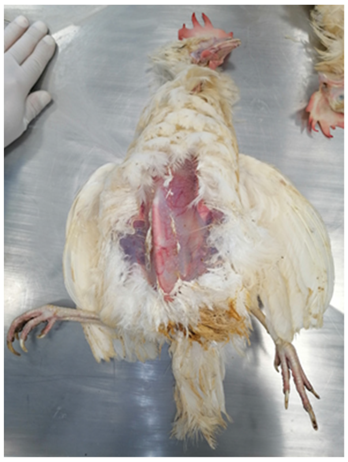
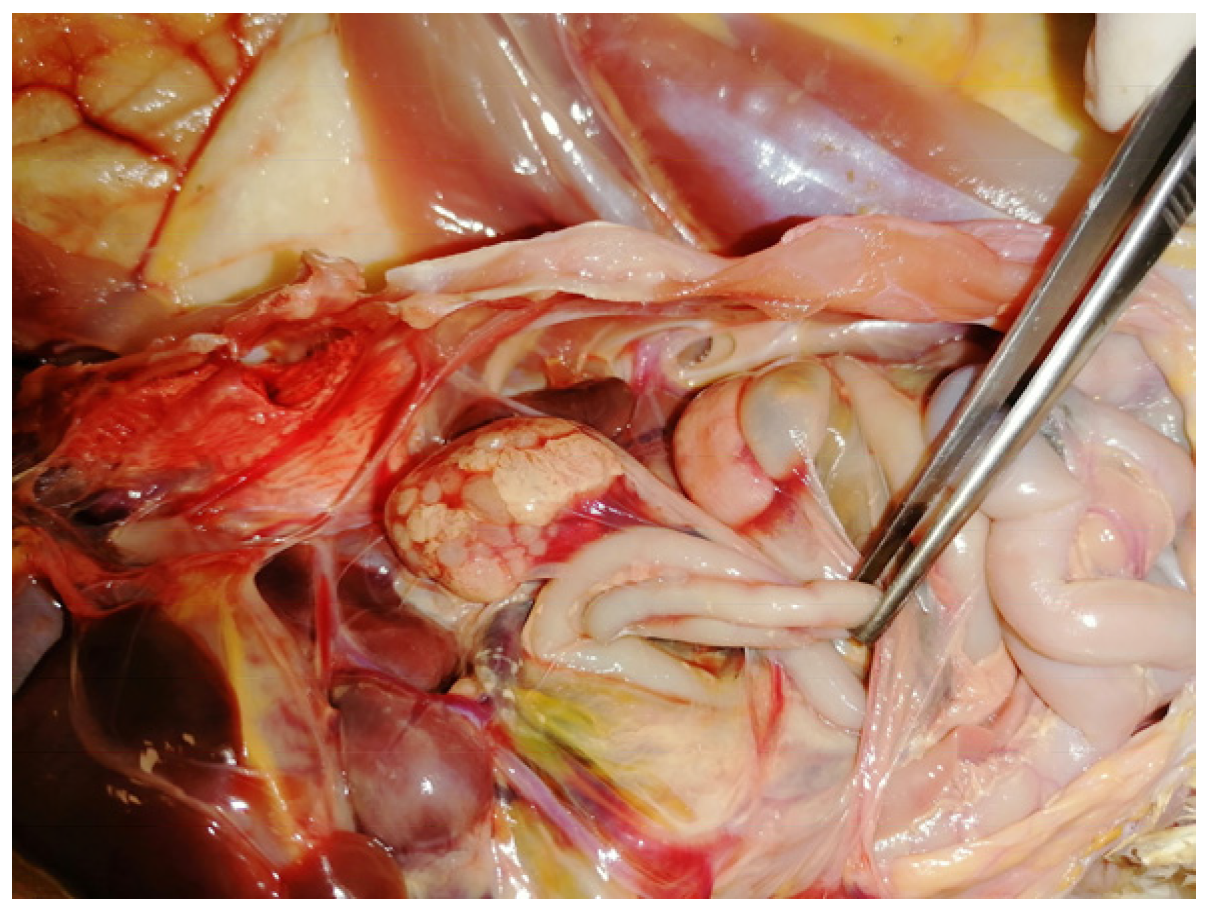
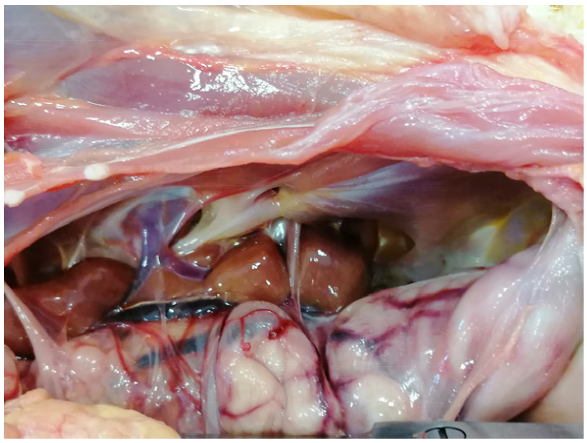
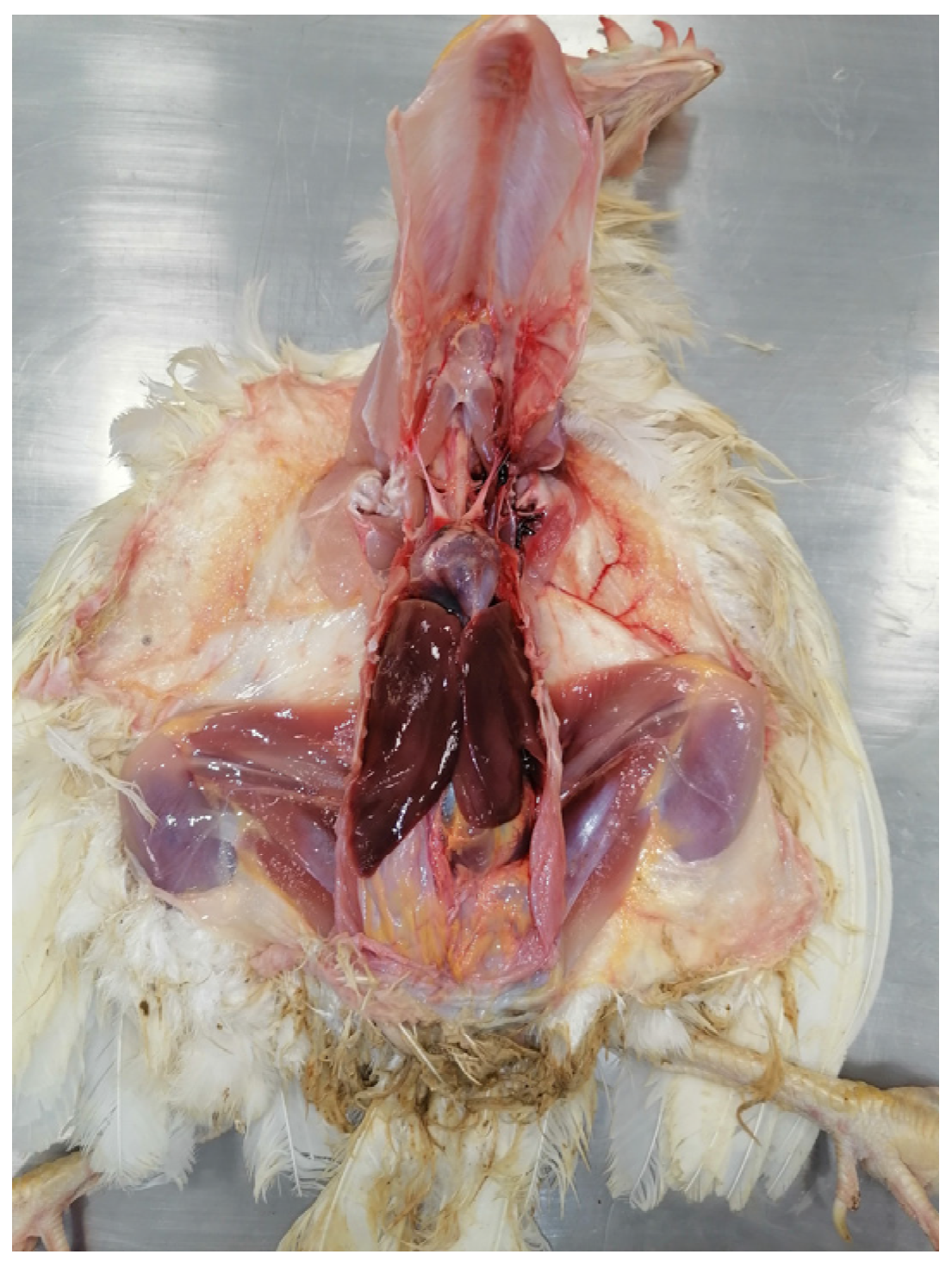
5. Post Mortem Examination

During the necropsy of the layer hens, careful macroscopic evaluation was made on the organs (liver and kidney) typically targeted by OTA and/or AFB1-induced pathology. For histological examination, pieces of tissue were collected and fixed in 10% neutral buffered formalin. The samples were embedded in paraffin wax, sectioned at 4 µm, and stained by Haematoxylin Eosin (HE).

6. Results and Discussion

At intravital examination, some animals were lethargic with anemia signs, such as pale appearance of combs and wattles (Figure 1 and Figure 2a). The necropsy examination highlighted a poor state of nutrition (Figure 3) and confirmed anemia by evidencing the discoloration of the content of the femoral medullary cavity (Figure 2b).
The muscular stomach appeared smaller than normal, suggesting a poor feed intake. In agreement with other authors [42,43] who described OTA and AFs activities, the ovary and oviduct showed hypoplasia (Figure 4) and this was the most possible reason for the drop in egg production.
Kidneys were enlarged, discolored, and yellowish, with no fat in the renal chamber, thus indicating a state of cachexia (Figure 5). The renal impairment was also remarked by the presence of urate crystals in the peritoneum (Figure 6a) and in the abdominal air sac (Figure 6b) [28]. Finally, the liver appeared enlarged, firm, and covered with scattered necrotic foci (Figure 7).
Histological examination clearly confirmed renal and hepatic damage. In particular, kidney specimens showed lesions characteristic of OTA intoxication [43,44,45]. In fact, the epithelial cells were enlarged in the whole cross-section of the proximal tubules, with vacuolated cytoplasm, karyomegaly, and granular degeneration of the nucleus, in a pattern strongly suggestive of tubule-nephrosis (Figure 8a). Glomerular changes were marked as a uniform and homogenous thickening of the glomerular capillary basement membrane and the presence of glomerular atrophy (Figure 8b).
At the hepatic level, the histopathological picture revealed more significant lesions with respect to the macroscopic examination, consisting of moderate focal fibrosis (Figure 9a) with the presence of nodular inflammatory cells. Numerous hepatocytes with fatty infiltration were clearly visible, as well as congestion and hemorrhage (Figure 9b). Additionally, in this case, the anatomopathological picture was consistent with those previously described for AFB1 intoxication [46].
The toxicological investigations detected OTA in the kidney and liver with mean (n = 10) values equal to 47 ± 3.03 µg/kg and 24 ± 1.92 µg/kg, respectively. While, in the same organs, AFB1 was detected with mean values (n = 10) equal to 1.4 ± 0.3 µg/kg and 3.6 ± 0.44 µg/kg, respectively.
Both mycotoxins, OTA and AFB1, were detected in the five samples of feed that included supplementation with 4% linseed, and the mean values were equal to 31 ± 3.08 µg/kg and 5.6 ± 0.33 µg/kg dry weight, respectively. While in the five samples of feed without supplementation, only OTA was detected at a mean concentration of 1.1 ± 0.15 µg/kg dry weight: AFB1 was not detectable.
Those values were far below the contamination levels needed for retrieving mycotoxins in eggs. By investigating the dynamics of OTA accumulation in eggs placed by laying hens experimentally exposed to OA, it was demonstrated that after exposure at the concentrations admitted by the current European legislation (100 μg/kg) and at concentrations 20-folds as much the European Legislation limit (2000 μg/kg), OTA was not detectable in the eggs [43]. Moreover, pioneering studies ascertained that AFB1 residues in eggs were detectable only when contamination of feedstuff reached 5000 μg/kg [47]. More recent studies, carried out with more sensitive equipment, found that detection of AFB1 from eggs was only possible when the feedstuff contamination reached 500 μg/kg [48]. However, it should be underlined that oviposition was found to be reduced when laying hens were fed with feedstuff contaminated by 700 μg/kg AFB1, ceasing completely at d 5000 μg/kg [49].
In the present case, the toxic effects arose at much lower AFB1 and OTA concentrations in feedstuff. Moreover, our findings showed that infectious diseases can be excluded. Indeed, in the episode we described, anemia and weight loss were not associated with the typical lesions induced by the viruses responsible for avian neoplasms (Marek’s Disease Virus or avian leukosis virus) [50,51], nor with the medullary hypoplasia described in the case of gyrovirus infections [52]. Furthermore, although infectious bronchitis can induce kidney lesions similar to those described [53], the animals were treated with a vaccine with a wide antigenic panel.
Such a finding is not surprising, since it is well-known that mixed feed may be more prone to be contaminated by different mycotoxins [54,55]. Additionally, Gruber-Dorninger et al. [56], showed that AFB1 and OTA were the most commonly identified combination in complete feed.
As shown by other studies [57,58,59] 75% of animal feed samples may contain more than one mycotoxin. Although in our study the contamination levels of the two mycotoxins detected were below the limits established by the current legislation, there seems to have a sort of synergistic action that could impact animal health even at minimal doses.
The simplest explanation for such action relies on the renal failure caused by OTA, previously evidenced in an experimental study [43], which might have impaired the excretion capacity of the affected animals. It is possible that such a defect would have increased the terminal half-life of the active metabolites of AFB1, thus enhancing their effects on the target organs such as liver and bone marrow (with consequent anemia and coagulopathy), also compromising the general health status of the animals, with consequent undernutrition and deposition impairment.
Additionally, the persistent binding of OTA to serum proteins may have triggered a positive feedback effect, in which the high bioaccumulation might be enhanced by the increased elimination time, as previously suggested [60].
All those considering, it is advisable that more efforts should be driven to provide more detailed knowledge about the synergistic effects of mycotoxins, from different points of view. On one hand, the scientific community should be interested in retrieving both qualitative and quantitative data and information about the mechanism triggered by the concurrent assumption of AFB1 and OTA in poultry by performing kinetic and dynamic studies, in order to build knowledge that may pave the way to the definition of new proper limits. On the other hand, the operators of the poultry system should be aware that, when controlling feedstuff or their ingredients, the acceptability thresholds might not be enough safe in the case of concurrent contamination by more than one mycotoxin. It is noteworthy that, in the here described case, the farmer demanded mycotoxin-free certified linseed, which was determinant for solving the problem and maintaining the omega-3 enrichment certification. Thus, while waiting for potential future institutional determinations, scientists and poultry stakeholders could evaluate new strategies for avoiding the synergic effects of multiple mycotoxin contaminations.

7. Conclusions

The main vehicle of mycotoxins is feed. For this reason, EU Regulation 574/2011 and EU Recommendation 576/2006 define the maximum acceptable levels of AFs and OTAs in the finished product. Compliance with these limits should prevent the occurrence of toxic phenomena by reducing the risk of contamination of human food. The presence of both investigated mycotoxins in the mixed feed including 4% linseed would indicate a limitation of the current regulations. Therefore, it would be appropriate to review the acceptable limits, especially in the case of mixed feed produced with many raw ingredients. The search for high-quality productions capable of occupying increasingly larger niche markets, due to their ability to attract health-conscious consumers, must stimulate producers to greater attention to the quality of the raw materials used. In this sense, the new EU Reg. No. 2017/625 [61], which regulates the Official Controls, represents an innovative tool available to Official veterinarians [62]. In fact, through the correct legal interpretation of the regulation, controls will not stop at the concept of the food chain but will be extended to the much broader concept of the agri-food chain.

Author Contributions

Conceptualization, G.B. and A.C.; writing-original draft preparation, G.B., A.C., N.P., M.M.D. and R.S.; writing-review and editing, G.B., A.C., M.M.D., R.S., E.C. (Elena Circella), A.S. and N.P.; supervision, G.B. and A.C.; methodology, E.C. (Edmondo Ceci) and E.B.; data curation E.C. (Edmondo Ceci), funding acquisition, G.B. and A.C. All authors have read and agreed to the published version of the manuscript.

Funding

This research received no external funding.

Institutional Review Board Statement

Ethical review and approval were waived for this study because of it was conducted on animals already dead in the laying hens farm.

Data Availability Statement

Data sharing is not applicable to this article.

Acknowledgments

The authors would like to thank Anthony Green for kindly reviewing the English in the manuscript.

Conflicts of Interest

The authors declare no conflict of interest.

References

  1. Feijó Corrêa, J.A.; Orso, P.B.; Bordin, K.; Vaz Hara, R.; Luciano, F.B. Toxicological effects of fumonisin B1 in combination with other Fusarium toxins. Food Chem. Toxicol. 2018, 121, 483–494. [Google Scholar] [CrossRef] [PubMed]
  2. Rodríguez-Blanco, M.; Marín, S.; Sanchis, V.; Ramos, A.J. Fusarium mycotoxins in total mixed rations for dairy cows. Mycotoxin Res. 2020, 36, 277–286. [Google Scholar] [CrossRef] [PubMed]
  3. Medina, A.; Rodríguez, A.; Magan, N. Climate change and mycotoxigenic fungi: Impacts on mycotoxin production. Current Opinion in Food Sci. 2015, 5, 99–104. [Google Scholar] [CrossRef]
  4. Van der Fels-Klerx, H.; Focker, M.; De Rijk, T.; Liu, C. Mycotoxins in wheat cultivated in the Netherlands: Results from eight years of field surveys. Mycotoxin Res. 2021, 37, 183–192. [Google Scholar] [CrossRef] [PubMed]
  5. Bozzo, G.; Corrente, M.; Testa, G.; Casalino, G.; Dimuccio, M.M.; Circella, E.; Brescia, N.; Barrasso, R.; Celentano, F.E. Animal Welfare, Health and the Fight against Climate Change: One Solution for Global Objectives. Agriculture 2021, 11, 1248. [Google Scholar] [CrossRef]
  6. Van Egmond, H.P.; Schothorst, R.C.; Jonker, M.A. Regulations relating to mycotoxins in food. Anal. Bioanal. Chem. 2007, 389, 147–157. [Google Scholar] [CrossRef] [Green Version]
  7. EFSA CONTAM Panel (EFSA Panel on Contaminants in the Food Chain). Opinion of the Scientific Panel on Contaminants in the Food Chain on a request from the Commission related to the potential increase of consumer health risk by a possible increase of the existing maximum levels for aflatoxins in almonds, hazelnuts and pistachios and de-rived products. EFSA J. 2007, 446, 1–127. [Google Scholar] [CrossRef]
  8. EFSA CONTAM Panel (EFSA Panel on Contaminants in the Food Chain). Scientific Opinion on the risk as-sessment of ochratoxin A in food. EFSA J. 2020, 18, e06113. [Google Scholar] [CrossRef]
  9. Afsah-Hejri, L.; Jinap, S.; Hajeb, P.; Radu, S.; Shakibazadeh, S. A Review on Mycotoxins in Food and Feed: Malaysia Case Study. Compr. Rev. Food Sci. Food Saf. 2013, 12, 629–651. [Google Scholar] [CrossRef]
  10. Bozzo, G.; Ceci, E.; Pinto, P.; Bonerba, E.; Martella, V.; Terio, E.; Tantillo, G. Ochratoxin A in avicultural meat production: Chemical and histological effects. World Mycotoxin J. 2009, 2, 61–69. [Google Scholar] [CrossRef]
  11. IARC. Agents Classified by the IARC Monographs. 2022, Volumes 1–132. Available online: http://monographs.iarc.fr/ENG/Classification/ (accessed on 20 May 2022).
  12. Bozzo, G.; Ceci, E.; Bonerba, E.; Desantis, S.; Tantillo, G. Ochratoxin a in laying hens: High-performance liquid chromatography detection and cytological and histological analysis of target tissues. J. Appl. Poult. Res. 2008, 17, 151–156. [Google Scholar] [CrossRef]
  13. Armorini, S.; Al-Qudah, K.M.; Altafini, A.; Zaghini, A.; Roncada, P. Biliary ochratoxin A as a biomarker of ochratoxin exposure in laying hens: An experimental study after administration of contaminated diets. Res. Vet. Sci. 2015, 100, 265–270. [Google Scholar] [CrossRef] [PubMed]
  14. Galtier, P.; Alvinerie, M.; Charpenteau, J. The pharmacokinetic profiles of ochratoxin A in pigs, rabbits, and chickens. Food Cosmet. Toxicol. 1981, 19, 735–738. [Google Scholar] [CrossRef] [PubMed]
  15. Chang, C.F.; Hamilton, P.B. Increased severity and new symptoms of infectious bursal disease during aflatoxicosis in broiler chickens. Poult. Sci. 1982, 61, 1061–1068. [Google Scholar] [CrossRef] [PubMed]
  16. Gibson, R.M.; Bailey, C.A.; Kubena, L.F.; Huff, W.E.; Harvey, R.B. Ochratoxin A and dietary protein. 1. Effects on body weight, feed conversion, relative organ weight, and mortality in three-week-old broilers. Poult. Sci. 1989, 68, 658–663. [Google Scholar] [CrossRef]
  17. Huff, W.E.; Kubena, L.F.; Harvey, R.B.; Doerr, J.A. Mycotoxin interactions in poultry and swine. J. Anim. Sci. 1988, 66, 2351–2355. [Google Scholar] [CrossRef] [Green Version]
  18. Huff, W.E.; Wyatt, R.D.; Hamilton, P.B. Nephrotoxicity of dietary ochratoxin A in broiler chickens. Appl. Microbiol. 1975, 30, 48–51. [Google Scholar] [CrossRef]
  19. Glahn, R.P.; Wideman, R.F.J.; Evangelisti, J.W.; Huff, W.E. Effects of ochratoxin A alone and in combination with citrinin on kidney function. Of single comb white leghorn. Poult. Sci. 1988, 67, 1034–1042. [Google Scholar] [CrossRef]
  20. Glahn, R.P.; Shapiro, R.S.; Vena, V.E.; Wideman, R.F., Jr.; Huff, W.E. Effects of chronic ochratoxin A and citrinin toxicosis on kidney function of single comb White Leghorn pullets. Poult. Sci. 1989, 68, 1205–1212. [Google Scholar] [CrossRef]
  21. Ochieng, P.E.; Scippo, M.; Kemboi, D.C.; Croubels, S.; Okoth, S.; Kang’ethe, E.K.; Doupovec, B.; Gathumbi, J.K.; Lindahl, J.F.; Antonissen, G. Mycotoxins in Poultry Feed and Feed Ingredients from Sub-Saharan Africa and Their Impact on the Production of Broiler and Layer Chickens: A Review. Toxins 2021, 13, 633. [Google Scholar] [CrossRef]
  22. Rawal, S.; Kim, J.E.; Coulombe, R., Jr. Aflatoxin B1 in poultry: Toxicology, metabolism and prevention. Res. Vet. Sci. 2010, 89, 325–331. [Google Scholar] [CrossRef]
  23. Hussein, H.S.; Brasel, J.M. Toxicity, metabolism, and impact of mycotoxins on humans and animals. Toxicology 2001, 167, 101–134. [Google Scholar] [CrossRef] [PubMed]
  24. Haschek, W.M.; Voss, K.A.; Beasley, V.A. Selected Mycotoxins Affecting Animal and Human Health. Handb. Toxicol. Pathol. 2002, 1, 645–699. [Google Scholar] [CrossRef]
  25. Klein, P.J.; Buckner, R.; Kelly, J.; Coulombe, R.A., Jr. Biochemical basis for the extreme sensitivity of turkeys to aflatoxin B-1. Toxicol. Appl. Pharmacol. 2000, 165, 45–52. [Google Scholar] [CrossRef] [PubMed]
  26. Quist, C.F.; Bounous, D.I.; Kilburn, J.V.; Nettles, V.F.; Wyatt, R.D. The effect of dietary aflatoxin on wild turkey poults. J. Wildl. Dis. 2000, 36, 436–444. [Google Scholar] [CrossRef] [PubMed] [Green Version]
  27. Casteel, S.W.; Rottinghouse, G.E. Mycotoxicoses. Encycl. Microbiol. 2000, 3, 337–348. [Google Scholar]
  28. Murugesan, G.R.; Ledoux, D.R.; Naehrer, K.; Berthiller, F.; Applegate, T.J.; Grenier, B.; Phillips, T.D.; Schatzmayr, G. Prevalence and effects of mycotoxins on poultry health and performance, and recent development in mycotoxin counteracting strategies. Poult. Sci. 2015, 94, 1298–1315. [Google Scholar] [CrossRef]
  29. Yang, C.; Song, G.; Lim, W. Effects of mycotoxin-contaminated feed on farm animals. J. Hazard. Mater. 2020, 389, 122087. [Google Scholar] [CrossRef]
  30. Leeson, S.; Diaz, G.J.; Summers, J.D. Poultry Metabolic Disorders and Mycotoxins; University Books: Guelph, SO, Canada, 1995. [Google Scholar]
  31. Devegowda, G.; Murthy, T.N.K. Mycotoxins: Their effects in poultry and some practical solutions. In The Mycotoxin Blue Book; Diaz, D.E., Ed.; Nottingham University Press: Nottingham, UK, 2005; pp. 25–56. [Google Scholar]
  32. Reed, K.M.; Mendoza, K.M.; Coulombe, R.A., Jr. Differential Transcriptome Responses to Aflatoxin B1 in the Cecal Tonsil of Susceptible and Resistant Turkeys. Toxins 2019, 11, 55. [Google Scholar] [CrossRef] [Green Version]
  33. Emmanuel, K.T.; Els, V.P.; Bart, H.; Evelyne, D.; Els, V.H.; Els, D. Carry-over of some Fusarium mycotoxins in tissues and eggs of chickens fed experimentally mycotoxin-contaminated diets. Food Chem. Toxicol. 2020, 145, 111715. [Google Scholar] [CrossRef]
  34. Stefanaki, I.; Foufa, E.; Tsatsou-Dritsa, A.; Dais, P. Ochratoxin A concentrations in Greek domestic wines and dried vine fruits. Food Addit. Contam. 2003, 20, 74–83. [Google Scholar] [CrossRef]
  35. Commission Recommendation 2006/576/EC of 17 August 2006 on the Presence of Deoxynivalenol, Zearalenone, Ochratoxin A, T-2 and HT-2 and Fumonisins in Products Intended for Animal Feeding. O J L 229, 23.8. 2006, pp. 7–9. Available online: https://eur-lex.europa.eu/legal-content/EN/TXT/?uri=CELEX%3A32006H0576 (accessed on 13 October 2022).
  36. Commission Regulation (EU) 2011/574 of 16 June 2011 Amending Annex I to Directive 2002/32/EC of the European Parliament and of the Council as Regards Maximum Levels for Nitrite, Melamine, Ambrosia spp. and Carry-Over of Certain Coccidiostats and Histomonostats and Consolidating. O J L 159, 17.6. 2011, pp. 7–24. Available online: https://eur-lex.europa.eu/legal-content/EN/ALL/?uri=CELEX%3A32011R0574 (accessed on 13 October 2022).
  37. Alassane-Kpembi, I.; Schatzmayr, G.; Taranu, I.; Marin, D.; Puel, O.; Oswald, I.P. Mycotoxins co-contamination: Methodological aspects and biological relevance of combined toxicity studies. Crit. Rev. Food Sci. Nutr. 2017, 57, 3489–3507. [Google Scholar] [CrossRef] [PubMed]
  38. Lohmann Breeders. Available online: https://lohmann-breeders.com/strains/lohmann-lsl-classic-cage-housing/ (accessed on 15 May 2022).
  39. Monaci, L.; Tantillo, G.; Palmisano, F. Determination of Ochratoxin A in Portuguese Rice Samples by High Performance Liquid Chromatography with Fluorescence Detection. Anal. Bioanal. Chem. 2004, 378, 1777–1782. [Google Scholar] [CrossRef]
  40. Losito, I.; Monaci, L.; Palmisano, F.; Tantillo, G. Determination of ochratoxin A in meat products by high-performance liquid chromatography coupled to electrospray ionisation sequential mass spectrometry. Rapid Commun. Mass Spectrom. 2004, 18, 1965–1971. [Google Scholar] [CrossRef] [PubMed]
  41. Matrella, R.; Monaci, L.; Milillo, M.A.; Palmisano, F.; Tantillo, M.G. Ochratoxin A determination in paired kidneys and muscle samples from swines slaughtered in southern Italy. Food Control 2006, 17, 114–117. [Google Scholar] [CrossRef]
  42. Trucksess, M.W.; Stoloff, L.; Young, K.; Wyatt, R.D.; Miller, B.L. Aflatoxicol and aflatoxins B1 and M1 in eggs and tissues of laying hens consuming aflatoxin-contaminated feed. Poult. Sci. 1983, 62, 2176–2182. [Google Scholar] [CrossRef]
  43. Bozzo, G.; Bonerba, E.; Ceci, E.; Colao, V.; Tantillo, G. Determination of ochratoxin A in eggs and target tissues of ex-perimentally drugged hens using HPLC–FLD. Food Chem. 2011, 126, 1278–1282. [Google Scholar] [CrossRef]
  44. Dwivedi, P.; Burns, R.B. Pathology of ochratoxicosis in young broiler chicks. Res. Vet. Sci. 1984, 36, 92–103. [Google Scholar] [CrossRef]
  45. Kozaczyński, W. Experimental ochratoxicosis A in chickens. Histopathological and histochemical study. Arch. Vet. Pol. 1994, 34, 205–219. [Google Scholar] [PubMed]
  46. Ali, A.; Khatoon, A.; Almohaimeed, H.M.; Al-Sarraj, F.; Albiheyri, R.; Alotibi, I.; Abidin, Z.U. Mitigative Potential of Novel Lactobacillus plantarum TISTR 2076 against the Aflatoxins-Associated Oxidative Stress and Histopathological Alterations in Liver and Kidney of Broiler Chicks during the Entire Growth Period. Toxins 2022, 14, 689. [Google Scholar] [CrossRef]
  47. Lötzsch, R.; Leistner, L. Transmission of aflatoxins into eggs and egg products. Ann. Nutr. L’alimenta-Tion 1977, 31, 499–508. [Google Scholar]
  48. Oliveira, C.A.; Kobashigawa, E.; Reis, T.A.; Mestieri, L.; Albuquerque, R.; Corrêa, B. Aflatoxin B1 residues in eggs of laying hens fed a diet containing different levels of the mycotoxin. Food Addit. Contam. 2000, 17, 459–462. [Google Scholar] [CrossRef] [PubMed]
  49. Exarchos, C.C.; Gentry, R.F. Effect of aflatoxin B1 on egg production. Avian Dis. 1982, 26, 191–195. [Google Scholar] [CrossRef]
  50. Song, B.; Zeb, J.; Hussain, S.; Aziz, M.U.; Circella, E.; Casalino, G.; Camarda, A.; Yang, G.; Buchon, N.; Sparagano, O. A Review on the Marek’s Disease Outbreak and Its Virulence-Related meq Genovariation in Asia between 2011 and 2021. Animals 2022, 12, 540. [Google Scholar] [CrossRef]
  51. Dunn, J. Lymphoid Leukosis in Poultry. MSD Veterinary Manual 2022. Available online: https://www.msdvetmanual.com/veterinary/poultry/neoplasms/lymphoid-leukosis-in-poultry (accessed on 28 December 2022).
  52. Schat, K.A.; van Santen, V.L. Chicken infectious anemia and circovirus infections in commercial flocks. In Diseases of Poultry, 14th ed.; Swayne, D.E., Boulianne, M., Logue, C.M., McDougald, L.R., Nair, V., Suarez, D.L., Wit, S., Grimes, T., Johnson, D., Kromm, M., et al., Eds.; John Wiley & Sons: Hoboken, NJ, USA, 2019; pp. 284–320. [Google Scholar] [CrossRef]
  53. Jackwood, M.W.; de Wit, S. Infectious bronchitis. In Diseases of Poultry, 14th ed.; Swayne, D.E., Boulianne, M., Logue, C.M., McDougald, L.R., Nair, V., Suarez, D.L., Wit, S., Grimes, T., Johnson, D., Kromm, M., et al., Eds.; John Wiley & Sons: Hoboken, NJ, USA, 2019; pp. 167–188. [Google Scholar] [CrossRef] [Green Version]
  54. Smith, M.C.; Madec, S.; Coton, E.; Hymery, N. Natural Co-occurrence of mycotoxins in foods and feeds and their in vitro combined toxicological effects. Toxins 2016, 8, 94. [Google Scholar] [CrossRef] [PubMed]
  55. Santos-Pereira, C.; Cunha, S.C.; Fernandes, J.O. Prevalent mycotoxins in animal feed: Occurrence and analytical methods. Toxins 2019, 11, 290. [Google Scholar] [CrossRef] [PubMed] [Green Version]
  56. Gruber-Dorninger, C.; Jenkins, T.; Schatzmayr, G. Global mycotoxin occurrence in feed: A ten-year survey. Toxins 2019, 11, 375. [Google Scholar] [CrossRef] [Green Version]
  57. Zachariasova, M.; Dzuman, Z.; Veprikova, Z.; Hajkova, K.; Jiru, M.; Vaclavikova, M.; Zachariasova, A.; Pospichalova, M.; Florian, M.; Hajslova, J. Occurrence of multiple mycotoxins in European feedingstuffs, assessment of dietary intake by farm animals. Anim. Feed Sci. Technol. 2014, 193, 124–140. [Google Scholar] [CrossRef]
  58. Pinotti, L.; Ottoboni, M.; Giromini, C.; Dell’Orto, V.; Cheli, F. Mycotoxin contamination in the EU feed supply chain: A focus on cereal Byproducts. Toxins 2016, 8, 45. [Google Scholar] [CrossRef] [Green Version]
  59. Castaldo, L.; Graziani, G.; Gaspari, A.; Izzo, L.; Tolosa, J.; Rodríguez-Carrasco, Y.; Ritieni, A. Target analysis and ret-rospective screening of multiple mycotoxins in pet food using UHPLC-Q-orbitrap HRMS. Toxins 2019, 11, 434. [Google Scholar] [CrossRef]
  60. Pozzo, L.; Cavallarin, L.; Antoniazzi, S.; Guerre, P.; Biasibetti, E.; Capucchio, M.T.; Schiavone, A. Feeding a diet con-taminated with ochratoxin A for broiler chickens at the maximum level recommended by the EU for poultry feeds (0.1 mg/kg). 2. Effects on meat quality, oxidative stress, residues and histological traits. J. Anim. Physiol. Anim. Nutr. 2013, 97 (Suppl. 1), 23–31. [Google Scholar] [CrossRef] [PubMed]
  61. Regulation (EU) No 2017/625 of the European Parliament and of the Council of 15 March 2017 on Official Controls and Other Official Activities Performed to Ensure the Application of Food and Feed Law, Rules on Animal Health and Welfare, Plant Health and Plant Protection Products. O J L 95, 7.4.2017. Available online: https://eur-lex.europa.eu/legal-content/EN/TXT/?uri=celex%3A32017R0625 (accessed on 24 October 2022).
  62. Bozzo, G.; Dimuccio, M.M.; Casalino, G.; Ceci, E.; Corrente, M. New approaches for risk assessment and management of bovine protothecosis. Saudi J. Biol. Sci. 2022, 29, 103368. [Google Scholar] [CrossRef] [PubMed]
Figure 1. Less responsive and anaemic animal.
Agriculture 13 00138 g001
Figure 2. Necropsy: anemic animal. (a) Detail paleness of crest and wattle. (b) Lighter color of femoral medullary cavity.
Agriculture 13 00138 g002
Figure 3. Necropsy: poor state of nutrition.
Agriculture 13 00138 g003
Figure 4. Necropsy: hypoplasia of the ovary and oviduct.
Agriculture 13 00138 g004
Figure 5. Necropsy: detail of enlarged and discolored kidneys.
Agriculture 13 00138 g005
Figure 6. Necropsy. (a) Urate crystals in the peritoneum. (b) Urate crystals in the abdominal air sac.
Agriculture 13 00138 g006
Figure 7. Necropsy: detail of liver.
Agriculture 13 00138 g007
Figure 8. Kidney. (a) Epithelial cells of the proximal tubules appearing enlarged with vacuolated cytoplasm, karyomegaly and granular degeneration of the nucleus (HE) 20×. (b) Glomerular atrophy (HE) 40×.
Agriculture 13 00138 g008
Figure 9. Liver. (a) Nodular inflammatory cells and moderate focal fibrosis. Great congestive area constituted by red corpuscles and inflammatory cells surrounded by fibrosis (HE) 20×. (b) Particular of hepatocytes with fatty infiltration (HE) 40×.
Agriculture 13 00138 g009
Disclaimer/Publisher’s Note: The statements, opinions and data contained in all publications are solely those of the individual author(s) and contributor(s) and not of MDPI and/or the editor(s). MDPI and/or the editor(s) disclaim responsibility for any injury to people or property resulting from any ideas, methods, instructions or products referred to in the content.

