Article

Geraniol: A Potential Defense-Related Volatile in “Baiye No. 1” Induced by Colletotrichum camelliae

1
College of Tea Science, Guizhou University, Guiyang 550025, China
2
Guizhou Tea Research Institute, Guizhou Academy of Agricultural Sciences, Guiyang 550006, China
3
Tongren Bureau of Agriculture and Rural Affairs, Tongren 554300, China
*
Author to whom correspondence should be addressed.
These authors contributed equally to this work.
Agriculture 2023, 13(1), 15; https://doi.org/10.3390/agriculture13010015
Submission received: 14 November 2022 / Revised: 16 December 2022 / Accepted: 19 December 2022 / Published: 21 December 2022
(This article belongs to the Section Crop Production)

Abstract

:
Plants produce and emit a large variety of volatiles that have multiple defense-related functions in response to abiotic or biotic stresses. In comparison with studies on plant volatile–herbivore interactions, little research has been carried out on plant volatile–microbe interactions. In the present paper, tea volatile–Colletotrichum camelliae interactions were studied. The results of emitted volatiles following infection with C. camelliae in “Baiye No. 1” showed that healthy tea plants contained 68 kinds of volatiles, while infected tea plants contained 76 kinds of volatiles. Five volatiles, namely, geraniol, linalool, methyl salicylate, (E)-3-hexen-1-ol, and α-farnesene, were found to have a relatively large content variation in infected tea plants, with increments of 7.903%, −2.247%, 2.770%, −6.728%, and 3.848%, respectively. The fungicidal activity results of the five volatiles against C. camelliae showed that geraniol had the best activity, with MIC and MBC values of 0.5 and 1 mg·mL−1, respectively. Thus, geraniol was selected for subsequent studies. The effects of geraniol on the mycelia and cell structures of C. camelliae were investigated by scanning electron microscopy (SEM) and transmission electron microscope (TEM). The results showed that the mycelia were significantly disrupted, and the cell structures were damaged. The effects of geraniol on the related enzymes of C. camelliae were assessed. The results showed that cellulase activity increased, malondialdehyde content increased, and the activity of defense enzymes was inhibited, thereby inhibiting the growth of pathogens. This study provides the first evidence that geraniol is a defense-related function volatile of “Baiye No. 1” in response to C. camelliae stress. It also provides valuable information and enriches the chemical ecology of tea plant diseases for the research field on defensive substances of microbe-induced plant volatiles.

1. Introduction

Plants can induce and emit a large variety of volatile substances through abiotic or biotic stresses [1]. Plant volatiles are secondary metabolites of plants that are associated with plant disease defense [2]. When a pathogen infects a plant, the plant releases volatiles that affect organisms or differ from healthy plants [3,4,5,6]. This volatile substance induces the expression of defense-related gene and enzyme to defend against pathogens; it may also act as an alarm signal or increase resistance to microorganisms to reduce damage and protect itself [7,8,9]. Volatiles could function to prevent microbial attack. The direct action of plant volatiles on microorganisms is one of the important processes in its plant self-defense system [10,11,12]. Plant volatiles have long been known for their antimicrobial activity, which could inhibit the germination of pathogenic conidia and protect plants from pathogen, and their antimicrobial effects have been well studied [13,14,15]. Prost reported that many volatile dxylipins have broad chemical bactericidal potential associated with plant disease resistance [16]. Wright revealed that the n-decanal, hexenal and octenal in the volatile components of corn could inhibit the synthesis of aflatoxin by Aspergillust [17]. Zhang found that the bound volatiles in the form of glycosides in tea inhibit pathogenic fungi [18]. Green leaf volatiles are released from leaves following infection by plant pathogens. These compounds may have a defensive function, such as that C6 aldehydes and alcohols have substantial antimicrobial effects against bacteria or fungi in vitro [16,19,20].
Tea tree [Camellia sinensis (L.) O. Kuntze] is an important economic crop that is widely grown in tropical and subtropical regions [21]. Tea tree diseases are diverse, and they occur seriously all over the world. Tea plant growth is also affected by various diseases. Anthracnose, caused by the genus Anthrax, is the most widespread fungal disease in tea plantation [22]. Anthracnose showed water-soaked lesions in the early stage and irregular lesions in the later stage, and the lesions were scattered with small black dots [23]. When anthracnose occurs seriously, it affects the growth of tea leaves, causing poor quality and a significant reduction in yield [23]. The control of anthracnose mainly relies on chemical pesticides, and the long-term use of chemical pesticides destroys the ecological balance of tea gardens, affects human health, and restricts the development of the tea economy. Therefore, green and environmentally friendly prevention and control technologies are urgently needed to replace chemical control.
Plant volatiles have been a research hot spot in the fields of chemical ecology and plant protection in recent years. In comparison with studies on plant volatile–herbivore interactions, little research has been carried out on plant volatile–microbe interactions [2]. On the basis of the chemical ecological strategy of tea tree diseases, the volatile substances in “Baiye No. 1” induced by Colletotrichum camelliae were determined, and the differences in the content of volatile substances in tea trees were compared. The inhibitory activity of several volatiles with great variation in content against C. camelliae was studied, and the effects of volatiles with excellent inhibitory activities on the related enzymes of C. camelliae were screened and studied to provide new options for agriculture and forestry applications to reduce disease.

2. Materials and Methods

2.1. Materials

Geraniol, linalool, methyl salicylate, (E)-3-hexen-1-ol, and α-farnesene were purchased from AladdinReagent Co., Ltd. (Shanghai, China). Cellulase (CE), malondialdehyde (MDA), succinate dehydrogenase (SDH), malate dehydrogenase (MDH), peroxidase (POD), superoxide dismutase (SOD), and soluble protein were purchased from Maisha Industrial Co., Ltd. (Wuxi, China). The emulsifier Tween 80 was purchased from Tianjin Damao Chemical Reagent Factory. All chemicals were of analytical reagent grade, and ultrapure water (18 MΩ/cm) was used throughout the experiment. Volatiles were analyzed by an HP6890/5975C GC/MS coupler (Agilent, USA). Colletotrichum camelliae was obtained from Guizhou Tea Research Institute, Guizhou Academy of Agricultural Sciences, Guiyang, China.

2.2. Sample Processing and Testing

C. camelliae was incubated in PDA medium for 5 days, and mycelial blocks with a diameter of 5 mm were taken with a hole punch for use. The mycelial block was connected to the leaf of potted “Baiye No. 1” and placed in an artificial climate incubator at 25 °C. The infected leaves were cut into pieces, and about 1.0 g of the mixed sample was weighed. The sample was placed in a sampling bottle in a solid-phase microextraction instrument, inserted into a manual injector equipped with a 2 cm–50/30 μm DVB/CAR/PDMS StableFlex fiber head, and removed after head space extraction for 50 min under the condition of flat plate heating at 80 °C. The extraction head was immediately inserted into the inlet gas chromatograph (temperature 250 °C), and the sample was injected with thermal desorption.
Chromatographic conditions: the chromatographic column was an elastic quartz capillary column HP-5MS (60 m × 0.25 mm × 0.25 μm), and the initial temperature was 40 °C (retention for 2 min). The temperature was then increased to 208 °C at 3.5 °C/min and heated to 10 °C/min to 308 °C. (retention for 2 min). The running time was 62 min; the vaporization chamber temperature was 250 °C; the carrier gas was high-purity He (99.999%); the pre-column pressure was 15.98 psi; the carrier gas flow was 1.0 mL/min, splitless; and the solvent delay time was 3 min.
Mass spectrometry conditions: the ion source was EI source, the ion source temperature was 230 °C, the quadrupole temperature was 150 °C, the electron energy was 70 eV, the emission current was 34.6 μA, and the multiplier voltage was 2259 V.

2.3. Determination of Minimal Inhibitory Concentration (MIC) and Minimal Bactericide Concentration (MBC)

Different dilution concentrations of PDA were prepared by the serial dilution method [24]. Each concentration was repeated three times. The control group was composed of 0.1% Tween 80 and 5% absolute ethanol solution. The prepared mycelial suspension (80 µL) was evenly coated on the PDA surface and cultured in an incubator at 25 °C. After the growth of mycelia was observed for 2 days, the minimum concentration of completely sterile mycelial growth was determined as the MIC agent against C. camelliae. On the basis of the MIC experiment, the growth of mycelia was observed for 7 days, and the growth of the minimum concentration of sterile filaments was determined to be the minimum bactericidal concentration (MBC) of the agent against C. camelliae.

2.4. Mycelial Morphology and Structure of Mycelial Cell

The external morphology of the mycelia and the structural changes in the cell were observed by scanning electron microscopy (SEM) and transmission electron microscope (TEM). The mycelia of C. camelliae were placed in the MBC of volatiles and cultured at 25 °C. The electron microscopic samples were prepared as follows: a small amount of mycelia was fixed with 2.5% glutaraldehyde at 4 °C for 12 h. The samples were washed three times with phosphate buffer solution for 15 min each and then subjected to a series of ethanol gradients (30%, 50%, 70%, 90%, and 95%) in one dehydration for 15 min. Finally, they were dehydrated with absolute ethanol three times for 15 min each time. The final samples were freeze-dried and gold sprayed, and the mycelial morphology and microstructure of fungal cells were observed in SU8010 SEM and JEM-1200 TEM, respectively [25].

2.5. Determination of Enzymatic Activity and Content of C. camelliae

The mycelia (1 g) of C. camelliae were treated with the MIC concentration of volatiles and cultured on a shaker at 25 °C for 0, 3, 6, 9, 12, and 24 h. The samples were removed and centrifuged (6000 r/min, 5 min), and the supernatant was discarded for later use. The enzyme activity and content of CE, SDH, MDH, POD, SOD, MDA, and soluble protein were determined using a detection kit, and each treatment was replicated three times.

3. Results and Discussion

3.1. Difference Analysis of Volatiles

The changes in volatile substances before and after the induction of “Baiye No. 1” by C. camelliae were compared. Solid-phase microextraction and gas chromatography were used to detect volatile substances before and after infection, and the total ion flow is shown in Figure 1. The Nist17 and Wiley 275 standard mass spectrograms were retrieved and checked for each peak in the total ion flow diagram through the mass spectrum computer data system, and the relative content of each component was determined by the peak area normalization method. Figure 2 shows that the types of volatiles in leaves infected with C. camelliae were more than those in healthy leaves. A total of 68 and 76 volatile components were identified from healthy leaves and leaves infected with C. camelliae, respectively. Compared with healthy tea leaves, 37 volatile compounds were upregulated and 39 volatile compounds were downregulated in leaves infected with C. camelliae. Eight volatile substances were also detected in infected leaves but not in healthy leaves. As shown in Figure 1, five volatile species had relative content change values greater than 2%, namely, geraniol, linalool, methyl salicylate, and (E)-3-hexen-1-olandα-farnesene, with increments of 7.903%, −2.247%, 2.770%, −6.728%, and 3.848%, respectively. α-Farnesene was only detected in susceptible tea plants, whereas the rest were seen in healthy and infected tea plants.
Zhang reported that tea volatiles geraniol, linalool, methyl salicylate, benzyl alcohol, and phenylethanol had a strong inhibitory effect on C. camelliae Massea [18]. After maize and cotton were reported to be infected by Asperillus flavus, C6 alcohol and C6 aldehyde were found to inhibit the pathogen growth of A. flavus [26]. Combined with the above reports, five volatiles with a content difference of more than 2% were speculated to be involved in antimicrobial activities through differential analysis of volatiles induced by C. camelliae. For further explanation, these five volatiles were chosen as the main research object to illustrate the antimicrobial of action through the following experiments.

3.2. Determination of MIC and MBC

The best antifungal volatiles were screened from five volatiles, namely, geraniol, linalool, methyl salicylate, hexenol and α-farnesene, by a serial dilution method. The data revealed that different volatile components had different inhibitory effects on C. camelliae, indicating that volatile compounds had selective inhibitory effects. As shown in Figure 3A, only geraniol, linalool, and methyl salicylate showed activity 2 days after treatment, with MIC values of 0.5, 1, and 2 mg·mL−1, respectively. As shown in Figure 3B, geraniol and linalool had MBC values of 1 and 2 mg·mL−1, respectively, whereas the MBC of methyl salicylate was greater than 2 mg·mL−1. The three volatiles exhibited a certain inhibitory effect on C. camelliae. In accordance with the MIC and MBC values, geraniol with excellent inhibitory effects against C. camelliae was selected and further studied.

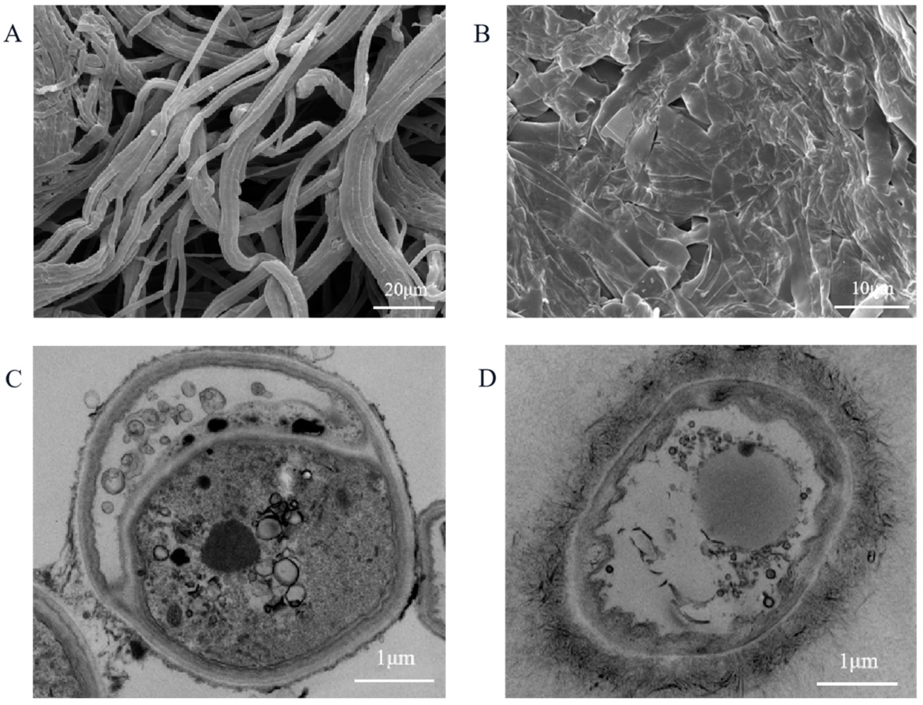
3.3. Effect of Geraniol on Mycelial Morphology and Cell Structures of C. camelliae

Fungal inhibition experiments showed that geraniol could inhibit the growth of C. camelliae, so whether its internal morphology changes were investigated. The effects of geraniol on the mycelia and cell structures of C. camelliae were explored by SEM and TEM. The SEM results showed that the mycelial surface of C. camelliae was smooth, plump, and regular flat in the control group (Figure 4A). Meanwhile, the mycelial surface was rougher and the mycelia were contracted, adhered, and deformed in the MBC concentration treatment group of geraniol (Figure 4B). The TEM results demonstrated that the normal cell structures were intact, the organelles were evenly distributed in the cytoplasm, and the cell wall thicknesses were consistent in the control group (Figure 4C). The cell membrane structures were destroyed, the cells overflowed, the contents were severely dissolved, and the cells were no longer saturated due to shrinkage in the MBC concentration treatment group of geraniol (Figure 4D). Moreover, geraniol could seriously shrink the mycelia and destroy the morphological structures, inhibiting or killing pathogens. SEM observations showed that geraniol could damage cell surface structures, and TEM observations found that it could alter membrane systems (swelling and leakage), such as cell walls, cell membranes, and cytoplasm, resulting in irreversible changes. These findings are consistent with those of Di and Li [27,28]. These results suggested that geraniol has an inhibitory effect on the mycelia and cell structures of C. camelliae. It is speculated that geraniol may act on cell wall, cell membrane, and cytoplasm of C. camelliae. In order to further confirm the role and targeting of geraniol, the activity and content of related enzymes of C. camelliae were further studied.

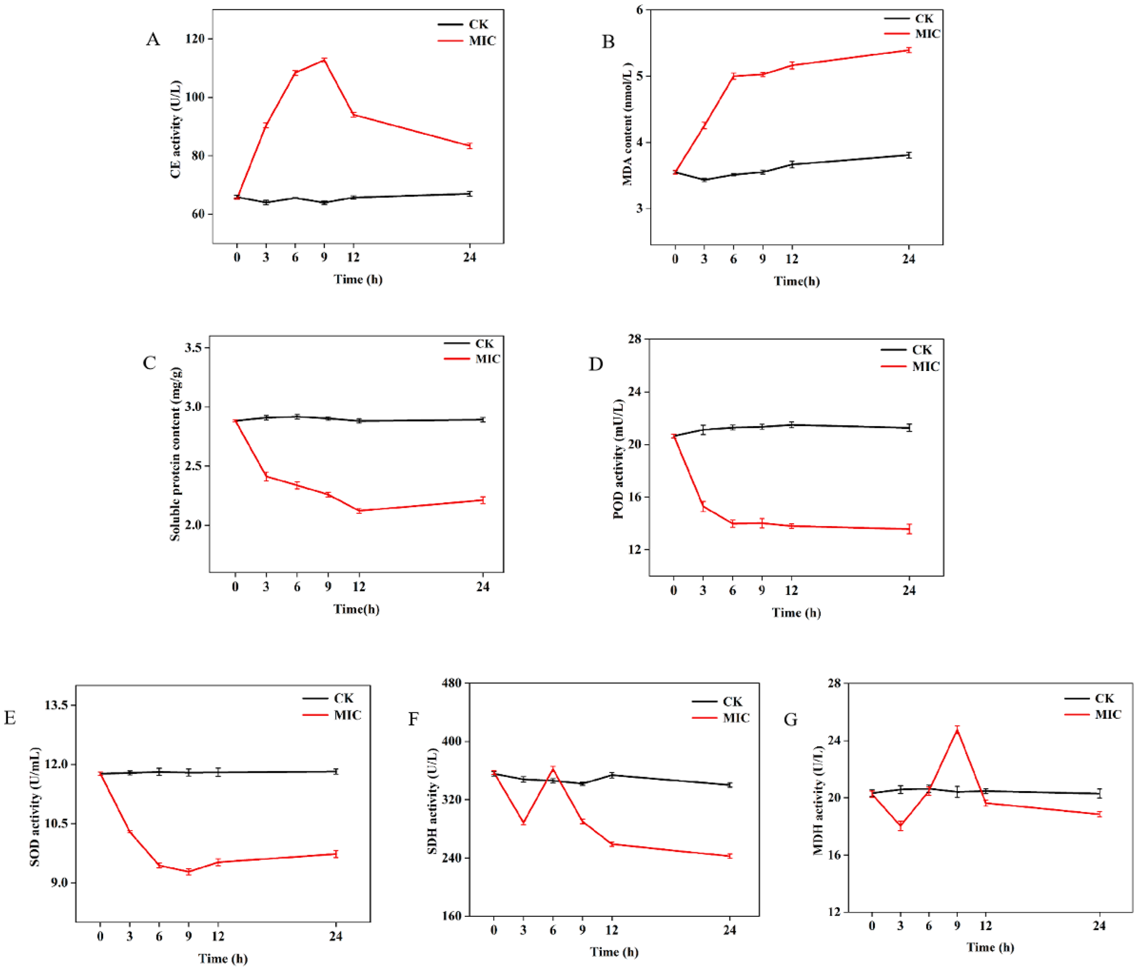
3.4. Analysis of the Activity and Content of C. camelliae-Related Enzymes

The CE activity and MDA content of mycelia in the geraniol MIC group was affected, and the overall showed an upward trend. The CE activity of mycelia increased significantly from 0 h to 9 h, and after 9 h, it decreased and then leveled off (Figure 5A). The MDA content of mycelia increased from 0 h to 6 h and gradually increased after 6 h. In addition, the MIC concentration treatment group of geraniol was higher than that of the control group (Figure 5B). The soluble protein content of mycelia treated with geraniol concentration generally showed a downward trend. It notably decreased from 0 h to 12 h and became flat after 12 h (Figure 5C). The POD and SOD activities of geraniol treatment generally showed a downward trend. It markedly decreased from 0 h to 6 h and tended to be flat after 6 h. The enzyme activity of the MIC concentration treatment group of geraniol was lower than that of the control group (Figure 5D,E). The SDH and NAD-MDH overall activity of geraniol treatment showed a trend of first decreasing, increasing, and then decreasing. The SDH activity showed a downward trend at 0–3 h and an upward trend at 3–6 h, and it overall showed a downward trend after 6 h (Figure 5F). The NAD-MDH activity decreased at 0–3 h, increased significantly at 3–9 h, and showed a decreasing trend after 9 h (Figure 5G).
Under the observation of SEM and TEM, the cell wall and membrane were obviously destroyed. As expected, geraniol could significantly increase the enzyme activity of CE and damage the integrity of the cell wall structures. MDA is an important indicator of membrane lipid peroxidation, and elevated levels of MDA could directly lead to cell damage [29]. Soluble protein is one of the components of the cell membrane, which reflects the integrity of the cell membrane [30]. The increase of MDA content and the decrease of soluble protein content showed that geraniol damaged the cell membrane to a considerable extent. The reduction in POD and SOD activities indicated that geraniol treatment of C. camelliae inhibited the activities of defense enzymes, resulting in excessive accumulation of intracellular H2O2 and interfering cellular metabolism. SDH is a mitochondrial marker enzyme and one of the critical links between respiratory electron transport and oxidative phosphorylation [31]. MDH is an essential enzyme in the TCA cycle and a key enzyme in the process of glucose metabolism, which exists in the cytoplasm and mitochondria of fungal cells [32]. When the activities of SDH and NAD-MDH were decreased overall, the mitochondrial membrane and plasma membrane of C. camelliae were damaged, which may affect the level of respiratory metabolism and interfere with the TCA cycle pathway. In addition, consistent studies have shown that Curcuma longa could decrease the activity of SDH, thus inhibiting the TCA cycle in mitochondria, interfering with ATP synthesis, and inhibiting the respiratory chain of Fusarium graminearum [33].
Plant volatiles play an important role in the inhibition of fungi. The inhibitory effect of plant volatiles on fungi usually acts on cell wall, cell membrane, respiration, and metabolism [34,35,36]. Geraniol could inhibit C. camelliae by breaking down cellulose in the cell wall and damaging the permeability of cell membranes. Geraniol could also adhere to the cell membrane lipids of the microorganism and make them more permeable, thus damaging their structure [37]. Geranol could damage cell membranes by interfering with ergosterol biosynthesis and inhibiting the critical PM-ATPase [38]. It also increases the potassium leakage rate of the whole cell, increases membrane permeability, and inhibits the growth of C. albicans and S. cerevisiae [39]. In conclusion, the main functions of geraniol against C. camelliae may be to destroy the cell wall and cell membrane, inhibit the activity of defense enzymes, damage cell function, and interfere with the respiratory chain to achieve inhibition of mycelial growth. Therefore, geraniol has high potential in plant fungal disease control.

4. Conclusions

This study showed that “Baiye No. 1” was induced by C. camelliae to produce and emit volatiles, among which five volatiles with large differences in content were selected to determine their activity. Geraniol had excellent antifungal activities, with MIC and MBC values of 0.5 and 1 mg·mL−1, respectively. The mycelia of C. camelliae were deformed by SEM and TEM, and the cell structures were destroyed. The activities and contents of seven related enzymes were determined to further study the effect of geraniol on C. camelliae. The results demonstrated that the main functions of geraniol are to destroy the integrity of cell membranes, inhibit the activity of defense enzymes, and interfere with cell respiratory metabolism function, thus inhibiting the growth of pathogen. Meanwhile, a geraniol–C.camelliae interaction mechanism was preliminarily illustrated. The results provide the first evidence that C. camelliae inducing “Baiye No. 1” to produce more geraniol is a potential defense-related function. More knowledge on volatile–microbe interactions could provide valuable information for disease control in agriculture and forestry and enrich the chemical ecology of tea plant diseases. Differences may exist in the metabolism of volatiles on different tea varieties following infection with C. camelliae, thereby requiring further study.

Author Contributions

Conceptualization, W.Y. and W.C.; methodology, W.C. and W.Y.; software, W.C.; validation, H.L., C.M. and Y.L.; investigation, W.Y., H.L. and Y.C. (Yao Chen); resources, Y.C. (Yao Chen); data curation, Y.C. (Yongjia Cheng), C.M. and Y.L.; writing—original draft preparation, W.C.; writing—review and editing, W.C. and W.Y.; visualization, W.C. and H.L.; supervision, W.Y. and W.C.; project administration, H.L. and Y.C. (Yao Chen). All authors have read and agreed to the published version of the manuscript.

Funding

This work was funded by the Ministry of Finance and Ministry of Agriculture and Rural Affairs supported by the National Modern Agricultural Industry Technology System and the Youth Science and Technology Fund of Guizhou Academy of Agricultural Sciences No. [2020]02, Natural Science Foundation of Guizhou Province, ZK [2022]224.

Data Availability Statement

Not applicable.

Acknowledgments

This work was funded by the Ministry of Finance and Ministry of Agriculture and Rural Affairs supported by the National Modern Agricultural Industry Technology System and the Youth Science and Technology Fund of Guizhou Academy of Agricultural Sciences No. [2020]02, natural Science Foundation of Guizhou Province, ZK [2022]224.

Conflicts of Interest

The authors declare no conflict of interest.

References

  1. Loreto, F.; Schnitzler, J.P. Abiotic stresses and induced BVOCs. Trends Plant Sci. 2010, 15, 154–166. [Google Scholar] [CrossRef] [PubMed]
  2. Hammerbacher, A.; Coutinho, T.A.; Gershenzon, J. Roles of plant volatiles in defence against microbial pathogens and microbial exploitation of volatiles. Plant Cell Environ. 2019, 42, 2827–2843. [Google Scholar] [CrossRef] [PubMed] [Green Version]
  3. Clavijo Mccormick, A.; Unsicker, S.B.; Gershenzon, J. The specificity of herbivore-induced plant volatiles in attracting herbivore enemies. Trends Plant Sci. 2012, 17, 303–310. [Google Scholar] [CrossRef]
  4. Arimura, G.; Kost, C.; Boland, W. Herbivore-induced, indirect plant defences. Biochim. Biophys. Acta-Mol. Cell Biol. Lipids 2005, 1734, 91–111. [Google Scholar] [CrossRef]
  5. Hu, L.; Ye, M.; Erb, M. Integration of two herbivore-induced plant volatiles results in synergistic effects on plant defence and resistance. Plant Cell Environ. 2019, 42, 959–971. [Google Scholar] [CrossRef] [Green Version]
  6. Song, G.C.; Ryu, C.M. Evidence for volatile memory in plants: Boosting defence priming through the recurrent application of plant volatiles. Mol. Cells 2018, 41, 724–732. [Google Scholar] [PubMed]
  7. Arimura, G.; Ozawa, R.; Shimoda, T.; Nishioka, T.; Boland, W.; Takabayashi, J. Herbivory-induced volatiles elicit defence genes in lima bean leaves. Nature 2000, 406, 512–515. [Google Scholar] [CrossRef]
  8. Fang, T.; Zhao, W.L.; Gao, X.W. Communication between plants: Induced resistance in poplar seedlings following herbivore infestation, mechanical wounding, and volatile treatment of the neighbors. Entomol. Exp. Appl. 2013, 149, 110–117. [Google Scholar]
  9. Sharifi, R.; Lee, S.M.; Ryu, C.M. Microbe-induced plant volatiles. New Phytol. 2018, 220, 655–658. [Google Scholar] [CrossRef] [Green Version]
  10. Pickett, J.A.; Khan, Z.R. Plant volatile-mediated signalling and its application in agriculture: Successes and challenges. New Phytol. 2016, 212, 856–870. [Google Scholar] [CrossRef] [Green Version]
  11. Kaori, S.; Kyutaro, K.; Rika, O.; Soichi, K.; Soichi, U.; Genichiro, A.; Junichiro, H.; Takaaki, N.; Kenji, M.; Junji, T. Changing green leaf volatile biosynthesis in plants: An approach for improving plant resistance against both herbivores and pathogens. Proc. Natl. Acad. Sci. USA 2006, 103, 16672–16676. [Google Scholar]
  12. Frago, E.; Mala, M.; Weldegergis, B.T.; Yang, C.; Mclean, A.; Godfray HC, J.; Gols, R.; Dicke, M. Symbionts protect aphids from parasitic wasps by attenuating herbivore-induced plant volatiles. Nat. Commun. 2017, 8, 1860. [Google Scholar] [CrossRef]
  13. Shen, Q.; Liu, L.; Wang, L.; Wang, Q. Indole primes plant defense against necrotrophic fungal pathogen infection. PLoS ONE 2018, 13, e207607. [Google Scholar] [CrossRef] [PubMed] [Green Version]
  14. Hammer, K.A.; Carson, C.F.; Riley, T.V. Antifungal activity of the components of Melaleuca alternifolia (tea tree) oil. J. Appl. Microbiol. 2003, 95, 853–860. [Google Scholar] [CrossRef] [PubMed] [Green Version]
  15. Huang, M.; Sanchez-Moreiras, A.M.; Abel, C.; Sohrabi, R.; Lee, S.; Gershenzon, J.; Tholl, D. The major volatile organic compound emitted from Arabidopsis thaliana flowers, the sesquiterpene (E)-β-caryophyllene, is a defense against a bacterial pathogen. New Phytol. 2012, 193, 997–1008. [Google Scholar] [CrossRef] [PubMed]
  16. Prost, I.; Dhondt, S.; Rothe, G.; Vicente, J.; Rodriguez, M.J.; Kift, N.; Carbonne, F.; Griffiths, G.; Hamberg, M.; Fournier, J. Evaluation of the antimicrobial activities of plant oxylipins supports their involvement in defense against pathogens. Plant Physiol. 2005, 139, 1902–1913. [Google Scholar] [CrossRef] [Green Version]
  17. Wright, M.S.; Greene-McDowelle, D.M.; Zeringue, H.J.; Bhatnagar, D.; Cleveland, T.E. Effects of volatile aldehydes from Aspergillus-resistant varieties of corn on Aspergillus parasiticus growth and aflatoxin biosynthesis. Toxicon 2000, 38, 1215–1223. [Google Scholar] [CrossRef]
  18. Zhang, Z.Z.; Li, Y.B.; Qi, L.; Wan, X.C. Antifungal activities of major tea leaf volatile constituents toward Colletorichum camelliae massea. J. Agric. Food Chem. 2006, 54, 3936–3940. [Google Scholar] [CrossRef]
  19. Quintana-Rodriguez, E.; Morales-Vargas, A.T.; Molina-Torres, J.; ádame-Alvarez, R.M.; Acosta-Gallegos, J.A.; Heil, M. Plant volatiles cause direct, induced and associational resistance in common bean to the fungal pathogen Colletotrichum lindemuthianum. J. Ecol. 2015, 103, 250–260. [Google Scholar] [CrossRef]
  20. Kishimoto, K.; Matsui, K.; Ozawa, R.; Takabayashi, J. Volatile C6-aldehydes and Allo-ocimene activate defense genes and induce resistance against Botrytis cinerea in Arabidopsis thaliana. Plant Cell Physiol. 2005, 46, 1093–1102. [Google Scholar] [CrossRef] [Green Version]
  21. Chen, Z.M.; Yang, Y.J. The Classic of Tea in China; Shanghai Culture Press: Shanghai, China, 2011; p. 390. [Google Scholar]
  22. Liu, F.; Weir, B.S.; Damm, U.; Crous, P.W.; Wang, Y.; Liu, B.; Wang, M.; Zhang, M.; Cai, L. Unravelling Colletotrichum species associated with Camellia: Employing ApMat and GS loci to resolve species in the C. Gloeosporioides complex. Persoonia 2015, 35, 63–86. [Google Scholar] [CrossRef] [PubMed] [Green Version]
  23. Guo, M.; Pan, Y.; Dai, Y.; Gao, Z. First report of brown blight disease caused by Colletotrichum gloeosporioides on Camellia sinensis in Anhui province, China. Plant Dis. 2014, 98, 284. [Google Scholar] [CrossRef] [PubMed]
  24. Wu, Z.; Wang, Y.; Ai, Q. Inhibitory effect and antimicrobial mechanism of pyrolin on Monilinia fructicola in peach. Sci. Agric. Sin. 2009, 42, 2784–2792. [Google Scholar]
  25. Zhang, J.; Du, C.; Li, Q.; Hu, A.; Peng, R.; Sun, F.; Zhang, W. Inhibition mechanism and antibacterial activity of natural antibacterial agent citral on bamboo mould and its anti-mildew effect on bamboo. R. Soc. Open Sci. 2021, 8, 202244. [Google Scholar] [CrossRef]
  26. Zeringue, H.J.; Brown, R.L.; Neucere, J.N.; Cleveland, T.E. Relationships between C-6-C-l 2 alkanal and alkenal volatile contents and resistance of maize genotypes to Aspergillus flavus and aflatoxin production. J. Agric. Food Chem. 1996, 44, 403–407. [Google Scholar] [CrossRef]
  27. Li, W.R.; Shi, Q.S.; Dai, H.Q.; Liang, Q.; Xie, X.B.; Huang, X.M.; Zhang, L.X. Antifungal activity, kinetics and molecular mechanism of action of garlic oil against Candida albicans. Sci. Rep. 2016, 6, 22805. [Google Scholar] [CrossRef]
  28. Di Pasqua, R.; Betts, G.; Hoskins, N.; Edwards, M.; Ercolini, D.; Mauriello, G. Membrane toxicity of antimicrobial compounds from essential oils. J. Agric. Food Chem. 2007, 55, 4863–4870. [Google Scholar] [CrossRef]
  29. Tsikas, D. Assessment of lipid peroxidation by measuring malondialdehyde (MDA) and relatives in biological samples: Analytical and biological challenges. Anal. Biochem. 2017, 524, 13–30. [Google Scholar] [CrossRef]
  30. Mo, F.X.; Hu, X.F.; Ding, Y.; Li, R.Y.; Long, Y.H.; Wu, X.M.; Li, M. Naturally produced magnolol can significantly damage the plasma membrane of Rhizoctonia solani. Pestic. Biochem. Phys. 2021, 178, 104942. [Google Scholar] [CrossRef]
  31. Chen, C.J.; Li, Q.Q.; Ma, Y.N.; Wang, W.; Cheng, Y.X.; Xu, F.R.; Dong, X. Antifungal effect of essential oils from five kinds of rutaceae Plants-Avoiding pesticide residue and resistance. Chem. Biodivers. 2019, 16, e1800688. [Google Scholar] [CrossRef]
  32. Miller, S.S.; Driscoll, B.T.; Gregerson, R.G.; Gantt, J.S.; Vance, C.P. Alfalfa malate dehydrogenase (MDH): Molecular cloning and characterization of five different forms reveals a unique nodule-enhanced MDH. Plant J. 1998, 15, 173–184. [Google Scholar] [CrossRef] [PubMed]
  33. Higuchi, M.; Yamamoto, Y.; Kamio, Y. Molecular biology of oxygen tolerance in lactic acid bacteria: Functions of NADH oxidases and Dpr in oxidative stress. J. Biosci. Bioeng. 2000, 90, 484–493. [Google Scholar] [CrossRef]
  34. Zhao, Q.; Ding, Y.; Song, X.; Liu, S.; Li, M.; Li, R.; Ruan, H. Proteomic analysis reveals that naturally produced citral can significantly disturb physiological and metabolic processes in the rice blast fungus Magnaporthe oryzae. Pest. Biochem. Physiol. 2021, 175, 104835. [Google Scholar] [CrossRef] [PubMed]
  35. Kujur, A.; Kumar, A.; Yadav, A.; Prakash, B. Elucidation of antifungal and aflatoxin B1 inhibitory mode of action of Eugenia caryophyllata L. Essential oil loaded chitosan nanomatrix against Aspergillus flavus. Pest. Biochem. Physiol. 2021, 172, 104755. [Google Scholar] [CrossRef] [PubMed]
  36. Yoon, M.Y.; Kim, Y.S.; Shi, Y.R.; Choi, G.J.; Yong, H.C.; Jang, K.S.; Cha, B.; Han, S.S.; Kim, J.C. In vitro and in vivo antifungal activities of decursin and decursinol angelate isolated from Angelica gigas against Magnaporthe oryzae, the causal agent of rice blast. Pestic. Biochem. Physiol. 2011, 101, 118–124. [Google Scholar] [CrossRef]
  37. Coutinho, H.D.M.; de Freitas, M.A.; Gondim, C.N.F.L.; de Albuquerque, R.S.; de Alencar Ferreira, J.V.; Andrade, J.C. In vitro antimicrobial activity of Geraniol and Cariophyllene against Staphylococcus aureus. Rev. Cuba. Plantas Med. 2015, 20, 98–105. [Google Scholar]
  38. Sharma, Y.; Khan, L.A.; Manzoor, N. Anti-Candida activity of geraniol involves disruption of cell membrane integrity and function. J. Mycol. Médicale 2016, 26, 244–254. [Google Scholar] [CrossRef]
  39. Bard, M.; Albrecht, M.R.; Gupta, N.; Guynn, C.J.; Stlllwell, W. Geraniol interferes with membrane functions in strains of Candida and Saccharomyces. Lipids 1988, 23, 534–538. [Google Scholar] [CrossRef]
Figure 1. GC-MS total ion chromatogram of volatile compounds in tea leaves (A) healthy; (B) infected.
Agriculture 13 00015 g001
Figure 2. Heat map analysis of relative percentage of healthy and infected on “Baiye No.1” (A) shows the volatile content less than 2%; (B) The volatiles in the red boxes show the volatile content greater than 2%.
Agriculture 13 00015 g002
Figure 3. (A,B) show inhibitory effects of different volatile components on C. camelliae for 2 and 7 days, respectively; different volatile components are (a) geraniol, (b) linalool, (c) methyl salicylate; different treatment concentrations are (1) 2 mg·mL−1, (2) 1 mg·mL−1, (3) 0.5 mg·mL−1, (4) 0.25 mg·mL−1, (5) 0.125 mg·mL−1.
Agriculture 13 00015 g003
Figure 4. The effect of geraniol on C. camelliae mycelia and internal structure; (A) the SEM of C. camelliae; (B) the SEM of C. camelliae were treated with geraniol; (C) the SEM of C. camelliae; (D) the TEM of C. camelliae was treated with geraniol. Scale bar (A) = 20 µm, (B) = 10 µm, (C,D) = 1 µm.
Agriculture 13 00015 g004
Figure 5. Effects of geraniol on related enzymes activity and content of C. camelliae; (A) effects of geraniol on CE activity; (B) effects of geraniol on MDA content; (C) effects of geraniol on soluble protein content; (D) effects of geraniol on POD activity; (E) effects of geraniol on SOD activity; (F) effects of geraniol on SDH activity; (G) effects of geraniol on MDH activity.
Agriculture 13 00015 g005
Disclaimer/Publisher’s Note: The statements, opinions and data contained in all publications are solely those of the individual author(s) and contributor(s) and not of MDPI and/or the editor(s). MDPI and/or the editor(s) disclaim responsibility for any injury to people or property resulting from any ideas, methods, instructions or products referred to in the content.

