Article

Myristic Acid Regulates Triglyceride Production in Bovine Mammary Epithelial Cells through the Ubiquitination Pathway

College of Life Science, Southwest Forestry University, Kunming 650224, China
*
Author to whom correspondence should be addressed.
Agriculture 2023, 13(10), 1870; https://doi.org/10.3390/agriculture13101870
Submission received: 21 July 2023 / Revised: 6 September 2023 / Accepted: 6 September 2023 / Published: 25 September 2023
(This article belongs to the Section Farm Animal Production)

Abstract

:
This study investigated the regulatory mechanism of myristic acid on milk fat synthesis in cows. An association between myristic acid and high milk fat content in Zhongdian yaks’ guts was found through combined metagenomic and metabolomic analysis. Bovine mammary epithelial cells (MAC-T) were cultured and treated with various myristic acid concentrations. After 24 h, the protein expression levels of CD36 (membrane glycoprotein CD36), ADFP (adipose differentiation-related protein), and UB (ubiquitin) were analyzed, along with cellular proteasome activity, triglyceride content, lipid droplets, and cell viability. Myristic acid at 200 μM significantly upregulated CD36, ADFP, UB, the content of triglyceride content and lipid droplets, and cell viability, but did not affect proteasome activity. Pathway analysis revealed that myristic acid regulates milk fat synthesis through ubiquitination–lysosome and ubiquitination–proteasome pathways. The study demonstrates myristic acid’s role in regulating triglyceride synthesis in MAC-T cells and its potential application as a feed additive for cattle, benefitting the dairy industry’s milk production efficiency and economic outcomes.

1. Introduction

Myristic acid is a medium-chain saturated fatty acid containing fourteen carbon atoms. It is widely found in coconut oil, palm oil, and mammalian breast milk. Research has shown that myristic acid has various health benefits, including antimicrobial and anti-inflammatory effects [1]. In our previous research, a combined analysis of metagenomics and metabolomics revealed a significant correlation between myristic acid in the gut of the Yunnan yak and high milk fat content, suggesting its potential role in bovine milk fat synthesis [2].
The primary component of milk is triglyceride (TG), which is synthesized mainly by mammary epithelial cells either via de novo synthesis from precursor molecules like acetate or via the direct utilization of long-chain fatty acids present in the blood [3]. Myristic acid, a 14-carbon fatty acid, may could be directly employed in the synthesis of triglycerides. Myristic acid, a 14-carbon fatty acid, could potentially be utilized directly in triglyceride synthesis, although there is limited existing research on this topic.
Previous studies have indicated that the ubiquitination pathway affects the final milk’s fat production by regulating the degradation and turnover of milk’s fat-related proteins and rate-limiting enzymes [3]. Hence, the focus of our study revolves around the hypothesis that myristic acid potentially influences triglyceride (TG) synthesis through the ubiquitination pathway. Our research strategy entails cultivating MAC-T cells within varying concentrations of myristic acid solution. This approach aims to explore the influence of myristic acid on the production of bovine milk fat TG, as well as its potential correlation with the ubiquitination signaling pathway. Furthermore, our objective extends to delving into the intricate molecular mechanisms through which myristic acid exercises regulation over milk fat synthesis, specifically through its interaction with the ubiquitination pathway. This comprehensive investigation aims to provide deeper insights into myristic acid’s role in the generation of triglycerides within mammary epithelial cells. Additionally, we aspire to discern its viability as a potential feed additive that could enhance the milk fat content in cattle.

2. Materials and Methods

2.1. Reagents

The immortalized bovine mammary gland epithelial cell line MAC-T cells, of 40 generations, were provided by the laboratory of the Chinese Academy of Sciences, Kunming Institute of Zoology. The high glucose Dulbecco’s modified Eagle’s medium (DMED, Gibco™ 11965092, 99%), fetal bovine serum (FBS, Gibco™ 10099-141, 98%), and penicillin-streptomycin (Gibco™ 15140122, concentrated: 100×) were purchased from Thermo Fisher Scientific (Waltham, MA, USA). The CD36 antibody (No. ab252922) and ADFP antibody (No. a181452) were acquired from Abcam (Cambridge, UK). The ubiquitin antibody (Ubiquitin, sc53509) was purchased from Santa Cruz (Santa Cruz, CA, USA). The beta-actin antibody (66009-Ig) was obtained from Protein tech (Chicago, IL, USA). The HRS-conjugated secondary antibody (7076S) was purchased from Cell Signaling Technology (Danvers, MA, USA). The myristic acid powder (CAS: 544-63-8, 99%) was acquired from Thermo Fisher Scientific (Waltham, MA, USA). The proteasome activity assay kit (Proteasome-GloTM cell-based Assays, G1180) was purchased from Promega (Madison, WI, USA).

2.2. Cell Culture and Addition of Myristic Acid Solution

MAC-T cells were cultured in DMEM complete medium supplemented with 10% FBS, 100 U/mL penicillin, and 100 μg/mL streptomycin in a constant temperature incubator set at 37 °C and 5% CO2. The culture medium was changed every 2–3 days. When the cell density reached 70–80%, indicating good growth status, the cells were subcultured at a density of 2.5 × 10⁵ cells per well in a 6-well plate. The old culture medium was aspirated, and the cells were washed once with PBS. A small amount of 0.25% trypsin was added for digestion. After centrifugation (1000 rpm, 5 min) and removal of the supernatant, fresh complete culture medium was added and gently mixed. The cells were then transferred to a 6-well plate for continued culture for subsequent experiments.
Myristic acid powder was added to an appropriate amount of cell culture medium and filtered through a sterile filter to remove insoluble particles. The prepared culture medium containing different concentrations of myristic acid (100, 150, 200 μmol/L) was used to incubate MAC-T cells for 24 h. In contrast, the control group (CT) was cultured in complete DMEM medium supplemented with 10% FBS, 100 U/mL penicillin, and 100 μg/mL streptomycin, without the presence of myristic acid. Total cellular proteins were extracted and quantified.

2.3. Total Protein Extraction and Western Blotting

Cells were lysed using RIPA buffer containing SDS, Tris-HCl, HCl, and EDTA to release the proteins from the cells. The cell lysate was then subjected to centrifugation to obtain the total cellular proteins. Protein quantification was performed using the BCA method.
A 10% SDS-PAGE polyacrylamide gel was prepared, and the protein samples to be tested (30 μg) were loaded onto the SDS-PAGE gel. Electrophoresis was carried out at 70 V for 0.5 h for separation and at 120 V for 1 h for quantification. The separated proteins were then transferred to polyvinylidene fluoride (PVDF) nanofiber membranes using the electrotransfer technique at 200 mA for 1 h. The PVDF membrane was then placed in Tris-buffered saline containing 5% skim milk powder or 3% BSA and incubated at room temperature for 30–60 min to block nonspecific binding. The PVDF membrane was incubated with the desired primary antibodies (UB: 1:500; CD36: 1:1000; ADFP: 1:1000) in TBS-T containing 1% BSA overnight at 4 °C. Afterward, the membrane was washed 3–5 times for 10–15 min each with TBS-T containing 1% BSA. The PVDF membrane was then incubated with the appropriate secondary antibody that specifically binds to the primary antibody in TBS-T containing 1% BSA for 1–2 h at 4 °C. Finally, the PVDF membrane was washed 3–5 times for 10–15 min each with TBS-T and subjected to ECL chemiluminescence detection. The grayscale analysis of the protein bands was performed using Image J software (ImageJ Release J Version 1.53t).

2.4. Proteasome Activity Assay

Proteasome activity was measured in MAC-T cells cultured with different concentrations of myristic acid using a proteasome activity assay kit, which included activities of trypsin-like, chymotrypsin-like, and caspase-like proteasomes. The cell suspensions from each group were added to a 96-well plate. The Proteasome-Glo™ Cell-Based Reagent was prepared and the reagent was allowed to stand to equilibrate to room temperature. The 96-well plate containing cells was removed from the incubator, and the plate was allowed to equilibrate to room temperature. A total of 100 μL of Proteasome-Glo™ Cell-Based Reagent was added to each 100 μL of the sample and control. The plate was covered using a plate lid. The contents of the wells were mixed at 700 rpm using a plate shaker for 2 min. They were incubated at room temperature for 10 min. The relative fluorescence signal value was measured to analyze the proteasome activity of the three types of proteasomes in different cell groups.

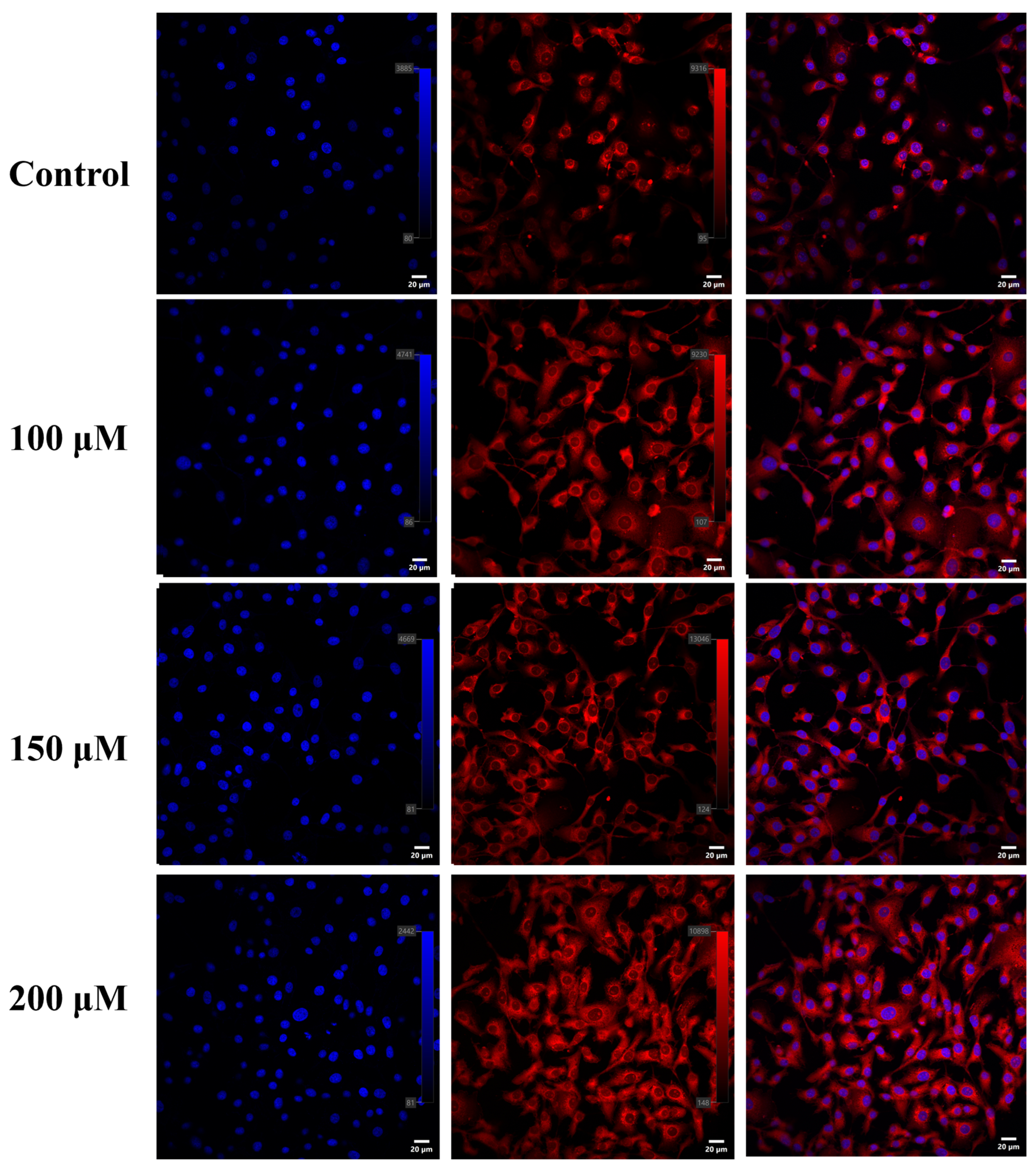
2.5. Nile Red Staining

After seeding the cells into a 12-well plate and incubating them in culture medium containing myristic acid for 24 h, the cells were fixed with 4% paraformaldehyde at room temperature for 15 min, followed by three washes with PBS containing 1% BSA. Then, the cells were stained with 10 µg/mL Nile Red at 4 °C for 1 h and washed three times with PBS containing 1% BSA. The stained cells were air-dried after incubation with DAPI (P0131, Beyotime, Shanghai, China). Finally, the cells were covered with a coverslip, and images of the lipids were captured using a confocal microscope (Olympus IX81-FV1000, Oxford, UK).

2.6. Triglyceride Content Assay

TG content in the cells was extracted and quantified using a TG detection kit (Nanjing Jiancheng Bioengineering Institute, Nanjing, China). Cells were homogenized in chloroform/methanol (2:1) and centrifuged at 3000 rpm for 10 min. The supernatant was collected and dried under nitrogen, and the residue was dissolved in isopropanol. TG content was measured based on TG enzymatic hydrolysis and colorimetric assay. The absorbance was read at 510 nm using an enzyme-linked immunosorbent assay (ELISA) reader, and TG content was expressed as µg/mg protein.

2.7. Cell Viability Assay

The CCK-8 method was used to detect the viability of MAC-T cells. MAC-T cells were seeded in a 96-well cell culture plate and cultured until the cell confluence reached 70%. Then, 200 μM myristic acid solution was added to the cells for 24 h incubation. After incubation, the culture medium was replaced with 10% CCK-8 basic medium, and the cells were incubated for an additional 3 h. The absorbance of each cell group at 450 nm wavelength was measured using an automated microplate reader. Cell viability was calculated based on the absorbance values.

2.8. Detection of mRNA Expression Levels of Relevant Genes

A total of 38 relevant genes involved in lipid synthesis and the ubiquitin–proteasome and ubiquitin–lysosome signaling pathways were selected based on the NCBI (https://www.ncbi.nlm.nih.gov/, accessed on 15 July 2023) and KEGG websites (KEGG—Table of Contents, http://www.genome.jp/kegg/kegg2.html, accessed on 15 July 2023). The mRNA expression levels of these 38 relevant genes were detected using qRT-PCR with GAPDH as the reference gene. Vazyme SYBR Green Mix was used for quantitative expression analysis (reaction system and program followed the SYBR Green Mix manual), and amplification was performed using the ABI QuantStudio 5 fluorescence quantitative PCR instrument. The relative expression levels of target gene mRNA were calculated using the 2-ΔΔCt method (ΔΔCt = ΔCt treatment group − ΔCt control group).

2.9. Data Analysis

All experimental data were represented as “mean ± standard deviation” based on three independent replicates. Differences between two groups were analyzed using independent sample t-tests in SPSS software (V.21.0). p < 0.05 was considered to be statistically significant, and p < 0.01 was considered to be highly significant.

3. Results

3.1. Effects of Different Concentrations of Myristic Acid on the Expression of Lipid-Synthesis-Related Proteins and Ubiquitin Proteins in MAC-T Cells

MAC-T cells were cultured with different concentrations of myristic acid, and the expression levels of lipid-synthesis-related proteins were measured, as shown in Figure 1. The expression levels of fatty acid transporter protein CD36 and lipid droplet marker protein ADFP were significantly upregulated 1.49-fold (p = 0.002) and 2.69-fold (p = 0.000), respectively, in MAC-T cells cultured with 200 μM myristic acid. The expression levels of UB were significantly upregulated 1.47-fold (p = 0.152), 2.27-fold (p = 0.000), and 2.42-fold (p = 0.003) at concentrations of 100 μM, 150 μM, and 200 μM myristic acid, respectively. These results indicate that all three different concentrations of myristic acid can significantly increase the level of protein ubiquitination in MAC-T cells. However, only the concentration of 200 μM myristic acid significantly increases the expression level of lipid-synthesis-related proteins in MAC-T cells. This suggests that myristic acid may affect lipid synthesis in MAC-T cells through the ubiquitination signaling pathway.

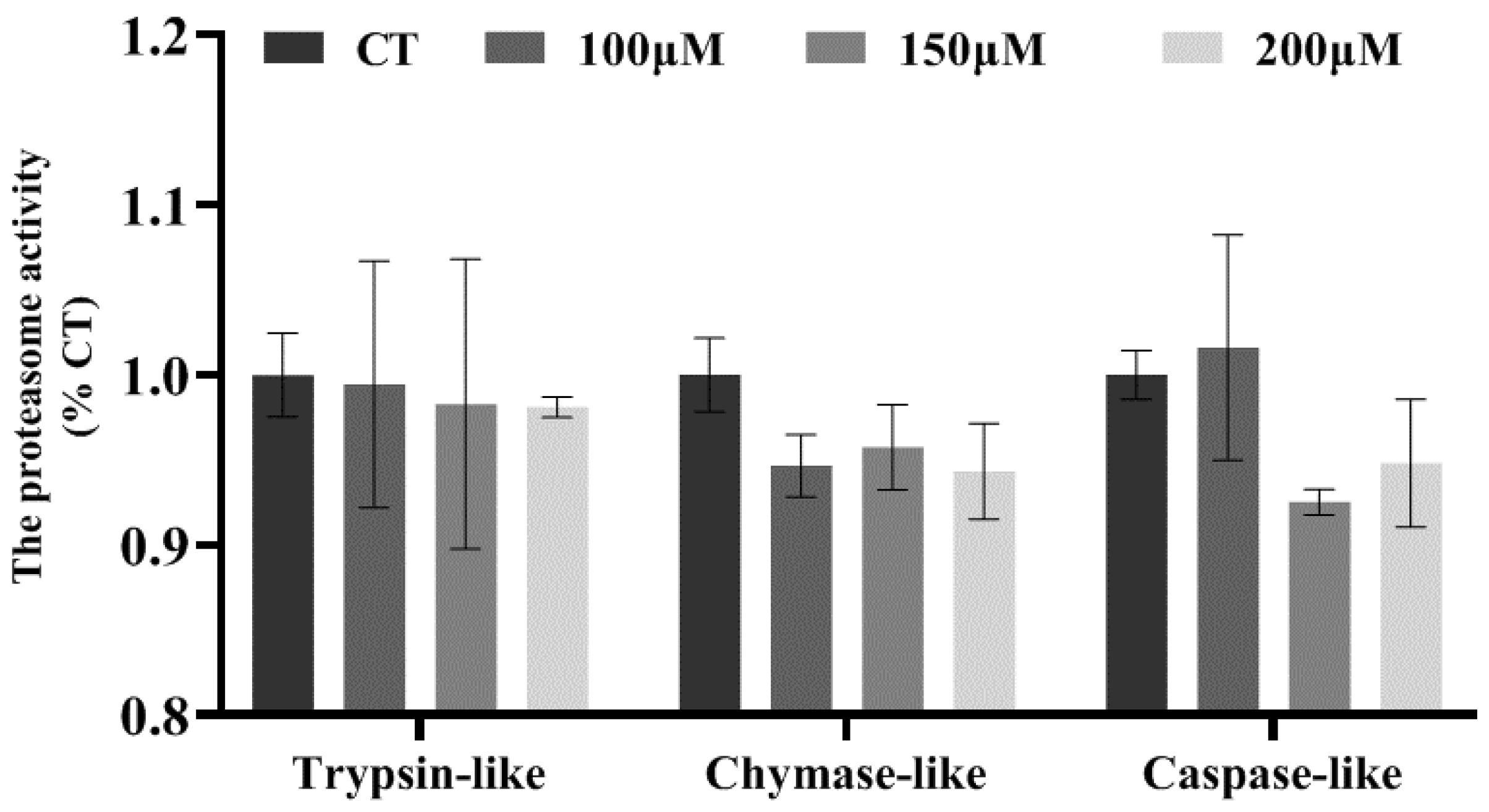
3.2. Effects of Different Concentrations of Myristic Acid on Proteasome Activity in MAC-T Cells

To verify whether myristic acid can affect lipid synthesis in MAC-T cells through the ubiquitination signaling pathway, MAC-T cells were cultured with three different concentrations of myristic acid, and the proteasome activity in the cells was measured. The results, shown in Figure 2, indicate that all three concentrations of myristic acid do not affect the proteasome activity in MAC-T cells. This suggests that the three concentrations of myristic acid may alter the protein ubiquitination level in MAC-T cells but do not affect protein homeostasis maintained by proteasome activity.

3.3. Effects of Different Concentrations of Myristic Acid on Cell Viability and Triglyceride Synthesis in MAC-T Cells

To further determine the effects of different concentrations of myristic acid on cell viability and triglyceride synthesis in MAC-T cells, cell viability was tested using CCK-8 assay. The results (Figure 3A) showed that compared to the control group, 100 μM (p = 0.001), 150 μM (p = 0.001), and 200 μM (p = 0.011) myristic acid all significantly promoted MAC-T cell viability, with 200 μM myristic acid showing the most significant effect. Triglyceride content in MAC-T cells was measured (Figure 3B), and it was found that 200 μM myristic acid significantly increased the total triglyceride content in the cells (p = 0.012). Nile red staining (Figure 3C) also revealed that MAC-T cells cultured with 200 μM myristic acid contained more and larger lipid droplets. These results indicate that 200 μM myristic acid significantly increases triglyceride content and lipid droplet size in MAC-T cells.

3.4. Regulation of Relevant Genes in MAC-T Cells by Myristic Acid

To further analyze how myristic acid regulates triglyceride synthesis in MAC-T cells through the ubiquitination signaling pathway, this study used fluorescence quantitative PCR to detect the relative expression levels of 37 relevant genes in MAC-T cells treated with 200 μM myristic acid, with GAPDH as the reference gene. Table 1 shows that in the lipid synthesis pathway, the expression levels of CD36, ACSL1, and ADFP (PLIN2) were significantly upregulated (p < 0.05), which is consistent with the Western blot results. In the ubiquitination signaling pathway, the expression levels of HERC3, PIP5K, CP1, UBC, UBA7, ISG15, and SPP1 were significantly upregulated (p < 0.05), which is also consistent with the Western blot results. However, the expression levels of genes associated with ESCRTs and 26S proteasome subunits, such as VPS45, CHMP3, CHMP2B, EEA1, and PSMC5, were significantly downregulated (p < 0.05).
To clarify the effects of myristic acid on triglyceride synthesis, the regulatory pathways of candidate genes were constructed using STRING online software (version 11b, Beijing, China). As shown in Figure 4, myristic acid can affect the expression of genes related to the ubiquitination–lysosome and ubiquitination–proteasome signaling pathways in MAC-T cells, ultimately regulating triglyceride synthesis.

4. Discussion

The content of triglycerides in milk fat accounts for about 99%, making it the most important substance in milk fat. Therefore, the changes in triglyceride content in mammary epithelial cells can directly reflect variations in milk fat synthesis. In previous research, we discovered a significant positive correlation between the content of myristic acid in the intestines of Tibetan yaks and milk fat percentage. Thus, in this study, we explored the function of myristic acid at the mammary epithelial cell level, aiming to elucidate its crucial role and mechanism in regulating milk fat production and provide important theoretical evidence for the specific mechanisms and application prospects of myristic acid in regulating milk fat synthesis.
MAC-T cells are a commonly used bovine mammary epithelial cell line and an essential cell model for studying mammary gland development and milk secretion [4]. In mammary epithelial cells, there are two main pathways for triglyceride synthesis [5,6,7]. Firstly, fatty acids from the external environment are transported into the cells through transport proteins on the cell membrane, and then, through processes like fatty acid activation, they are eventually synthesized into triglycerides. Secondly, cells directly utilize small molecules such as acetate to synthesize triglycerides with the assistance of enzymes like fatty acid synthase.
Myristic acid is a saturated fatty acid that serves as a precursor for fatty acid synthesis [8,9]. Specifically, myristic acid is first converted to myristoyl-CoA by the action of carboxylase, and then enters the fatty acid synthesis pathway, where it undergoes a series of reactions involving enzymes like acetyl-CoA carboxylase and phosphatidylcholine synthase, to finally synthesize triglycerides [10]. Additionally, research has shown that myristic acid can affect cell metabolism through different pathways. Firstly, myristic acid can promote the generation of triglycerides in cells by activating the fatty acid synthesis pathway [11]. The fatty acid synthesis pathway is a complex metabolic pathway involving the regulation of multiple enzymes and metabolites. Myristic acid can activate key enzymes in the fatty acid synthesis pathway, such as acetyl-CoA carboxylase and fatty acid synthase, to increase fatty acid synthesis and triglyceride generation. Secondly, myristic acid can also promote triglyceride synthesis by influencing other cellular metabolic pathways, such as the glucose metabolism pathway and triglyceride pathway. Specifically, myristic acid can promote the phosphorylation of key molecules, such as phosphatidylinositol 4-phosphate 5-kinase, and activate multiple key molecules in the insulin signaling pathway, enhancing its own activation and downstream signaling. Moreover, myristic acid can directly bind to phosphatidylinositol 4-phosphate 5-kinase, enhancing its activity [12,13]. This study also found that 200 μM myristic acid significantly increased the expression levels of PIP5K and other related genes in MAC-T cells. These effects ultimately lead to the smooth transmission of the insulin signaling pathway, promoting the absorption and utilization of glucose and other nutrients in cells, and further stimulating fatty acid synthesis, thereby promoting triglyceride synthesis. Additionally, myristic acid can covalently bind to specific amino acids, such as glycine at the N-terminus of proteins [14], regulating the structure and function of proteins through N-myristoylation, and thereby controlling triglyceride synthesis. In recent years, research has found that ubiquitin proteins can bind to some N-myristoylated proteins and participate in regulating their degradation and metabolism. Specifically, ubiquitin proteins can form a covalent bond with the lysine residues on N-myristoylated proteins, known as ubiquitination, and then tag and send them to the ubiquitin-dependent proteasome for degradation [15]. In conclusion, myristic acid plays a crucial role in maintaining normal lipid metabolism and cell growth and development, and is associated with various metabolic pathways through the ubiquitination pathway.
This study also found that different concentrations of myristic acid can significantly increase the level of protein ubiquitination and the expression of ubiquitin-related genes in MAC-T cells. Therefore, this study speculates that myristic acid may regulate triglyceride synthesis in MAC-T cells through the ubiquitination pathway. To explore whether myristic acid can regulate fatty acid transport through the ubiquitination pathway, this study first cultured MAC-T cells with different concentrations of myristic acid solution, then detected the expression levels of the fatty acid transport protein CD36 and ubiquitin (UB) in the cells, and also measured the activity of proteasomes in the cells. The results showed that 200 μM myristic acid significantly increased the expression levels of CD36 and UB in the cells, but the three concentrations of myristic acid had no significant effect on proteasome activity, and they all significantly increased cell viability. CD36 is a transport protein on the membrane of MAC-T cells, which can absorb long-chain fatty acids (LCFAs) and directly participate in the regulation of lipid intake [16]. As a transmembrane glycoprotein, CD36 contains multiple post-translational modification sites, such as glycosylation [17], phosphorylation [18], palmitoylation [19], acetylation [20], and ubiquitination [21], which regulate the stability, protein folding, and localization of CD36. Studies have found that CD36 can directly bind to ubiquitin proteins [22]. In summary, the results of this study indicate that appropriate concentrations of myristic acid can regulate fatty acid transport in MAC-T cells through the ubiquitination pathway, thereby promoting triglyceride synthesis.
Proteasomes are large multi-subunit complexes present in eukaryotic and prokaryotic organisms. They have various enzymatic activities and selectively degrade proteins in cells that are not needed or are incorrectly folded, breaking them down into individual amino acid residues, which are then used for new protein synthesis. Therefore, proteasome activity plays an important role in maintaining protein homeostasis in cells [23]. Studies have found that when cells experience proteotoxic stress, proteasome activity is inhibited, leading to the overactivation of cyclin-dependent kinases as the cyclin levels increase, which may cause the irreversible accumulation of protein aggregates and ultimately result in cell cycle disorder [24]. On the other hand, when proteasome activity is activated, it triggers the formation of proteasome liquid droplets in cells, leading to reduced cell volume and nucleolar stress [23,24]. This study found that the three concentrations of myristic acid solution did not affect the activity of proteasomes in the cells and could increase cell viability, indicating that myristic acid does not cause stress to the cells.
To further determine whether myristic acid can promote the synthesis of milk fat in MAC-T cells, this study further detected the expression level of adipose differentiation-related protein (ADFP), a marker protein for lipid droplets, as well as the content of triglycerides and lipid droplets in the cells.
The outcomes unveiled a significant correlation: the exposure of MAC-T cells to a concentration of 200 μM myristic acid yielded a substantial augmentation in the expression level of ADFP. Positioned on the surface of lipid droplets within cells, ADFP assumes a pivotal role in fostering the creation of intracellular lipid droplets [25,26]. Notably, existing research, albeit unpublished, has pointed out that ADFP possesses the capability to directly interact with ubiquitin proteins, governed by the ubiquitination–proteasome signaling pathway. The noticeable surge in ADFP expression effectively signifies a concurrent increase in the count of lipid droplets housed within the cells. Moreover, our investigation brought to light a compelling finding: the infusion of 200 μM myristic acid notably amplified the triglyceride content within MAC-T cells. This conclusion was fortified by the utilization of Nile red staining, which revealed a conspicuous augmentation in the lipid droplet content within the cells. This concurrence between the two observations serves to reinforce the hypothesis that myristic acid significantly stimulates triglyceride synthesis within MAC-T cells. As a cumulative result, this process ultimately propels the production of milk fat.
To summarize, our results lay a biological foundation that connects the elevation in ADFP expression, the surge in lipid droplet count, and the augmented triglyceride content to the administration of 200 μM myristic acid. This interplay underscores myristic acid’s role in bolstering intracellular lipid accumulation, subsequently contributing to the enhancement of milk fat production in MAC-T cells.

5. Conclusions

This study aimed to evaluate how myristic acid influences triglyceride synthesis in bovine milk fat. We cultured MAC-T cells with varying myristic acid concentrations and observed that 200 μM myristic acid notably increased protein expressions of CD36, ADFP, and UB. Myristic acid did not disrupt proteasome activity, enhancing cell viability and indicating minimal cellular stress. Subsequent tests on triglyceride content and lipid droplets confirmed the substantial role of 200 μM myristic acid in triglyceride synthesis and lipid content within MAC-T cells. This underscores the safe nature of myristic acid and its dose-dependent effect on triglyceride synthesis. These findings provide insights into myristic acid’s cellular-level contribution to milk fat regulation, awaiting validation at the organismal level. The study significantly bolsters our understanding of myristic acid’s mechanisms and potential in governing milk fat production.

Author Contributions

Conceptualization, L.L. and M.H.; methodology, L.L., M.H., P.W. and A.G.; software, L.L. and M.H.; validation, L.L. and M.H.; formal analysis, L.L. and M.H.; investigation, L.L. and M.H.; resources, L.L. and M.H.; data curation, L.L. and M.H.; writing—original draft preparation, L.L. and M.H.; writing—review and editing, L.L. and M.H.; visualization, L.L. and M.H.; supervision, L.L. and M.H.; project administration, L.L. and M.H.; funding acquisition, L.L. All authors have read and agreed to the published version of the manuscript.

Funding

This research was funded by the National Natural Science Foundation (grant number: 31902152; 3210200137), Scientific Research Foundation of Southwest Forestry University (grant number: 112119), and Ten Thousand Talent Plans for Young Top-notch Talents of Yunnan Province (grant number: 20221116), Anhui Natural Science Foundation (grant number: 2108085QC131).

Institutional Review Board Statement

Not applicable.

Data Availability Statement

All data measured or analyzed during this work are available from the corresponding author upon reasonable request.

Acknowledgments

The authors would like to thank the Chinese Academy of Sciences, Kunming Institute of Zoology, for the technical and platform support.

Conflicts of Interest

The authors declare no conflict of interest.

References

  1. Oepen, K.; Ozbek, H.; Schuffler, A.; Liermann, J.C.; Thines, E.; Schneider, D. Myristic Acid Inhibits the Activity of the Bacterial ABC Transporter BmrA. Int. J. Mol. Sci. 2021, 22, 13565. [Google Scholar] [PubMed]
  2. Liu, L.; Wu, P.; Chen, F.; Zhou, J.; Guo, A.; Shi, K.; Zhang, Q. Multi-omics analyses reveal that the gut microbiome and its metabolites promote milk fat synthesis in Zhongdian yak cows. PeerJ 2022, 10, e14444. [Google Scholar]
  3. Liu, L.; Zhang, Q. Comparative proteome analysis reveals VPS28 regulates milk fat synthesis through ubiquitylation in bovine mammary epithelial cells. PeerJ 2020, 8, e9542. [Google Scholar] [PubMed]
  4. Ogunnaike, M.; Wang, H.; Zempleni, J. Bovine mammary alveolar MAC-T cells afford a tool for studies of bovine milk exosomes in drug delivery. Int. J. Pharm. 2021, 610, 121263. [Google Scholar] [PubMed]
  5. Zhao, Y.; Guo, X.; Yan, S.; Shi, B.; Sheng, R. Acetate regulates milk fat synthesis through the mammalian target of rapamycin/eukaryotic initiation factor 4E signaling pathway in bovine mammary epithelial cells. J. Dairy Sci. 2021, 104, 337–345. [Google Scholar] [PubMed]
  6. Thering, B.J.; Graugnard, D.E.; Piantoni, P.; Loor, J.J. Adipose tissue lipogenic gene networks due to lipid feeding and milk fat depression in lactating cows. J. Dairy Sci. 2009, 92, 4290–4300. [Google Scholar]
  7. Bionaz, M.; Loor, J.J. Gene networks driving bovine milk fat synthesis during the lactation cycle. BMC Genom. 2008, 9, 366. [Google Scholar] [CrossRef]
  8. Hristov, A.N.; Lee, C.; Cassidy, T.; Long, M.; Heyler, K.; Corl, B.; Forster, R. Effects of lauric and myristic acids on ruminal fermentation, production, and milk fatty acid composition in lactating dairy cows. J. Dairy Sci. 2011, 94, 382–395. [Google Scholar]
  9. Odongo, N.E.; Or-Rashid, M.M.; Kebreab, E.; France, J.; McBride, B.W. Effect of supplementing myristic acid in dairy cow rations on ruminal methanogenesis and fatty acid profile in milk. J. Dairy Sci. 2007, 90, 1851–1858. [Google Scholar]
  10. Machmuller, A.; Kreuzer, M. Influence of myristic acid supplementation on energy, fatty acid and calcium metabolism of sheep as affected by dietary calcium and forage: Concentrate ratio. J. Anim. Physiol. Anim. Nutr. 2005, 89, 284–296. [Google Scholar] [CrossRef] [PubMed]
  11. Kummrow, E.; Hussain, M.M.; Pan, M.; Marsh, J.B.; Fisher, E.A. Myristic acid increases dense lipoprotein secretion by inhibiting apoB degradation and triglyceride recruitment. J. Lipid Res. 2002, 43, 2155–2163. [Google Scholar] [CrossRef] [PubMed]
  12. Saraswathi, V.; Kumar, N.; Ai, W.; Gopal, T.; Bhatt, S.; Harris, E.N.; Talmon, G.A.; Desouza, C.V. Myristic Acid Supplementation Aggravates High Fat Diet-Induced Adipose Inflammation and Systemic Insulin Resistance in Mice. Biomolecules 2022, 12, 739. [Google Scholar] [CrossRef] [PubMed]
  13. Chen, K.; Jia, H.; Liu, Y.; Yin, P.; Wei, Y. Insulin-Sensitizing Activity of Sub-Nanoscaled Polyalkoxyvanadate Clusters. Adv. Biosyst. 2020, 4, e1900281. [Google Scholar] [CrossRef]
  14. Jia, M.; Wang, Y.; Wang, J.; Qin, D.; Wang, M.; Chai, L.; Fu, Y.; Zhao, C.; Gao, C.; Jia, J.; et al. Myristic acid as a checkpoint to regulate STING-dependent autophagy and interferon responses by promoting N-myristoylation. Nat. Commun. 2023, 14, 660. [Google Scholar] [CrossRef] [PubMed]
  15. Kwon, H.; Choi, M.; Ahn, Y.; Pak, Y. N-myristoylation regulates insulin-induced phosphorylation and ubiquitination of Caveolin-2 for insulin signaling. Biochem. Biophys. Res. Commun. 2020, 532, 535–540. [Google Scholar] [CrossRef] [PubMed]
  16. Huang, C.C.; Chou, C.A.; Chen, W.Y.; Yang, J.L.; Lee, W.C.; Chen, J.B.; Lee, C.T.; Li, L.C. Empagliflozin Ameliorates Free Fatty Acid Induced-Lipotoxicity in Renal Proximal Tubular Cells via the PPARgamma/CD36 Pathway in Obese Mice. Int. J. Mol. Sci. 2021, 22, 12408. [Google Scholar] [CrossRef]
  17. Hoosdally, S.J.; Andress, E.J.; Wooding, C.; Martin, C.A.; Linton, K.J. The Human Scavenger Receptor CD36, glycosylation status and its role in trafficking and function. J. Biol. Chem. 2009, 284, 16277–16288. [Google Scholar] [CrossRef]
  18. Chu, L.Y.; Silverstein, R.L. CD36 ectodomain phosphorylation blocks thrombospondin-1 binding: Structure-function relationships and regulation by protein kinase C. Arter. Thromb. Vasc. Biol. 2012, 32, 760–767. [Google Scholar] [CrossRef]
  19. Zeng, S.; Wu, F.; Chen, M.; Li, Y.; You, M.; Zhang, Y.; Yang, P.; Wei, L.; Ruan, X.Z.; Zhao, L.; et al. Inhibition of Fatty Acid Translocase (FAT/CD36) Palmitoylation Enhances Hepatic Fatty Acid beta-Oxidation by Increasing Its Localization to Mitochondria and Interaction with Long-Chain Acyl-CoA Synthetase 1. Antioxid. Redox Signal. 2022, 36, 1081–1100. [Google Scholar] [CrossRef]
  20. Han, L.; Yang, Q.; Li, J.; Cheng, F.; Zhang, Y.; Li, Y.; Wang, M. Protocatechuic Acid-Ameliorated Endothelial Oxidative Stress through Regulating Acetylation Level via CD36/AMPK Pathway. J. Agric. Food Chem. 2019, 67, 7060–7072. [Google Scholar] [CrossRef]
  21. Fang, Y.; Shen, Z.Y.; Zhan, Y.Z.; Feng, X.C.; Chen, K.L.; Li, Y.S.; Deng, H.J.; Pan, S.M.; Wu, D.H.; Ding, Y. CD36 inhibits beta-catenin/c-myc-mediated glycolysis through ubiquitination of GPC4 to repress colorectal tumorigenesis. Nat. Commun. 2019, 10, 3981. [Google Scholar] [CrossRef] [PubMed]
  22. Srikanthan, S.; Li, W.; Silverstein, R.L.; McIntyre, T.M. Exosome poly-ubiquitin inhibits platelet activation, downregulates CD36 and inhibits pro-atherothombotic cellular functions. J. Thromb. Haemost. 2014, 12, 1906–1917. [Google Scholar] [CrossRef] [PubMed]
  23. Davidson, K.; Pickering, A.M. The proteasome: A key modulator of nervous system function, brain aging, and neurodegenerative disease. Front. Cell Dev. Biol. 2023, 11, 1124907. [Google Scholar] [CrossRef] [PubMed]
  24. Yasuda, S.; Tsuchiya, H.; Kaiho, A.; Guo, Q.; Ikeuchi, K.; Endo, A.; Arai, N.; Ohtake, F.; Murata, S.; Inada, T.; et al. Stress- and ubiquitylation-dependent phase separation of the proteasome. Nature 2020, 578, 296–300. [Google Scholar] [CrossRef] [PubMed]
  25. Liu, M.; Qi, L.; Zeng, Y.; Yang, Y.; Bi, Y.; Shi, X.; Zhu, H.; Zhou, Z.; Sha, J. Transient scrotal hyperthermia induces lipid droplet accumulation and reveals a different ADFP expression pattern between the testes and liver in mice. PLoS ONE 2012, 7, e45694. [Google Scholar] [CrossRef] [PubMed]
  26. Chang, B.H.; Chan, L. Regulation of Triglyceride Metabolism, I.I.I. Emerging role of lipid droplet protein ADFP in health and disease. Am. J. Physiol. Gastrointest. Liver. Physiol. 2007, 292, G1465–G1468. [Google Scholar] [CrossRef] [PubMed]
Figure 1. Effects of myristic acid at different concentrations on the expression levels of CD36, ADFP, and UB proteins in MAC-T cells. ** denotes p < 0.01. (A): Western blot analysis of the protein levels of CD36, ADFP, and UB in MAC-T cells. (B): The relative protein levels of CD36, ADFP, and UB in MAC-T cells.
Agriculture 13 01870 g001
Figure 2. Effects of different concentrations of myristic acid on the three proteasome activities in MAC-T cells.
Agriculture 13 01870 g002
Figure 3. Effects of different concentrations of myristic acid on cell viability and triglyceride content in MAC-T cells. * denotes p < 0.05, ** denotes p < 0.01. (A,B): The effects of different concentrations of myristic acid on cell viability and triglyceride content in MAC-T cells. (C): Nile red staining of lipid droplets in MAC-T cells cultured with different concentrations of myristic acid (200×).
Agriculture 13 01870 g003
Figure 4. Regulatory pathways of candidate genes affected by myristic acid in MAC-T cells.
Agriculture 13 01870 g004
Table 1. The information of primers of the candidate genes and their relative expression.
GeneNamePrimer (5′→3′)Relative
Expression
GAPDHGlyceraldehyde-3-phosphate dehydrogenaseAGATGGTGAAGGTCGGAGTG/
CGTTCTCTGCCTTGACTGTG
LPLLipoprotein lipaseAGCTCCAAGTCGCCTTTCTC1.076
TCCTGGTTGGAAAGTGCCTC
LDLRLow-density lipoprotein receptorTGTTGGACACACGTACCCAG0.834 **
AAGGTCGCGACTTGTCTCAG
CD36Platelet glycoprotein 4GACGGATGTACAGCGGTGAT1.192 *
GAAAAAGTGCAAGGCCACCA
ACSL1Acyl-CoA synthetase long-chain family member 1 AGCCGCATTTCACTTTTACTGC1.568 **
AGCTCTTTAGGGCAAACCCC
FABP3Fatty-acid-binding protein 3ACGCGTTCTCTGTCGTCTTT0.488 **
AACCGACACCGAGTGACTTC
ACACAAcetyl-coa carboxylase/biotin carboxylase 1ACGGCTGACTGGAGTTGAAG0.914
AACGTCTGCTTGTCCGTCTT
ACBPAacyl-coa-binding protein TGGAATCTTTGCAACACCGC0.955 *
TGTCACCCACAGTTGCTTGT
FASNFatty acid synthaseCCTCAAGATGAAGGTGGTGCT0.742
GGCCCTGGGTTATATCGAGC
SCDStearoyl-CoA desaturase TCCTGATCATTGGCAACACCA1.05
CCAACCCACGTGAGAGAAGAA
DGAT1Diacylglycerol o-acyltransferase 1TACCCCGACAACCTGACCTA1.05
GGGAAGTTGAGCTCGTAGCA
LPIN1Lipin 1 CTTCGATTCCCAAACCGGGA0.262
TCACAGTGACGAACACCTGG
ADFPPerilipin-2GCGTCTGCTGGCTGATTTC2.03 **
(PLIN2)AGCCGAGGAGACCAGATCATA
APOEApolipoprotein ECGGTTTCTGGAGGCGAAGAA0.796
CTCCATATCCGCCTGGCATC
PRKCACalcium-activated, phospholipid- and diacylglycerol-dependent serineGACTTCGGGATGTGCAAGGA1.005
CGTACGGCTGATAGGCGATT
MAPK1Mitogen-activated protein kinase 1AACAAAGTCCGAGTCGCCAT0.854
CGATGGTCGGTGCTCGAATA
ARF6ADP-ribosylation factor 6AACTGGTATGTGTCAGCCCTC0.921
GAAAGAGGTGATGGTGGCGA
STAM1Signal transducing adapter molecule 1CCTGGTACTGCGGCTAACAA1.277 *
ACGAACTTTCCGGCCTTCAT
EEA1Early endosome antigen 1 isoform X4x4CAGGCCCAGGACAGCTTAAA0.91 *
GCAAGTTCCTGTGCTGCTTG
HERC3HECT- and RLD-domain-containing E3 ubiquitin protein ligase 3 CTCGAGGGCCTAGCTGTCT1.163 **
TTTGTCAGAAGGGTCTGGCG
VPS45Vacuolar protein sorting 45 homolog CCCCAAAGATGCTGTGGCTA0.922 *
AGTGTGCTGGGGCCTAGATA
CHMP2BCharged multivesicular body protein 2bACGAGGTACACAGAGGGCTA0.868 **
AGCTGTTTGGCTAAAACTCTGC
CHMP3Charged multivesicular body protein 3GTTTGAAATCACCGCAGGGG0.75 **
CTAAAGGTTCAGGCTCCGGG
PIP5KPhosphatidylinositol-4-phosphate 5-kinase, putativeCTCAGCACCTGGAAGAGCAA1.623 *
TTCTTCTTTCCCCGAGCCAC
CYHR1Cysteine and histidine rich 1 GCCAACCTGCTTTTGGGAAG1.006
GGTTGTGAAAACGGCCACAA
CPUbiquitin-like domain-containing CTD phosphatase 1CATGGTGGCCAAAGGTGTTG1.156 **
CATCTGCTGGAGATTTTTGGCA
PSMC1Proteasome 26S subunit, ATPase 1GGTACGACTCCAACTCAGGC0.893 **
ATCCGGTTTGTGGCCATGAT
PSMC326S protease regulatory subunit 6ATGAACAAGACGCTGCCGTAT0.89
TGCCGCGTAGAGGTTTTGAT
PSMC526S proteasome regulatory subunit 8CTCTGCACAAGATCCTGCCT0.828 **
ATGCTTCACAGGCAGCTCAA
PSMD1226S proteasome non-ATPase regulatory subunit 12ATACGTCAGGCATCTCGCAG0.966
GGCCATGTTGTAGGGGACAA
UBCUbiquitin-CGGGAGGTGTTTTAAGTTCTCCCT1.682 **
TTGAACTCTAACCCACCCCTAAC
UBA52Ubiquitin-60S ribosomal protein L40GCCCAGTGACACCATTGAGA0.832 **
GCAGGGTGGACTCTTTCTGG
UBA7Ubiquitin-like modifier-activating enzyme 7TCAGCAGGATGGTCTGAGGA1.3 *
AGTTCCAATACCAGCACCCG
TUBATubulin beta chain GTCTACTCCTGTTGCCTGC1.024
AGGCATTGCCGATCTGGAC
ISG15Ubiquitin-like protein ISG15CCATCCTGGTGAGGAACGAC1.496 **
GTCTGCTTGTACACGCTCCT
MX1Interferon-induced GTP-binding protein Mx1TGCCAACTAGTCAGCACTACATT0.639 **
TGTACAGGTTGCTCTTGGACTC
SPP1Secreted phosphoprotein 1TCCGCCCTTCCAGTTAAACC1.945 **
GCTTCTGAGATGGGTCAGGC
RPS27ARibosomal protein S27aTTTCGTGAAGACCCTGACGG0.819 *
GTCTTTGCTGGTCAGGAGGAA
* denotes p < 0.05; ** denotes p < 0.01.
Disclaimer/Publisher’s Note: The statements, opinions and data contained in all publications are solely those of the individual author(s) and contributor(s) and not of MDPI and/or the editor(s). MDPI and/or the editor(s) disclaim responsibility for any injury to people or property resulting from any ideas, methods, instructions or products referred to in the content.

