Article

Evaluation of the Differences in the Serum Protein Electrophoretic Pattern in Precolostral Serum of Farm Animal Neonates

1
Clinic of Ruminants, University of Veterinary Medicine and Pharmacy, 041 81 Košice, Slovakia
2
Clinic of Swine, University of Veterinary Medicine and Pharmacy, 041 81 Košice, Slovakia
*
Author to whom correspondence should be addressed.
Agriculture 2023, 13(5), 1035; https://doi.org/10.3390/agriculture13051035
Submission received: 18 April 2023 / Revised: 7 May 2023 / Accepted: 8 May 2023 / Published: 10 May 2023
(This article belongs to the Special Issue Welfare, Behavior and Health of Farm Animals)

Abstract

:
The objective of this study was to compare the electrophoretic pattern of serum proteins in newborn calves, lambs, goat kids and piglets in precolostral blood samples. Blood samples were collected within 30 min of birth, prior to ingestion of first colostrum, and the concentrations of total proteins and protein fractions were analyzed using electrophoresis on agarose gel. The size and shape of the protein fractions on the electrophoretograms differed among the investigated animal species. Significant differences were found in the total protein values and all the separated protein fractions, as well as albumin to globulin ratio (p < 0.01 and p < 0.001). The mean total protein concentration in piglets was lower compared to ruminants. Albumin concentrations were markedly higher, and the concentrations of α1-globulins were lower, in neonatal ruminants compared to piglets. The values of α2-globulins were higher in lambs and goat kids, and lower in calves and piglets. An opposite tendency was found in the values of β-globulins: a detectable amount of γ-globulins was recorded in all evaluated animal species. Presented results suggest marked species related differences in the shape and size of protein fraction among neonates of farm animal species, and the importance of the evaluation of electrophoretograms with regard to these findings.

1. Introduction

The placenta connects the maternal and fetal circulation systems and has important functions in the protection, nutrition, respiration of the fetus, regulation of its development, and especially in the transfer of antibodies [1,2]. It is composed of several tissue layers depending on animal species [3]. The physiology of a ruminant’s placenta (i.e., bovine, ovine, caprid, cervidae) differs from other species because it is classified as a synepitheliochorial type consisting of six tissue membranes, which impedes the effective transfer of maternal serum proteins—especially immunoglobulins to the fetal circulation during the intrauterine life [4,5]. Therefore, newborn calves, lambs, as well as goat kids have low concentrations of serum proteins at birth and are practically agammaglobulinemic or with minimal concentrations of immunoglobulins and are thus without immune protection against pathogens [6,7,8,9]. Similarly, piglets are born with low concentrations of serum proteins and are immunologically incompetent because the epitheliochorial type of placenta of sows does not allow the transfer of maternal antibodies [10,11]. Therefore, in these animal species, the survival of newborn and the acquirement of immunity is dependent on passive immune transfer through the intake of colostrum and its absorption [12,13]. Despite previous studies dealing with serum proteins in newborn animals, the differences in the composition of neonatal serum proteins and the proportion of protein fractions among animal species are still not fully described.
The electrophoretic technique is one of the most important methods in clinical biochemistry to separate blood serum proteins and quantify protein fractions. This method separates serum proteins or a group of proteins according to their size and electrical charge [14]. In companion animals, serum protein electrophoresis is more frequently used as a laboratory method to detect protein abnormalities, especially in the gamma region [15]. In farm animals it is not commonly used, although it can yield important information about the proportion and migration of protein fractions in various diseases and pathological processes associated with the altered values of total serum proteins. This method may also be of great importance in newborn animals to evaluate gamma globulin status and immunoglobulin transfer. Wide changes were obtained in the values of total serum proteins, as well as their fractions in the neonatal period and during the first months of life as newborns of various animal species [16,17,18,19]. However, the differences in the electrophoretic pattern of serum proteins among neonates of different animal species have not been previously described. Therefore, this study was aimed at the determination of differences in the electrophoretograms, especially in the shape and size of protein fractions separated by electrophoresis, as well as differences in the values of total proteins and individual protein fractions in precolostral serum samples of newborn farm animals.

2. Material and Methods

2.1. Ethical Declaration

The blood samples from the neonates, as per standard sampling procedure, were collected and were used without any harm to the animals. All procedures with animals were conducted in accordance with the common ethical standards and guidelines approved by the institutional Committee on protection of animals used for scientific purposes and complied with the requirements of the Code of Ethics for Scientists (Directive 74/2019/UVLF).

2.2. Animals and Sample Collection

Blood samples from twenty full-term newborn calves, ten lambs, ten goat kids and thirty-five piglets of both sexes were selected and were included into the study. The calves (8 females and 12 males) were of Slovak spotted and low-land black spotted breeds, and their crossbreeds were born from primiparous and multiparous dairy cows from conventional dairy farms with similar management. They were born as singles and their mean body weight at birth was 36.1 ± 2.3 kg. The neonate lambs (6 males, 4 females) were of the merino breed, born from ewes from two farms with a similar standard method of rearing. The average body weight of the lambs at birth was 3.3 ± 0.4 kg. The goat kids were born as singles (6 kids) and twins (4 kids) from dams of the white shorthaired breed with a mean body weight of 3.2 ± 0.3 kg. The piglets were crossbreeds of Large White × Landrace born from three sows (first to third gestation) with an average body weight 1.59 ± 0.27 kg at birth. The health status of the dams was continuously checked during pregnancy. The pregnancies were of normal length and the parturitions took place under controlled conditions at the University Veterinary Hospital. The births were uncomplicated, and all newborns were clinically in good health condition.
Blood samples from all the evaluated newborns were collected within 30 min of birth, prior to the ingestion of first colostrum. The samples from calves, lambs and goat kids were taken from the jugular vein into 4.4 mL serum gel separator plastic tubes without anticoagulants and other additives (Sarstedt, Nümbrecht, Germany). Blood samples from piglets were taken from the orbital sinus into 1.1 mL tubes of the same type as mentioned above. The piglets for the blood collection from sinus ophtalmicus were restrained in the dorsal recumbent position and placed on a V-board table. The heads of the animals were in front of the edge of the table and both legs were held to point backwards.

2.3. Laboratory Analyses

After clotting, the blood samples were centrifuged at 4000× g for 15 min and then were separated into Eppendorf tubes. The aliquots of sera were frozen at −20 °C and stored until further processing and laboratory measurements. The serum samples were analyzed for the total protein concentrations (TP, g.L−1) and serum protein electrophoresis. The TP values were determined according to the biuret method with commercial diagnostic kits (Randox, Crumlin, UK) on an automated chemistry analyzer Alizé (Lisabio, Poully en Auxois, France). The serum protein fractions were separated by zone electrophoresis on agarose gel using an automated electrophoresis system, Hydrasys, and using the commercial diagnostic kit, Hydragel 7 Proteine (Sebia Corporate, Lisses, Evry Cedex, France), according to the application instructions of the manufacturer. The gels were then stained, and the staining intensity of the separated protein bands was examined with a densitometer Epson Perfection V700 (Epson America Inc., Long Beach, CA, USA) and the image analysis software Phoresis version 5.50 (Sebia Corporate, France). The protein fractions were presented in relative values (%) based on the optical density, and their absolute values (g.L−1) were calculated from the TP concentrations. Albumin to globulin ratios (A/G) were calculated as well.

2.4. Statistical Analysis

Mean values and standard deviations were determined by the methods of descriptive statistics in the computer program GraphPad Prism V5.02 (GraphPad Software Inc., San Diego, CA, USA). The Shapiro–Wilk test for normality was applied to estimate the distribution of the values. In order to assess the significance of differences between the groups of newborn animals, the results were analyzed by the ANOVA test and Tukey’s multiple comparisons post hoc test in case of normally distributed values and using the Kruskal–Wallis test with Dunn’s test in case of not-normally-distributed values. The statistical significance of the differences between the animal groups was estimated at a 5% probability level (p < 0.05).

3. Results

The relative values of all protein fractions presented in Table 1 showed significant differences among the evaluated animal species (p < 0.001).
The significantly highest relative values of albumin were observed in newborn small ruminants, with a significant difference in mean values also observed between lambs and kids (p < 0.05). The mean relative albumin value obtained in calves was significantly lower than in newborn small ruminants (p < 0.05), but still accounted for almost 60% of total serum proteins. The significantly lowest relative albumin concentrations were found in neonatal piglets (p < 0.05), and the mean value was more than four times lower when compared to lambs (Figure 1a). The relative values of α1-globulins, on the other hand, were significantly the lowest in lambs and goat kids, while those found in calves were significantly higher than in small ruminants (p < 0.05). The highest average relative value of α1-globulins was recorded in piglets and represented almost 70% of total serum proteins. These values were approximately two and a half times higher compared to calves and three to four times higher than in small ruminants (p < 0.05; Figure 1b). The mean relative values of α2-globulins were significantly more than two-fold lower in neonatal calves and piglets compared to lambs and goat kids (p < 0.05; Figure 1c). An opposite tendency of the difference in values was found in the relative values of β-globulins. The means were significantly lower in lamb and goat kids (p < 0.05) and were approximately four to five-fold higher in calves and piglets (Figure 1d). The relative values of γ-globulins were very low in all the evaluated newborn animal species. The significantly lowest average concentration was recorded in newborn calves (p < 0.05; Figure 1e). The evaluation of A/G ratios revealed significant differences among the investigated animal species (p < 0.001) with the lowest average concentration in piglets. The mean values in ruminants were higher by more than seven times in calves and approximately ten times in newborn small ruminants (Figure 1f).
The values of total proteins and all serum protein fractions given in Table 2 showed significant differences among the evaluated animal species (p < 0.01 and p < 0.001).
The mean total serum protein values in newborn ruminants before colostrum intake were approximately similar. The values obtained in piglets were significantly and about 1.5 times lower (p < 0.05; Figure 2).
A similar tendency of differences was found in the absolute concentrations of albumin with significantly higher values in precolostral serum samples of ruminant species (p < 0.05). The mean value found in piglets was approximately six times lower than in ruminants. On contrary to albumin values the significantly highest α1-globulin concentrations were recorded in piglets (p < 0.05). In addition, differences in α1-globulin values were found also between the newborn ruminants with the highest mean value in calves and the lowest average concentration in lambs. The values of α2-globulins were significantly lower in newborn calves and piglets (p < 0.05). The mean values obtained in lambs and goat kids were almost four times higher. An opposite tendency of differences was recorded in the values of β-globulins. The significantly lowest mean values were found in newborn small ruminants (p < 0.05). The highest average value of β-globulins was recorded in calves. Although the concentrations of γ-globulins in precolostral serum were very low in all the evaluated animal species, the mean values differed significantly (p < 0.01). The significantly lowest mean values were found in calves, and the highest in lambs and goat kids.
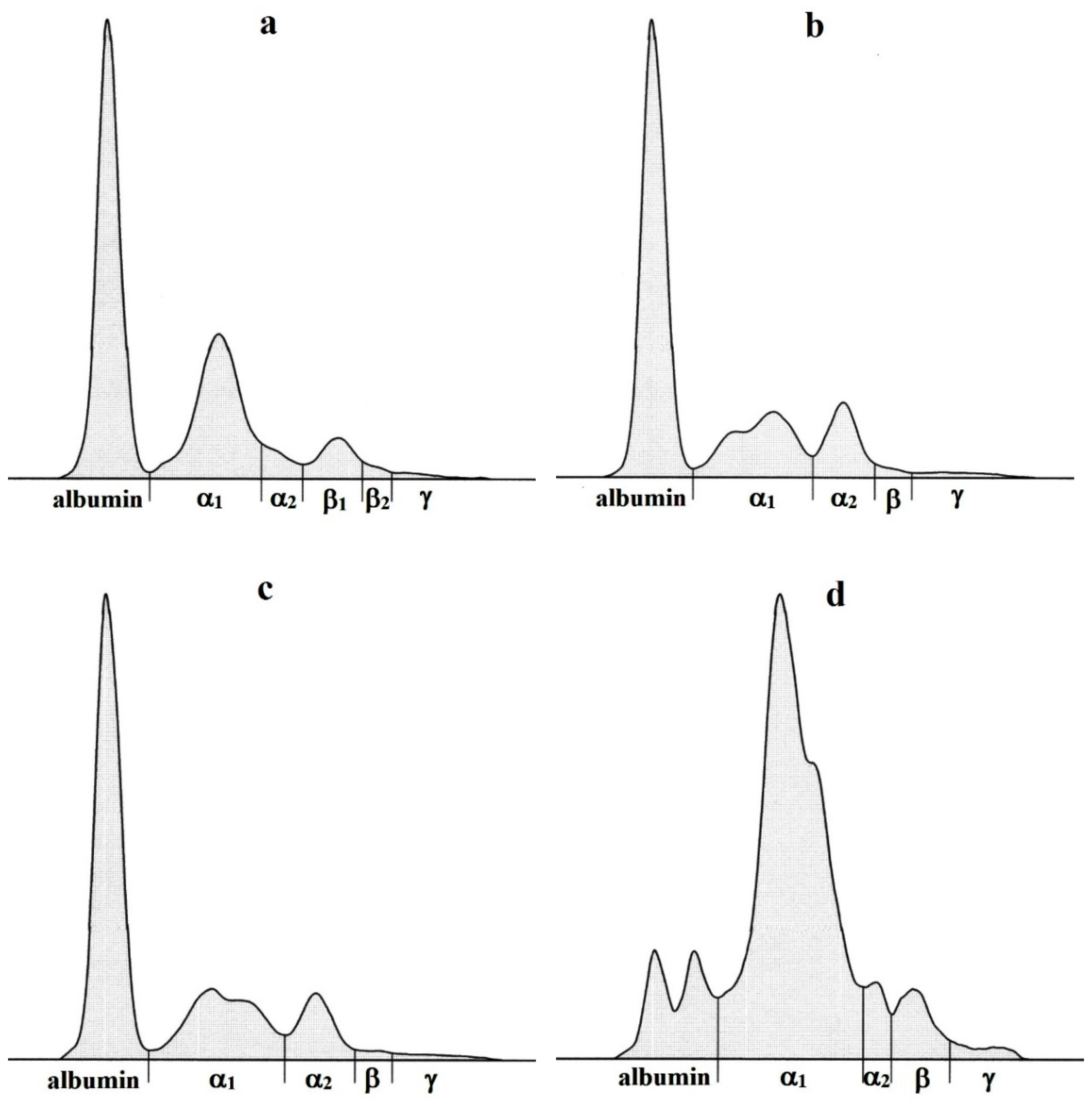
The size and shape of separated serum protein fractions on the electrophoretograms differed according to the evaluated animal species (Figure 3a–d). In newborn calves, six protein fractions were recognized, including albumin, α1-, α2-, β1-, β2- and γ-globulins. Serum proteins in lambs, goat kids and piglets were separated into five fractions: albumin, α1-, α2-, β- and γ-globulins. Albumin was identified as the most marked fraction in calves, lambs and goat kids, and was characterized by high and narrow peak on the electrophoretogram. Electrophoresis in newborn piglets showed a double peak in the albumin zone with two approximately equivalent lower peaks. The α-globulins migrated in α1- and α2-subfractions in all the investigated neonates. While in calves the α1-globulin fraction was presented as a single peak with moderate and sharper electrophoretographic amplitude, in lambs and goat kids this fraction was flatter and tended to show a twin peak. In neonatal piglets, the most prominent fraction observable as a wide and very high peak was the α1-globulin fraction. The α2-globulin zone in all the evaluated neonates was characterized by one peak, but in calves it was not clearly demarcated from the α1-globulin fraction. In the β-globulin zone, the serum protein electrophoresis identified one overall distinct band in all the evaluated animal species and the γ-globulin fraction was presented on the electrophoretograms as a very low and flat zone.

4. Discussion

Most of the studies dealing with the fractionation of serum proteins in neonatal ruminants were oriented to the interpretation of dynamic changes of serum protein fraction in a short period after birth [16,18]. Precolostral serum samples were not standardly collected and possible species-related differences among newborns of farm animals were not yet described. Therefore, the present study may contribute to the description and clarification of differences in the electrophoretic pattern of precolostral serum proteins among selected animals. The transfer of various substances from maternal circulation during gestation, especially serum proteins and antibodies, varies according to species and is related to the type of placenta and number of tissue layers between the maternal and fetoplacental circulation systems [20]. It has been stated in most animal species that the values of serum proteins at birth are low due to the ineffective transfer of immunoglobulins through the placenta and low values of albumin [21,22]. In the present study, no marked differences in the values of total proteins were observed in newborn ruminants and the values were comparable to those detected by Franklin et al. [23] and Piccione et al. [16] in calves, and by Chen et al. [24] and Nagy et al. [25] in small ruminants. On the other hand, the values recorded in neonatal piglets were significantly lower and about the half of values measured in newborn ruminants. Similarly, Ingvarsson et al. [26] and Cavanagh et al. [27] obtained very low total protein values in piglets directly after birth, which were only around 20 g.L−1. This may be related to the lower birth weight of piglets compared to calves or lambs, the higher size of piglets in the litter, a physiologically immature body construction and not having fully developed liver functions to produce an adequate amount of proteins [28]. A very similar pattern of differences with higher values in newborn ruminants and lower in piglets was found in the albumin values. The proportion of albumin was higher than 50% in all the evaluated ruminant species, but the study showed differences in its concentrations between calves, lambs, as well as goat kids. Although the measured values were comparable to those presented previously [29,30,31], comparison studies between different animal species were not yet performed. Variations in the amino acid composition of the albumin, structure and binding sites for ligands were observed among different animal species [32]. Therefore, the differences in measured albumin values may be attributed to these structural differences or other species differences in metabolic pathways that need to be further investigated. In contrast to ruminants, the albumin concentrations in neonatal piglets were significantly lower and constituted only 16% of total proteins. Similarly, Szymeczko, et al. [33] recorded low albumin values in newborn piglets, which represented in average 14.2% of total serum proteins, while Ingvarsson et al. [26] and Martin et al. [34] detected even lower values (6–7% of total proteins). This indicates that piglets are unique species of animals, which at birth have very low albumin values. The synthesis of albumin in the fetuses of piglets starts in the later stages of development in comparison to other serum proteins and, similarly to immunoglobulins, the epitheliochorial type of the placenta does not allow its transfer to the fetus that might contribute to low albumin concentrations in newly born piglets [35,36]. Furthermore, electrophoresis in newborn piglets showed a double peak in the albumin zone with two approximately similar lower peaks that were previously not presented in any other species. The second peak on the cathodal side of the albumin zone probably represents the so called postalbumin fraction and its shape and size diminishes with advancing age [31]. According to Lardinois and Page [37], postalbumin could be a fetal-specific protein that disappears with age. However, further studies are needed to clarify the biochemical properties of albumin fractions and their metabolic functions in neonatal piglets.
Species-related differences were observed also in the values of α1-globulins with the highest average concentration in piglets, in which α1-globulins represented almost 70% of total proteins. In newborn piglets, α1-acid glycoprotein is the predominant serum protein in the perinatal period before colostrum intake, with concentrations around 12 mg.mL−1 [38,39,40]. Lampreave and Piñeiro [41] stated that α1-acid glycoprotein has important functions in the adaptive processes of newly born organism, which have to prepare the piglets for the extrauterine life, but its exact role in physiology and the significance of the aforementioned very high concentrations are not yet known. Other proteins from the α1-globulin fraction are α1-antitrypsin and fetuin that may be found in the serum of piglets after birth in higher concentrations (representing around 18% of total proteins at birth). These proteins have important functions in the inhibition of the effect of proteases absorbed by the gastrointestinal tract in the first days of life [41,42]. On the other hand, the concentrations of α1-fetoprotein in piglets after birth are lower when compared to other animal species [43,44]. Higher values of alpha1-fetoprotein were recorded in newborn calves, contributing to higher values of α1-globulins after birth, while its values in neonatal lambs and goat kids have not been defined [45]. Alpha1-fetoprotein is synthesized by the developing fetus and then by parenchymal cells of the liver, but its production decreases with advancing age [46]. The biological role of higher concentrations in neonates was not completely understood, but it shows similarities to albumin [47]. Furthermore, our study showed differences in the values of α1-globulins also among the evaluated ruminant species, with the highest in calves and the lowest in lambs. These differences probably reflect the different composition of the α1-globulin fraction in the evaluated ruminants and different physiological concentrations of serum proteins from this fraction.
The analyses of the values of α2-globulins revealed their highest proportion in newborn lambs and goat kids, which represented approximately 10% of total serum proteins. The values obtained in calves and piglets were significantly lower (approximately four-fold) compared to small ruminants. The α2-globulin fraction is composed of several important proteins, including haptoglobin, α2-macroglobulin, α2-lipoprotein and ceruloplasmin. All these proteins are minor components with low serum concentrations [48]. Hiss-Pesch et al. [49] obtained low concentrations of haptoglobin (5.16 ± 0.86 µg.mL−1) in neonatal piglets right after birth before the first colostrum. These data were comparable with those presented by Martin et al. [34] and Llamas Moya et al. [50]. Slightly higher serum Hp concentrations were recorded by Rocha et al. [51] and Erkiliç et al. [52] in neonatal calves before colostrum intake (6.63–8.34 mg.dL−1), while the values found in neonatal lambs were markedly higher [53]. The precolostral ceruloplasmin values in the blood serum of neonatal calves ranged from 13.25 to 31.5 mg.dL−1 [51,52], and the values obtained in neonatal lambs were in the range of 2.42–9.99 mg.dL−1 [54]. Nevertheless, there is little information on the concentrations of other proteins from this fraction in newborns of farm animals.
An opposite tendency was found in the values of β-globulins, with higher concentrations in neonatal calves and piglets, and lower concentrations in small ruminants. The values obtained in precolostral serum samples are comparable to those recorded by Marc et al. [55] in calves prior to colostrum intake and by Nagy et al. [19,25] in neonatal lambs and goat kids. Complement components (especially C3 and C4), transferrin and ferritin are the main proteins belonging to this fraction [56]. Transferrin plays central role in the metabolism of iron, especially in the transport of iron ions from the intestine and the reticuloendothelial system to various cells and tissues of the liver, spleen and bone marrow [57]. According to Moser et al. [58], its values are higher in calves with iron deficiency (above 8 g.L−1) and it shows a negative correlation with hemoglobin. Thoren-Tolling and Martinsson [59] found transferrin concentrations around 1 mg.mL−1 in piglets at birth, while Asadi et al. [60] recorded an average transferrin concentration of 2.98 mg.mL−1 in newborn lambs. The serum concentrations of ferritin obtained by Atyabi et al. [61] in newborn calves before colostrum intake were comparable with values in postpartum cows (in average 32.93 ng.mL−1), but decreased significantly in 1- and 2-day-old calves to 21.46 ng/mL and correlated with body iron status. In newborn piglets, the serum concentrations of ferritin at birth differed according to the amount of iron in the diet of sows and ranged from 106.83 to 122.47 ng.mL−1 [62]. No published data are available for the concentrations of ferritin in neonatal lambs and goat kids, but the aforementioned species differences in the concentrations of selected proteins may be responsible for the differences recorded in the concentrations of β-globulin fraction in neonates of various farm animals. Furthermore, some immunoglobulins may migrate into the β-globulin fraction (mainly immunoglobulins of the classes A and M), but their concentrations in newborns may differ according to the type of placenta in each given species. All the aforementioned differences in the values of these mentioned individual serum proteins might contribute to differences in the protein fractions recorded in our study in precolostral serum samples.
It has been stated in some previous studies that the epitheliochorial placenta in sows and the synepitheliochorial placenta in cows and other ruminants inhibits the passage of immunoglobulins and other immunological factors to the fetus, and the neonates of these species do not have immunoglobulins prior to colostrum ingestion [2,10]. The results of γ-globulins obtained in neonates of the evaluated species before the intake of colostrum indicate that the newborns of farm animals are not born absolutely agammaglobulinemic, as we found a detectable number of γ-globulins in all the evaluated newborns as result of selective transport. However, the comparison of results showed differences between the evaluated species, being lower in neonatal calves and piglets while the values in lambs and goat kids were higher. This pattern of low γ-globulin fractions is related to the minimal quantities of immunoglobulins transferred through the placenta of ruminants and sows, but the concentrations passed depend on the thickness and extent of the barrier [63,64]. In these animals, antibodies are secreted in the maternal milk and subsequently must be passively received by the fetus from colostrum [65]. The γ-globulin concentrations obtained in this study are comparable to those presented previously in neonatal ruminants [29,31], but comparative studies were not yet performed. The results suggest that electrophoresis may be an important laboratory diagnostic aid to monitor perinatal serum protein electrophoretic pattern and the distribution of serum proteins in newborns of farm animals. The presence of γ-globulins in higher concentrations in precolostral serum samples indicate placental lesions, which allow the transfer of antibodies through the placenta [66,67]. As result of differences in the distribution of protein fractions, the A/G ratios also differed significantly among the evaluated animal species and were the highest in neonatal lambs and the lowest in piglets. These variations reflect the higher proportion of the albumin fraction in newborns of ruminants and its very low concentrations in neonatal piglets, but there is very high proportion of α-globulins in this species.

5. Conclusions

The presented results indicate marked differences in the precolostral serum protein electrophoretic pattern among the evaluated farm animal species and in the shape, size and distribution of serum protein fractions in the serum samples collected from neonates of these species prior to colostrum intake. Significant differences were observed in all serum protein fractions (p < 0.001), with the most marked differences in the values of albumin and α1-globulin fractions. While the albumin values were higher in the neonates of ruminants and lower in piglets, the values of α1-globulins presented an opposite tendency with lower values in precolostral serum samples of ruminant species and very high values in piglets. A detectable number of γ-globulins was obtained in all the evaluated species with marked differences among them. The γ-globulin concentrations were lower in neonatal calves and piglets, while lambs and goat kids before colostrum intake had higher values. Knowledge of the characteristics of the precolostral serum protein electrophoretic pattern and the distribution of serum proteins in neonates of farm animals prior to colostrum intake is of fundamental importance within farm animal medicine, especially to perinatal care. It provides information about the physiological pattern of serum proteinograms in neonates of farm animals and points out significant differences among the evaluated animal species.

Author Contributions

Investigation, Methodology, Resources, Writing—Original Draft Preparation, C.T.; Resources, Supervision, R.L.; Methodology, Resources, V.G.; Conceptualization, Project Administration, Validation, Writing—Review & Editing, O.N. All authors have read and agreed to the published version of the manuscript.

Funding

This work was supported by the grants No. VEGA 1/0177/22 and VEGA 1/0314/20 from the Ministry of Education, Science, Research and Sport of the Slovakia.

Institutional Review Board Statement

All procedures with animals in the study were conducted in accordance with the common ethical standards and guidelines approved by the Institutional Committee of the University of Veterinary Medicine and Pharmacy in Košice on protection of animals used for scientific purposes and complied with the requirements of the Code of Ethics for Scientists (Directive 74/2019/UVLF).

Data Availability Statement

The data that support the findings of this study are available from the corresponding author upon reasonable request.

Conflicts of Interest

The authors declare no conflict of interest.

References

  1. King, G.J. Comparative placentation in ungulates. J. Exp. Zool. 1982, 31, 588–602. [Google Scholar] [CrossRef]
  2. Chucri, T.M.; Monteiro, J.M.; Lima, A.R.; Salvadori, M.L.B.; Kfoury, J.R.; Miglino, M.A. A review of immune transfer by the placenta. J. Reprod. Immunol. 2010, 87, 14–20. [Google Scholar] [CrossRef] [PubMed]
  3. Leiser, R.; Kaufmann, P. Placental structure: In a comparative aspect. Exp. Clin. Endocrinol. 1994, 102, 122–134. [Google Scholar] [CrossRef] [PubMed]
  4. Wooding, F.B.P.; Flint, A.P.F.; Heap, R.B.; Morgan, G.; Buttle, H.L.; Young, I.R. Control of binucleate cell immigration in the placenta of ruminants. J. Reprod. Fertil. 1986, 76, 499–512. [Google Scholar] [CrossRef] [PubMed]
  5. Wooding, F.B.P. Current topic: The synepitheliochorial placenta of ruminants: Binucleate cell fusions and hormone production. Placenta 1992, 13, 101–113. [Google Scholar] [CrossRef]
  6. Halliday, R.; Williams, M.R. The absorption of immunoglobulin from colostrum by bottle-fed lambs. Ann Rech. Vet. 1979, 10, 549–556. [Google Scholar]
  7. Constant, S.B.; Leblanc, M.M.; Klaptein, E.F.; Beebe, D.E.; Leneau, H.M.; Nunier, C.J. Serum immunoglobulin G concentration in goat kids fed colostrum or a colostrum substitute. J. Am. Vet. Med. Assoc. 1994, 205, 1759–1762. [Google Scholar]
  8. Arguello, A.; Castro, N.; Zamorano, M.J.; Castroalonso, A.; Capote, J. Passive transfer of immunity in kid goats fed refrigerated and frozen goat colostrum and commercial sheep colostrum. Small Rum. Res. 2004, 54, 237–241. [Google Scholar] [CrossRef]
  9. Castro, N.; Capote, J.; Álvarez, S.; Argüello, A. Effects of lyophilized colostrum and different colostrum feeding regimens on passive transfer of immunoglobulin G in Majorera goat kids. J. Dairy Sci. 2005, 88, 3650–3654. [Google Scholar] [CrossRef]
  10. Moffett, A.; Loke, C. Immunology of placentation in eutherian mammals. Nature 2006, 6, 584–594. [Google Scholar] [CrossRef]
  11. Wilson, M.E.; Biensen, N.J.; Ford, S.P. Novel insight in to control of litter size in pigs, using placental efficiency as a selection tool. J. Anim. Sci. 1999, 77, 1654–1658. [Google Scholar] [CrossRef] [PubMed]
  12. Giguère, S.; Polkes, A.C. Immunologic disorders in neonatal foals. Vet. Clin. Equine 2005, 21, 241–272. [Google Scholar] [CrossRef]
  13. Crisman, M.V.; Scarratt, W.K. Immunodeficiency disorders in horses. Vet. Clin. Equine 2008, 24, 299–310. [Google Scholar] [CrossRef]
  14. O’Connell, T.X.; Horita, T.J.; Kasravi, B. Understanding and interpreting serum protein electrophoresis. Am. Fam. Physician 2005, 71, 105–112. [Google Scholar] [PubMed]
  15. Tappin, S.W.; Taylor, S.S.; Tasker, S.; Dodkin, S.J.; Papasouliotis, K.; Murphy, K.F. Serum protein electrophoresis in 147 dogs. Vet. Rec. 2011, 168, 456. [Google Scholar] [CrossRef] [PubMed]
  16. Piccione, G.; Casella, S.; Giannetto, C.; Vazzana, I.; Niutta, P.P.; Giudice, E. Influence of age on profile of serum proteins in the calf. Acta Vet. 2009, 59, 413–422. [Google Scholar]
  17. Teixeira, W.T.; Fonteque, G.V.; Ramos, A.F.; da Silva Mariante, A.; do Egito, A.A.; Martins, V.M.V.; Saito, M.E.; Fonteque, J.H. Transfer of passive immunity and serum proteinogram in the first six months of life of Criollo Lageano and Black and White Holstein calves. Pesq. Vet. Bras. 2012, 32, 980–986. [Google Scholar] [CrossRef]
  18. Piccione, G.; Arfuso, F.; Faggio, C.; Casella, S.; Zumbo, A.; Panzera, M. Serum proteins profile in Comisana lambs during the first month of life. Arch. Tierz. 2013, 56, 742–750. [Google Scholar] [CrossRef]
  19. Nagy, O.; Tóthová, C.; Nagyová, V.; Kováč, G. Serum protein electrophoretic pattern in goat kids after colostrum intake during the first week of life. J. Vet. Sci. Med. Diagn. 2016, 5, 2. [Google Scholar] [CrossRef]
  20. Enders, A.C.; Blankenship, T.N. Comparative placental structure. Adv. Drug Deliv. Rev. 1999, 38, 3–15. [Google Scholar] [CrossRef]
  21. Sato, T.; Oda, K.; Kubo, M. Hematological and biochemical values of thoroughbred foals in the first six months of life. Cornell Vet. 1979, 69, 3–19. [Google Scholar] [PubMed]
  22. Hammon, H.M.; Schiessler, G.; Nussbaum, A.; Blum, J.W. Feed intake patterns, growth performance and metabolic and endocrine traits in calves fed unlimited amounts of colostrums and milk by automate strating in the neonatal period. J. Dairy Sci. 2002, 85, 3352–3362. [Google Scholar] [CrossRef] [PubMed]
  23. Franklin, S.T.; Ameral-Philips, D.M.; Jackson, J.A.; Campbell, A.A. Health and performance of Holstein calves that suckled or were hand-fed colostrums and were fed one of three physical forms of starter. J. Dairy Sci. 2003, 86, 2145–2153. [Google Scholar] [CrossRef] [PubMed]
  24. Chen, J.C.; Chang, C.J.; Peh, H.C.; Chen, S.Y. Serum protein levels and neonatal growth rate of Nubian goat kids in Taiwan area. Small Rum. Res. 1999, 32, 153–160. [Google Scholar] [CrossRef]
  25. Nagy, O.; Tóthová, C.; Nagyová, V.; Kováč, G.; Pošivák, J. Changes in the serum protein electrophoretic pattern in lambs during the first month of life. Acta Vet. Brno 2014, 83, 187–193. [Google Scholar] [CrossRef]
  26. Ingvarsson, B.I.; Carlsson, R.N.K.; Karlsson, B.W. Synthesis of α-fetoprotein, albumin and total serum protein in neonatal pigs. Biol. Neonate 1978, 34, 259–268. [Google Scholar] [CrossRef]
  27. Cavanagh, M.E.; Cornelis, M.E.; Dziegielewska, K.M.; Luft, A.J.; Lai, P.C.W.; Lorscheider, F.L.; Saunders, N.R. Proteins in cerebrospinal fluid and plasma of fetal pigs during development. Dev. Neurosci. 1982, 5, 492–502. [Google Scholar] [CrossRef]
  28. Svendsen, L.S.; Weström, B.R.; Svendsen, J.; Olsson, A.; Karlsson, B.W. Blood serum characteristics of newborn pigs. Comparison of unaffected pigs with pigs belonging to five mortality groups. Acta Vet. Scand. 1991, 32, 287–299. [Google Scholar] [CrossRef]
  29. Nagyová, V.; Tóthová, C.; Nagy, O. The impact of colostrum intake on the serum protein electrophoretic pattern in newborn ruminants. J. Appl. Anim. Res. 2017, 45, 498–504. [Google Scholar] [CrossRef]
  30. Souza, D.C.; Silva, D.G.; Rocha, T.G.; Monteiro, B.M.; Pereira, G.T.; Fiori, L.C.; Viana, R.B.; Fagliari, J.J. Serum biochemical profile of neonatal buffalo calves. Arq. Bras. Med. Vet. Zootec. 2019, 71, 187–196. [Google Scholar] [CrossRef]
  31. Tóthová, C.; Link, R.; Kyzeková, P.; Nagy, O. Serum protein electrophoretic pattern in piglets during the early postnatal period. Sci. Rep. 2021, 11, 17539. [Google Scholar] [CrossRef] [PubMed]
  32. Bujacz, A.; Talaj, J.A.; Zielinski, K.; Pietrzyk-Brzezinska, A.J.; Neumann, P. Crystal structures of serum albumins from domesticated ruminants and their complexes with 3,5-diiodosalicylic acid. Acta Crystallogr. D Struct. Biol. 2017, 73, 896–909. [Google Scholar] [CrossRef]
  33. Szymeczko, R.; Kapelaǹski, W.; Piotrowska, A.; Dybala, J.; Boguslawska-Tryk, M.; Burlikowska, K.; Hertig, I.; Sassek, M.; Pruszyǹska-Oszmalek, E.; Maćkowiak, P. Changes in the content of major proteins and selected hormons in the blood serum of piglets during the early postnatal period. Folia Biol. 2009, 57, 97–103. [Google Scholar] [CrossRef] [PubMed]
  34. Martin, M.; Tesouro, M.A.; González-Ramón, N.; Piňeiro, A.; Lampreave, F. Major plasma proteins in pig serum during postnatal development. Reprod. Fertil. Dev. 2005, 17, 437–445. [Google Scholar] [CrossRef] [PubMed]
  35. Stone, R.T. In vitro liver synthesis and serum levels of alpha-fetoprotein and albumin in the fetal pig. Biol. Reprod. 1981, 20, 573–580. [Google Scholar] [CrossRef]
  36. Le Dividich, J.; Rooke, J.A.; Herpin, P. Nutritional and immunological importance of colostrum for the new-born pig. J. Agric. Sci. 2005, 143, 469–485. [Google Scholar] [CrossRef]
  37. Lardinois, R.; Page, L.A. Serum albumin, prealbumin and postalbumin in perinatal pigs. Dev. Biol. 1969, 19, 261–280. [Google Scholar] [CrossRef]
  38. Lampreave, F.; Piñeiro, A. The major serum protein of fetal and newborn pigs: Biochemical properties and identification as a fetal form of α1-acid glycoprotein. Int. J. Biochem. 1984, 16, 47–53. [Google Scholar] [CrossRef]
  39. Itoh, H.; Tamura, K.; Izumi, M.; Motoi, Y.; Kidoguchi, K.; Funayama, Y. The influence of age and health status on the serum α1-acid glycoprotein level of conventional and specific pathogen-free pigs. Can. J. Vet. Res. 1993, 57, 74–78. [Google Scholar]
  40. Heegaard, P.M.H.; Miller, I.; Sorensen, N.S.; Soerensen, K.E.; Skovgaard, K. Pig α1-glycoprotein: Characterization and first description in any species as a negative acute phase protein. PLoS ONE 2013, 8, e68110. [Google Scholar] [CrossRef]
  41. Lampreave, F.; Piñeiro, A. Concentrations of major plasma proteins in serum and whole-tissue extracts in porcine fetuses during development. J. Reprod. Fertil. 1992, 95, 441–449. [Google Scholar] [CrossRef] [PubMed]
  42. Weström, B.R.; Karlsson, B.W.; Svendsen, J. Levels of serum protease inhibitors during fetal and postnatal development of the pig. Biol. Neonate 1982, 41, 22–31. [Google Scholar] [CrossRef] [PubMed]
  43. Gitlin, D.; Boesman, M. Sites of serum α-fetoprotein synthesis in the human and in the rat. J. Clin. Investig. 1967, 46, 1010–1016. [Google Scholar] [CrossRef] [PubMed]
  44. Calvo, M.; Naval, J.; Lampreave, F.; Piñeiro, A. Fatty acids bound to α-fetoprotein and albumin during rat development. Biochim. Biophys. Acta 1988, 959, 238–246. [Google Scholar] [CrossRef] [PubMed]
  45. Kaneko, J.J. Serum proteins and the dysproteinemias. In Clinical Biochemistry of Domestic Animals, 5th ed.; Kaneko, J.J., Ed.; Academic Press: London, UK, 1997; pp. 117–138. [Google Scholar]
  46. Bishop, M.L.; Fody, E.P.; Schoeff, L.E. Clinical Chemistry: Techniques, Principles, Correlations; Lippincott Williams & Wilkins: Philadelphia, PA, USA, 2010; pp. 205–245. [Google Scholar]
  47. Bader, D.; Riskin, A.; Vafsi, O.; Tamir, A.; Peskin, B.; Israel, N.; Merksamer, R.; Dar, H.; David, M. Alpha-fetoprotein in the early neonatal period—A large study and review of the literature. Clin. Chim. Acta 2004, 349, 15–23. [Google Scholar] [CrossRef] [PubMed]
  48. Bossuyt, X. Advances in serum protein electrophoresis. Adv. Clin. Chem. 2006, 42, 43–80. [Google Scholar]
  49. Hiss-Pesch, S.; Daniel, F.; Bunkelberg-Denk, S.; Mielenz, M.; Sauerwein, H. Transfer of maternal haptoglobin in suckling piglets. Vet. Immunol. Immunopathol. 2011, 144, 104–110. [Google Scholar] [CrossRef]
  50. Llamas Moya, S.; Boyle, L.A.; Lynch, P.B.; Arkins, S. Age-related changes in pro-inflammatory cytokines, acute phase proteins and cortisol concentrations in neonatal pigs. Neonatology 2007, 91, 44–48. [Google Scholar] [CrossRef]
  51. Rocha, T.G.; Silva, F.D.F.; Bortoletto, C.; Silva, D.G.; Buzinaro, M.G.; Zafalon, L.F.; Fagliari, J.J. Serum concentrations of acute phase proteins and immunoglobulins of calves with rotavirus diarrhea. Arq. Bras. Med. Vet. Zootec. 2016, 68, 865–872. [Google Scholar] [CrossRef]
  52. Erkiliçc, E.E.; Merhan, O.; Kirmizigül, A.H.; Öğün, M.; Akyüz, E.; Çitil, M. Salivary and serum levels of serum amyloid A, haptoglobin, ceruloplasmin and albumin in neonatal calves with diarrhea. Kafkas Univ. Vet. Fak. Derg. 2019, 25, 583–586. [Google Scholar]
  53. Dinler, C.; Ulutas, B.; Voyvoda, H.; Ulutas, P.A.; Ural, K.; Karagenc, T. Haptoglobin and serum amyloid-A concentrations and their relationship with oocyst count in neonatal lambs experimentally infected with Cryptosporidium parvum. Vet. Parasitol. 2017, 247, 49–56. [Google Scholar] [CrossRef] [PubMed]
  54. Dinler, C.; Tuna, G.E.; Ay, E.; Ulutas, B.; Voyvoda, H.; Ulutas, P.A. Reference intervals for serum amyloid A, haptoglobin, ceruloplasmin, and fibrinogen in apparently healthy neonatal lambs. Vet. Clin. Pathol. 2020, 49, 484–490. [Google Scholar] [CrossRef] [PubMed]
  55. Marc, S.; Kirovski, D.; Mircu, C.; Hutu, I.; Otavă, G.; Paul, C.; Boldura, O.M.; Tulcan, C. Serum protein electrophoretic pattern in neonatal calves treated with clinoptilolite. Molecules 2018, 23, 1278. [Google Scholar] [CrossRef] [PubMed]
  56. Cerón, J.J.; Caldin, M.; Martinez-Subiela, S. Electrophoresis and acute phase protein measurement. In Schalm’s Veterinary Hematology, 6th ed.; Douglas, J., Weiss, K., Wardrop, J., Eds.; Blackwell Publishing Ltd.: Ames, IA, USA, 2010; pp. 1157–1161. [Google Scholar]
  57. De Jong, G.; van Dijk, J.P.; van Eijk, H.G. The biology of transferrin. Clin. Chim. Acta 1990, 190, 1–46. [Google Scholar] [CrossRef] [PubMed]
  58. Moser, M.; Pfister, H.; Bruckmaier, R.M.; Rehage, J.; Blum, J.W. Blood serum transferrin concentration in cattle in various physiological states, in veal calves fed different amounts of iron, and in cattle affected by infectious and non-infectious diseases. Zentralbl. Veterinarmed. A 1994, 41, 413–420. [Google Scholar] [CrossRef] [PubMed]
  59. Thoren-Tholling, K.; Martinsson, K. On the transferrin concentration in serum of sows and growing pigs and in sow colostrum. Acta Vet. Scand. 1974, 15, 120–134. [Google Scholar] [CrossRef]
  60. Asadi, M.; Toghdory, A.; Hatami, M.; Ghassemi Nejad, J. Milk supplemented with organic iron improves performance, blood hematology, iron metabolism parameters, biochemical and immunological parameters in suckling Dalagh lambs. Animals 2022, 12, 510. [Google Scholar] [CrossRef]
  61. Atyabi, N.; Gharagozloo, F.; Nassiri, S.M. The necessity of iron supplementation for normal development of commercially reared suckling calves. Comp. Clin. Pathol. 2006, 15, 165–168. [Google Scholar] [CrossRef]
  62. Li, Y.; Yang, W.; Dong, D.; Jiang, S.; Yang, Z.; Wang, Y. Effect of different sources and levels of iron in the diet of sows on iron status in neonatal pigs. Anim. Nutr. 2018, 4, 197–202. [Google Scholar] [CrossRef]
  63. Jones, H.N.; Powell, T.L.; Jansson, T. Regulation of placental nutrient transport: A review. Placenta 2007, 28, 763–774. [Google Scholar] [CrossRef]
  64. Borghesi, J.; Mario, L.C.; Nogueira Rodrigues, M.; Oliveira Favaron, P.; Miglino, M.A. Immunoglobulin transport during gestation in domestic animals and humans—A review. Open J. Anim. Sci. 2014, 4, 323–336. [Google Scholar] [CrossRef]
  65. Baintner, K. Transmission of antibodies from mother to young: Evolutionary strategies in a proteolytic environment. Vet. Immunol. Immunopathol. 2007, 117, 153–161. [Google Scholar] [CrossRef] [PubMed]
  66. Poitras, B.J.; Miller, R.B.; Wilkie, B.N.; Bosu, W.T. The maternal to fetal transfer of immunoglobulins associated with placental lesions in sheep. Can. J. Vet. Res. 1986, 50, 68–73. [Google Scholar] [PubMed]
  67. Gabriel, S.; Geldhof, P.; Phiri, I.K.; Cornillie, P.; Goddeeris, B.M.; Vercruysse, J. Placental transfer of immunoglobulins in cattle infected with Schistosoma mattheei. Vet. Immunol. Immunopathol. 2005, 104, 265–272. [Google Scholar] [CrossRef]
Figure 1. The distribution of the precolostral serum relative values of albumin (a), α1- (b), α2- (c), β- (d), γ-globulins (e) and albumin/globulin ratios (f) in calves, lambs, kids and piglets. The plots show the median (line within the box), 25th and 75th percentiles (box), as well as minimal and maximal concentrations (whiskers).
Agriculture 13 01035 g001
Figure 2. The distribution of the precolostral total serum protein values in calves, lambs, kids and piglets during the early postnatal period of life. The plots show the median (line within the box), 25th and 75th percentiles (box), as well as minimal and maximal concentrations (whiskers).
Agriculture 13 01035 g002
Figure 3. Electrophoretograms representing the differences in precolostral serum protein fractions in calf (a), lamb (b), kid (c) and piglet (d).
Agriculture 13 01035 g003
Table 1. Relative values of the precolostral protein fractions (%) and albumin to globulin ratio (A/G) in the blood serum of the monitored newborn animals.
ParametersCalvesLambsKidsPigletsp Value
Albumin58.1 ± 2.8 a68.0 ± 1.6 b64.4 ± 3.7 c16.1 ± 2.9 d<0.001
α1-globulins27.5 ± 1.8 a16.6 ± 2.0 b21.1 ± 2.9 c68.5 ± 3.3 d<0.001
α2-globulins2.93 ± 0.66 a11.39 ± 0.92 b10.91 ± 1.43 b4.69 ± 0.86 c<0.001
β-globulins10.11 ± 1.33 a1.93 ± 0.45 b1.40 ± 0.31 b8.20 ± 1.29 c<0.001
γ-globulins1.40 ± 0.58 a2.13 ± 0.43 b2.20 ± 1.11 a,b2.53 ± 1.11 b<0.001
A/G1.40 ± 0.17 a2.13 ± 0.16 b1.84 ± 0.30 c0.19 ± 0.04 d<0.001
a,b,c,d–values with different superscripts in rows indicate significant differences in means among species of animals (p < 0.05).
Table 2. Absolute concentrations of the precolostral total protein concentrations (TP) and serum protein fractions (g.L−1) in the evaluated newborn animals.
ParametersCalvesLambsKidsPigletsp Value
TP40.0 ± 2.1 a41.9 ± 2.7 a40.8 ± 4.8 a25.9 ± 2.4 b<0.001
Albumin23.2 ± 1.6 a28.5 ± 1.8 b26.4 ± 3.7 c4.2 ± 0.9 d<0.001
α1-globulins11.0 ± 0.9 a6.9 ± 1.0 b8.5 ± 1.0 c17.7 ± 1.8 d<0.001
α2-globulins1.16 ± 0.30 a4.79 ± 0.57 b4.47 ± 0.89 b1.22 ± 0.22 a<0.001
β-globulins4.06 ± 0.62 a0.80 ± 0.18 b0.57 ± 0.15 b2.12 ± 0.37 c<0.001
γ-globulins0.56 ± 0.22 a0.89 ± 0.19 b0.90 ± 0.53 a,b0.65 ± 0.29 a<0.01
a,b,c,d–means with different superscripts in rows indicate significant differences between species of animals (p < 0.05).
Disclaimer/Publisher’s Note: The statements, opinions and data contained in all publications are solely those of the individual author(s) and contributor(s) and not of MDPI and/or the editor(s). MDPI and/or the editor(s) disclaim responsibility for any injury to people or property resulting from any ideas, methods, instructions or products referred to in the content.