Article

Effect of Melatonin on the Production Performance, Blood Biochemical Parameters, Nutrient Digestibility, and Gastrointestinal Microbiome of Liaoning Cashmere Goats

State Key Laboratory of Animal Nutrition and Feeding, College of Animal Science and Technology, China Agricultural University, Beijing 100193, China
*
Author to whom correspondence should be addressed.
These authors contributed equally to this work.
Agriculture 2024, 14(11), 1983; https://doi.org/10.3390/agriculture14111983
Submission received: 23 September 2024 / Revised: 28 October 2024 / Accepted: 4 November 2024 / Published: 5 November 2024
(This article belongs to the Section Farm Animal Production)

Abstract

:
Melatonin’s capacity to improve cashmere production and quality in goats is well established, but its underlying mechanisms, particularly those concerning the gastrointestinal microbiome, remain inadequately understood. This study aims to elucidate the effects of melatonin implantation on the production performance, blood biochemical parameters, nutrient digestibility, and gastrointestinal microbiome of Liaoning cashmere goats. Thirty newborn Liaoning cashmere goat lambs were selected and randomly assigned to control and melatonin groups using a paired test design. The melatonin group received three melatonin implantations at 15, 75, and 135 days of age, respectively, with a dosage of 2 mg/kg body weight, while the control group received no treatment. Digestive metabolism tests were conducted at 150 and 300 days of age; prior to these tests, blood, rumen fluid, and rectal feces were collected. Apparent nutrient digestibility and blood biochemical indexes were determined, and rumen fluid and rectal feces were analyzed using microbial 16S rRNA sequencing. The results indicated that melatonin significantly reduced daily weight gain and body weight at 60 days (p < 0.05) while significantly increasing daily weight gain at 300 days (p < 0.05). Additionally, it significantly increased cashmere length and reduced its fineness (p < 0.05). Melatonin significantly enhanced nitrogen deposition (p < 0.05), elevated plasma levels of T-AOC, CAT, GSH-PX, and BUN (p < 0.05), and reduced plasma levels of MDA, GOT, GPT, and AKP (p < 0.05). Moreover, melatonin significantly elevated the microbial Ace and Chao1 indices in rectal feces (p < 0.05), increasing genera beneficial for feed digestion and absorption, including Prevotella, Lachnospiraceae, Ruminococcus, and Synergistaceae (p < 0.05); the abundance of these beneficial genera were positively correlated with improved cashmere production performance, antioxidant activity, and liver and kidney function. In conclusion, melatonin enhances cashmere production by modulating gastrointestinal microbiota, antioxidant activity, liver and kidney function, and nitrogen metabolism in cashmere goats. This study provides a theoretical foundation for melatonin’s role in microbiota modulation, which is essential for promoting high-quality and sustainable development in the cashmere goat industry.

1. Introduction

Melatonin is a potent free radical scavenger with antioxidant properties, anti-aging properties, and the ability to promote cell proliferation and differentiation. Melatonin’s effect on cashmere production has been demonstrated in both adult and young Inner Mongolia cashmere goats [1,2]. In adult Inner Mongolia cashmere goats, melatonin increases cashmere production while reducing fineness by synchronizing the growth and resting phases of the cashmere cycle [1]. In young Inner Mongolia cashmere goats, melatonin promotes morphogenesis and the development of secondary hair follicles by increasing the organism’s antioxidant level, reducing oxidative stress damage, inhibiting hair follicle apoptosis, and increasing the number of secondary hair follicles [2], thereby decreasing cashmere fineness and improving cashmere production efficiency. Furthermore, nutrient digestion and absorption are critical to the development of cashmere in goats. Proteins, amino acids, and nitrogen deposition are crucial components of cashmere production, as proteins form the primary constituent of cashmere, which is rich in sulfur-containing amino acids [3]. Recent studies have indicated that melatonin enhances nutrients digestion and absorption by improving the activity of digestive enzymes in pregnant ewes, such as small intestine glucoamylase, isomaltase, and maltase [4], and melatonin administration in ewes improves fetal uptake of branched-chain amino acids [5]. Therefore, melatonin might increase cashmere production in goats by enhancing the efficiency of nutrient digestion and absorption.
The gastrointestinal microbiota of cashmere goats is a large and diverse community that plays an essential role in nutritional digestion and absorption, as well as immunological function and metabolism [6,7]. The host digestive system and intestinal microorganisms work synergistically to digest and absorb the ingested diet, in which carbohydrate fermentation produces short-chain fatty acids; these not only provide energy to the host, but also act as signaling molecules to activate cellulases, proteases, lipases, etc., and improve the body’s efficiency in the utilization of protein and energy [8,9]. Amino acid-utilizing bacteria are extensively distributed in the digestive tracts of both humans and animals, playing a key role in protein metabolism [10]. Both dietary and endogenous proteins supply amino acids to microorganisms for microbial protein synthesis and energy generation, while microorganisms, in turn, provide amino acids for the host’s protein synthesis [11,12]. These reciprocal exchanges of amino acids occur during protein metabolism. Enhancing the gastrointestinal microflora in cashmere goats may improve the efficiency of amino acid metabolism and absorption, ultimately enhancing cashmere quality [13].
Melatonin is produced in large quantities in the gastrointestinal system, with the gut microbiota playing a direct or indirect role in this process [14,15]. Melatonin is produced by microbial metabolism directly on the one hand, and is converted into melatonin by arylalkyl N-acetyltransferase (AANAT) and acetylserotonin O-methyltransferase (ASMT) action on the other hand, due to the stimulation of 5-hydroxytryptamine production by SCFAs produced by intestinal microorganisms [16,17]. Melatonin is inextricably linked to gastrointestinal tract function, but few studies focus on the relationship between melatonin, gastrointestinal microbiota, and cashmere goat production performance. Building upon the foundational insights gained from prior research, this study aims to comprehensively investigate the effects of melatonin on nutritional metabolism throughout the digestive tract of Liaoning cashmere goats, with a particular focus on microbial composition and functional alterations, as well as melatonin’s impact on antioxidant capacity, liver function, and kidney function in these animals.

2. Materials and Methods

2.1. Animals, Diets, and Experimental Design

Thirty newborn cashmere goat lambs were selected as experimental subjects and were randomly assigned into control and experimental groups using a paired design. The average birth weights were 3.53 ± 0.29 kg and 3.51 ± 0.15 kg for the control and experimental groups, respectively. The control group received no treatment, whereas the experimental group was administered melatonin implants. Digestive and metabolic tests were conducted at 150 and 300 days of age. The pre-feeding phase lasted 5 days, and the formal period lasted 5 days. A comprehensive fecal collection process, including urine collection for nitrogen analysis, was implemented. Eight goats with comparable average body weights from each group were selected and housed in digestion and metabolism cages. Diet, fecal, and urine samples were collected. Prior to the experiment, rumen fluid, rectal fecal, and blood samples were collected. Blood biochemical and antioxidant parameters were measured, and microbial 16S rRNA sequencing was performed on rumen fluid and rectal fecal samples. The basal diets used in the experiment are detailed in Table 1, with water provided ad libitum throughout the study.

2.2. Melatonin Administration

Lambs in the melatonin group were administered subcutaneous melatonin implants (Kangtai Biotechnology Co., Ltd., Beijing, China) behind the left ear on three specific dates: 1 April, 1 June, and 31 July 2022. The melatonin dosage of 2 mg/kg body weight was determined based on results from our previous studies [1,2]. The melatonin release period is two months, with its effective slow-release phase lasting until 180 days of age in the cashmere goats used in this study.

2.3. Apparent Digestibility of Nutrients

Daily collections of urine and excrement samples were performed at 8:00 and 20:00, and precise measurements of the study goats’ urine and feces volumes were conducted. A uniform sample of 10–15% of the excretion volume was taken after the feces were precisely weighed. Urine was collected at a 10–15% concentration and placed into a brown bottle along with 2 mL of 10% diluted sulfuric acid after an exact measurement of the flow of urine was made. The −20 °C refrigerator was used to store the feces and urine samples that were collected.
The diets and feces were analyzed for dry matter (DM), crude protein (CP), crude fat (EE), crude fiber (CF), neutral detergent fiber (NDF), acid detergent fiber (ADF), and fecal energy. Urine measures included CP and urine energy. CP in feces and urine was determined by trace Kjeldahl nitrogen determination, EE by Soxhlet extraction, NDF and ADF by the Van Soest detergent fiber determination method, and total energy (GE) in feed ration, feces, and urine samples was determined by a fully automatic isothermal oxygen bomb calorimeter (5E-AC8018, Changsha Kaid Measurement and Control Instrument Co., Ltd., Changsha, China) [18]. Calculations were carried out as follows:
N u t r i e n t s   a p p a r e n t   d i g e s t i b i l i t y = N u t r i e n t   i n t a k e F e c a l   n u t r i e n t N u t r i e n t   i n t a k e * 100 %
Nitrogen intake = dry matter intake × nitrogen content of feed dry matter; deposited nitrogen = ingestion nitrogen—fecal nitrogen—urine nitrogen; digestible nitrogen = ingested nitrogen—fecal nitrogen; total energy intake (MJ/d) = daily feed intake × dietary total energy; digestive energy (MJ/d) = total ingestion energy—fecal energy; metabolizable energy (MJ/d) = total ingestion energy—fecal energy—urine energy.

2.4. Blood Sampling and Biochemical Analyses

One week prior to the digestive metabolism test, 10 mL of blood was collected from the jugular vein before morning feed in an anticoagulant tube containing sodium heparin (750 IU). The plasma samples were separated by centrifugation at 3500 rpm for 10 min at 4 °C, and they were dispensed into 2 mL centrifugal tubes (Beijing North Institute of Biological Technology, Beijing, China), stored at −80 °C.
Plasma biochemical assay kits were obtained from Nanjing Jiancheng Bioengineering Institute (Nanjing Jiancheng), and all procedures were carried out in strict adherence with the manufacturer’s protocols. Total antioxidant capacity (T-AOC) was assessed using the ABTS method [19]. Catalase (CAT) activity was evaluated using the ultraviolet method [19]. Glutathione peroxidase (GSH-PX) activity was determined using a colorimetric assay [19]. Malondialdehyde (MDA) levels were measured using the thiobarbituric acid (TBA) assay [19]. Creatinine (CRE) concentration was measured using the sarcosine oxidase method [20]. Blood urea nitrogen (BUN) was assessed using the urease method [21]. Glutamate aminotransferase (GOT) and alanine aminotransferase (GPT) activities were measured using the microplate method [22]. Alkaline phosphatase (AKP) activity was determined using a microplate enzyme-labeling method [22]. All assays were conducted in strict accordance with the manufacturer’s instructions, and absorbance was measured using a microplate reader (ELx800™, BioTek®® Instruments, Inc., Highland Park, Winooski, VT, USA).

2.5. Cashmere Sample Collection and Determination

To determine cashmere length and diameter, a 5 × 5 cm2 sample was obtained from the goat’s left scapular area, near the skin’s surface, one week prior to the digestive metabolism test. At the conclusion of the final digestive metabolic test, all fleeces were collected and weighed to determine total fleece production.
The collected cashmere samples were first cleaned with tap water and then with distilled water, dried at 65 °C in an oven, and soaked in carbon tetrachloride for an entire night in a fume hood. The cashmere was then removed and allowed to naturally dry in the fume hood for the purpose of analyzing its length, diameter, and density. The assay methodology was detailed in our previous study [2].

2.6. Gastrointestinal Microbiota Analysis

Microbial genomic DNA extraction from ruminal fluid and rectal feces of cashmere goats was conducted using a TGuide S96 Magnetic Universal DNA Kit (Tiangen, DP812, Tiangen Biochemical Technology (Beijing) Co., Ltd., Beijing, China) [23]. The nucleic acid concentration was measured using an enzyme-linked immunosorbent assay (ELISA) reader (GeneCompany Limited, Synergy HTX, El Cajon, CA, USA) with 1X dsDNA HSWorking Solution (Yeasen Biotechnology (Shanghai) Co., Ltd., Shanghai, China). Based on the concentration and amplification region, detection and amplification were performed on a BL1000automated PCR system (RevvityCo., Ltd., Shanghai, China). QC-qualified samples were utilized to construct a target region PCR system to amplify the V3~V4 region of the bacterial 16S rRNA gene from each genomic DNA sample with primers 338F (5′-ACTCCTACGGGGAGGCAGCA-3′) and 806R* (5′-GGACTACHVGGGTWTCTAAT-3′). For the constructed libraries, on-board sequencing was performed using illumina novaseq6000 (novaseq6000, Illumina, San Diego, CA, USA) (Beijing Bimac Biotechnology Co., Ltd., Beijing, China). Non-repetitive sequences were analyzed by operational taxonomic units (OTUs), and species matching was performed on representative OTU sequences using the RPD database [23].

2.7. Statistical Analyses

Statistical analyses of body weight, nutrient digestion, blood biochemical indices, and cashmere phenotyping data were performed using SPSS 25.0 (IBM, New York, NY, USA) followed by an unpaired two-tailed Student’s t-test. GraphPad Prism 7 (GraphPad Inc., Solana Beach, CA, USA) software was used for graphing, and data results were expressed as mean ± standard deviation, with p < 0.05 considered significant.

3. Results

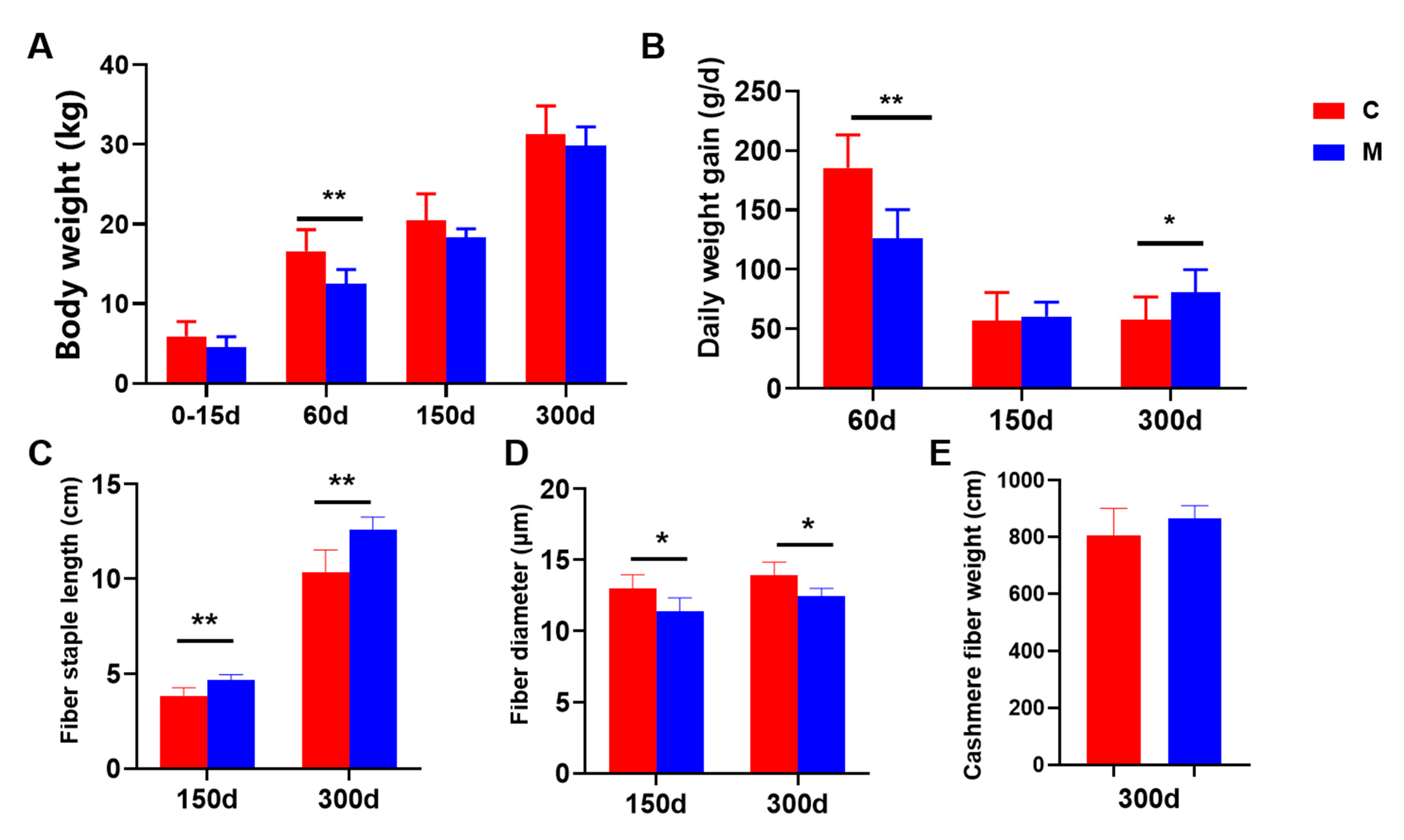
3.1. Liaoning Cashmere Goats’ Production Performance

The effects of melatonin implantation on the production performance of 0–15-day-old, 60-day-old, 150-day-old, and 300-day-old Liaoning cashmere goats are presented in Figure 1. The body weight of Liaoning cashmere goats showed a declining trend following melatonin administration, and by 60 days, both the daily weight gain and body weight had significantly decreased (p < 0.05). The daily weight gain increased significantly at 300 days (p < 0.05), and there was no significant effect in the other time periods. The length of the goats’ cashmere increased significantly (p < 0.05), and the fineness of the goats’ cashmere decreased significantly following melatonin administration (p < 0.05); the overall weight of the cashmere did not significantly change.

3.2. Apparent Nutritional Digestion

To investigate the effects of melatonin on the apparent nutritional digestion of Liaoning cashmere goats, nitrogen metabolism, energy metabolism, and nutrients’ apparent digestibility were determined. The intake and fecal nitrogen content of 150- and 300-day-old Liaoning jersey goats were not significantly affected by melatonin (p > 0.05), but the urine nitrogen content was significantly reduced (p < 0.05) (Table 2). The deposited nitrogen of the 150-day-old velvet goats in the melatonin group was significantly higher than that of the control group by 14.79% (p < 0.05), the urine nitrogen/feeding nitrogen of the 300-day-old cashmere goats in the control group was significantly higher than that of the melatonin group by 20.13% (p < 0.05), and the deposited nitrogen/feeding nitrogen of the melatonin group was significantly higher than that of the control group by 16.54% (p < 0.05) (Table 2). In 150-day-old Liaoning velvet goats, melatonin significantly (p < 0.05) reduced urine energy; no significant effect was observed on the other aspects of energy metabolism (p > 0.05) (Table 3). Melatonin implantation did not significantly affect the dry matter intake (p > 0.05) or the apparent digestibility of nutrients (ether extract, crude protein, crude fiber, neutral detergent fiber, and acid detergent fiber) in 150-day and 300-day-old Liaoning cashmere goats (p > 0.05) (Table 4).

3.3. Blood Biochemical Parameters

The effect of melatonin implantation on the blood biochemical parameters of Liaoning cashmere goats is presented in Figure 2. Melatonin implantation significantly improved blood antioxidant activity in 150- and 300-day-old Liaoning cashmere goats. In the melatonin group, plasma T-AOC, CAT, and GSH-PX were significantly increased (p < 0.05), and MDA was significantly decreased (p < 0.05). Plasma creatinine content tended to decrease with melatonin treatment administration, but not significantly (p > 0.05). Plasma BUN content increased significantly in the 300-day-old cashmere goat melatonin group (p < 0.05). Plasma liver injury indexes tended to decrease in 150-day-old and 300-day-old cashmere goats, and plasma GOT, GPT, and AKP decreased significantly (p < 0.05) during the effective slow-release time of melatonin (150 days old) (Figure 2).

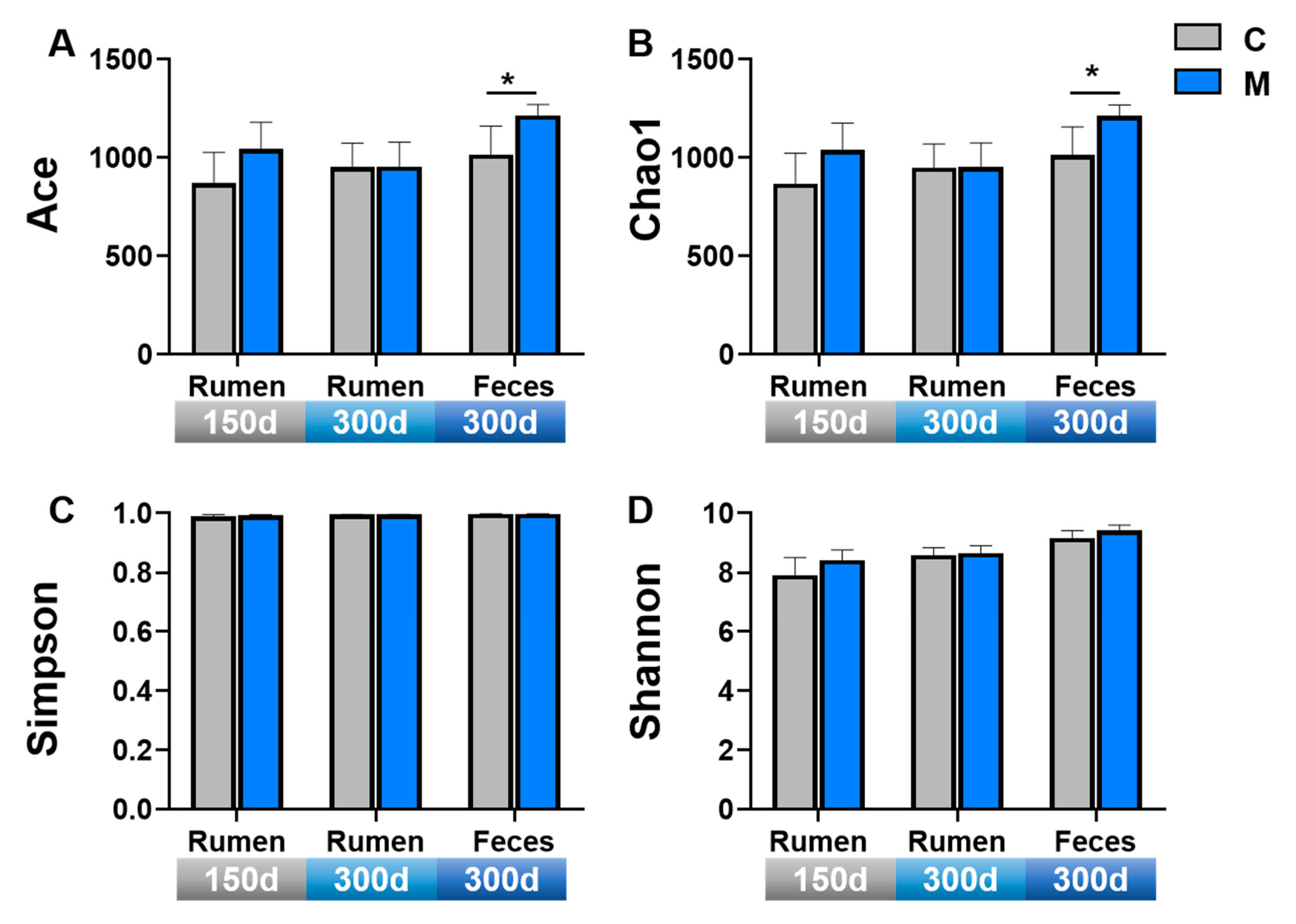
3.4. Gastrointestinal Microbiota Diversity

The α-diversity of microorganisms in the rumen fluid and rectal feces of cashmere goats are presented in Figure 3. Melatonin implantation increased the Ace index, Chao1 index, and Shannon index of rumen fluid in cashmere goats at 150 days. At 300 days, melatonin administration significantly increased the microbial Ace index and Chao1 index of rectal feces (p < 0.05) (Figure 3).
To determine whether the gastrointestinal tract microbial community structure in cashmere goats differed between the control and melatonin groups, principal coordinate analysis (PCoA) was performed. Melatonin implantation did not significantly change the β-diversity of microorganisms in the rumen fluid of cashmere goats (p > 0.05) (Figure 4A,B). However, it did significantly change the β-diversity of fecal microorganisms in the rectum of 300-day-old goats, with fecal microorganisms in the melatonin and control groups exhibiting a significant separation (p < 0.05) (Figure 4C). UPGMA clustering confirmed that the control and melatonin groups were clearly separated in the feces of 300-day-old Liaoning velvet goats. At the level of fecal microbial phyla, Firmicutes and Bacteroidota were the major phyla at the level of fecal microbial phyla (Figure 4H); Prevotella and UCG-005 were the prominent genera at the genus level (Figure 4I).
The microbial composition of rumen fluid and rectal feces of 150- and 300-day-old goats is presented in Figure 5. At the phylum level, the dominant phyla of Liaoning cashmere goats’ rumen fluid were Bacteroidota, Firmicutes, Synergistota, and Proteobacteria, and the dominant phyla of Liaoning cashmere goats feces were Firmicutes and Bacteroidota (Figure 5A,C,E). At the genus level, the dominant genera in the rumen fluid of 150-day-old Liaoning cashmere goats were prevotella, uncultured_rumen _bacterium, Quinella, Rikenellaceae_ RC9_gut_group, Fretibacterium, and Succiniclasticum (Figure 5B). The dominant genera of rumen fluid in 300-day-old Liaoning cashmere goats were prevotella, uncultured_rumen_bacterium, Succiniclasticum, Rikenellaceae_ RC9_gut_group, Butyrivibrio, and Ruminococcus (Figure 5D). The dominant fecal bacteria genera of 300-day-old Liaoning cashmere goats were UCG_010, UCG_005, unclassified_Lachnospiraceae, Rikenellaceae_RC9_gut_group, unclassified_[Eubacterium]_coprostanoligenes_group, and Alistipes (Figure 5F).
At the phylum level of the fecal microbiome, Lefse analysis revealed a significant increase in the relative abundance of Myxococcota, Acidobacteria, and Patescibacteria in the melatonin group (Figure 6A). Microbial genus levels in the rumen fluid of 150-day-old Liaoning cashmere goats showed a significant increase in uncultured rumen bacteria, unclassified Synergistaceae, and unclassified Bacteroids__BS11_gut_group in the melatonin group (p < 0.05) (Figure 6B), which showed a significant correlation with antioxidant activity, nitrogen deposition, liver and kidney function, and cashmere production performance (Figure 6E). The relative abundance of Prevotellaceae_UCG_001 in rumen fluid of 300-day-old Liaoning cashmere goats was significantly increased (p < 0.05), and was positively correlated with cashmere production performance, antioxidant activity, and liver and kidney function (Figure 6F). The relative abundances of unclassified_ Absconditabacteriales_SR1 and Eubacterium_ylanophilum_group were significantly decreased (p < 0.05) (Figure 6C). Microbial genus levels in Liaoning cashmere goat feces at 300 days of age and the relative abundance of Ruminococcus, unclassified_Myxococcaceae, unclassifed_ Bacteroidales, and Anaerofustis were significantly increased (p < 0.05) (Figure 6D). These were positively correlated with cashmere production performance, antioxidant activity, liver function, and kidney function (Figure 6F).

4. Discussion

4.1. Effect of Melatonin Implantation on Production Performance of Liaoning Cashmere Goats

Animal body weight and average daily weight gain are direct indicators of growth and economic performance. Cashmere growth is an important component in establishing the economic value of cashmere goats, and it is also indicative of overall growth and development in goats. The results of this study demonstrated that in the early period (0–60 d), melatonin administration significantly reduced body weight and daily weight gain in cashmere goats. Over the entire study period (0–300 days), melatonin had no significant influence on body weight or daily weight gain. The cashmere length of goats increased significantly at 150 and 300 days of age, the fineness decreased significantly, and the cashmere production had an upward trend. Recent research indicates that administering lambs a subcutaneous 18 mg melatonin implant at the base of their left ear did not significantly change these lambs’ body weight at weaning or ADG as a result of the melatonin administration [24]. Melatonin reduced the Inner Mongolian cashmere goats’ daily body weight after it was injected once or twice at a dose of 2 mg/kg, though the effect was not statistically significant [1]. Melatonin has been shown to reduce body weight and low-grade inflammation while also improving insulin resistance in mice fed a high-fat diet (HFD) [25]. Further investigations have demonstrated that melatonin enhances thermogenesis in intramuscular adipocytes by promoting mitochondrial biogenesis and increasing mitochondrial respiratory activity [26]. Furthermore, melatonin is essential for the proper synthesis, secretion, and action of insulin. It plays a critical role in establishing an appropriate energy balance by regulating energy flux to and from storage sites, directly modulating energy expenditure via the activation of brown adipose tissue, and facilitating the browning of white adipose tissue [27]. Therefore, we hypothesize that melatonin reduces body weight and stimulates cashmere growth in young goats, potentially due to its role in redistributing energy and protein resources toward cashmere production.
Over the past decade, numerous studies have confirmed that melatonin promotes secondary hair follicle proliferation in cashmere goats, reduces cashmere fiber diameter, and enhances overall cashmere quality [1,2,28,29]. Early studies demonstrated that melatonin increased cashmere production and decreased cashmere fineness by inducing a combination of cashmere-growth and cashmere-non-growth periods in adult goats [1]. Subsequent findings indicated that melatonin administration in young goats could reduce oxidative stress damage, inhibit hair follicle cell apoptosis, promote skin secondary hair follicle morphogenesis and maturation, and increase secondary hair follicle number by improving body antioxidants and reducing oxidative stress damage, thus reducing cashmere fineness, increasing cashmere yield, and improving fleece production performance [2]. In recent years, novel mechanisms have been identified (e.g., the MAPK and Keap1-Nrf2 pathways) that mediate melatonin’s role in regulating secondary hair follicle genesis and development in adult and juvenile velvet goats, respectively [28,29]. All these findings are consistent with this study and indirectly support the role of melatonin in promoting the proliferation of secondary hair follicle numbers, decreasing cashmere fineness, and increasing cashmere production.

4.2. Effect of Melatonin Implantation on Liaoning Cashmere Goats’ Apparent Nutritional Digestion

The digestion of nutrients is indicative of livestock feed utilization efficiency and overall growth and development, and protein is an important nutrient not only for a cashmere goat’s health, but also for the high efficiency of production performance [30]. This study found that melatonin significantly improved deposited nitrogen in cashmere goats, while melatonin administration greatly reduced urine nitrogen content and urinary energy, without significantly affecting the digestion of other nutrients. Melatonin implantation and light time manipulation were used in studies on Inner Mongolia velvet goats to examine the effects of melatonin on nitrogenous material distribution and cashmere production performance. The study indicated that during the cashmere-non-growth periods, short light exposure and melatonin implantation enhanced nitrogen digestibility and deposition. Under short light, long light, and natural light conditions, cashmere goats’ nitrogen intake increased sequentially. Nitrogen apparent digestion was highest under short light conditions, followed by long and natural light, with a corresponding sequential increase in urine nitrogen content. Nitrogen deposition increased as the duration of light exposure decreased [31]. This study also discovered that extended light exposure promoted the deposition of dietary nitrogen toward body protein, while short light exposure and melatonin implantation favored the deposition of dietary nitrogen towards cashmere protein and raised the body fat content of cashmere goats [31]. In contrast to Inner Mongolian cashmere goats under natural light conditions, a study demonstrated that light control reduced light duration, resulting in slightly lower nutrient digestibility compared to the control group midway through the light control period, and the apparent digestibility of crude protein was significantly higher than that of the control group at the end of the light control period [32]. These findings are consistent with this study, which implies that melatonin regulates energy partitioning to augment body fat content and promote the deposition of dietary nitrogen into cashmere proteins.

4.3. Effect of Melatonin Implantation on Blood Biochemical Parameters in Liaoning Cashmere Goats

Indicators of antioxidant activity in animal blood are a direct reflection of an organism’s antioxidant capacity. In this study, melatonin significantly improved the organism’s potential for antioxidants, evidenced by a considerable decrease in MDA content and a significant elevation in T-AOC, CAT, and GSH-PX levels. Melatonin is synthesized and metabolized by both human and rodent skin [33]. Melatonin’s metabolites, including AFMK, AMK, 4-hydroxymelatonin, and 2-hydroxymelatonin, have potent antioxidant properties [34]. Melatonin and its metabolites perform biological functions through receptor-dependent or non-receptor-dependent mechanisms [35]. Lower concentrations of melatonin exert biological effects by binding to receptors (MT1 and MT2) on cell membranes, whereas pharmacological concentrations of melatonin exert their antioxidant functions by promoting the expression of antioxidant enzyme genes or increasing the activity of antioxidant enzymes [36]. In the blood and skin tissue of young Inner Mongolian cashmere goats, melatonin was demonstrated to significantly increase the activity of GSH-PX and T-AOC levels while dramatically lowering MDA levels [2]. Research indicates that the induction of additional skin hair follicles can be facilitated enhanced through antioxidant supplementation, which boosts the body’s antioxidant capacity and diminishes the build-up of free radicals. The administration of selenium supplements to ewes between 30 days prior to breeding and 110 days of gestation has been shown to mitigate the build-up of free radicals and oxidative stress by enhancing the activities of antioxidant enzymes such as SOD and GSH-Px, as well as total antioxidant power, thereby encouraging the development of secondary hair follicles in cashmere goats’ skin and augmenting the quantity of secondary hair follicles in the skin following delivery [37]. These findings support the study’s hypothesis that melatonin enhances the body’s capacity to produce antioxidants; lessens the harm caused by free radicals and oxidative stress that occurs during the development of secondary hair follicles in the skin, thereby promoting their growth both quantitatively and qualitatively; and increases cashmere production.
Blood BUN levels can provide insight into the body’s nitrogen balance, while GOT, GPT, and AKP levels serve as indicators of liver health and function. The present study’s findings showed that melatonin administration significantly elevated BUN levels in blood and significantly decreased levels of GOT, GPT, and AKP. Related research has demonstrated that melatonin decreases fibrosis in the CLP (cecum ligation puncture) model and improves BUN and SCR (serum creatinine) to alleviate renal insufficiency [38]. Melatonin inhibits LPS-induced ROS accumulation and reduces mRNA levels of inflammatory factors such as Il-1α, Il-1β, Mcp-1, and Tgf-β1, reducing kidney injury by downregulating SOD2 and upregulating Nox4 [38]. Melatonin pretreatment was found to significantly increase ileal farnesol X receptor (FXR) protein expression and improve liver function, as evidenced by the reduction of ALT through the downregulation of proteins (TLR4, MyD88, p-p65, and p-IκBα) and signaling RNAs (mRNAs) linked to the TLR4/NF-κB signaling pathway in the liver of mice exposed to aflatoxin, and in reducing AST and ALP levels in mice’s livers [39]. These results are consistent with those of this study, indicating that melatonin may improve liver and kidney function by regulating the inflammatory effect and antioxidant activity of the body and improve cashmere goat health to increase cashmere production.

4.4. Effect of Melatonin Implantation on Gastrointestinal Microbiome Diversity in Liaoning Cashmere Goats

The gastrointestinal microbiome in mammals maintains a symbiotic relationship with its host and plays important roles in the host’s nutrient metabolism, resistance to pathogenic bacteria, and immune system maturation [40]. For ruminants, the ability of gastrointestinal microorganisms to break down plant fibers in feed into soluble small-molecule carbohydrates like butyric acid, propionic acid, and acetic acid enables these carbohydrates to be absorbed and metabolized by the animals, meeting their energy needs [41]. In this study, melatonin significantly increased the ACE index and CHAO1 index of the cashmere goats’ fecal microorganisms and significantly changed the β-diversity of fecal microorganisms, with a tendency observed towards increased rumen fluid microbial diversity. Related studies indicated that the addition of 0.5 g melatonin to the basal diet did not significantly affect the alpha diversity of rumen microbes in dairy cows [42]. Melatonin administration modifies colonic microbes in sleep-deprived mice, leads to an increase in beneficial bacteria such as Akkermansia, Bacteroides, and Faecalibacterium, and increases colonic microbial diversity [43]. These findings suggest that melatonin may enhance the diversity of the rectal fecal microbiome and alter gut microorganisms to enhance body health.
Furthermore, this study revealed that the predominant gastrointestinal microbial phyla in Liaoning cashmere goats at 150 and 300 days of age were Firmicutes and Bacteroidota, with Prevotella, uncultured rumen bacteria, Succiniclasticum, and Rikenellaceae_RC9_gut_group constituting the majority of the dominant genera. At the phylum level, there were distinct phyla Myxococcota, Acidobacteria, and Patescibacteria in the fecal microbiota. The relative abundance of Patescibacteria was significantly reduced in mice with colonic inflammation induced by sodium dextran sulfate (DSS) compared to the control group, and Patescibacteria has the ability to restore intestinal damage [44]. The 150-day-old cashmere goat rumen fluid showed a significantly higher relative abundance of unclassified Bacteroidales __BS11_gut_group, uncultured rumen bacteria, and unclassified Synergistaceae at the genera level, which were positively correlated with sedimentary nitrogen, cashmere length, and antioxidant indices. Gram-negative bacteria Bacteroidales are the major dietary polysaccharide metabolizers in the gut [45], uncultured_rumen_bacterium is the main genus of cellulose-degrading bacteria in the rumen of ruminants [46], and Synergistaceae is the main acetic acid-oxidizing bacterium, which also functions in amino acid fermentation [47]. The increased abundance of beneficial bacteria further enhanced nutrient uptake in cashmere goats. The relative abundance of Prevotellaceae_UCG_001 increased significantly, while unclassified_Absconditabacteriales_SR1 and Eubacterium_ylanophilum_group decreased significantly in the rumen fluid of cashmere goats at 300 days of age. Strong cellulose degradation abilities allow Prevotellaceae to break down complex carbohydrates and plant cellulose, produce short-chain fatty acids and other advantageous metabolites, and aid in nitrogen utilization, ammonia metabolism, and the maintenance of nitrogen balance and microecological stability in the gastrointestinal tract [48]. Absconditaacteriales were significantly enriched in chronic gastritis [49]. In 300-day-old cashmere goat feces, the relative abundance of Ruminococcus, unclassified_Myxococcaceae, unclassifed_Bacteroidales, and Anaerofustis increased significantly. Ruminococcus is rich in propionic acid and butyric acid, which are involved in dietary digestion and the maintenance of gastrointestinal barrier function [50]. Anaerofustis was significantly positively linked with the expression of ileal barrier-related genes [51]. These results indicated that melatonin enhances cashmere quality and yield by improving the gastrointestinal microbiota in Liaoning cashmere goats.

5. Conclusions

The findings of this study suggest that melatonin implantation enhances liver and kidney metabolism, antioxidant capacity, and nitrogen metabolism in cashmere goats by promoting the richness and diversity of the gastrointestinal microbiota. Specifically, the increased abundance of beneficial microbial genera, including Prevotella, Lachnospiraceae, Ruminococcus, and Synergistaceae, contributes to improved feed digestion and nutrient absorption, as well as enhanced systemic antioxidant activity and metabolic functions of the liver and kidneys. Notably, significant increases were observed in plasma levels of T-AOC, CAT, and GSH-PX, while levels of MDA, GOT, GPT, and AKP were markedly reduced. Consequently, these physiological improvements translate into enhanced cashmere quality and higher yield. These findings offer a theoretical basis for understanding the mechanisms by which melatonin boosts cashmere production through the regulation of gastrointestinal microbiota.

Author Contributions

Z.Z. and D.H.: writing—original draft, writing—review and editing, data curation, formal analysis, and methodology. Z.S.: software, validation, and visualization. L.H.: conceptualization and investigation. W.Z.: writing—review and editing, funding acquisition, project administration, and methodology. All authors have read and agreed to the published version of the manuscript.

Funding

This research was funded by the earmarked fund for China Agriculture Research System-39 (CARS-39).

Institutional Review Board Statement

All animal experiments in this study were approved by the Animal Welfare Committee of the Agricultural Research Organization, China Agricultural University (Approval no.: AW11703202-1-1).

Data Availability Statement

All raw data and sequencing information can be requested by contacting the corresponding author Wei Zhang (wzhang@cau.edu.cn).

Conflicts of Interest

The authors declare no conflicts of interest.

References

  1. Duan, C.; Xu, J.; Sun, C.; Jia, Z.; Zhang, W. Effects of melatonin implantation on cashmere yield, fibre characteristics, duration of cashmere growth as well as growth and reproductive performance of Inner Mongolian cashmere goats. J. Anim. Sci. Biotechnol. 2015, 6, 22. [Google Scholar] [CrossRef] [PubMed]
  2. Yang, C.H.; Xu, J.H.; Ren, Q.C.; Duan, T.; Mo, F.; Zhang, W. Melatonin promotes secondary hair follicle development of early postnatal cashmere goat and improves cashmere quantity and quality by enhancing antioxidant capacity and suppressing apoptosis. J. Pineal Res. 2019, 67, e12569. [Google Scholar] [CrossRef]
  3. Wang, W.; Ye, L.; Dou, X.; Liu, H.; Han, D. Effects of Rumen-Protected Methionine Supplementation on Growth Performance, Nutrient Digestion, Nitrogen Utilisation and Plasma Amino Acid Profiles of Liaoning Cashmere Goats. Animals 2023, 13, 2995. [Google Scholar] [CrossRef] [PubMed]
  4. Trotta, R.J.; Lemley, C.O.; Vonnahme, K.A.; Swanson, K.C. Effects of nutrient restriction and melatonin supplementation from mid-to-late gestation on maternal and fetal small intestinal carbohydrase activities in sheep. Domest. Anim. Endocrinol. 2021, 74, 106555. [Google Scholar] [CrossRef] [PubMed]
  5. Lemley, C.O.; Camacho, L.E.; Meyer, A.M.; Kapphahn, M.; Caton, J.S.; Vonnahme, K.A. Dietary melatonin supplementation alters uteroplacental amino acid flux during intrauterine growth restriction in ewes. Anim. Int. J. Anim. Biosci. 2013, 7, 1500–1507. [Google Scholar] [CrossRef]
  6. Cao, Y.; Feng, T.; Wu, Y.; Xu, Y.; Du, L.; Wang, T.; Luo, Y.; Wang, Y.; Li, Z.; Xuan, Z.; et al. The multi-kingdom microbiome of the goat gastrointestinal tract. Microbiome 2023, 11, 219. [Google Scholar] [CrossRef] [PubMed]
  7. Chai, J.; Zhuang, Y.; Cui, K.; Bi, Y.; Zhang, N. Metagenomics reveals the temporal dynamics of the rumen resistome and microbiome in goat kids. Microbiome 2024, 12, 14. [Google Scholar] [CrossRef]
  8. Ma, L.; Tao, S.; Song, T.; Lyu, W.; Li, Y.; Wang, W.; Shen, Q.; Ni, Y.; Zhu, J.; Zhao, J.; et al. Clostridium butyricum and carbohydrate active enzymes contribute to the reduced fat deposition in pigs. iMeta 2024, 3, e160. [Google Scholar] [CrossRef] [PubMed]
  9. Dalile, B.; Van Oudenhove, L.; Vervliet, B.; Verbeke, K. The role of short-chain fatty acids in microbiota-gut-brain communication. Nature reviews. Gastroenterol. Hepatol. 2019, 16, 461–478. [Google Scholar]
  10. Yu, Y.; Yang, J.; Zheng, L.Y.; Sheng, Q.; Li, C.Y.; Wang, M.; Zhang, X.Y.; McMinn, A.; Zhang, Y.Z.; Song, X.Y.; et al. Diversity of D-Amino Acid Utilizing Bacteria from Kongsfjorden, Arctic and the Metabolic Pathways for Seven D-Amino Acids. Front. Microbiol. 2020, 10, 2983. [Google Scholar] [CrossRef]
  11. Torrallardona, D.; Harris, C.I.; Fuller, M.F. Lysine synthesized by the gastrointestinal microflora of pigs is absorbed, mostly in the small intestine. American journal of physiology. Endocrinol. Metab. 2003, 284, E1177–E1180. [Google Scholar]
  12. Metges, C.C. Contribution of microbial amino acids to amino acid homeostasis of the host. J. Nutr. 2000, 130, 1857s–1864s. [Google Scholar] [CrossRef] [PubMed]
  13. Wu, T.; Liang, J.; Wang, T.; Zhao, R.; Ma, Y.; Gao, Y.; Zhao, S.; Chen, G.; Liu, B. Cysteamine-supplemented diet for cashmere goats: A potential strategy to inhibit rumen biohydrogenation and enhance plasma antioxidant capacity. Front. Vet. Sci. 2022, 9, 997091. [Google Scholar] [CrossRef]
  14. Bubenik, G.A.; Brown, G.M.; Grota, L.J. Immunohistological localization of melatonin in the rat digestive system. Experientia 1977, 33, 662–663. [Google Scholar] [CrossRef]
  15. Liu, Y.; Li, L.; Feng, J.; Wan, B.; Tu, Q.; Cai, W.; Jin, F.; Tang, G.; Rodrigues, L.R.; Zhang, X.; et al. Modulation of chronic obstructive pulmonary disease progression by antioxidant metabolites from Pediococcus pentosaceus: Enhancing gut probiotics abundance and the tryptophan-melatonin pathway. Gut Microbes 2024, 16, 2320283. [Google Scholar] [CrossRef] [PubMed]
  16. Zheng, J.; Zhou, Y.; Zhang, D.; Ma, K.; Gong, Y.; Luo, X.; Liu, J.; Cui, S. Intestinal melatonin levels and gut microbiota homeostasis are independent of the pineal gland in pigs. Front. Microbiol. 2024, 15, 1352586. [Google Scholar] [CrossRef]
  17. Liu, W.; Huang, Z.; Zhang, Y.; Zhang, S.; Cui, Z.; Liu, W.; Li, L.; Xia, J.; Zou, Y.; Qi, Z. ASMT determines gut microbiota and increases neurobehavioral adaptability to exercise in female mice. Commun. Biol. 2023, 6, 1126. [Google Scholar] [CrossRef] [PubMed]
  18. Abdelsattar, M.M.; Vargas-Bello-Pérez, E.; Zhuang, Y.; Fu, Y.; Zhang, N. Impact of dietary supplementation of β-hydroxybutyric acid on performance, nutrient digestibility, organ development and serum stress indicators in early-weaned goat kids. Anim. Nutr. 2021, 9, 16–22. [Google Scholar] [CrossRef]
  19. Chen, L.; Sun, J.; Pan, Z.; Lu, Y.; Wang, Z.; Yang, L.; Sun, G. Analysis of Chemical Constituents of Chrysanthemum morifolium Extract and Its Effect on Postprandial Lipid Metabolism in Healthy Adults. Molecules 2023, 28, 579. [Google Scholar] [CrossRef]
  20. Fabiny, D.L.; Ertingshausen, G. Automated reaction-rate method for determination of serum creatinine with the CentrifiChem. Clin. Chem. 1971, 17, 696–700. [Google Scholar] [CrossRef]
  21. Henry, R.J. Clinical Chemistry, Principles and Techniques, 2nd ed.; Harper and Row: Hagerstown, MD, USA, 1974. [Google Scholar]
  22. Reitman, S.; Frankel, S. A colorimetric method for the determination of serum glutamic oxalacetic and glutamic pyruvic transaminases. Am. J. Clin. Pathol. 1957, 28, 56–63. [Google Scholar] [CrossRef] [PubMed]
  23. Ma, L.; Lyu, W.; Zeng, T.; Wang, W.; Chen, Q.; Zhao, J.; Zhang, G.; Lu, L.; Yang, H.; Xiao, Y. Duck gut metagenome reveals the microbiome signatures linked to intestinal regional, temporal development, and rearing condition. Imeta 2024, 3, e198. [Google Scholar] [CrossRef] [PubMed]
  24. Abecia, J.A.; Luis, S.; Canto, F. Implanting melatonin at lambing enhances lamb growth and maintains high fat content in milk. Vet. Res. Commun. 2021, 45, 181–188. [Google Scholar] [CrossRef]
  25. Xu, P.; Wang, J.; Hong, F.; Wang, S.; Jin, X.; Xue, T.; Jia, L.; Zhai, Y. Melatonin prevents obesity through modulation of gut microbiota in mice. J. Pineal Res. 2017, 62, e12399. [Google Scholar] [CrossRef]
  26. Liu, K.; Yu, W.; Wei, W.; Zhang, X.; Tian, Y.; Sherif, M.; Liu, X.; Dong, C.; Wu, W.; Zhang, L.; et al. Melatonin reduces intramuscular fat deposition by promoting lipolysis and increasing mitochondrial function. J. Lipid Res. 2019, 60, 767–782. [Google Scholar] [CrossRef] [PubMed]
  27. Cipolla-Neto, J.; Amaral, F.G.; Afeche, S.C.; Tan, D.X.; Reiter, R.J. Melatonin, energy metabolism, and obesity: A review. J. Pineal Res. 2014, 56, 371–381. [Google Scholar] [CrossRef] [PubMed]
  28. Diao, X.; Yao, L.; Duan, T.; Qin, J.; He, L.; Zhang, W. Melatonin promotes the development of the secondary hair follicles by regulating circMPP5. J. Anim. Sci. Biotechnol. 2023, 14, 51. [Google Scholar] [CrossRef] [PubMed]
  29. Diao, X.; Duan, C.; Yao, L.; Qin, J.; He, L.; Zhang, W. Melatonin Promotes the Development of Secondary Hair Follicles in Adult Cashmere Goats by Activating the Keap1-Nrf2 Signaling Pathway and Inhibiting the Inflammatory Transcription Factors NFκB and AP-1. Int. J. Mol. Sci. 2023, 24, 3403. [Google Scholar] [CrossRef]
  30. Wang, X.H.; Li, Q.; Zheng, Z.B.; Diao, X.G.; He, L.W.; Zhang, W. Supplementary Feeding of Grazing Inner Mongolian Cashmere Goats during Pregnancy-Based on “Nutrient Requirements of Cashmere Goats”. Animals 2023, 13, 473. [Google Scholar] [CrossRef]
  31. Wang, L.; Lu, D.; Sun, H.; Zhao, X.; Shan, D.; Yang, G.; Fang, L. Effects of photoperiod and melatonin on nitrogen partitioning and production performance of Inner Mongolia White Cashmere Goats. Front. Agric. China 2007, 1, 229–236. [Google Scholar] [CrossRef]
  32. Zhang, C.Z.; Sun, H.Z.; Li, S.L.; Sang, D.; Zhang, C.H.; Jin, L.; Antonini, M.; Zhao, C.F. Effects of photoperiod on nutrient digestibility, hair follicle activity and cashmere quality in Inner Mongolia white cashmere goats. Asian-Australas. J. Anim. Sci. 2019, 32, 541–547. [Google Scholar] [CrossRef] [PubMed]
  33. Slominski, A.; Tobin, D.J.; Zmijewski, M.A.; Wortsman, J.; Paus, R. Melatonin in the skin: Synthesis, metabolism and functions. Trends Endocrinol. Metab. TEM 2008, 19, 17–24. [Google Scholar] [CrossRef]
  34. Kim, T.K.; Kleszczynski, K.; Janjetovic, Z.; Sweatman, T.; Lin, Z.; Li, W.; Reiter, R.J.; Fischer, T.W.; Slominski, A.T. Metabolism of melatonin and biological activity of intermediates of melatoninergic pathway in human skin cells. FASEB J. Off. Publ. Fed. Am. Soc. Exp. Biol. 2013, 27, 2742–2755. [Google Scholar] [CrossRef]
  35. Slominski, A.T.; Semak, I.; Fischer, T.W.; Kim, T.K.; Kleszczyński, K.; Hardeland, R.; Reiter, R.J. Metabolism of melatonin in the skin: Why is it important? Exp. Dermatol. 2017, 26, 563–568. [Google Scholar] [CrossRef] [PubMed]
  36. Slominski, R.M.; Reiter, R.J.; Schlabritz-Loutsevitch, N.; Ostrom, R.S.; Slominski, A.T. Melatonin membrane receptors in peripheral tissues: Distribution and functions. Mol. Cell. Endocrinol. 2012, 351, 152–166. [Google Scholar] [CrossRef]
  37. Zhao, C.; Duan, Y.; Diao, X.; He, L.; Zhang, W. Effects of Dietary Selenium Yeast Supplementation in Pregnant Cashmere Goats on the Development of Offspring Hair Follicles. Animals 2024, 14, 477. [Google Scholar] [CrossRef] [PubMed]
  38. Chen, L.; Han, Z.; Shi, Z.; Liu, C.; Lu, Q. Melatonin Alleviates Renal Injury in Mouse Model of Sepsis. Front. Pharmacol. 2021, 12, 697643. [Google Scholar] [CrossRef] [PubMed]
  39. Liu, S.; Kang, W.; Mao, X.; Ge, L.; Du, H.; Li, J.; Hou, L.; Liu, D.; Yin, Y.; Liu, Y.; et al. Melatonin mitigates aflatoxin B1-induced liver injury via modulation of gut microbiota/intestinal FXR/liver TLR4 signaling axis in mice. J. Pineal Res. 2022, 73, e12812. [Google Scholar] [CrossRef]
  40. Ridlon, J.M.; Harris, S.C.; Bhowmik, S.; Kang, D.J.; Hylemon, P.B. Consequences of bile salt biotransformations by intestinal bacteria. Gut Microbes 2016, 7, 22–39. [Google Scholar] [CrossRef]
  41. Monteiro, H.F.; Zhou, Z.; Gomes, M.S.; Peixoto, P.M.G.; Bonsaglia, E.C.R.; Canisso, I.F.; Weimer, B.C.; Lima, F.S. Rumen and lower gut microbiomes relationship with feed efficiency and production traits throughout the lactation of Holstein dairy cows. Sci. Rep. 2022, 12, 4904. [Google Scholar] [CrossRef]
  42. Fu, Y.; Yao, S.; Wang, T.; Lu, Y.; Han, H.; Liu, X.; Lv, D.; Ma, X.; Guan, S.; Yao, Y.; et al. Effects of melatonin on rumen microorganisms and methane production in dairy cow: Results from in vitro and in vivo studies. Microbiome 2023, 11, 196. [Google Scholar]
  43. Gao, T.; Wang, Z.; Dong, Y.; Cao, J.; Lin, R.; Wang, X.; Yu, Z.; Chen, Y. Role of melatonin in sleep deprivation-induced intestinal barrier dysfunction in mice. J. Pineal Res. 2019, 67, e12574. [Google Scholar] [CrossRef] [PubMed]
  44. Zheng, J.; Li, H.; Zhang, P.; Yue, S.; Zhai, B.; Zou, J.; Cheng, J.; Zhao, C.; Guo, D.; Wang, J. Paeonol Ameliorates Ulcerative Colitis in Mice by Modulating the Gut Microbiota and Metabolites. Metabolites 2022, 12, 956. [Google Scholar] [CrossRef]
  45. Park, S.Y.; Rao, C.; Coyte, K.Z.; Kuziel, G.A.; Zhang, Y.; Huang, W.; Franzosa, E.A.; Weng, J.K.; Huttenhower, C.; Rakoff-Nahoum, S. Strain-level fitness in the gut microbiome is an emergent property of glycans and a single metabolite. Cell 2022, 185, 513–529.e521. [Google Scholar] [CrossRef]
  46. Fakih, I.; Got, J.; Robles-Rodriguez, C.E.; Siegel, A.; Forano, E.; Muñoz-Tamayo, R. Dynamic genome-based metabolic modeling of the predominant cellulolytic rumen bacterium Fibrobacter succinogenes S85. mSystems 2023, 8, e0102722. [Google Scholar] [CrossRef] [PubMed]
  47. Meng, X.; Cao, Q.; Sun, Y.; Huang, S.; Liu, X.; Li, D. 16S rRNA genes- and metagenome-based confirmation of syntrophic butyrate-oxidizing methanogenesis enriched in high butyrate loading. Bioresour. Technol. 2022, 345, 126483. [Google Scholar] [CrossRef]
  48. Zhao, C.; Hu, X.; Qiu, M.; Bao, L.; Wu, K.; Meng, X.; Zhao, Y.; Feng, L.; Duan, S.; He, Y.; et al. Sialic acid exacerbates gut dysbiosis-associated mastitis through the microbiota-gut-mammary axis by fueling gut microbiota disruption. Microbiome 2023, 11, 78. [Google Scholar] [CrossRef]
  49. Chen, Y.; Xia, S.Y.; Ru, F.X.; Feng, J.J.; Tao, J.; Wei, Z.Y.; Li, X.; Qian, C.; Lin, Q.; Chen, J.H. Gastric juice microbiota in pediatric chronic gastritis that clinically tested positive and negative for Helicobacter pylori. Front. Microbiol. 2023, 14, 1112709. [Google Scholar] [CrossRef]
  50. Crost, E.H.; Coletto, E.; Bell, A.; Juge, N. Ruminococcus gnavus: Friend or foe for human health. FEMS Microbiol. Rev. 2023, 47, 14. [Google Scholar] [CrossRef]
  51. Song, Y.; Cui, Y.; Wang, Y.; Yu, J.; Wang, B.; Wen, Q.; Zheng, X. Donor selection for fecal bacterial transplantation and its combined effects with inulin on early growth and ileal development in chicks. J. Appl. Microbiol. 2023, 134, 99. [Google Scholar] [CrossRef]
Figure 1. Effect of melatonin implantation on production performance in 150- and 300-day-old Liaoning cashmere goats. (A) Body weights of Liaoning cashmere goats in control and melatonin groups at 0–15 d, 60 d, 150 d, and 300 d. (B) Daily weight gain of Liaoning cashmere goats at 60 d, 150 d, and 300 d. (C) Cashmere length of Liaoning cashmere goats at 150 d and 300 d. (D) Cashmere diameter of Liaoning cashmere goats at 150 d and 300 d. (E) Cashmere fiber weight of Liaoning cashmere goats. Data are expressed as mean ± SD (n = 15) and were analyzed by Student’s t-test. * p < 0.05; ** p < 0.01. C, control group; M, melatonin group.
Agriculture 14 01983 g001
Figure 2. Effect of melatonin implantation on blood biochemical parameters in 150 and 300 day old Liaoning cashmere goats. (A) Triglyceride, TG; (B) Total cholesterol, T-CHO; (C) Low density lipoprotein-cholesterol, LDL-C; (D) High density lipoprotein-cholesterol, HDL-C; (E) Creatinine, CRE; (F) Blood Urea Nitrogen, BUN; (G) Glutamic oxaloacetic transaminase, GOT; (H) Glutamic pyruvic transaminase, GPT; (I) Alkaline phosphatase, AKP. Data was expressed as mean ± SEM (n = 6) and analyzed by Student’s t-test. * p < 0.05, ** p < 0.01.
Agriculture 14 01983 g002
Figure 3. Microbial alpha diversity includes indicators of species richness ((A) Ace index; (B) Chao1 index) and species evenness ((C) Simpson index; (D) Shannon index). CR, rumen fluid microbiome in control group; MR, rumen fluid microbiome in melatonin group; CF, rectal feces microbiome in control group; MF, rectal feces microbiome in melatonin group. Data are expressed as mean ± SD (n = 6) and were analyzed by Student’s t-test. * p < 0.05; C, control group; M, melatonin group.
Agriculture 14 01983 g003
Figure 4. Principal component analysis (PCoA) of gastrointestinal microorganisms in Liaoning cashmere goats based on the Bray–Curtis distances method. (A) PCoA analysis of rumen fluid in 150-day-old Liaoning cashmere goats; (B) PCoA analysis of rumen fluid in 300-day-old Liaoning cashmere goats; (C) PCoA analysis of rectal feces in 300-day-old Liaoning cashmere goats. (DI) Gastrointestinal tract microorganisms of Liaoning cashmere goats were analyzed by arithmetic mean unweighted pair group method with arithmetic mean (UPGMA) at the phylum (D,F,H) level and genus (E,G,I) level.
Agriculture 14 01983 g004
Figure 5. The gastrointestinal bacterial community of the Liaoning cashmere goats at the phylum and genus levels. (A,B) Composition of rumen fluid microbial phylum (A) and genus (B) levels in 150-day-old Liaoning cashmere goats in control and melatonin groups (TOP10). (C,D) Composition of rumen fluid microbial phylum (C) and genus (D) levels in 300-day-old Liaoning cashmere goats in control and melatonin groups (TOP10). (E,F) Composition of feces microbial phylum (E) and genus (F) levels in 300-day-old Liaoning cashmere goats in control and melatonin groups (TOP10).
Agriculture 14 01983 g005
Figure 6. Microbial Lefse analysis of differences in phylum and genus levels of gastrointestinal microbiota in Liaoning cashmere goats (LDA > 2, p < 0.05). (A) Microbial Lefse analysis of differences in fecal microbiota phylum levels in Liaoning cashmere goats at 300 days of age. (B,C) Microbial Lefse analysis of differences in rumen fluid levels in Liaoning cashmere goats at 150 (B) and 300 (C) days of age. (D) Microbial Lefse analysis of differences in fecal genus levels in 300-day-old Liaoning cashmere goats. (E,F) Correlation between microbiological and phenotypic characteristics of variations in the control and melatonin groups of 150- (F) and 300-day-old (E) goats.
Agriculture 14 01983 g006
Table 1. Ingredients and nutritional level of basal diets (DM basis).
Ingredient(%)Nutrient Levels 2
Alfalfa35.00ME, (MJ/Kg)9.69
Peanut straw35.00DM, (%)86.99
Corn16.00EE, (%)3.62
Soybean meal9.50CP, (%)14.80
Fermented soybean meal3.50Ca, (%)1.00
Dicalcium phosphate0.10P, (%)0.32
Salt0.50NDF, (%)24.66
Premix 10.30ADF, (%)47.38
Total100.00ASH, (%)6.70
Ingredient(%)Nutrient levels
1 Premix: FeSO4·7H2O 170 g/kg; CuSO4·5H2O 70 g/kg; MnSO4·5H2O 290 g/kg; ZnSO4·7H2O 240 g/kg; CoCl2·6H2O 510 mg/kg; KI 220 mg/kg; Na2SeO3 130 mg/kg; VA 1, 620,000 IU/kg; VD3 324,000 IU/kg; VE 540 IU/kg; VK3 150 mg/kg; VB1 60 mg/kg; VB2 450 mg/kg; VB12 0.9 mg/kg. 2 Nutrients are measured values except for metabolizable energy, which is calculated. ME, metabolizable energy; DM, dry matter; CP, crude protein; EE, ether extract; ADF, acid detergent fiber; NDF, neutral detergent fiber; Ca, calcium; P, phosphorus; ASH, Ashcontent.
Table 2. Effects of melatonin implantation on nitrogen metabolism of Liaoning cashmere goats.
Item150 d300 d
ControlMelatoninp-ValueControlMelatoninp-Value
Intake N (g/d)16.50 ± 0.7716.19 ± 0.360.405426.46 ± 2.0824.62 ± 1.190.1264
Feces N (g/d)4.38 ± 0.393.89 ± 0.600.14176.05 ± 0.625.73 ± 0.820.5461
Urine N (g/d)4.48 ± 0.83 a3.54 ± 0.48 b0.04037.85 ± 0.64 a6.13 ± 0.99 b0.0199
Digestible N (g/d)12.12 ± 0.8812.30 ± 0.640.696020.40 ± 1.8718.89 ± 1.300.6960
Retention N (g/d)7.64 ± 0.22 b8.77 ± 1.01 a0.031612.54 ± 1.7512.76 ± 1.180.8296
FN/NI (%)26.60 ± 2.7823.00 ± 3.660.216720.30 ± 1.9123.30 ± 3.400.2167
UN/NI (%)26.98 ± 4.21 a21.90 ± 3.30 b0.046429.84 ± 3.03 a24.84 ± 3.59 b0.0425
RN/NI (%)46.42 ± 2.17 b54.10 ± 5.74 a0.015947.21 ± 3.8451.86 ± 4.430.8296
NAD (%)73.40 ± 2.7876.00 ± 3.600.216777.04 ± 2.1376.70 ± 3.400.8713
FN, Feces Nitrogen. UN, urine nitrogen. NI, nitrogen intake. RN, Retention Nitrogen. NAD, nitrogen apparent digestion. Data are expressed as mean ± SD (n = 8) and were analyzed by using an unpaired two-tailed Student’s t-test; the different superscript letters in the table represent a significant difference (p < 0.05).
Table 3. Effects of melatonin implantation on energy metabolism of Liaoning cashmere goats.
Item150 d300 d
ControlMelatoninp-ValueControlMelatoninp-Value
GE intake (MJ/d)11.92 ± 0.5111.84 ± 0.370.892919.19 ± 0.4618.53 ± 0.100.5431
FE (MJ/d)3.85 ± 0.353.28 ± 0.530.31265.24 ± 0.104.84 ± 0.160.3743
UE (MJ/d)0.19 ± 0.06 a0.12 ± 0.03 b0.04440.42 ± 0.020.36 ± 0.090.3113
DE (MJ/d)8.34 ± 0.478.55 ± 0.560.709613.95 ± 0.4213.68 ± 0.190.7891
ME (MJ/d)8.15 ± 0.458.43 ± 0.580.635913.53 ± 0.4113.32 ± 0.180.8107
GE, Gross Energy. FE, Feces Energy. UE, urine energy. DE, Digestion Energy. ME, Metabolism Energy. Data are expressed as mean ± SD (n = 8) and were analyzed by using an unpaired two-tailed Student’s t-test; the different superscript letters in the table represent a significant difference (p < 0.05).
Table 4. Effects of melatonin implantation on nutrient digestibility of Liaoning cashmere goats.
Item150 d300 d
ControlMelatoninp-ValueControlMelatoninp-Value
Dry matter
Intake (g/d)699.14 ± 38.76676.88 ± 21.380.25501155.02 ± 27.331114.82 ± 6.210.5351
Digestive bulk (g/d)481.92 ± 38.91473.60 ± 33.300.6978849.77 ± 25.76830.83 ± 11.250.7787
Digestibility (%)68.89 ± 2.7069.97 ± 4.510.646673.36 ± 0.6974.51 ± 0.850.6566
Organic matter
Intake (g/d)651.95 ± 36.20631.06 ± 20.010.25321074.68 ± 25.511037.55 ± 5.760.5423
Digestive bulk (g/d)464.57 ± 36.80451.10 ± 26.030.4642808.22 ± 24.09789.80 ± 9.910.7532
Digestibility (%)71.23 ± 2.5671.50 ± 3.810.896175.00 ± 0.6776.11 ± 0.800.6461
Ether extract
Intake (g/d)28.06 ± 1.3227.02 ± 0.850.143028.98 ± 0.6427.82 ± 0.170.4503
Digestive bulk (g/d)24.02 ± 1.4022.55 ± 1.130.080422.90 ± 0.6621.90 ± 0.360.5651
Digestibility (%)85.57 ± 2.2183.45 ± 3.310.245578.81 ± 0.8078.76 ± 1.300.9834
Crude fiber
Intake (g/d)177.19 ± 14.02169.39 ± 7.620.2673296.71 ± 7.99289.61 ± 1.490.7036
Digestive bulk (g/d)102.3 ± 10.9995.48 ± 12.660.3668173.75 ± 7.10176.59 ± 4.780.8856
Digestibility (%)57.63 ± 3.0156.36 ± 7.070.714658.19 ± 1.2160.98 ± 1.630.5524
Neutral detergent fiber
Intake (g/d)355.69 ± 23.80342.11 ± 13.080.2576533.84 ± 13.62518.56 ± 2.700.6315
Digestive bulk (g/d)217.06 ± 6.03211.03 ± 13.520.6641338.22 ± 12.56338.26 ± 6.430.9936
Digestibility (%)61.01 ± 2.6361.65 ± 6.930.846863.03 ± 1.0465.24 ± 1.230.5538
Acid detergent fiber
Intake (g/d)245.08 ± 18.72234.34 ± 10.320.2554407.89 ± 10.83397.68 ± 2.040.6828
Digestive bulk (g/d)143.39 ± 12.65133.69 ± 18.240.3350254.87 ± 10.83397.68 ± 2.040.9035
Digestibility (%)58.58 ± 3.8657.00 ± 7.100.662662.04 ± 1.1364.90 ± 1.210.4614
Data are expressed as mean ± SD (n = 8) and were analyzed by using an unpaired two-tailed Student’s t-test.
Disclaimer/Publisher’s Note: The statements, opinions and data contained in all publications are solely those of the individual author(s) and contributor(s) and not of MDPI and/or the editor(s). MDPI and/or the editor(s) disclaim responsibility for any injury to people or property resulting from any ideas, methods, instructions or products referred to in the content.

