Review

Current Status of Haploidization in Cool-Season Grain Legume Crop Species

by
Wiktor Skrzypkowski
and
Agnieszka Kiełkowska
*
Department of Plant Biology and Biotechnology, Faculty of Biotechnology and Horticulture, University of Agriculture in Krakow, Al. 29-Listopada 54, 31-425 Krakow, Poland
*
Author to whom correspondence should be addressed.
Agriculture 2024, 14(7), 1031; https://doi.org/10.3390/agriculture14071031
Submission received: 8 May 2024 / Revised: 21 June 2024 / Accepted: 26 June 2024 / Published: 28 June 2024
(This article belongs to the Section Crop Genetics, Genomics and Breeding)

Abstract

:
Doubled haploid technology is, so far, the fastest route to induce a true homozygous state in plants. True homozygous plants are particularly important for breeders, as they can facilitate hybrid breeding and are useful in fixing traits in a breeding line. Fabaceae species are of great importance in food and feed production; however, they are far behind other families with respect to the development of effective haploidization protocols. Here, we present the most recent status of research on haploidization protocols in cool-season grain legume crops, including dry peas, chickpeas, faba beans, lentils, lupines, and grass peas. The first four species are primarily for human consumption; the latter are utilized as forage. All the mentioned species have been subject to haploidization trials; however, repeatable protocols, including the regeneration of confirmed haploid or doubled haploid plants, have not been elaborated. Research in field pea, chickpea, grass pea, and lupine is promising, with the reported regeneration of microspore-derived embryos in all four species. Repeatable plant regeneration has been reported only in field peas and chickpeas. The most recent achievements on haploidization through male and female gametophytes in faba bean are also presented. The key factors for the effective stimulation of haploid cell development in cool-season legumes are reviewed, providing a useful basis for future efforts toward haploidization in this group.

1. Introduction

The major cool-season grain legumes include dry pea (Pisum sativum L.), faba bean (Vicia faba L.), lentil (Lens culinaris Medik. subsp. culinaris), chickpea (Cicer arietinum L.), lupin (Lupinus spp.), and grass pea (Lathryus sativus L.). Dry pea, chickpea, broad bean, and lentil are the four major cool-season grain legume crops produced for human consumption. They are grown on all the continents except Antarctica [1]. The total world production of dry peas in 2001 was 10.3 million tons and rose to 12.4 million tons in 2021. The top five producers of dry peas are Canada, the Russian Federation, China, France, and India. In chickpeas, the total world production increased from 7 million tons in 2001 to 15.8 million tons in 2021, and 84% of the total world production was contributed in Asia. Vetches (genus Vicia) include edible broad bean (V. faba L.), but also common vetch (V. sativa L.), hairy vetch (V. villosa, also called fodder vetch), bard vetch (V. articulata), French vetch (V. serratifolia), and Narbon bean (V. narbonensis). Purple vetch (V. benghalensis) and Hungarian vetch (V. pannonica) are cultivated for forage and green manure. In vetches, world production increased from 3.2 million tons in 2001 to 5.6 million tons in 2021. The top five vetch producers are the Russian Federation, Ethiopia, Mexico, Turkey, and Spain. Lupin species are important for animal feed. The world production of lupins was constant over the period of 2001–2021, at approximately 1.4 million tons. The main producers of lupins are Australia, Poland, the Russian Federation, Morocco, and Chile [2]. Grass pea is used for human and animal food in countries in the Mediterranean basin, but its use is limited by the presence of the neurotoxin (oxalyldiaminopropionic acid) in its seed, responsible for lathyrism.
All the above-mentioned species form associations with specific soil bacteria (rhizobia) to produce root nodules that can convert atmospheric nitrogen into amino acids [3]. The fixation of atmospheric nitrogen gives cool-season grain legumes an advantage over most non-legume crops in low-soil nitrogen environments.
The process of gametic embryogenesis results in a plant having the gametic number of chromosomes (n) in somatic cells. Haploid itself is a weak and sterile plant; however, doubling the chromosome number of a haploid plant results in the restoration of vigor and fertility [4]. The genome duplication might be either spontaneous or induced. The spontaneous duplication of chromosomes occurs during the in vitro phase as a result of nuclear fusion and was described for paternally derived haploids mostly. For maternally derived haploids, the rate of spontaneous genome doubling is rare; therefore, an induction of this process is necessary. Chromosome doubling is induced chemically with the use of anti-microtubule drugs. The most popular anti-microtubule agent is colchicine. Its action is based on the inhibition of microtubule polymerization by binding to tubulin; however, it was proven to be highly toxic to plant tissues [4,5]. Alternatively, with colchicine, the use of trifluralin, oryzalin, or amiprophosmethyl was reported [6]. For genome doubling, antimitotic agents can be incorporated into the culture medium but their application to haploid embryos, shoots, or whole plantlets was reported [5,6,7,8,9]. Genome duplication, either spontaneous or induced, leads to the formation of a true homozygote, termed a doubled haploid plant (DH). When the DH plant is self-pollinated, the obtained seed progeny is a doubled haploid line (DH line). The main motivation for developing applicable and reproducible protocols for DH plant production in crop species is their potential utility in breeding, i.e., for the rapid fixation of desirable traits in a breeding line/variety or the facilitation of hybrid breeding [4,5]. The practical utilization of DH plants also differs between self- and cross-pollinated crops. The application of DH plants in the breeding of self-pollinated crops is based on their potential to produce true homozygous lines in one generation and perform early generation testing, which is very effective as DH possess additive variance only. Moreover, in self-pollinated crops, the DH lines with valuable traits can directly represent a new variety. The application of DH plants in cross-pollinated crops is also based on the rapid production of homozygous accessions but is also used as a replacement for inbreeding for the production of parental lines for hybrid varieties [7,8,9]. The practical application of haploid technology in breeding has been shown in several species, e.g., tobacco (Nicotiana tabacum L.), wheat (Triticum aestivum L.), barley (Hordeum vulgare L.), rapeseed (Brassica napus L.), rice (Oryza sativa L.), and maize (Zea mays L.), which has allowed to shorten the time necessary to breed new cultivars and release them on the market [5,10,11]. In addition to the above-mentioned practical applications, haploids can provide valuable material for other research such as genetic mapping, mutations, transformations, somatic hybridization, and biochemical and physiological studies [10].
Cool-seasoned leguminous species are categorized as self-pollinated. In this group, stable well performing double haploid lines with interesting morphological traits can represent new cultivars directly. The main breeding scheme in leguminous species starts with the crossings of desired genotypes, which result in hybrids that combine the chromosome sets of both parents. During gamete formation, recombination enables new gene combinations. If effective doubled haploid technology is introduced at this stage, these gene combinations will be fixed. The obtained doubled haploids will represent the recombinant products of parental genomes, however, in a completely homozygous state. They can be propagated as true breeding lines, facilitating the large-scale testing of agronomic performance over the years. Due to complete homozygosity, the efficiency of selection for both qualitative and quantitative characters is increased since recessive alleles are fixed in one generation and directly expressed [5,12].
There have been many attempts to induce haploidization in leguminous species. In 2006, Crosser et al. [12] published a review paper showing the efforts directed towards haploidization in leguminous species, and the highest success was reported for Glycine max, in which embryogenesis from microspores was observed and resulted in haploid plant regeneration [13,14]. The sporadic obtainment of haploid plants was also reported for Pisum sativum [15], Cicer arietinum [16], Medicago sativa [17,18] and Albizzia lebbeck [19]. Trials conducted for the other members of the family (including cold-seasoned legumes and arboreal legumes) resulted mainly in the obtainment of callus tissue or embryos, for which ploidy was often not analyzed, and available reports showed its various states from haploid to mixoploid [12,15]. Based on the obtained results, a whole family of leguminous species was classified as recalcitrant to haploidization [4,8,12,20]. This paper presents the most current compilation and analysis of results achieved so far in research on haploidization in cool-season grain species.

2. Origin of Haploids

2.1. In Vivo Haploidization

The development of haploid embryos in plants has been observed in several species, and cytological and histological analyses showed that they have various origins. Haploid embryos were observed in the seeds of Capsicum annuum [21] or Asparagus sp. [22] and their development was associated with polyembryony. Polyembryony refers to the simultaneous development of sexual and asexual embryos in the seed [23]. In Zea mays, haploid embryos develop through pseudogamy, which is the development of an unfertilized female gamete or cell after stimulation by the male nucleus [24]. Haploid embryos in Gossypium hirsutum and G. barbadense [25] developed through semigamy, whereby reduced male and female gametes participate in embryogenesis, but nuclear fusion does not occur; this process results in chimeral plants with sectors of maternal and paternal origin. In some species (Nicotiana, Anthirrinum), the maternal nucleus was eliminated or inactivated before the fertilization of the egg cell, and the haploid individual contained in its cells the chromosome set of the male gamete only [26]. The only legume in which in vivo haploid development pathway has been documented was the tetraploid form of Medicago sativa in which haploid parthenogenesis was observed [27]. However, the frequency of the above-mentioned phenomenon is generally extremely useful for practical purposes.

2.2. In Vitro Haploidization

To increase the effectiveness of haploid cell stimulation and haploid embryo development, a tissue culture-based approach was employed. There are two main methods for the induction of the in vitro development of the haploid cells of both male (androgenesis) and female (gynogenesis) gametophytes (Figure 1).

2.2.1. Androgenesis

Androgenesis is a process involving the development of a plant organism from male gametophyte cells. In plants, male gametophyte cells include microspores formed during microsporogenesis, and pollen representing the final stage of gametophyte formation and developed in the course of microgametogenesis. Pollen mother cells (PMCs) in anthers undergo meiosis, and four cells with a haploid number of chromosomes, called tetrads, are formed. These cells are surrounded by a thick layer of callose. Upon its breakdown, facilitated by enzymes secreted by the tapetum of the anther, individual microspores are released. Later, microspores undergo two rounds of mitoses and pollen grains are formed [28].
Androgenesis under in vitro conditions can be initiated either through anthers or isolated microspore cultures from microspores or early bicellular pollens. These cells are able to switch the developmental pathway to form multicellular structures, and as a result, haploid embryos can be formed (direct pathway, Figure 1(A)), or intensive divisions can lead to the development of callus tissue (indirect pathway, Figure 1(B)). In the first technique, anthers are isolated from floral buds and placed directly onto the induction medium. Plant regeneration can occur directly from microspore-derived embryos (direct embryogenesis) or indirectly through organogenesis involving callus tissue (indirect organogenesis). The inclusion of maternal somatic tissues (found in anthers walls) in the culture is a disadvantage of the anther culture technique. These tissues can also be induced to develop throughout the culture, which might result in regenerants having a somatic origin; therefore, often it is necessary to employ molecular markers in order to identify the origin of the regenerants. In the second technique (isolated microspore cultures), microspores are released from the anthers, purified, and suspended in a liquid culture medium. In this technique, all the development originates from haploid male gametophyte cells (direct pathway, gametic embryogenesis, Figure 1(A)).
The induction of androgenesis in plants is influenced by various factors, i.e., the growth conditions of donor plants (temperature and biotic and abiotic stress), the developmental stage of anthers, and stress factors. The optimal stage of anther development varies among different plant species and should be determined experimentally. However, the uninucleate microspore or the early binucleate stage was most commonly used for androgenesis in the majority of crops [29]. Stress factors, such as cold, heat, or osmotic stress, can be applied as a pretreatment to flower buds from which anthers/microspores will be isolated or can be delivered directly to the cultured explants. One of the most common triggers for switching the cell developmental pathway during androgenesis is temperature [30]. Studies in Brassica napus (a model plant for androgenesis) showed that the intensity of the stress and its duration must be experimentally determined, and in this species, the exposure of microspores to a temperature of 32 °C for a minimum of 8 h is essential for triggering androgenic development. In other species, i.e., triticale (× Triticosecale Wittm.) [31,32], Secale cereale [33], Capsicum annum [34,35], and Brassica oleracea L. var. italica [36,37], a positive effect on androgenesis was observed after the application of cold stress. The composition of the culture media also affects androgenesis; however, the main factor determining the response to androgenesis is genotype. Many species of crop plants, and even entire botanical families, were characterized as recalcitrant to the haploidization process. These include primarily Fabaceae, Solanaceae, and Apiaceae. Nevertheless, within these families, procedures for haploidization have been developed in recent years, and androgenetic haploids have been obtained in Solanum melongena [38], Capsicum sp. [39,40], and Daucus carota [9,41,42].

2.2.2. Gynogenesis

Gynogenesis in plants refers to embryo development from the unfertilized cells of the female gametophyte. Typically, embryos develop from the egg cell, although development through apogamy was also reported [43]. Gynogenic plants are completely of maternal origin. Gynogenesis is applied in species where androgenesis has been unsuccessful. It can be initiated by culturing unpollinated flowers, ovaries, or isolated ovules on solid media. In this method, preventing pollination and the subsequent fertilization of the egg cell is essential [4]. This is particularly problematic in self-pollinating species, such as many plants in the Solanaceae family, including tomatoes and eggplants, or a significant majority of the cultivated plants in the Fabaceae family. In such species, reproductive organs utilized to establish the in vitro culture must be collected before self-pollination, or removing the anthers or entire stamens must precede explant collection. A significant limitation of this method, similar to anther culture, is the presence of somatic tissues (i.e., ovary walls, placenta, integuments, and nucellus) surrounding the haploid cells that constitute the egg apparatus [5]. In in vitro conditions, somatic cells in these organs often undergo stimulation to intensive proliferation, leading to the formation of callus tissue or the initiation of somatic embryogenesis. In such cases, it is necessary to verify the origin of obtained regenerants [4,9]. The gynogenic plants have been successfully regenerated in Beta vulgaris [44], Cucumis sativus [45,46], Triticum sp. [47], Allium cepa [48], and even in some representatives of the Fabaceae family [49,50] which is considered recalcitrant to haploidization. In A. cepa, gynogenesis-based cultivars exhibiting heterosis have been also released on the market [11].

2.3. In Vivo-In Vitro Methods

It has been shown in several species that haploids can be induced by the stimulation of haploid cells in female gametophytes after pollination with pollen from distantly related species or by ineffective pollen. In this system, haploid embryos develop undisturbed in seeds or, due to abnormalities in endosperm development haploid embryos, have to be rescued by introduction to in vitro cultures at the early stages of their development.

2.3.1. Chromosome Elimination

After a normal double fertilization, a subsequent preferential elimination of the chromosomes of a specific genome occurs in the early stages of embryo development. During this process, the endosperm usually aborts early in the seed, and as a consequence, the haploid embryo must be rescued by in vitro culture. The most well-known example of using this method is the crossing of Hordeum vulgare (♀) with H. bulbosum (♂), where fertilization results in the formation of a hybrid embryo, followed by the successive elimination of the chromosomes of the paternal component. In consequence, an embryo is formed whose genome is solely derived from the maternal component. Named after the species, this method is known as the Bulbosum method. This very unique system besides Hordeum was also applied in some other cereals, i.e., triticale (× Triticosecale Wittm.) pollinated with Zea mays [51], rye (Secale cereale L.) pollinated with Zea mays [52], oats (Avena sativa L.) pollinated with Zea mays [53,54], and wheat (Tricticum aestivum) pollinated with maize Zea mays, Pennisetum americanum, or Oryza sativa [55,56]. However, the phenomenon of chromosome elimination after wide pollination was not observed in other species, including legumes [5,8,10,12].
The chromosomes of pollen donors in the above-mentioned crosses were eliminated several days after pollination. The mechanisms of uniparental chromosome elimination covered a difference in the timing of mitosis and asynchrony in nucleoprotein synthesis, the formation of multipolar spindles, different genome ratios, spatial separation between genomes at interphase and/or mitosis, the selective inactivation of centromeres, and the degradation of one set of chromosomes through nuclease activity [57,58].

2.3.2. Induced Parthenogenesis (Matromorphy)

As a result of specific treatments, such as pollination with foreign, inactive, or incompatible pollen, fertilization and hybrid embryo development do not occur. Instead, haploid cells within the embryo sac are stimulated to undergo divisions and form a haploid embryo. However, disruptions in endosperm development often occur, preventing in vivo embryo development and the formation of functional seeds. In such cases, it is necessary to resort to in vitro methods such as embryo rescue, which involves isolating embryos and further cultivating them on growth media [59].
The induction of parthenogenetic embryo development is possible through wide pollination (pollen of other species or genera); however, this method is very laborious (emasculation, hand pollination, pollen treatments, seed evaluation, embryo excision) [60,61]. Diploid parthenogenesis was observed in seeds resulting from crosses between Brassica oleracea and B. rapa, Eruca sativa, Raphanus sativus, Camelina sativa, and Crambe abyssinica. Plants derived from such seeds were termed matromorphs, or maternal-type plants. These organisms were identified as the potential sources of homozygous parental plants for hybrid varieties. The best results were achieved after pollinating B. oleracea var. sabauda × E. sativa (14.3 seeds per 100 pollinated buds) and B. oleracea var. capitata × R. sativus (2.9 seeds per 100 pollinated buds) [62].
The in vitro pollination of Mimulus luteus flowers with pollen from Torrenia fournieri resulted in parthenogenetic haploids with decorative values, characterized by reduced plant size and changed flower coloration [63]. Haploid parthenogenesis was also induced in cotton (Gossypium hirsutum) by pollination with pollen from Hibiscus cannabinus [64] and in Daucus carota pollinated with parsley (Petroselinum crispum) pollen [65,66] and in such cases, an embryo rescue technique was employed.
Pollination with the pollen collected from plants of different ploidy levels can also lead to the parthenogenetic development of the egg cell of the pollen acceptor. A classic example is the production of dihaploids in potatoes (Solanum tuberosum, 4x) after pollination with Solanum phureja (2x) pollen. Crosses between cultivated potato forms and their wild relatives result in hybrid and haploid seeds. To distinguish haploid seeds from hybrids, dominant marker genes in the homozygous pollinating form were utilized. Hybrid seeds had a visible spot, while dihaploid seeds lacked this spot [67]. Among the hybrid seeds obtained from crosses between diploid and tetraploid geraniums (Pelargonium zonale), approximately 4% of the seeds produced embryos that developed via parthenogenesis [68,69]. Crossing a tetraploid form of tuberous begonia (Begonia × tuberhybrida) with the diploid form Begonia semperflorens resulted in 100% maternal type F1 progeny [70]. Similarly, crossing a tetraploid form of Lilium longifolium with a diploid form of the same species allowed for the production of parthenogenetic plants from the egg cell stimulated to develop in the tetraploid form [71,72]. Haploid plants were also obtained in Citrus clementina by pollinating the pistils under in vitro conditions with triploid grapefruit pollen (C. paradisi Macf.) [73].
Parthenogenesis can also be stimulated by pollinating flowers with irradiated pollen. Typically, X-rays or gamma rays are used to deactivate the nuclear DNA in sperm cells. Pollination with pollen inactivated in this manner can lead to the development of haploid cells in the ovule sac. However, a lack of fertilization results in the absence of endosperm development, which is why this technique is also referred to as pseudo-fertilization [74]. Placing the ovules on artificial media 2–4 weeks after pollination allows for further embryo development and the formation of haploid plants [75]. Lofti and Kashi’s research [76] showed that radiation does not affect the percentage of germinating pollen grains; therefore, the pollen tubes penetrated the ovule sacs, inducing the formation of haploid embryos. About half of the pollinated flowers produced fruits, and some of the developing seeds contain embryos. Approximately 30 days after pollination, the embryos were isolated from the seeds and transferred to a suitable media for further development into plants. This method has been used to induce haploids in the Cucurbitaceae family, such as cucumber (Cucumis sativus L.) [77], melon (Cucumis melo L.) [78], watermelon (Citrullus vulgaris) [79], and pumpkin (Cucurbita pepo L.) [80], but also in other species, i.e., kiwi (Actinidia arguta) [81] and sunflower (Helianthus annuus) [82]; ornamental plants such as carnation (Dianthus caryophyllus) [74,83] and rose (Rosa x hybrida) [84]; and fruit trees such as apple (Malus communis) [85] or cherry (Prunus avium) [86].

3. Haploidization in Cool-Season Grain Legume Crops

3.1. Tribe Vicieae (Fabeae)

3.1.1. Field Pea

Field pea (Pisum sativum L. subsp. sativum var. arvense) is a diploid (2n = 14) and self-pollinated species with a very low percentage of outcrossing. Field pea is one of the most widely cultivated species belonging to the Fabaceae family worldwide, holding significant commercial and economic importance. Alongside cereals, chickpeas (Cicer arietinum), lentils (Lens culinaris), and bitter vetch (Vicia ervilia), peas were one of the first domesticated plant species, cultivated as early as the Neolithic period in the Near East [87,88,89].
P. sativum, commonly known as the garden pea, has a wide range of culinary uses. It can be consumed as immature pods and seeds, and its seeds can also be eaten dried. There are many varieties, some harvested for their green pods, while others are destined for drying. Additionally, peas are used as animal feed, providing valuable fodder. With a short growing season and minimal cultivation requirements, peas are a valuable component of crop rotation as a leguminous plant, enriching the soil with nitrogen and being particularly beneficial for low-quality soils. The largest cultivation areas of this species are found in Europe, followed closely by the Americas [90]. P. sativum contains approximately 23–25% protein in dried seeds and about 5% in fresh seed mass. Additionally, the seeds are rich in starch (about 48–51% of the dry seed mass) and fiber (approximately 6–6.8% of the dry seed mass). Moreover, they contain small amounts of fat and non-nutritive components such as minerals (including Ca, P, K, Na, Mg, Fe, Mn, Zn, and Cu) and vitamins (such as niacin, riboflavin, beta-carotene, biotin, pantothenic acid, tocopherol, and ascorbic acid), as well as phytochemicals like phenolic compounds, phytates, saponins, and oxalates [91,92,93,94].
P. sativum is a well-studied species widely used in research and regarded as one of the model species in genetic studies. All the published reports on haploidization in P. sativum are given in Table 1. The first attempts at haploidization in this species date back to the early 1970s. Gupta and his coworkers [95,96] induced androgenesis in anther cultures. In both of these studies, the anthers containing uninucleate microspores were cultured on White’s medium [97] supplemented with 2,4-dichlorophenoxyacetic acid (2,4-D) or naphthaleneacetic acid (NAA). The anthers produced mainly calli which were found to be haploid, and few embryos. The development of shoots was induced from calli and embryos, although plant regeneration was not achieved. Further studies on anther culture in P. sativum were conducted by Gosal and Bajaj [98]. Anthers were subjected to low-temperature stress (4 °C) for 72 h and cultured on MS [99] medium. They observed the development of mixoploid callus tissue from the obtained embryos but did not regenerate plants. The first successful study resulting in plant regeneration from the anther cultures of P. sativum took place in 2005 [100]. In this experiment, the researchers observed an embryogenic response after applying stress factors in the form of high temperature (37 °C) for 4 h to freshly isolated anthers. In another study, various combinations of thermal shocks, including low (4 °C) and high temperatures (35–38 °C) on androgenesis were analyzed. Isolated buds were pretreated at low temperature (4 °C), while anther cultures were subjected to higher temperatures (ranging from 35 to 38 °C). In these studies, a modified MSB medium containing MS salts and B5 [101] vitamins supplemented with 2,4-D or indole-3-butyric acid (IBA) was used. The result was an embryogenic callus from which shoots were regenerated. However, molecular analysis revealed that these shoots originated from the somatic tissue of the anthers rather than haploid cells [102,103]. Bermejo et al. [104] cultured anthers containing the uninucleate microspores of 11 different genotypes belonging to the genus Pisum, including the wild forms of P. sativum subsp. elatius and P. fulvum. They applied cold stress, keeping the flower buds at 4 °C for 3 days. Then, they cultured the anthers on MS medium supplemented with 6% sucrose and various concentrations of 2,4-D (ranging from 0 to 1 mg/L). They obtained embryogenic calli from which embryos were derived, and upon maturation, these embryos regenerated into plants. The highest callus production in all the genotypes occurred in the medium supplemented with 1 mg/L 2,4-D and in the groups that were not subjected to cold stress. Although they succeeded in regenerating plants from embryogenic calli, these plants were so weak that they did not survive acclimatization, and their ploidy analysis was not conducted; thus, their origin remains unknown.
Reports on isolated microspore cultures in P. sativum are scarce. Croser and Lulsdorf [15] cultured uninucleate microspores in a modified ML6 medium [105] supplemented with 9% sucrose. Before isolation, they applied cold stress by incubating flower buds at 4 °C for 48 h. The application of cold shock proved to be the most effective in inducing the symmetric divisions of microspores. These divisions led to the formation of multinucleate structures. These structures then underwent cellularization, leading to the formation of globular embryos, which, in some cases, continued to develop, reaching the torpedo stage. However, the regeneration of plants was not observed. Recently, a small number of plants were recovered from the isolated microspores of a few P. sativum genotypes [106]. In these studies, various stress factors were utilized, such as low-temperature shock (at 4 or 10 °C) for varying durations (flower buds stored in the dark for 2, 4, 7, 14, 21 days, and 1 month or longer), or alternatively high-temperature exposure (flower buds stored at 32 or 36 °C for 3, 6, 12, 24, or 48 h). Additionally, the effect of osmotic stress was tested by culturing microspores in media with various concentrations of mannitol and sucrose (3%, 5%, 10%, 13%, 15%, 17%, 19%, or 21%) for 1, 2, 3, 5, or 7 days. Another stress factor applied in this study was electrical shock using electroporation but with a minor effect. Microspore cultures, which were not subjected to low-temperature shock and those exposed to heat stress, showed lower viability by approximately 25%, with the highest percentage of microspore divisions observed in the group subjected to cold stress. As a result, five plants were obtained through organogenesis from microspore-derived calli, and three more plants were produced via embryogenesis from the microspores. Bobkov et al. [107] employed cold stress (4 °C) on the induction of androgenesis using isolated microspore culture in P. sativum. This stress was applied to flower buds for a period ranging from 0 to 28 days. Additionally, heat stress was applied for 18 h. In their study, micro-callus development was observed in two nutrient media, KM-ap1 (containing 1% sucrose, 13% PEG, charcoal, and supplemented with 0.5 mg/L glutamine, 0.25 mg/L NAA, and 0.2 mg/L 6-benzylaminopurine (BAP)) and MSB-M3 (containing 0.6% sucrose, 5.47% mannitol, charcoal, and supplemented with 0.5 mg/L glutamine, 0.5 mg/L 2,4-D, and 0.5 mg/L BAP). The above-cited research underlined some difficulties in producing androgenic haploids in P. sativum; however, the regeneration of shoots and plants was reported in several studies. It seems that the cold pretreatment of P. sativum floral buds subjected to androgenesis as well as auxin supplementation into the culture medium is beneficial.
There are also studies showing attempts at the induction of parthenogenesis by distant pollination in P. sativum. Gritton and Wierzbicka [108] focused on embryological analysis following a cross between P. sativum and V. faba. They pollinated P. sativum flowers after prior emasculation at the bud stage to prevent self-pollination. They observed V. faba pollen germination on the stigma of P. sativum pistils, and after 3 days of pollination—zygote development; after 4 days a 2-celled embryo with the accompanying development of a 2-nucleate endosperm; after 5 days a 4-celled embryo with an unorganized mass of endosperm cells; and after 6 days from pollination, due to developmental abnormalities, especially regarding the development of the endosperm, there was a collapse of embryos development. Following distant pollination, the percentage of viable embryos ranged from 2 to 8%. The initial development of P. sativum embryos after pollination with V. faba pollen indicated potential for this method, but it does not provide certainty about the nature of the embryos, i.e., whether they result from the hybridization of both species or solely from the P. sativum egg cell without prior fertilization. In another study, in order to induce diploid parthenogenesis, P. sativum flowers were pollinated with Lathyrus odoratus pollen [109]. As a result of the crosses, the authors obtained up to 20% parthenogenetic seeds, which were evaluated for homozygosity using morphological marker genes (seed shape and plant height).
Table 1. Reports on haploid research in Pisum sativum L.
Method/
Technique
Material/Stage of DevelopmentConditions and TreatmentsDevelopmentRemarksReferences
Androgenesis/
Anther culture
Anthers
Uninucleate microspore
No treatments, darkCallusA total of 91% of the callus was identified as haploid
No plant regeneration
[95]
Anthers
Uninucleate microspore
No treatments, darkCallus
Embryo-like structures
Embryos (globular and heart)
Shoot regeneration
Some embryos developed into plants but no haploid plants were obtained
Callus was a mixoploid. No plant regeneration from the callus
[96]
Anthers
Stage not specified
Anther culture kept at 4 °C for 72 h, darkEmbryoid formation and dedifferentiation into callusMixoploid callus
No plant regeneration
[98]
Anthers
Stage not specified
Anthe cultures kept at 37 °C for 4 h, darkCallus
Embryo-like structures
Shoot regeneration
Embryo-like structures and shoots were obtained after treating an isolated anther culture at a temperature of 37 °C for 4 h
Regenerated shoots and plants.
[100]
Anthers
Uninucleate microspore
Flower buds kept at 4 °C for 0 to 4 days.
Anther culture kept at 35 °C or 38 °C for 18 h, dark
Callus
Embryos (globular)
Callus multiplication
No plant regeneration
[102]
Uninucleate microsporeAnther culture kept at 4 °C and 35 to 38 °C, darkCallus
Shoot regeneration
Regenerated plants were derived from the somatic cells of anthers[103]
Uninucleate microsporeFlower buds kept at 4 °C for 3 days, darkCallus
Embryos
Shoot regeneration
Plant regeneration from callus-derived embryos. The ploidy level was not analyzed[104]
Androgenesis/Isolated microspore cultureUninucleate microsporeFlower buds kept at 4 °C for 48 h, darkMicrospore-derived multinucleate structure
Embryos (torpedo)
Single diploid plant regenerated from embryo[15]
Tetrads, uninucleate microspores, and the later stages of microsporogenesisFlower buds kept at 4 °C or 10 °C for 2, 4, 7, 14, 21 days or 1 month;
osmotic stress provided by using mannitol or sucrose in various concentrations; electric shock
Microspore-derived micro-calli
Regenerated plants
A small number of haploid plants obtained after applying stresses
The best androgenic response for uninucleate microspores
[106]
Uninucleate microsporeFlower buds kept at 4 °C for 0 to 28 days, later isolated microspore culture treated at 35 °C for 0 to 18 h, darkMicrospore-derived micro-calliMicro-calli was produced after applying cold pretreatment for 10 and 16 days[107]
Induced parthenogenesis (Matromorphy)Vicia faba pollenNo additional treatmentEmbryosAfter pollination with V. faba pollen, abnormal endosperm was formed[108]
Lathyrus odoratus pollen, another variety of P. sativum pollenGamma irradiation,
flower buds treated with IAA on the fifth day following emasculation
SeedsThe homozygosity of some progeny obtained from seeds[109]

3.1.2. Faba Bean

Faba bean (Vicia faba L.), also known as fava bean, broad bean, field bean, or horse bean, is one of the earliest domesticated vegetable species in the world, being cultivated in ancient China 5000 years ago and 3000 years ago in ancient Egypt. Over the centuries, its cultivation spread to other regions, especially around the Mediterranean Sea. The probable primary center of origin for the wild ancestors of V. faba was the Mediterranean basin: North Africa and Northwestern Asia [110,111]. However, this matter is still subject to debate, especially considering that the wild ancestors of this crop have not been fully identified. Among its probable ancestors, closely related species such as V. narbonensis or V. galilea are mentioned. However, there is a difference in the number of chromosomes between these wild species and the cultivated forms of V. faba which has a chromosome number of 2n = 12, while its wild relatives have 2n = 14 [112,113]. V. faba, among other legumes, has the highest cultivation and production potential, primarily due to the high nutritional value of its seeds. This is particularly attributed to the high protein content in the seeds, ranging from 25 to 34% of the dry seed mass, as reported by various sources. Additionally, the seeds contain starch (approximately 40%), dietary fiber (approximately 5–8.8%), and a small amount of fat (1–3.2%). Due to its high nutritional content, this species is widely utilized not only as an element of the human diet but also as feed for animals, including pigs, poultry, and ruminants [114,115,116]. This species exhibits diversity in terms of cultivated forms. Traditionally, based on seed size, four main groups are distinguished: major with the largest seeds, minor and paucijuga with small seeds, and equina with intermediate-sized seeds [117,118].
V. faba is very recalcitrant to various tissue culture methods, including micropropagation, agrobacterium transfection, and haploid technology [106]. So far, an efficient procedure for obtaining haploid/DH regenerants in this species has not been developed, even though initial attempts began in the late 1970s. All the publications related to haploidization in this species focus on androgenesis (Table 2). The initial attempts were based on the use of anther cultures. The first report on anther cultures in V. faba was published by Paratisilpin [119]. He isolated and cultured anthers containing tetrads and microspores in uninucleate and binucleate stages on agar MS medium containing kinetin, NAA, and 2,4-D and reported the formation of multinuclear structures obtained from the anthers containing microspores at both uni- and binucleate stages. Based on the obtained results, he identified the uninucleate microspore stage as the most responsive and suitable for androgenesis in V. faba. Shortly after, Hesemann [120] isolated and cultured anthers containing microspores and obtained a haploid callus; however, plant regeneration was not observed. For years, these two reports were the only ones addressing haploidization attempts in V. faba. The next report on androgenesis in the anther cultures of V. faba was published in 2012 by Shlahi et al. [121]. The authors obtained haploid calli from anthers cultured on the medium supplemented with BAP, NAA, and 2,4-D. These studies also confirmed that the most appropriate developmental stage for androgenesis induction in V. faba was the uninucleate microspore. They obtained callus formation on anthers and a cytological analysis of the callus samples showed the presence of diploid and haploid cells. This has also been confirmed in the studies by Küçükrecep and Tekdal [122], which represents the latest literature report on haploidization through androgenesis in V. faba. They cultured anthers containing microspores and young pollen on media supplemented with kinetin and 2,4-D. In these studies, an embryogenic callus, embryoids, and heart-stage embryos were obtained; however, no plant regeneration was observed. They did not report the ploidy status of the obtained structures.
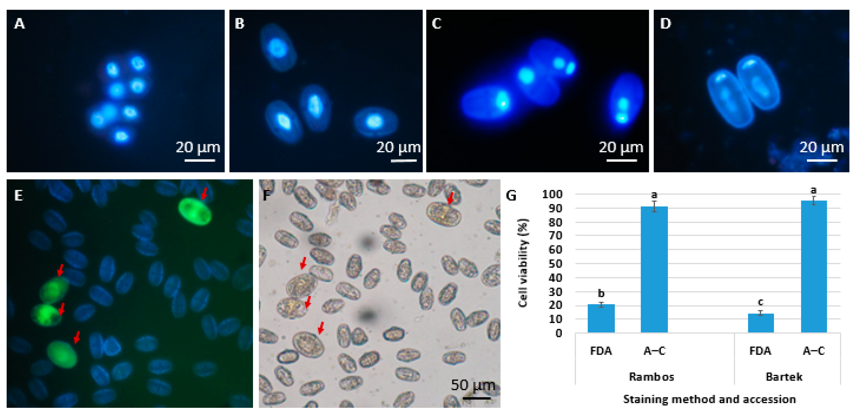
In our labs (UAK Krakow), we conducted trials on androgenesis in microspore cultures in two V. faba cultivars (Bartek and Rambos). We determined the stages of pollen development in the anthers of both cultivars using DAPI (Figure 2A–D), and selected floral buds in which anthers contained uninucleate microspores. From the selected buds, microspores were isolated with a protocol elaborated in our labs for Brassiceace [7]. Briefly, at a single round, approximately 20 floral buds were placed into a glass beaker with 4 mL of culture medium. Microspores were released by gently squeezing the buds with a syringe piston. The suspension then was filtered through a nylon net (100 μm pore size) and centrifuged twice (101× g for 5 min). The released microspores were subjected to culture in liquid MS or B5 medium. Both media were supplemented with growth regulators (2,4-D, NAA, or kinetin) and contained 80–120 g/L sugars (sucrose, or a mixture of sucrose and maltose). The analysis of microspore viability (Figure 2G), performed on the day of isolation, showed surprising results. Average viability after acetocarmine staining was very high (91–95%), while results obtained after fluorescein diacetate staining (FDA) (Figure 2E,F) were much lower (14–21%). Further observations showed that in a 30-day-old culture, the majority of the microspores did not change (40–70%), some of the microspores were enlarged (20%), and only 2–4% of the microspores divided mitotically; however, the mitoses were arrested and no further development was observed (unpublished data).
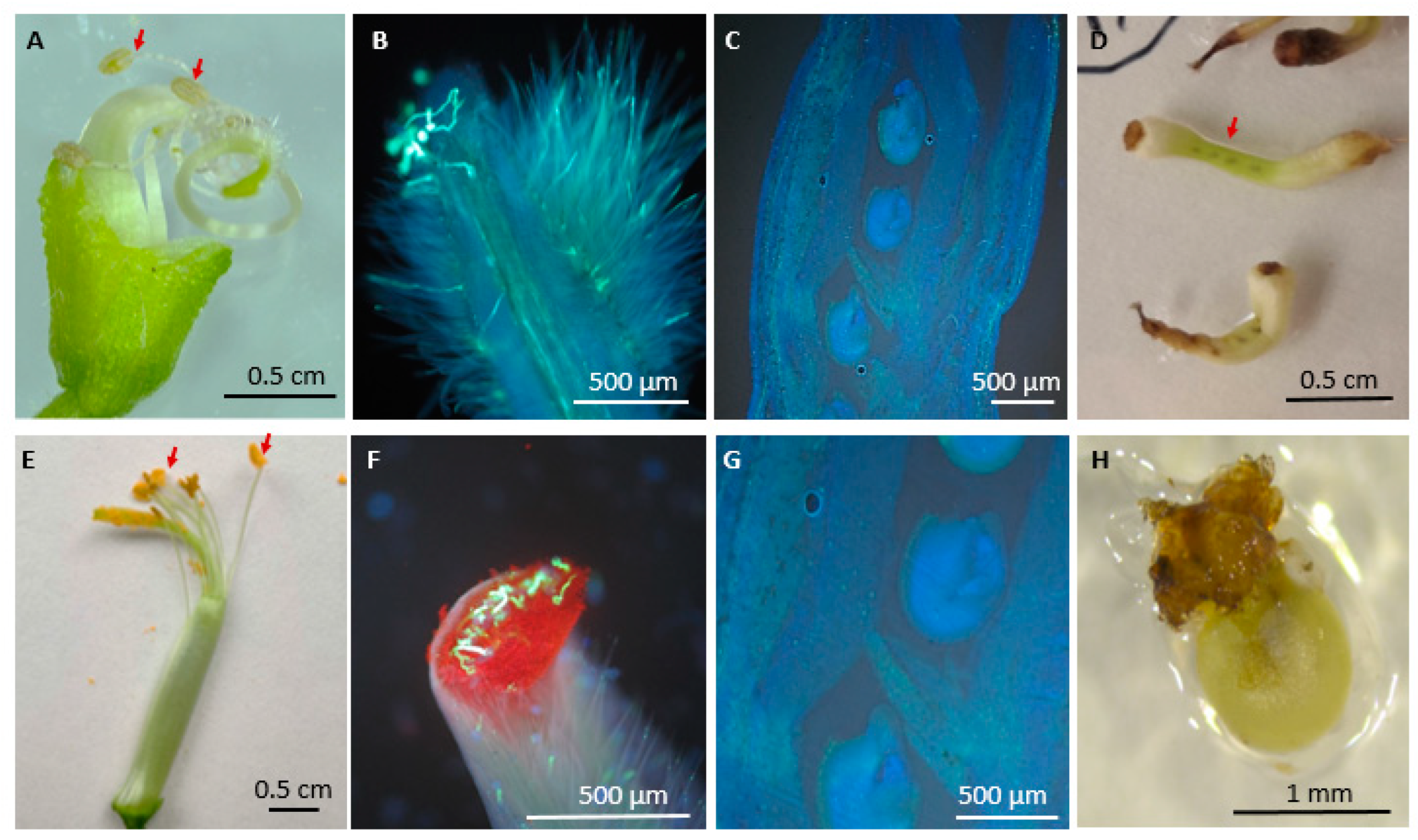
To date, there have been no reports on the induction of haploidization by the stimulation of female gametophyte in V. faba. Such research was also undertaken in our labs. We attempted to stimulate the haploid cells of the female gametophyte of V. faba by foreign pollination. As a pollen source, we used Phaseolus vulgaris and Lathyrus odoratus. Firstly, our goal was to evaluate if the foreign pollen germinates on V. faba stigmas penetrates the transmission track in style, reaching the ovules for fertilization. The results showed that the pollens of P. vulgaris (Figure 2A) and L. odoratus (Figure 2E) germinate on V. faba stigma (Figure 2B–F); however, they do not enter the transmission track nor reach the ovules (Figure 2C,G) even after 48 h after pollination. These results showed that potentially both pollen sources could be suitable for further investigations; however, we experienced some technical difficulties with obtaining the pollen of P. vulgaris. The corolla of P. vulgaris is composed of two wings and two fused keel petals. The keel petals are coiled around the reproductive organ [123], which makes manipulation with its pollen very difficult. After establishing the culture of V. faba ovaries, isolated from flowers pollinated with P. vulgaris pollen, we observed callus tissue development. The microscopical analyses of the cultured ovaries showed that callus developed exclusively from the ovary walls, not from ovules (Figure 3D).
Based on the obtained results, further experiments were conducted using L. odoratus pollen exclusively. A set of pollinations of V. faba flowers with the pollen of L. odoratus was performed, and an in vitro culture of isolated ovules was established. Most recent results showed that ovules produced callus tissue mainly at the micropylar site (Figure 3H) which is promising. Unfortunately, the majority of the developed calli get brown and die within 30 days of culture. Single callus samples were subjected to ploidy analysis with flow cytometry, which showed the presence of haploid nuclei. Future efforts will be focused on finding the proper media composition in order to culture and multiply the obtained calli and subject them to plant regeneration trials.

3.1.3. Grass Pea

Grass pea (Lathyrus sativus L.), also known as chickling pea, is an annual leguminous herb with a diploid chromosome number of 2n = 14. This species is one of the earliest cultivated crop species in human history. Archaeological excavations indicate that it was used as early as 8000 years BC in the Balkan region, potentially making it the first domesticated species in Europe. The Mediterranean region is traditionally considered the center of the origin of this species, from where it spread to further areas of Asia, Africa, and Europe [124]. Today, it is primarily cultivated in certain regions of Asia and Africa, where it is locally utilized as a food source for people, including Bangladesh, China, Ethiopia, India, Nepal, and Pakistan, and on a small scale in some regions of both Americas as a fodder crop and for human consumption. Alongside some other species from the Fabaceae family such as cowpea (Vigna unguiculata), groundnut (Arachis hypogaea), and bambara groundnut (Vigna subterranea), L. sativus is classified as an “orphan crop” [125]. Orphan crops are very important in the countries where they’re grown (Africa, Asia, and South America). They provide income for the poorest farmers and serve as staples in the local diet. At the same time, they get less research attention.
L. sativus possesses several important characteristics that make it potentially valuable as a crop, both for human consumption, as a source of animal feed, and as a valuable component in crop rotation. Its seeds contain approximately 30% protein, while its low cultivation requirements and resistance to adverse environmental conditions such as water scarcity or nutrient-poor soil make it a species with high potential for the intensification of its utilization. Furthermore, being a plant from the Fabaceae family, it has beneficial effects on improving the physicochemical properties of soil, enhancing its structure and nutrient and mineral content, which is particularly valuable in areas characterized by the presence of poor and depleted soils [126,127]. However, despite the many positive attributes of this species and its high nutritional value, the primary limiting factor that hinders its utilization as a potential food source for both animals and humans is the presence of the neurotoxin β-N-oxalyl-L-α,β-diaminopropanoic acid (ODAP) in its seeds, which has neurodegenerative effects, causing a condition known as lathyrism [128,129].
For L. sativus, there are reports regarding callus induction from internode explants [128,130], shoot regeneration from epicotyl segments [131], shoot regeneration from stem nodes and induction of rhizogenesis [132], in vitro plant regeneration from callus derived from root explants [133], and protoplast cultures in this species [134]. However, among the published data, there is only one known report on haploidization in this species (Table 3), and it focuses on isolated microspore cultures [106]. In this study, microspores were isolated from buds containing microspores in the uninucleate stage and were subjected to in vitro culture in different liquid media: NLN [135], HSO [106], or LMJ [136] supplemented with various concentrations of NAA, 2,4-D, or gibberellic acid (GA3), added at 0.1, 0.5, 1.0, or 2.0 mg/L and in various combinations. The authors also applied temperature treatments; however, the application of heat shock was found to have a negative impact on microspore viability, reducing it to approximately 35%. Similarly, low viability and a lack of cell divisions were observed in the cultures that were not subjected to low-temperature shock. The best response in culture, characterized by cell divisions leading to callus formation, was obtained in the microspore cultures isolated from flower buds stored for 2 days at 4 °C. In these cultures, multicellular structures, followed by micro-callus and callus, were first formed, from which plants were subsequently regenerated. In the study, two fractions of calli were observed, one with a potential for organogenesis, from which one plant was regenerated, and the other with a potential for embryogenesis, from which two plants were regenerated.

3.1.4. Lentil

Lentil (Lens culinaris Medik. subsp. culinaris) is diploid (2n = 14) and predominantly self-pollinated [137]. Lentil is one of the oldest crops cultivated by humans and one of the most widely grown, traditionally in the Middle East. As with many legumes, the main nutritional value of lentils lies in their high protein content, ranging from 21% to 27.1% of fresh seed mass. Additionally, they serve as a source of complex carbohydrates such as starch, dietary fiber, and vitamins (including ascorbic acid, thiamine, niacin, riboflavin, pyridoxine, folate, vitamins A, E, K, tocopherol, and choline), as well as bioactive phytochemicals like tannins and tannin-related compounds. The total tannin content in lentil seeds ranges around 915 mg/100 g, making it one of the richest legumes in this regard [138,139]. L. culinaris, as a species with high commercial and economic value, has been subjected to various research efforts aimed at improving its cultivation and enhancement, including the following among others: in vitro embryo cultures [140], in vitro flowering and shoot formation [141], as well as direct [142] and indirect [143] regeneration. The Agrobacterium-mediated genetic transformation of this species [144] as well as the application of embryo rescue technique in the hybridization of L. culinaris with other species within the same genus [145] were also reported.
L. culinaris is considered one of the most recalcitrant crop species to haploidization within the Fabaceae family [146]. Attempts at haploidization in this species, despite its significant value and importance, are strictly limited and mainly focused on androgenesis using anther cultures and isolated microspores (Table 4). Keller and Ferrie [147] cultured anthers containing uninucleate microspores on ML6 medium supplemented with 2 mg/L of trichlorophenoxyacetic acid (2,4,5-T), 1 mg/L BAP, and 6% sucrose. They managed to obtain callus, with this response observed in culture for over 57% of the anthers, as well as a small number of embryos. However, they were unable to regenerate plants, and the stress factors applied in this experiment (heat and cold) were found to have no positive effect on the androgenic response. Croser and Lulsdorf [15] attempted to induce androgenesis using isolated microspore culture. They subjected flower buds to pretreatment with cold (4 °C) for 96 h. Cell divisions leading to the formation of 8-cell structures were observed, but embryo development did not occur, and as a result, plant regeneration was not achieved. Desmal and Vijaipandurangam [146,148] initiated the isolated microspore cultures of L. culinaris using microspores at the uninucleate stage. They subjected flower buds to cold treatment (4 °C) for 72 h. Two types of media were used in this experiment: CHU (N6) medium [149] and LS medium [150], both supplemented with 2,4-D and BAP at concentrations ranging from 0.1 mg/L to 0.5 mg/L. In the study, no significant differences were observed depending on the various concentrations of growth hormones used. Thus, structures that the authors referred to as “callus-like projections” were obtained in cultures; these were most likely the calli that developed as a result of microspore divisions. In their investigation, no plant regeneration was reported.

3.2. Tribe Genisteae

Lupins

The genus Lupinus L. comprises several commercially important species, among which the most significant are white lupin (L. albus, 2n = 50), narrow-leaved lupin (L. angustifolius, 2n = 40), and yellow lupin (L. luteus, 2n = 52). The main producers of lupins are Australia and Poland [151]. Andean lupin (L. mutabilis) and ornamental species such as summer lupin (L. hartwegii) or large-leaved lupin (L. polyphyllus) have lesser economic significance. Lupin has been cultivated since ancient times, mainly in the Mediterranean region, where it was primarily grown as a fodder crop and used as green manure by the Ancient Egyptians, Greeks, and Romans [152]. The high value of lupin seeds is primarily determined by their high protein content, which ranges from 39% to 55% of the dehulled seed’s dry matter depending on the species and cultivar. This makes lupin a competitor to soy, which contains an average of 49% protein in the dry matter of its dehulled seeds. Moreover, in the dry matter of dehulled lupin seeds, there is an average of 7.8% carbohydrates, 8.8% fats, and 33.7% total dietary fiber [153]. Lupin seeds are also a source of biologically active compounds, including polyphenols from the subclass of flavonoids, phenolic acids, and isoflavonoids, as well as phytosterols and tocopherols [154,155]. Despite the high content of nutrients and biologically active compounds, lupin seeds cannot be considered an entirely safe dietary ingredient, especially for human consumption, due to their high content of quinolizidine alkaloids, including lupanine, albine, hydroxylupanine, sparteine, anagyrine, lupinine, and angustifolin [156,157,158]. In response, breeding efforts have been ongoing for years aimed at developing varieties characterized by low levels of these alkaloids while simultaneously maintaining high levels of nutritional and dietary components, known as sweet lupin [159,160].
Considering the application of tissue cultures and biotechnological methods to lupin, progress is limited [161] and lupins are considered recalcitrant species [162]. Among over 200 lupin species, only eight (Lupinus albus L., L. angustifolius L., L. hartwegii Lindl., L. hispanicus Boiss. et Reut., L. luteus L., L. mutabilis Sweet, L. polyphyllus Lyndl., L. texensis Hook.) have been studied so far, and the following subjects were considered: vegetative propagation (including callus formation, shoot differentiation, and meristem culture), embryo culture (including shoot multiplication and somatic embryogenesis), embryo rescue culture, the hybridization of protoplasts and somatic cells, transformation, and haploidization [163]. Table 5 lists research conducted towards haploidization in lupin. There is a single report on the haploid plant development as a result of crossing L. albus x L. graceus, which was then backrossed to L. albus. In BC4 generation from such crosses 2 plants identified as haploids were observed [164]. The first works on the in vitro haploidization of plants belonging to the Lupinus genus were initiated in the 1980s, and these were focused on androgenesis exclusively. There were relatively few trials on anthers cultures. The first report was given by Palada and Sator [165] and later studies were continued [166,167]. They established anther culture in various lupin species (L. polyphyllus, L. hartwegii, L. angustifolius, and L. luteus), and reported regenerants from the anther-derived callus of L. polyphyllus. However, these plants were diploid, and the authors suggested their somatic origin. Later, Campos-Andrada and Mota [168] using L. hartwegii anthers and Skrzypek et al. [169] using L. albus, L. luteus, and L. angustifolius anthers reported the development of anther-derived callus tissue. According to the authors of the latter report, the addition of 5% maltose to the induction medium and the absence of stress factors promoted microspore division. However, plant regeneration was not achieved in both of these studies. Kozak et al. [170] employed anther cultures in L. angustifolius. They observed an embryogenic response in the anthers containing microspores in the uninucleate stage, with the highest response occurring when the anthers were subjected to cold stress (4 °C) for 4 days and cultured on MS medium containing 2.0 mg/L 2,4-D and 0.5 mg/L kinetin, as indicated by the highest number of embryo-like structures and multicellular structures per anther. In these studies, organogenesis from callus was also achieved on a medium containing 2.0 mg/L BAP and 0.5 mg/L NAA; however, it should be noted that it was rhizogenesis.
Almost simultaneously with other culture trials, several researchers worked on the isolated microspore cultures of Lupinus sp. The first trial in L. polyphyllus was not successful [164]. Later, Ormerod and Caligari [171] established microspore cultures in L. albus and obtained embryo-like structures (ELSs), which, according to the authors, were embryogenic in origin and proliferated into embryos; however, no ploidy was analyzed and no plants were regenerated. In another trial, the cultured microspores of L. albus, L. angustifolius, and L. luteus underwent cell divisions, and multicellular structures as well as proembryos were observed. In these studies, the microspores were subjected to the stress conditions of cold (4 °C) and heat (32 °C) for 24 h [172]. Simoniuc et al. [173] in L. albus established both anther and isolated microspore cultures. They concluded that the optimal stage of microspore for androgenesis in this species is the late uninucleate stage. Although they observed embryogenesis in both anther and isolated microspore cultures, they found that a higher embryogenic response occurred in isolated microspore cultures, as indicated by a higher percentage of embryo-like structures (ELSs) when using this method.
Table 5. Reports on haploid research in genus Lupinus L.
Method/
Technique
Material/Stage of DevelopmentConditions and TreatmentsDevelopmentRemarksReferences
In vivo/spontaneousL. albus-seedsTwo sterile plants[164]
Androgenesis/Anther cultureL. polyphyllus anthersNot specifiedCallusNo further development of callus[165]
L. polyphyllus anthersNot specifiedCallus-[166]
L. polyphyllus anthersNot specifiedCallus
Regenerated plants
Chromosome counts showed diploid calli[167]
L. hartwegii anthersNot specifiedCallus-[168]
L. albus anthersNo application of stress treatment, liquid culture mediumEmbryo-like structures (ELSs)secondary somatic embryogenesis on ELS[171]
L. angustifolius, L. luteus, and L. albus anthers containing microspores in uninucleate stageCold or heat pretreatment: flower buds kept at 4 °C for 2 or 5 days in darkness or at 32 °C for 1 or 3 daysCallusPlant regeneration was not obtained[169]
L. albus anthers containing microspores in the late mononucleate developmental stageHeat stress treatment: anther cultured at 30 °C in the dark,Embryo-like structures (ELSs)ELSs were obtained on media containing 2,4-D and BAP[173]
L. angustifolius anthers in the uninucleate stageCold pretreatment: inflorescences kept at 4 °C for 4 days in the darknessMulticellular structures
Embryo-like structures
Callus
Calli produced roots on MS medium containing BAP and NAA[170]
Androgenesis/Isolated microspore cultureL. polyphyllus microspore. But unknown developmental stageNot specifiedNo development-[165]
L. albus microsporeNo application of stress treatment, liquid culture mediumNo development-[171]
L. albus,
L. angustifolius, and L. luteus microspores
Cold or heat treatment: microspore culture was kept at 4 °C or 32 °C for 24 h in darknessInduction of embryogenesis
Multicellular structures
Proembryos
No plant regeneration[172]
L. albus,
uninucleate microspores (late stage)
Microspore culture treated at 32 °C for 3 daysEmbryo-like structures (ELSs)ELSs were obtained on media containing 2,4-D and BAP[173]

3.3. Tribe Cicereae

Cicer arietinum L. (chickpea) is a diploid (2n = 16) self-pollinating species. C. arietinum is one of the species cultivated by humans for the longest time. It has been cultivated since the Neolithic period [174]. The origin of this species is debatable. Among the proposed centers of origin have been the region between Greece and the Himalayas, the Mediterranean and southwestern regions of Asia, and Ethiopia. Currently, the most likely place of origin for this species is considered to be the southeastern regions of Turkey and northern Syria. Due to their significant commercial importance, chickpeas are primarily valued for their high nutritional and dietary qualities, especially their protein content ranging from 18 to 22%, which, compared to other legumes, exhibits superior digestibility. Additionally, C. arietinum seeds contain a high content of complex carbohydrates (50–66.9%) and proportionally high levels of fat (2.7–7.42%) compared to other legumes [175]. These exceptional nutritional qualities make chickpeas highly valued in trade and a fundamental component of the diet in many regions of Asia and Africa.
Table 6 lists the reports on the studies on haploidization in C. arietinum. The first report on in vitro androgenesis in C. arietinum was given by Khan and Ghosh [176]. As a result of anther culture, three embryoids were regenerated from calli, but plants were not obtained. Altaf and Ahmad [177] used a cold pretreatment of floral buds at 4 °C for 3–7 days and observed callus development on anthers. However, organogenesis was not observed, and the ploidy status of the callus cells was not determined. Bajaj and Gosal [178] induced callus from cold-treated anthers (for 3 days) and reported a few multicellular embryoids, which did not develop further. Embryogenesis occurred on MS medium containing 2.0 mg/L indole-3-acetic acid (IAA) and 0.5 mg/L kinetin. The highest callus formation was developed on MS medium supplemented with 4 mg/L IAA and 2 mg/L kinetin and rhizogenesis on MS medium containing 0.5 mg/L NAA and 3 mg/L kinetin was reported. Later, callus development from the cold-treated anthers cultured on B5 medium with 2,4-D or NAA was reported, but the ploidy level was not determined. The highest callus formation was obtained on B5 media containing 2 mg/L NAA and 2 mg/L BAP and 2 mg/L NAA and 1 mg/L BAP [179]. Vessal et al. [180] obtained mature embryos from cold-treated buds for 7–10 days, followed by anther culture on MS medium with 1 mg/L 2,4-D and 0.2 mg/L kinetin. The embryos were induced from haploid callus on a medium with 0.5 mg/L kinetin and 10% sucrose. Plants were not regenerated from embryos. In the callus samples, chromosome counts were performed and they consisted of cells with haploid to polyploid chromosome numbers. Grewal et al. [181] applied cold stress by incubating chickpea flower buds containing uninucleate microspores for 3–4 days at 4 °C. Additionally, they applied two physical stresses: electroporation and centrifugation, along with osmotic stress. They achieved callus development, embryo formation, and plant regeneration, with the highest embryogenic response obtained through the combination of cold stress, centrifugation, and electroporation. Panchangam et al. [182] typed flower buds containing uninucleate microspores and pretreated them with cold (4 °C) for 2–4 days. Then, they isolated anthers and centrifuged them at varying speeds (168, 200, 500, 671, and 1000 g for 3, 5, 10, and 15 min). Further, the anthers were subjected to osmotic stress for 4 days in three different liquid media containing either 170 or 89 g/L sucrose. Only multicellular structures were obtained in this experiment. In another study [183], haploid plants were regenerated using media containing 2,4-D and silver nitrate. Media containing 10 mg/L 2,4-D and 15 mg/L silver nitrate or 10 mg/L 2,4-D and 15 or 50 mg/L silver nitrate proved to be the most effective for embryo induction and plant regeneration. It was also noted that the incubation of cultured anthers at 32 °C for 2 days significantly increased the number of embryos, while a beneficial effect on plant regeneration was observed with the combination of cold stress (4 °C for 7 days) and heat (30 °C for 10 days). There are significantly fewer reports regarding the culture of isolated microspores in C. arietinum [15,184,185]. In the known studies where cultures were established from uninucleate microspores, cell divisions or early-stage embryo development were achieved, but none of them resulted in plant regeneration.
In C. arietinum, a single report on the induction of haploids from female gametophytes was also reported. Reddy and Reddy [16] performed a cross between a male-sterile mutant (used as the maternal component) and a fertile cultivar. As a result, they obtained 149 seeds, out of which 51 plants exhibited weaker growth and narrow leaves. A cytological analysis of the obtained plants indicated their haploidy.
A very interesting approach was presented in C. arietinum by Mallikarjuna et al. [186]. They performed a crossing between C. arietinum and C. pinnatifidum. As a result of the cross, hybrid plants were obtained. These hybrids were fragile and weak and flowered only when the cytokinin zeatin (1 mg/L) was added to the sterilized tap water used to water the plants. All the whorls of the flower were present, although the anthers did not dehisce. The cytological observation of the anthers (in acetocarmine) revealed cell divisions in many of the microspores within the anther. The authors concluded that such microspores can lead to the development of androgenic plants, as division in these microspores was a consequence of the wide cross, as the anthers were not cultured in vitro. The mode of division was not identified, and the multicellular cells occurred in conjunction with sterile pollen grains. However, these studies were not confirmed by other authors.

4. Conclusions

The review of the literature has enabled us to identify some general factors affecting the success of the stimulation of haploid cell development in cool-season legumes and they are discussed below.

4.1. Method of Haploidization

The first important aspect is the selection of a proper and effective method of haploid induction. The majority of the studies on cool-season legumes focused on androgenesis. The best results were achieved in high-quality donor plants grown in a controlled environment with minimized stress conditions. In this method, a key factor is the selection of the proper stage of cells in anthers from which the development will be induced. Bud size and microspore stage are important in androgenesis, and they are usually closely related and relatively easy to determine in legumes. However, cytological studies confirm that pollen development in legumes is not synchronous and in single anther cells at various stages of development it will occur (i.e., tetrads and uninucleate microspores, or uninucleate microspores and binucleate pollens) [187]. This implies the need for the selection of floral buds in which the ordered stage (i.e., uninucleate microspores) is dominant. Based on published studies, embryogenic competence in cool-season legumes aims between the mid-unicellular and early bicellular stages [12,106,169,182]. Some authors also underlined that androgenesis in legumes might be limited by the thick exine of microspores or young pollens [20,172]. It has been shown that the exine thickness varies significantly in legumes, and in Vicia pollen, it is relatively thick, measuring 1.5–1.7 µm in the polar region, while in P. sativum pollen ranges from 0.2 to 0.4 µm [188,189]. Considering that the androgenesis in P. sativum resulted in androgenic plants [96,100,102], while in V. faba microspore cultures were ineffective and anther cultures resulted mainly in callus development [119,120,121,122], the theory of the exine thickness effect seems reliable.
The isolated microspore cultures are advocated as a more effective technique of androgenesis compared to anther cultures due to the lack of somatic tissue during culturing. In the Fabaceae family, this technique was applied, though it proved to be difficult and ineffective. Essentially, among the published reports on cool-season legumes, only one indicates its effectiveness reflected in embryo development, and it was observed in Lathyrus sativus and P. sativum [106]. In other cases, isolated microspore cultures resulted only in early mitoses that did not progress further. Recently, in our laboratory, we started research on haploidization in V. faba using isolated microspore cultures. So far, the results have not yielded significant progress due to low microspore viability and very low division frequency.
A literature review revealed that there were barely a few reports regarding attempts at haploidization using female gametophyte cells in cool-season grain legume crop species. Some trials have been made in P. sativum [108,109]. However, these are old reports from 40–50 years ago, and nothing new in this regard has been published since. Our research using the distant hybridization of V. faba with pollen from other species within the Fabaceae family (Phaseolus vulgaris and Lathyrus odoratus) shows potential when combined with in vitro methods, as they resulted in the obtainment of haploid callus (unpublished data). Therefore, this method is worth developing and optimizing. Perhaps another avenue is also to explore other candidates, such as the other representatives of the Fabaceae family as pollen donors, which could be utilized to induce egg cell development. Also, methods involving gynogenesis in unpollinated ovaries and ovules would be worth trying.

4.2. Genotype

The significance of genotype on haploidization in cool-season legumes is a limiting factor. In the majority of the published studies, the authors used one or two accessions, from which only some responded positively to developing calli or embryos, while the others were unresponsive [95,96,97,98,107,108,109,119,120,121,122,165,166,167,168,169,170,171,172,173,177,178,179,180,181,182]. The effect of genotype was most profoundly shown in the studies of Ochatt et al. [106]. They tested ten P. sativum genotypes, and among them, only three resulted in the regeneration of haploid plants from the cultured microspores. This was even more sound, as the authors included in their study three single loci EMS-mutants of Frisson accession (namely P64, P79, and P90). These mutants, when cultured in vitro from various explants, were capable of proliferating into calli, differentiating shoots, and early-stage embryos, but failed to regenerate any plants in haploidization studies.

4.3. Stress Factors

The application of different stresses is shown to be of great importance, especially during androgenesis [5,10], which was most widely studied in cool-season legumes. The effect of a cold storage period on flower buds prior to anther culture has been studied in these species, and it has been proven more beneficial compared to its application to cultured anthers or microspores. The anthers of P. sativum cv. Bonneville and the breeding lines T163 and P88 were subjected to a 72 h cold pretreatment whereby callus and heart-shaped-stage embryos were obtained even if plants were not recovered [98] For P. sativum [12], Cicer arietinum [12,182], and Lupinus sp. [169], the cold pretreatment of flower buds was necessary to induce divisions in microspores. High and low temperatures with increasing lengths of time were tested on the flower buds of P. sativum prior to their culture [106]. It was shown that high temperatures were detrimental to microspore viability, even if they were applied for a short treatment (a few hours). In contrast, the cold storage of buds was always beneficial, and even storage for several weeks did not cause any detrimental effect on the viability of cultured anthers or the division competence of cultured microspores [12,15,106,180,181,182]. The vast majority of early studies on cool-season grain legumes used mostly temperature (heat or cold) as stress pretreatments, although other stresses had also been reported. Electroporation has proved to be useful for P. sativum, Lathyrus sativus [106], and C. arietinum [182]. For some species, a combination of several stress factors was proven to be effective. In P. sativum, the key to success was combining a cold treatment of flower buds with electrostimulation and an osmotic shock [106]. In C. arietinum, the centrifugation of isolated anthers (at 168× g for 15 min) was also beneficial for their development [182].

4.4. Culture Medium

There is no consensus in the literature on the culture media required for androgenesis in cool-season legumes [12]. Various authors reported the effects of medium composition, and most studies have used various modifications of the MS or B5 formula with various growth regulators, mostly auxins and cytokins. Usually, media for the induction of androgenesis in other crops are recommended to have high osmoticum supplied mostly by sucrose in the concentrations of 90–130 g/L [5,7,8,9]. In cool-season legumes, the stimulation of androgenic development was effectively achieved in media supplemented with sucrose and/or maltose in concentrations ranging from 80 to170 g/L [12,15,107,181,185], compared to the media supplemented with 30–50 g/L [167,168,169].
Ochatt et al. [106] compared three different basal media: NLN medium [135,190], LMJ medium [136], and HSO (original) on androgenesis. They found that medium composition was not crucial for responses, as all three media supported reproducible and comparable responses in the absence of any treatment of microspores but following the cold storage of the donor flower buds. Conversely, some genotypes showed recalcitrance regardless of the treatments or culture conditions, indicating that the genotype is the primary factor influencing androgenetic potential in legume species.
In recent years, a lot of attention has been directed towards searching for media supplements that have the ability to stimulate cell proliferation. One such agent is the oligopeptide phytosulfokine alpha (PSK-α). It was shown that PSK stimulated microspore embryogenesis in triticale (x Triticosecale Wittm.) and Triticum aestivum [191] or Brassica napus [192]. PSK at concentrations of 10−10 to 10−6 M added to culture media has been shown to induce regeneration in P. sativum and highly recalcitrant V. faba during micropropagation [193]. Another interesting group of media supplements are the inhibitors of DNA methylation and histone acetylation. Studies on microspore embryogenesis in B. napus, H. vulgare, and triticale (x Triticosecale Wittm.) have shown that induced epigenetic modifications regulate the activity of genes involved in these processes, which affects their final efficiency [194,195]. Future efforts toward haploid induction in cold-seasoned legumes should take these media supplements into consideration.

4.5. Plant Regeneration

Plant regeneration remains the main factor limiting haploid production in the majority of legume crops in general. As has been shown above, reproducible protocol and repeatable plant regeneration have been reported for P. sativum [100,104,106] and C. arietinum [183,184,186] while for the remaining cool-season legumes, plant regeneration was occasional. The obtainment of embryo-like structures or embryos in different developmental stages (globular to heart and even mature) has been reported; however, it seems that for embryos, regeneration should be performed on hormone-free or low-hormone-containing media. The regeneration from callus tissue was also problematic, as it was often non-effective [15,102,147], and organogenesis from callus involved mainly rhizogenesis [98,170,177] and rarely shoot organogenesis [15,96,100,106]. In legumes, a common problem during regeneration was the development of shoots without roots [15,177]. An interesting approach to solving that problem was in vitro grafting [196,197,198].

4.6. Origin of Regenerants

The most popular techniques for verifying ploidy include chromosome counting, flow cytometry, or the cytological monitoring of microspore embryogenesis. Cool-season legumes were the subject of all these techniques. Most research demonstrating the ploidy status of cool-seasoned legumes focused on callus tissue. Research revealed that the obtained callus tissue was diploid [167,177] or haploid [95,120,121], but mixoploid samples were also reported [96,98]. The reports on the analysis of ploidy in whole regenerated plants are rare, as the conversion of embryos into plants and organogenesis of shoots from calli is very difficult in legumes. A small number of regenerated plants were identified as haploids [184,186] and diploids [15,103,184,185].
The verification of homozygosity in regenerants obtained from self-pollinated plants is indeed problematic, as these plants are autogamous and therefore highly homozygous. The use of heterozygous starting material followed by the molecular or morphological confirmation of the status of progeny obtained after in vitro haploidization methods is a standard route in other crops [9,54]. However, the majority of the publications on cool-season legumes completely omit this step although it is vital as anthers, ovaries and ovules consist of both haploid and diploid tissues. Histological studies in a member of the leguminous family, namely G. max, have shown that callus developed from the epidermis and the middle layer of cultured anthers [199]. In the case of legumes, this step would involve making crosses between selected homozygous parents, and the manipulations must have been performed on heterozygous F1 progeny. However, such studies have not been reported so far.
Regardless of the explant used, the majority of studies on haploidization in legumes report development mostly through the callus tissue (Table 1, Table 2, Table 3, Table 4, Table 5 and Table 6). Its development in the recalcitrant species may be considered as a success, but this tissue is known as genetically unstable [200]. Some researchers have reported increased ploidy levels with the increasing age of the callus obtained after the application of haploidization methods [96,182,190,201]. Given this, it seems reasonable to maximally shorten the phase needed to increase callus mass in order to proceed as much as possible to the regeneration stage.

5. Perspectives

Considering standard methods for haploidization, androgenesis seems to be the most promising for cool-season legumes. However, it is coupled with problems, i.e., the effect of genotype, low callus/embryo development, plant regeneration, and the determination of the origin of regenerants. Combining different stresses seems to be the pathway to androgenesis, especially a combination of cold pretreatment of floral buds and high osmotic medium. Future efforts should be directed towards the induction of organogenesis from calli. That might be achieved by searching for effective media supplements other than classic growth regulators, and advancing to the regeneration stage as soon as possible after the induction of development from the used explant.
Although traditional methods commonly used in the breeding programs of other species (e.g., in the Brassicaceae family) do not yield as significant results in species belonging to the Fabaceae family, new biotechnological methods developed in recent years may prove helpful. One such method involves utilizing mutations in the gene encoding the protein CENH3 (Centromere-Specific Histone H3). CENH3 is a specific histone protein located in centromeres, which is crucial for chromosome segregation during cell division. In Arabidopsis thaliana, crossing a mutant expressing altered CENH3 proteins with a wild-type individual resulted in the elimination of the mutant genome, leading to the obtainment of haploid seeds [202]. This method has also been applied in maize [203] and is a promising approach showing potential for genotypes resistant to traditional haploidization methods. Among the challenges researchers face in the upcoming period is the ability to implement this method and develop effective protocols for its application in species such as those classified as cool-season grain legume crop species and others recognized as recalcitrant to haploidization. The reports on the identification of centromeric repeats in Pisum sativum revealed centromeres consisting of multiple separated domains of CENH3 [204]. Similar chromosome organization was reported in Lathyrus but not in genera Vicia and Lens [205]. Relative to Vicia and Lens, Pisum and Lathyrus species possess an additional copy of the CENH3 gene. However, the subsequent study of Vicia faba, a species with simple centromeres and only one copy of CENH3, also revealed multiple centromeric satellites [204]. Therefore, Fabeae is a taxon with unusual distribution patterns and a possibly highly dynamic turnover of centromeric repeats. Research on CENH3-mediated haploid induction in legumes has already been initiated, and hopefully, stable haploid inducer lines can be generated in the future.

Author Contributions

Conceptualization, A.K.; resources, W.S. and A.K.; writing—original draft preparation, W.S. and A.K.; writing—review and editing, W.S. and A.K.; visualization, W.S. and A.K.; supervision, A.K. All authors have read and agreed to the published version of the manuscript.

Funding

This research received no external funding.

Institutional Review Board Statement

Not applicable.

Data Availability Statement

Not applicable.

Conflicts of Interest

The authors declare no conflicts of interest.

References

  1. Andrews, M.; Hodge, S. Climate Change, a Challenge for Cool Season Grain Legume Crop Production. In Climate Change and Management of Cool Season Grain Legume Crops; Yadav, S., Redden, R., Eds.; Springer: Dordrecht, The Netherlands, 2010. [Google Scholar] [CrossRef]
  2. FAOSTAT. Access Protocol. 2021. Available online: https://www.fao.org/faostat/en/#data/QCL/visualize (accessed on 30 April 2024).
  3. Andrews, M.; Raven, J.A. Root or shoot nitrate assimilation in terrestrial vascular plants–does it matter? Plant Soil. 2022, 476, 31–62. [Google Scholar] [CrossRef]
  4. Forster, B.P.; Heberle-Bors, E.; Kasha, K.J.; Touraev, A. The resurgence of haploids in higher plants. Trends Plant Sci. 2007, 12, 368–375. [Google Scholar] [CrossRef]
  5. Murovec, J.; Bohanec, B. Haploids and doubled haploids in plant breeding. In Plant Breeding; Abdurakhmonov, I., Ed.; InTech: Rijeka, Croatia, 2012; pp. 87–106. [Google Scholar]
  6. Dhooghe, E.; Van Laere, K.; Eeckhaut, T.; Leus, L.; Van Huylenbroeck, J. Mitotic chromosome doubling of plant tissues in vitro. Plant Cell Tiss Organ Cult. 2011, 104, 359–373. [Google Scholar] [CrossRef]
  7. Adamus, A.; Szklarczyk, M.; Kiełkowska, A. Haploid and doubled haploid plant production in Brassica rapa L. subsp. Pekinensis via microspore culture. In Doubled Haploid Technology; Methods in Molecular Biology; Segui-Simarro, J.M., Ed.; Humana Press: New York, NY, USA, 2021; Volume 2288, pp. 181–199. [Google Scholar] [CrossRef]
  8. Seguí-Simarro, J.M.; Jacquier, N.M.A.; Widiez, T. Overview of in vitro and in vivo doubled haploid technologies. In Doubled Haploid Technology; Methods in Molecular Biology; Segui-Simarro, J.M., Ed.; Humana: New York, NY, USA, 2021; Volume 2287, pp. 3–22. [Google Scholar] [CrossRef]
  9. Kiełkowska, A.; Kiszczak, W. History and current status of haploidization in carrot (Daucus carota L.). Agronomy 2023, 13, 676. [Google Scholar] [CrossRef]
  10. Maluszynski, M.; Kasha, K.J.; Forster, B.P.; Szarejko, I. Doubled Haploid Production in Crop Plants; Kluwer Academic Publishers: Dordrecht, The Netherlands, 2003. [Google Scholar] [CrossRef]
  11. Żur, I.; Adamus, A.; Cegielska-Taras, T.; Cichorz, S.; Dubas, E.; Gajecka, M.; Juzon, K.; Kiełkowska, A.; Malicka, M.; Oleszczuk, S.; et al. Doubled haploids: Contribution of Poland’s academies in recognizing the mechanism of gametophyte cell reprogramming and their utilization in breeding agricultural and vegetable crops. Acta Soc. Bot. Pol. 2022, 91, 9128. [Google Scholar] [CrossRef]
  12. Croser, J.S.; Lülsdorf, M.M.; Davies, P.A.; Clarke, H.J.; Bayliss, K.L.; Mallikarjuna, N.; Siddique, K.H.M. Toward doubled haploid production in the Fabaceae: Progress, constraints, and opportunities. Crit. Rev. Plant Sci. 2006, 25, 139–157. [Google Scholar] [CrossRef]
  13. Zhao, G.; Liu, Y.; Li, J. Germination of embryo in soybean anther culture. Chin. Sci. Bull. 1998, 43, 1991–1995. [Google Scholar] [CrossRef]
  14. Kaur, P.; Bhalla, J.K. Regeneration of haploid plants from microspore culture of pigeonpea (Cajanus cajan L.). Indian J. Exp. Biol. 1998, 36, 736–738. [Google Scholar]
  15. Croser, J.S.; Lulsdorf, M.M. Progress towards haploid division in chickpea (Cicer arietinum L.), field pea (Pisum sativum L.) and lentil (Lens culinaris Medik.) using isolated microspore culture. In Proceedings of the European Grain Legume Conference, Paris/Dijon, France, 7–11 June 2004; AEP: Wilmington, DE, USA, 2004; p. 189. [Google Scholar]
  16. Reddy, V.D.; Reddy, G.M. In vivo production of haploids in chickpea (Cicer arietinum L.). J. Genet. Breed. 1996, 51, 29–32. [Google Scholar]
  17. Zagorska, N.; Dimitrov, B. Induced androgenesis in alfalfa (Medicago sativa L.). Plant Cell Rep. 1995, 14, 249–252. [Google Scholar] [CrossRef]
  18. Zagorska, N.; Dimitrov, B.; Gadeva, P.; Robeva, P. Regeneration and characterization of plants obtained from anther cultures in Medicago sativa L. In Vitro Cell. Dev. Biol.-Plant 1997, 33, 107–110. [Google Scholar] [CrossRef]
  19. Gharyal, P.K.; Rashid, A.; Maheshwari, S.C. Production of haploid plantlets in anther cultures of Albizzia lebbeck L. Plant Cell Rep. 1983, 2, 308–309. [Google Scholar] [CrossRef]
  20. Lulsdorf, M.M.; Croser, J.S.; Ochatt, S. Androgenesis and Doubled-Haploid Production in Food Legumes. Biol. Breed. Food Legumes. 2011, 159, 159–177. [Google Scholar]
  21. Nowaczyk, P.; Nowaczyk, L. The influence of growth regulators on the frequency of polyembryony in pepper (Capsicum annuum L.). J. Appl. Gen. 1996, 37A, 204–207. [Google Scholar]
  22. Takeuchi, Y.; Kosaza, M.; Ozaki, Y.; Tomiyoshi, K.; Matsuishi, T.; Okubo, H. Origin of polyembryonic seeds and production of haploids in asparagus. Acta Hortic. 2020, 1301, 57–66. [Google Scholar] [CrossRef]
  23. Hand, M.L.; Koltunow, A.M. The genetic control of apomixis: Asexual seed formation. Genetics 2014, 197, 441–450. [Google Scholar] [CrossRef]
  24. Chase, S.S. Monoploids and monoploid-derivatives of maize (Zea mays L.). Bot. Rev. 1969, 35, 117–168. [Google Scholar] [CrossRef]
  25. Curtiss, J.; Rodriguez-Uribe, L.; Stewart, J.M.; Zhang, J. Identification of differentially expressed genes associated with semigamy in Pima cotton (Gossypium barbadense L.) through comparative microarray analysis. BMC Plant Biol. 2011, 16, 49. [Google Scholar] [CrossRef]
  26. McKone, M.J.; Halpern, S.L. The evolution of androgenesis. Am. Nat. 2003, 161, 641–656. [Google Scholar] [CrossRef]
  27. Palumbo, F.; Pasquali, E.; Albertini, E.; Barcaccia, G. A Review of Unreduced Gametes and Neopolyploids in Alfalfa: How to Fill the Gap between Well-Established Meiotic Mutants and Next-Generation Genomic Resources. Plants 2021, 10, 999. [Google Scholar] [CrossRef]
  28. Liu, L.; Wang, T. Male gametophyte development in flowering plants: A story of quarantine and sacrifice. J. Plant Physiol. 2021, 153365, 258–259. [Google Scholar] [CrossRef]
  29. Seguí-Simarro, J.M. Androgenesis revisited. Bot. Rev. 2010, 76, 377–404. [Google Scholar] [CrossRef]
  30. Dong, Y.Q.; Zhao, W.X.; Li, X.H.; Liu, X.C.; Gao, N.N.; Huang, J.H.; Tang, Z.H. Androgenesis, gynogenesis, and parthenogenesis haploids in cucurbit species. Plant Cell Rep. 2016, 35, 1991–2019. [Google Scholar] [CrossRef]
  31. Żur, I.; Dubas, E.; Golemiec, E.; Szechyńska-Hebda, M.; Gołębiowska, G.; Wędzony, M. Stress-related variation in antioxidative enzymes activity and cell metabolism efficiency associated with embryogenesis induction in isolated microspore culture of triticale (x Triticosecale Wittm.). Plant Cell Rep. 2009, 28, 1279–1287. [Google Scholar] [CrossRef] [PubMed]
  32. Würschum, T.; Tucker, M.R.; Maurer, H.P. Stress treatments influence efficiency of microspore embryogenesis and green plant regeneration in hexaploid triticale (× Triticosecale Wittmack, L.). In Vitro Cell. Dev. Biol.-Plant 2014, 50, 143–148. [Google Scholar] [CrossRef]
  33. Mikolajczyk, S.; Broda, Z.; Weigt, D. The effect of cold temperature stress on the viability of rye (Secale cereale L.) microspores. BioTechnologia. J. Biotechnol. Comput. Biol. Bionanotechnol. 2012, 93, 139–142. [Google Scholar]
  34. Supena, E.D.J.; Suharsono, S.; Jacobsen, E.; Custers, J.B.M. Successful development of a shed-microspore culture protocol for doubled haploid production in Indonesian hot pepper (Capsicum annuum L.). Plant Cell Rep. 2006, 25, 1–10. [Google Scholar] [CrossRef] [PubMed]
  35. Popova, T.; Grozeva, S.; Todorova, V.; Stankova, G.; Anachkov, N.; Rodeva, V. Effects of low temperature, genotype and culture media on in vitro androgenic answer of pepper (Capsicum annuum L.). Acta Physiol. Plantarum. 2016, 38, 273. [Google Scholar] [CrossRef]
  36. Yuan, S.X.; Liu, Y.M.; Fang, Z.Y.; Yang, L.M.; Zhuang, M.; Zhang, Y.Y.; Sun, P.T. Effect of combined cold pretreatment and heat shock on microspore cultures in broccoli. Plant Breed. 2011, 130, 80–85. [Google Scholar] [CrossRef]
  37. Yuan, S.; Su, Y.; Liu, Y.; Li, Z.; Fang, Z.; Yang, L.; Sun, P. Chromosome doubling of microspore-derived plants from cabbage (Brassica oleracea var. capitata L.) and broccoli (Brassica oleracea var. italica L.). Front. Plant Sci. 2015, 6, 1118. [Google Scholar] [CrossRef]
  38. Calabuig-Serna, A.; Camacho-Fernández, C.; Mir, R.; Porcel, R.; Carrera, E.; López-Díaz, I.; Seguí-Simarro, J.M. Quantitative and qualitative study of endogenous and exogenous growth regulators in eggplant (Solanum melongena) microspore cultures. Plant Growth Regul. 2021, 96, 345–355. [Google Scholar] [CrossRef]
  39. Kim, M.; Jang, I.C.; Kim, J.A.; Park, E.J.; Yoon, M.; Lee, Y. Embryogenesis and plant regeneration of hot pepper (Capsicum annuum L.) through isolated microspore culture. Plant Cell Rep. 2008, 27, 425–434. [Google Scholar] [CrossRef] [PubMed]
  40. Lantos, C.; Gémes Juhász, A.; Vági, P.; Mihály, R.; Kristóf, Z.; Pauk, J. Androgenesis induction in microspore culture of sweet pepper (Capsicum annuum L.). Plant Biotechnol. Rep. 2012, 6, 123–132. [Google Scholar] [CrossRef]
  41. Shmykova, N.; Domblides, E.; Vjurtts, T.; Domblides, A. Haploid embryogenesis in isolated microspore culture of carrots (Daucus carota L.). Life 2020, 11, 20. [Google Scholar] [CrossRef] [PubMed]
  42. Romanova, O.V.; Vjurtts, T.S.; Mineykina, A.I.; Tukuser, Y.P.; Kulakov, Y.V.; Akhramenko, V.A.; Domblides, E.A. Embryogenesis induction of carrot (Daucus carota L.) in isolated microspore culture. Foods Raw Mat. 2023, 11, 25–34. [Google Scholar] [CrossRef]
  43. Musiał, K.; Bohanec, B.; Jakse, M.; Przywara, L. The development of onion (Allium cepa L.) embryo sacs in vitro and gynogenesis induction in relation to flower size. In Vitro Cell Dev. Biol.-Plant 2005, 41, 446–452. [Google Scholar] [CrossRef]
  44. Lux, H.; Herrmann, L.; Wetzel, C. Production of haploid sugar-beet (Beta vulgaris L) by culturing unpollinated ovules. Plant Breed. 1990, 104, 177–183. [Google Scholar] [CrossRef]
  45. Wang, Y.; Gu, X.; Zhaing, S. Study the effect of pre-treatment and exogenous hormones on embryoids induced from unpollinated ovaries of cucumber. Acta Agric. Boreali-Sin. 2008, 23, 50–53. [Google Scholar] [CrossRef]
  46. Diao, W.P.; Bao, S.Y.; Jiang, B.; Cui, L.; Qian, C.T.; Chen, J.F. Cytogenetic studies on microsporogenesis and male gametophyte development in autotriploid cucumber (Cucumis sativus L.): Implication for fertility and production of trisomics. Plant Syst. Evol. 2009, 279, 87–92. [Google Scholar] [CrossRef]
  47. Sibi, M.L.; Kobaissi, A.; Shekafandeh, A. Green haploid plants from unpollinated ovary culture in tetraploid wheat (Triticum durum Defs.). Euphytica 2001, 122, 351–359. [Google Scholar] [CrossRef]
  48. Alan, A.R.; Brants, A.; Cobb, E.; Goldschmied, A.; Mutschler, M.A.; Earle, E.D. Fecund gynogenic lines from onion (Allium cepa L.) breeding materials. Plant Sci. 2004, 167, 1055–1066. [Google Scholar] [CrossRef]
  49. Chand, S.; Sahrawat, A.K. Embryogenesis and plant regeneration from unpollinated ovary culture of Psoralea corylifolia. Biol. Plant 2007, 51, 223–228. [Google Scholar] [CrossRef]
  50. Tekdal, D. In vitro Regeneration Studies of Vuralia turcica Using Unpollinated Ovaries. J. Agric. Sci. 2022, 28, 115–120. [Google Scholar] [CrossRef]
  51. Wędzony, M. Protocol for doubled haploid production in hexaploid triticale (x Triticosecale Wittm.) by crosses with maize. In Doubled Haploid Production in Crop Plants; Maluszynski, M., Kasha, K.J., Forster, B.P., Szarejko, I., Eds.; Springer: Dordrecht, The Netherlands, 2003. [Google Scholar] [CrossRef]
  52. Marcińska, I.; Czyczyło-Mysza, I.; Skrzypek, E.; Warchoł, M.; Zieliński, K.; Dubas, E. Obtaining of winter rye (Secale cereale L. ssp. cereale) haploid embryos through hybridization with maize (Zea Mays L.). Cereal Res. Commun. 2018, 46, 521–532. [Google Scholar] [CrossRef]
  53. Rines, H.W.; Dahleen, L.S. Haploid oat plants produced by application of maize pollen to emasculated oat florets. Crop Sci. 1990, 30, 1073–1078. [Google Scholar] [CrossRef]
  54. Devaux, P. The Hordeum bulbosum (L.) method. In Doubled Haploid Production in Crop Plants: A Manual; Springer: Dordrecht, The Netherlands, 2003; pp. 15–19. [Google Scholar] [CrossRef]
  55. Matzk, F.; Mahn, A. Improved techniques for haploid production in wheat using chromosome elimination. Plant Breed. 1994, 113, 125–129. [Google Scholar] [CrossRef]
  56. Bakos, F.; Jager, K.; Barnabás, B. Regeneration of haploid plants after distant pollination of wheat via zygote rescue. Acta Biol. Crac. 2005, 47, 167–171. [Google Scholar]
  57. Mochida, K.; Tsujimoto, H.; Sasakuma, T. Confocal analysis of chromosome behavior in wheat×maize zygotes. Genome 2004, 47, 199–205. [Google Scholar] [CrossRef]
  58. Ishii, T.; Karami-Ashtiyani, R.; Houben, A. Haploidization via Chromosome Elimination: Means and Mechanisms. Ann. Rev. Plant Biol. 2016, 67, 421–438. [Google Scholar] [CrossRef]
  59. Gałązka, J.; Niemirowicz-Szczytt, K. Review of research on haploid production in cucumber and other cucurbits. Folia Hortic. 2013, 25, 67–78. [Google Scholar] [CrossRef]
  60. Kasha, K.J.; Maluszynski, M. Production of doubled haploids in crop plants. An introduction. In Doubled Haploid Production in Crop Plants; Maluszynski, M., Kasha, K., Forster, B.P., Szarejko, I., Eds.; Springer: Berlin/Heidelberg, Germany, 2003; pp. 1–4. [Google Scholar]
  61. Conner, J.A.; Mookkan, M.; Huo, H.; Chae, K.; Ozias-Akins, P. A parthenogenesis gene of apomict origin elicits embryo formation from unfertilized eggs in a sexual plant. Proc. Natl. Acad. Sci. USA 2015, 112, 11205–11210. [Google Scholar] [CrossRef]
  62. Eeniuk, A.H. Matromorphy in Brassica oleracea L. II. Differences in parthenogenetic ability and parthenogenesis inducing ability. Euphytica 1974, 23, 435–445. [Google Scholar] [CrossRef]
  63. Hess, D.; Wagner, G. Induction of haploid parthenogenesis in Mimulus-luteus by in vitro pollination with foreign pollen. Z. Pflanzenphysiol. 1974, 72, 466–468. [Google Scholar] [CrossRef]
  64. Zhou, S.Q.; Qian, D.Q.; Cao, X.Y. Induction of parthenogenesis and chromosome behaviour in plants of parthenogenetic origin of cotton (Gossypium hirsutum). Genome 1991, 34, 255–260. [Google Scholar]
  65. Kiełkowska, A.; Adamus, A.; Baranski, R. An improved protocol for carrot haploid and doubled haploid plant production using induced parthenogenesis and ovule excision in vitro. In Vitro Cell Dev. Biol.–Plant 2014, 50, 376–383. [Google Scholar] [CrossRef] [PubMed]
  66. Kiełkowska, A.; Adamus, A.; Baranski, R. Haploid and Doubled Haploid Plant Production in Carrot Using Induced Parthenogenesis and Ovule Excision In Vitro. In Plant Cell Culture Protocols; Methods in Molecular Biology; Loyola-Vargas, V., Ochoa-Alejo, N., Eds.; Humana Press: New York, NY, USA, 2018; Volume 1815, pp. 301–315. [Google Scholar] [CrossRef]
  67. Yeh, B.P.; Peloquin, S.J. The nucleolus-associated chromosome of Solanum species and hybrids. Am. J. Bot. 1965, 52, 626. [Google Scholar] [CrossRef]
  68. Badr, M.; Horn, W. Genetische untersuchungen an diploiden und tetraploiden Pelargonium zonale-Hybriden. Z Pflanzenzuchtg. 1971, 66, 203–220. [Google Scholar]
  69. Horn, W. Interspecific crossability and inheritance in Pelargonium. Plant Breed. 1994, 113, 3–17. [Google Scholar] [CrossRef]
  70. Horn, W. Induktion und züchterische Nutzung der Parthenogenese. Z. Pflanzenzüchtg. 1972, 67, 39–44. [Google Scholar]
  71. Emsweller, S.L.; Uhring, J. Endosperm-embryo incompatibility in Lilium species hybrids. Adv. Hort. Sci. Theor. Appl. 1962, 2, 360–367. [Google Scholar]
  72. Emsweller, S.L.; Uhring, J. Parthenogenesis in tetraploid Lilium longiflorum. Am. J. Bot. 1962, 49, 978–984. [Google Scholar] [CrossRef]
  73. Germanà, M.A.; Chiancone, B. Gynogenetic haploids of Citrus after in vitro pollination with triploid pollen grains. Plant Cell Tiss. Organ Cult. 2001, 66, 59–66. [Google Scholar] [CrossRef]
  74. Sato, S.; Katoh, N.; Yoshida, H.; Iwai, S.; Hagimori, M. Production of doubled haploid plants of carnation (Dianthus caryophyllus L.) by pseudofertilized ovule culture. Sci. Hortic. 2000, 83, 301–310. [Google Scholar] [CrossRef]
  75. Vijverberg, K.; Ozias-Akins, P.; Schranz, M.E. Identifying and Engineering Genes for Parthenogenesis in Plants. Front. Plant Sci. 2019, 10, 128. [Google Scholar] [CrossRef] [PubMed]
  76. Lotfi, M.; Kashi, A.; Onsinejad, R. Induction of parthenogenetic embryos by irradiated pollen in cucumber. Int. Symp. Cucurbits 1997, 492, 323–328. [Google Scholar] [CrossRef]
  77. Faris, N.M.; Nikolova, V.; Niemirowicz-Szczytt, K. The effect of gamma irradiation dose on cucumber (Cucumis sativus L.) haploid embryo production. Acta Physiol. Plant 1999, 21, 391–396. [Google Scholar] [CrossRef]
  78. Lotfi, M.; Alan, A.R.; Henning, M.J.; Jahn, M.M.; Earle, E.D. Production of haploid and doubled haploid plants of melon (Cucumis melo L.) for use in breeding for multiple virus resistance. Plant Cell Rep. 2003, 21, 1121–1128. [Google Scholar] [CrossRef]
  79. Doré, C.; Boulidard, L.; Sauton, A.; Rode, J.C.; Cuny, F.; Niemirowiecz-Szczytt, K.; Dumas de Vaulx, R. Interest of irradiated pollen for obtaining haploid vegetables. Genetic Improvement of Horticultural Crops by Biotechnology. Acta Hort. 1994, 392, 123–128. [Google Scholar]
  80. Kurtar, E.S.; Sarı, N.; Abak, K. Obtention of haploid embryos and plants through irradiated pollen technique in squash (Cucurbita pepo L.). Euphytica 2002, 127, 335–344. [Google Scholar] [CrossRef]
  81. Musial, K.; Przywara, L. Influence of irradiated pollen on embryo and endosperm development in kiwifruit. Ann. Bot. 1998, 82, 747–756. [Google Scholar] [CrossRef]
  82. Todorova, M.; Ivanov, P.; Shindrova, P.; Christov, M.; Ivanova, I. Doubled haploid production of sunflower (Helianthus annuus L.) through irradiated pollen-induced parthenogenesis. Euphytica 1997, 97, 249–254. [Google Scholar] [CrossRef]
  83. Demmink, J.F.; Custers, J.B.M.; Bergervoet, J.H.W. Gynogenesis to bypass crossing barriers between diploid and tetraploid dianthus species. Acta Hortic. 1987, 216, 343–344. [Google Scholar] [CrossRef]
  84. Meynet, J.; Barrade, R.; Duclos, A.; Siadous, R. Dihaploid plants of roses (Rosa x hybrida, cv ‘Sonia’) obtained by parthenogenesis induced using irradiated pollen and in vitro culture of immature seeds. Agronomie 1994, 14, 169–175. [Google Scholar] [CrossRef]
  85. De Witte, K.; Keulemans, J. Homozygous plant production in apple by parthenogenesis in situ: Improvement of the pollination stimulus for parthenogenic egg cell development. Eucarpia Symp. Fruit Breed. Genet. 1999, 538, 315–320. [Google Scholar] [CrossRef]
  86. Höfer, M.; Grafe, C. Induction of doubled haploids in sweet cherry (Prunus avium L.). Euphytica 2003, 130, 191–197. [Google Scholar] [CrossRef]
  87. Myers, J.R.; Baggett, J.R.; Lamborn, C. Origin, history, and genetic improvement of the snap pea (Pisum sativum L.). Plant Breed. Rev. 2001, 21, 93–138. [Google Scholar]
  88. Zohary, D.; Hopf, M. Domestication of Plants in the Old World, 3rd ed.; Clarendon Press: Oxford, UK, 2000. [Google Scholar] [CrossRef]
  89. Abbo, S.; Rachamim, E.; Zehavi, Y.; Zezak, I.; Lev-Yadun, S.; Gopher, A. Experimental growing of wild pea in Israel and its bearing on Near Eastern plant domestication. Ann. Bot. 2011, 107, 1399–1404. [Google Scholar] [CrossRef] [PubMed]
  90. Uskutoğlu, D.; İdikut, L. Pea production statistics in the world and in Turkey. Inn. Res. Agric. For. Water Issues 2023, 2, 25–38. [Google Scholar]
  91. Ravindran, G.; Nalle, C.L.; Molan, A.; Ravindran, V. Nutritional and biochemical assessment of field peas (Pisum sativum L.) as a protein source in poultry diets. J. Poult. Sci. 2010, 47, 48–52. [Google Scholar] [CrossRef]
  92. Dahl, W.J.; Foster, L.M.; Tyler, R.T. Review of the health benefits of peas (Pisum sativum L.). Br. J. Nutr. 2012, 108, S3–S10. [Google Scholar] [CrossRef]
  93. Dhall, R.K. Pea cultivation. In Bulletin no PAU/2017/Elec/FB/E/29; Punjab Agricultural University: Ludhiana, India, 2017. [Google Scholar]
  94. Hacisalihoglu, G.; Beisel, N.S.; Settles, A.M. Characterization of pea seed nutritional value within a diverse population of Pisum sativum. PLoS ONE 2021, 16, e0259565. [Google Scholar] [CrossRef]
  95. Gupta, S.; Ghosal, K.K.; Gadgil, V.N. Haploid tissue culture of Triticum aestivum var. Sonalika and Pisum sativum var. B22. Indian Agric. 1972, 16, 277–278. [Google Scholar]
  96. Gupta, S. Morphogenetic response of haploid callus tissue of Pisum sativum (Var. B22). Indian Agric. 1975, 19, 11–21. [Google Scholar]
  97. White, P.R. The Cultivation of Animal and Plant Cells, 2nd ed.; Ronald Press: New York, NY, USA, 1963. [Google Scholar]
  98. Gosal, S.S.; Bajaj, Y.P.S. Pollen embryogenesis and chromosomal variation in anther of three food legumes—Cicer arietinum, Pisum sativum and Vigna mungo. SABRAO J. 1988, 20, 51–58. [Google Scholar]
  99. Murashige, T.; Skoog, F. A revised medium for rapid growth and bio assays with tobacco tissue cultures. Physiol. Plant. 1962, 15, 473–497. [Google Scholar] [CrossRef]
  100. Sidhu, R.; Davies, P. Pea anther culture: Callus initiation and production of haploid plants. In Proceedings of the Australian Branch of the IAPT&B, Perth, Australia, 21–24 September 2005; pp. 180–186. [Google Scholar]
  101. Gamborg, O.L.; Miller, R.; Ojima, K. Nutrient requirements of suspension cultures of soybean root cells. Exp. Cell Res. 1968, 50, 151–158. [Google Scholar] [CrossRef]
  102. Bobkov, S.V. Isolated pea anther culture. Russian Agric. Sci. 2010, 36, 413–416. [Google Scholar] [CrossRef]
  103. Bobkov, S. Obtaining calli and regenerated plants in anther cultures of pea. Czech J. Gen. Plant Breed. 2014, 50, 123–129. [Google Scholar] [CrossRef]
  104. Bermejo, C.; Guindon, M.F.; Palacios, L.T.; Cazzola, F.; Gatti, I.; Cointry, E. Comparative androgenetic competence of various species and genotypes within the genus Pisum L. Plant Cell Tiss. Organ Cult. 2020, 143, 487–497. [Google Scholar] [CrossRef]
  105. Kumar, A.S.; Gamborg, O.L.; Nabors, M.W. Plant regeneration from cell suspension cultures of Vigna aconitifolia. Plant Cell Rep. 1988, 7, 138–141. [Google Scholar] [CrossRef]
  106. Ochatt, S.; Pech, C.; Grewal, R.; Conreux, C.; Lulsdorf, M.; Jacas, L. Abiotic stress enhances androgenesis from isolated microspores of some legume species (Fabaceae). J. Plant Physiol. 2009, 166, 1314–1328. [Google Scholar] [CrossRef]
  107. Bobkov, S. Initiation of microcalli in culture of pea (Pisum sativum L.) isolated microspores. Asia Pac. J. Mol. Biol. 2018, 26, 20–26. [Google Scholar] [CrossRef]
  108. Gritton, E.T.; Wierzbicka, B. An embryological study of a Pisum sativum x Vicia faba cross. Euphytica 1975, 24, 277–284. [Google Scholar] [CrossRef]
  109. Virk, D.S.; Gupta, A.K. Matromorphy in Pisum sativum L. Theor. Appl. Genet. 1984, 68, 207–211. [Google Scholar] [CrossRef]
  110. Duc, G.; Bao, S.; Baum, M.; Redden, B.; Sadiki, M.; Suso, M.J.; Zong, X. Diversity maintenance and use of Vicia faba L. genetic resources. Field Crops Res. 2010, 115, 270–278. [Google Scholar] [CrossRef]
  111. Singh, A.K.; Bharati, R.C.; Manibhushan, N.C.; Pedpati, A. An assessment of faba bean (Vicia faba L.) current status and future prospect. Afr. J. Agric. Res. 2013, 8, 6634–6641. [Google Scholar]
  112. Caracuta, V.; Weinstein-Evron, M.; Kaufman, D.; Yeshurun, R.; Silvent, J.; Boaretto, E. 14, 000-year-old seeds indicate the Levantine origin of the lost progenitor of faba bean. Sci. Rep. 2016, 6, 37399. [Google Scholar] [CrossRef]
  113. Zong, X.; Yang, T.; Liu, R. Faba bean (Vicia faba L.) breeding. Advances in Plant Breeding Strategies. Legumes 2019, 7, 245–286. [Google Scholar] [CrossRef]
  114. Crépon, K.; Marget, P.; Peyronnet, C.; Carrouée, B.; Arese, P.; Duc, G. Nutritional value of faba bean (Vicia faba L.) seeds for feed and food. Field Crops Res. 2010, 115, 329–339. [Google Scholar] [CrossRef]
  115. Sathya Prabhu, D.; Devi Rajeswari, V. Nutritional and biological properties of Vicia faba L.: A perspective review. Intern. Food Res. J. 2018, 25, 1332–1340. [Google Scholar]
  116. Meng, Z.; Liu, Q.; Zhang, Y.; Chen, J.; Sun, Z.; Ren, C.; Huang, Y. Nutritive value of faba bean (Vicia faba L.) as a feedstuff resource in livestock nutrition: A review. Food Sci. Nutr. 2021, 9, 5244–5262. [Google Scholar] [CrossRef]
  117. Nadal, S.; Suso, M.J.; Moreno, M.T. Management of Vicia faba genetic resources: Changes associated to the selfing process in the major, equina and minor groups. Genet. Res. Crop Evol. 2003, 50, 183–192. [Google Scholar] [CrossRef]
  118. Maalouf, F.; Hu, J.; O’Sullivan, D.M.; Zong, X.; Hamwieh, A.; Kumar, S.; Baum, M. Breeding and genomics status in faba bean (Vicia faba). Plant Breed. 2019, 138, 465–473. [Google Scholar] [CrossRef]
  119. Paratasilpin, T. Vegetative development of field bean pollen grain cultured in vitro. J. Sci. Soc. Thai. 1978, 4, 139–145. [Google Scholar]
  120. Hesemann, C.U. Haploid cells in calli from anther cultures of Vicia faba. Z. Pflanzenziichtg. 1980, 84, 23–29. [Google Scholar]
  121. Shlahi, S.A.; Majeed, D.M.; Ismail, E.N. Effect of the developmental stage of microspores, growth regulator and medium type on callus indication from broad bean Vicia faba anthers culture. J. Biotechnol. Res. Center. 2012, 6, 81–90. [Google Scholar] [CrossRef]
  122. Küçükrecep, A.; Tekdal, D. Microsporogenesis in faba bean (Vicia faba L.) grown in Mersin, Turkey. Plant Introduct. 2022, 95/96, 68–74. [Google Scholar] [CrossRef]
  123. Chacón, S.M.I.; Pickersgill, B.; Debouck, D.G. Domestication patterns in common bean (Phaseolus vulgaris L.) and the origin of the Mesoamerican and Andean cultivated races. Theor. Appl. Genet. 2005, 110, 432–444. [Google Scholar] [CrossRef]
  124. Polignano, G.B.; Uggenti, P.; Olita, G.; Bisignano, V.; Alba, V.; Perrino, P. Characterization of grass pea (Lathyrus sativus L.) entries by means of agronomically useful traits. Lathyrus Lathyrism Newsl. 2005, 4, 10–14. [Google Scholar]
  125. Lambein, F.; Travella, S.; Kuo, Y.H.; Van Montagu, M.; Heijde, M. Grass pea (Lathyrus sativus L.): Orphan crop, nutraceutical or just plain food? Planta 2019, 250, 821–838. [Google Scholar] [CrossRef]
  126. Croft, A.M.; Pang, E.C.K.; Taylor, P.W.J. Molecular analysis of Lathyrus sativus L.(grasspea) and related Lathyrus species. Euphytica 1999, 107, 167–176. [Google Scholar] [CrossRef]
  127. Grela, E.R.; Rybiński, W.; Matras, J.; Sobolewska, S. Variability of phenotypic and morphological characteristics of some Lathyrus sativus L. and Lathyrus cicera L. accessions and nutritional traits of their seeds. Genet. Res. Crop Evol. 2012, 59, 1687–1703. [Google Scholar] [CrossRef]
  128. Yan, Z.Y.; Spencer, P.S.; Li, Z.X.; Liang, Y.M.; Wang, Y.F.; Wang, C.Y.; Li, F.M. Lathyrus sativus (grass pea) and its neurotoxin ODAP. Phytochem 2006, 67, 107–121. [Google Scholar] [CrossRef]
  129. Enneking, D. The nutritive value of grasspea (Lathyrus sativus) and allied species, their toxicity to animals and the role of malnutrition in neurolathyrism. Food Chem. Toxicol. 2011, 49, 694–709. [Google Scholar] [CrossRef]
  130. Tripathy, S.K.; Swain, D.; Mishra, P.K.; Baisakh, B.; Dash, S. Optimization of callus induction in Lathyrus sativus L. Afr. J. Food Sci. Technol. 2014, 5, 60–66. [Google Scholar]
  131. Barik, D.P.; Mohapatra, U.; Chand, P.K. High frequency in vitro regeneration of Lathyrus sativus L. Biol. Plant. 2005, 49, 637–639. [Google Scholar] [CrossRef]
  132. Barpete, S.; Khawar, K.M.; Özcan, S. Differential competence for in vitro adventitous rooting of grass pea (Lathyrus sativus L.). Plant Cell Tiss. Organ Cult. 2014, 119, 39–50. [Google Scholar] [CrossRef]
  133. Roy, P.K.; Barat, G.K.; Mehta, S.L. In vitro plant regeneration from callus derived from root explants of Lathyrus sativus. Plant Cell Tiss. Organ Cult. 1992, 29, 135–138. [Google Scholar] [CrossRef]
  134. Ochatt, S.; Durieu, P.; Jacas, L.; Pontécaille, C. Protoplast, cell and tissue cultures for the biotechnological breeding of grass pea (Lathyrus sativus L.). Lathyrus Lathyrism Newsl. 2001, 2, 35–38. [Google Scholar]
  135. Lichter, R. Induction of haploid plants from isolated pollen of Brassica napus. Z. Pflanzenphysiol. 1982, 105, 427–434. [Google Scholar] [CrossRef]
  136. Ochatt, S.J.; Mousset-Déclas, C.; Rancillac, M. Fertile pea plants regenerate from protoplasts when calluses have not undergone endoreduplication. Plant Sci. 2000, 156, 177–183. [Google Scholar] [CrossRef] [PubMed]
  137. Malaviya, D.R.; Shukla, R.S. Evolutionary trend in Lens culinaris and allied species: A cytological evidence. Cytologia 2000, 65, 305–312. [Google Scholar] [CrossRef]
  138. Wang, N.; Daun, J.K. Effects of variety and crude protein content on nutrients and anti-nutrients in lentils (Lens culinaris). Food Chem. 2006, 95, 493–502. [Google Scholar] [CrossRef]
  139. Faris, M.E.A.I.E.; Takruri, H.R.; Issa, A.Y. Role of lentils (Lens culinaris L.) in human health and nutrition: A review. Mediterranean J. Nutr. Metab. 2013, 6, 3–16. [Google Scholar] [CrossRef]
  140. Bermejo, C.; Gatti, I.; Cointry, E. In vitro embryo culture to shorten the breeding cycle in lentil (Lens culinaris Medik). Plant Cell Tissue Organ Cult. 2016, 127, 585–590. [Google Scholar] [CrossRef]
  141. Sarker, R.H.; Das, S.K.; Hoque, M.I. In vitro flowering and seed formation in lentil (Lens culinaris Medik.). In Vitro Cell Dev. Biol.–Plant 2012, 48, 446–452. [Google Scholar] [CrossRef]
  142. Sultana, T.; Majeed, N.; Khan, F.; Rehman, A.; Naqvi, S.S. Direct regeneration and efficient in vitro root development studies in lentil (Lens culinaris Medik). Pak. J. Bot. 2016, 48, 1999–2004. [Google Scholar]
  143. Bagheri, A.; Ghasemi Omraan, V.O.; Hatefi, S. Indirect in vitro regeneration of lentil (Lens culinaris Medik.). J. Plant Mol. Breed. 2012, 1, 43–50. [Google Scholar] [CrossRef]
  144. Das, S.K.; Shethi, K.J.; Hoque, M.I.; Sarker, R.H. Agrobacterium-mediated genetic transformation in lentil (Lens culinaris Medik.) followed by in vitro flowering and seed formation. Plant Tissue Cult. Biotechnol. 2012, 22, 13–26. [Google Scholar] [CrossRef]
  145. Saha, S.; Tullu, A.; Yuan, H.Y.; Lulsdorf, M.M.; Vandenberg, A. Improvement of embryo rescue technique using 4-chloroindole-3 acetic acid in combination with in vivo grafting to overcome barriers in lentil interspecific crosses. Plant Cell Tiss. Organ Cult. 2015, 120, 109–116. [Google Scholar] [CrossRef]
  146. Deswal, K. Progress and opportunities in double haploid production in lentil (Lens culinaris Medik.), soybean (Glycine max L. Merr.) and chickpea (Cicer arietinum L.). J. Pharmac. Phytochem. 2018, 7, 3105–3109. [Google Scholar]
  147. Gupta, D.; Dady, R.H.; Sambasivam, P.; Bar, i.; Azad, M.; Beera, N.; Ford, R.; Biju, S. Conventional and Biotechnological Approaches for Targeted Trait Improvement in Lentil. In Accelerated Plant Breeding; Gosal, S.S., Wani, S.H., Eds.; Springer: Cham, Switzerland, 2020; Volume 3. [Google Scholar] [CrossRef]
  148. Vijaipandurangam, K.D. Microspore culture effects on double haploid production in lentil (Lens culinaris Medik.). Int. J. Agric. Sci. Res. 2019, 8, 147–152. [Google Scholar]
  149. Bold, H.C.; Wynne, M.J. Introduction to the algae: Structure and reproduction. In Cultivation of Algae in the Laboratory; PrenticeHall INC: Englewood Cliffs, NJ, USA, 1978; pp. 571–578. [Google Scholar]
  150. Linsmaier, E.M.; Skoog, F. Organic growth factor requirements of tobacco tissue cultures. Physiol. Plant. 1964, 18, 100–127. [Google Scholar] [CrossRef]
  151. FAOSTAT. Access Protocol. 2022. Available online: https://www.fao.org/faostat/en/#data/QCL/visualize (accessed on 30 April 2024).
  152. Van de Noort, M. Lupin: An important protein and nutrient source. In Sustainable Protein Sources; Academic Press: Cambridge, MA, USA, 2024; pp. 219–239. [Google Scholar] [CrossRef]
  153. Bähr, M.; Fechner, A.; Hasenkopf, K.; Mittermaier, S.; Jahreis, G. Chemical composition of dehulled seeds of selected lupin cultivars in comparison to pea and soya bean. LWT-Food Sci. Technol. 2014, 59, 587–590. [Google Scholar] [CrossRef]
  154. Arnoldi, A.; Boschin, G.; Zanoni, C.; Lammi, C. The health benefits of sweet lupin seed flours and isolated proteins. J. Funct. Foods 2015, 18, 550–563. [Google Scholar] [CrossRef]
  155. Khan, M.K.; Karnpanit, W.; Nasar-Abbas, S.M.; Huma, Z.E.; Jayasena, V. Phytochemical composition and bioactivities of lupin: A review. Internat. J. Food Sci. Technol. 2015, 50, 2004–2012. [Google Scholar] [CrossRef]
  156. Resta, D.; Boschin, G.; D’Agostina, A.; Arnoldi, A. Quantification of quinolizidine alkaloids in lupin seeds, lupin-based ingredients and foods. In Lupins for Health and Wealth Proceedings of the 12th International Lupin Conference, 14–18 September 2008, Fremantle, Australia; Palta, J.A., Berger, J.B., Eds.; International Lupin Assocation: Canterbury, New Zealand, 2008; ISBN 0-86476-153-8. [Google Scholar]
  157. Pereira, A.; Ramos, F.; Sanches Silva, A. Lupin (Lupinus albus L.) Seeds: Balancing the Good and the Bad and Addressing Future Challenges. Molecules 2022, 27, 8557. [Google Scholar] [CrossRef] [PubMed]
  158. Boukid, F.; Pasqualone, A. Lupine (Lupinus spp.) proteins: Characteristics, safety and food applications. Eur. Food Res. Technol. 2022, 248, 345–356. [Google Scholar] [CrossRef]
  159. Vishnyakova, M.A.; Kushnareva, A.V.; Shelenga, T.V.; Egorova, G.P. Alkaloids of narrow-leaved lupine as a factor determining alternative ways of the crop’s utilization and breeding. Vavilov J. Genet. Breed. 2020, 24, 625. [Google Scholar] [CrossRef]
  160. Osorio, C.E.; Till, B.J. A bitter-sweet story: Unraveling the genes involved in quinolizidine alkaloid synthesis in Lupinus albus. Front. Plant Sci. 2022, 12, 795091. [Google Scholar] [CrossRef]
  161. Atkins, C.A.; Smith, P.H.C.; Gupta, S.; Jones, M.C.K.; Caligary, P.D.S. Genetics, cytology and biotechnology. In Lupins as Crop Plants: Biology, Production and Utilization; Gladstones, J.S., Atkins, C.A., Hamblin, J., Eds.; CAB International: Oxon, NY, USA, 1998; pp. 67–92. [Google Scholar]
  162. Rybczyński, J. An assessment of the present status of in vitro culture of lupins. In Proceedings of the 8th International Lupin Conference Asilomar, Asilomar, CA, USA, 11–16 May 1996; pp. 222–235. [Google Scholar]
  163. Święcicki, W.; Rybczyński, J.J.; Święcicki, W.K. Domestication and genetics of the yellow lupin (Lupinus luteus L.) and the biotechnological improvement of lupins. J. Appl. Genet. 2000, 41, 11–34. [Google Scholar]
  164. Kazmierski, T.; Kazmierska, E.M. Meiosis in spontaneous haploids of white lupin (Lupinus albus L.). Genet. Polon. 1989, 30, 47–60. [Google Scholar]
  165. Palada, M.; Sator, C. Observatii asupra incipients ale evolutiei androgenetice a polenului in vitro. Probl. Genet. Theor. App. XM/2 1981, 115–119. [Google Scholar]
  166. Sator, C.; Mix, G.; Menge, U. Investigations on anther culture of Lupinus polyphyllus. Plant Res. Dev. 1983, 18, 37–46. [Google Scholar]
  167. Sator, C. Regeneration of lupin plants from anthers. Landbauforsch. Volk. 1985, 35, 5–7. [Google Scholar]
  168. Campos-Andrada, M.P.; Mota, M. Callus differentiation from anther culture of Lupinus hartwegii Lindl. In Proceedings of the Advances in Lupin Research, Proceedings 7th International Lupin Conference, Evora, Portugal, 18–23 April 1994; Technical University of Lisbon: Lisbon, Portugal, 1994; Volume 262. [Google Scholar]
  169. Skrzypek, E.; Czyczyło-Mysza, I.; Marcińska, I.; Wędzony, M. Prospects of androgenetic induction in Lupinus spp. Plant Cell Tiss. Organ Cult. 2008, 94, 131–137. [Google Scholar] [CrossRef]
  170. Kozak, K.; Galek, R.; Waheed, M.T.; Sawicka-Sienkiewicz, E. Anther culture of Lupinus angustifolius: Callus formation and the development of multicellular and embryo-like structures. Plant Growth Regul. 2012, 66, 145–153. [Google Scholar] [CrossRef]
  171. Ormerod, A.J.; Caligari, P.D.S. Anther and microspore culture of Lupinus albus in liquid culture medium. Plant Cell Tiss. Organ Cult. 1994, 36, 227–236. [Google Scholar] [CrossRef]
  172. Bayliss, K.L.; Wroth, J.M.; Cowling, W.A. Pro-embryos of Lupinus spp. produced from isolated microspore culture. Aust. J. Agric. Res. 2004, 55, 589–593. [Google Scholar] [CrossRef]
  173. Simioniuc, D.; Burlacu-Arsene, M.C.; Morariu, A.; Lipșa, F.D. Induction of the embryogenesis process in anther and microspores cultures at the Lupinus albus species. Lucrări Științifice, Universitatea de Stiinte Agricole Și Medicină Veterinară “Ion Ionescu de la Brad” Iași, Seria. Agronomie 2010, 53, 60–63. [Google Scholar]
  174. Jukanti, A.K.; Gaur, P.M.; Gowda, C.L.L.; Chibbar, R.N. Nutritional quality and health benefits of chickpea (Cicer arietinum L.): A review. Br. J. Nutr. 2012, 108, S11–S26. [Google Scholar] [CrossRef]
  175. Kaur, R.; Prasad, K. Nutritional characteristics and value-added products of Chickpea (Cicer arietinum)—A review. J. Postharvest Technol. 2021, 9, 1–13. [Google Scholar]
  176. Khan, S.K.; Ghosh, P.D. In vitro induction of androgenesis and organogenesis in Cicer arietinum L. Curr. Sci. 1983, 52, 891–893. [Google Scholar]
  177. Altaf, N.; Ahmad, M.S. Plant regeneration and propagation of chickpea (Cicer arietinum L.) through tissue-culture techniques. In Nuclear Techniques and In Vitro Culture for Plant Improvement, Food and Agriculture Organisation and International Atomic Energy Agency; International Atomic Energy Agency: Vienna, Austria, 1986; pp. 407–417. [Google Scholar]
  178. Bajaj, Y.P.S.; Gosal, S.S. Pollen embryogenesis and chromosomal variation in cultured anthers of chickpea. Int. Chickpea Newsl. 1987, 17, 12–13. [Google Scholar]
  179. Huda, S.; Islam, R.; Bari, M.A.; Asaduzzaman, M. Anther culture of chickpea. Int. Chickpea Pigeonpea Newsl. 2001, 8, 24–26. [Google Scholar]
  180. Vessal, S.R.; Bagheri, A.; Safarnejad, A. The possibility of in vitro haploid production in chickpea (Cicer arietinum L.). J. Sci. Technol. Agric. Nat. Resour. 2002, 6, 67–76. [Google Scholar]
  181. Grewal, R.K.; Lulsdorf, M.; Croser, J.; Ochatt, S.; Vandenberg, A.; Warkentin, T.D. Doubled-haploid production in chickpea (Cicer arietinum L.): Role of stress treatments. Plant Cell Rep. 2009, 28, 1289–1299. [Google Scholar] [CrossRef]
  182. Panchangam, S.S.; Mallikarjuna, N.; Gaur, P.M.; Suravajhala, P. Androgenesis in chickpea: Anther culture and expressed sequence tags derived annotation. Ind. J. Exp. Biol. 2014, 52, 181–188. [Google Scholar]
  183. Abdollahi, M.R.; Rashidi, S. Production and conversion of haploid embryos in chickpea (Cicer arietinum L.) anther cultures using high 2, 4-D and silver nitrate containing media. Plant Cell Tiss. Organ Cult. 2018, 133, 39–49. [Google Scholar] [CrossRef]
  184. Croser, J. Haploid and Zygotic Embryo Culture of Chickpea (Cicer arietinum L.). Ph.D. Thesis, The University of Melbourne, Melbourne, Australia, 2002. [Google Scholar]
  185. Croser, J.S.; Lulsdorf, M.M.; Grewal, R.K.; Usher, K.M.; Siddique, K.H. Isolated microspore culture of chickpea (Cicer arietinum L.): Induction of androgenesis and cytological analysis of early haploid divisions. In Vitro Cell. Dev. Biol.–Plant 2011, 47, 357–368. [Google Scholar] [CrossRef]
  186. Mallikarjuna, N.; Jadhav, D.R.; Clarke, H.; Coyne, C.; Muehlbauer, F.J. Induction of Androgenesis as a Consequence of Wide crossing in chickpea. J. SAT Agric. Res. 2005, 1, 1–3. [Google Scholar]
  187. Skrzypkowski, W.; Galek, R.; Adamus, A.; Kiełkowska, A. Pollen Development and Stainability in Vicia faba L. and Lupinus angustifolius L. Agriculture 2023, 13, 2065. [Google Scholar] [CrossRef]
  188. Clarke, G.C.S.; Kupicha, F.K. The relationships of the genus Cicer, L. (Leguminosae): The evidence from pollen morphology. Bot. J. Linn. Soc. 1976, 72, 35–44. [Google Scholar] [CrossRef]
  189. Jiang, Y.; Lahlali, R.; Karunakaran, C.; Kumar, S.; Davis, A.R.; Bueckert, R.A. Seed set, pollen morphology and pollen surface composition response to heat stress in field pea. Plant Cell Environ. 2015, 38, 2387–2397. [Google Scholar] [CrossRef] [PubMed]
  190. Lichter, R. Anther culture of Brassica napus in a liquid culture medium. Z. Pflanzenphysiol. 1981, 103, 229–237. [Google Scholar] [CrossRef]
  191. Asif, M.; Eudes, F.; Randhawa, H.; Amundsen, E.; Spaner, D. Induction medium osmolality improves microspore embryogenesis in wheat and triticale. Vitr. Cell. Dev. Biol.–Plant 2014, 50, 121–126. [Google Scholar] [CrossRef]
  192. Mestinšek, M.Š.; Kunej, U.; Vogrinčič, V.; Jakše, J.; Murovec, J. The effect of phytosulfokine alpha on haploid embryogenesis and gene expression of Brassica napus microspore cultures. Front. Plant Sci. 2024, 15, 1336519. [Google Scholar] [CrossRef] [PubMed]
  193. Ochatt, S.; Conreux, C.; Moussa Mcolo, R.; Despierre, G.; Magnin-Robert, J.B.; Raffiot, B. Phytosulfokine-alpha, an enhancer of in vitro regeneration competence in recalcitrant legumes. Plant Cell 2018, 135, 189–201. [Google Scholar] [CrossRef]
  194. Solís, M.T.; El-Tantawy, A.A.; Cano, V.; Risueño, M.C.; Testillano, P.S. 5-azacytidine promotes microspore embryogenesis initiation by decreasing global DNA methylation, but prevents subsequent embryo development in rapeseed and barley. Front. Plant Sci. 2015, 6, 472. [Google Scholar] [CrossRef]
  195. Nowicka, A.; Juzoń, K.; Krzewska, M.; Dziurka, M.; Dubas, E.; Kopeć, P.; Zieliński, K.; Żur, I. Chemically-induced DNA de-methylation alters the effectiveness of microspore embryogenesis in triticale. Plant Sci. 2019, 287, 110189. [Google Scholar] [CrossRef] [PubMed]
  196. Ochatt, S.J.; Sangwan, R.S.; Marget, P.; Ndong, Y.A.; Rancillac, M.; Perney, P.; Röbbelen, G. New approaches towards the shortening of generation cycles for faster breeding of protein legumes. Plant Breed. 2002, 121, 436–440. [Google Scholar] [CrossRef]
  197. Gurusamy, V.; Bett, K.E.; Vandenberg, A. Grafting as a tool in common bean breeding. Can. J. Plant Sci. 2010, 90, 299–304. [Google Scholar] [CrossRef]
  198. Haddon, L.; Northcote, D.H. The effect of growth conditions and origin of tissue on the ploidy and morphogenic potential of tissue cultures of bean (Phaseolus vulgaris L.). J. Exp. Bot. 1976, 27, 1031–1051. [Google Scholar] [CrossRef]
  199. Rodrigues, L.R.; Oliveira, J.M.S.; Mariath, J.E.; Bodanese-Zanettini, M.A. Histology of embryogenic responses in soybean anther culture. Plant Cell Tiss. Organ Cult. 2005, 80, 129–137. [Google Scholar] [CrossRef]
  200. Mousa, M.A.A.; Abo-Elyousr, K.A.M.; Ibrahim, O.H.M. Evaluation of Genetic Variability within a Collection of Cumin Genotypes Using RAPD, ISSR, SRAP and SCoT Markers and Variability of In Vitro Callus Induced Therefrom. Horticulturae 2023, 9, 742. [Google Scholar] [CrossRef]
  201. Ravi, M.; Chan, S.W. Haploid plants produced by centromere-mediated genome elimination. Nature 2010, 464, 615–618. [Google Scholar] [CrossRef] [PubMed]
  202. Kelliher, T.; Starr, D.; Wang, W.; McCuiston, J.; Zhong, H.; Nuccio, M.L.; Martin, B. Maternal haploids are preferentially induced by CENH3-tailswap transgenic complementation in maize. Front. Plant Sci. 2016, 7, 414. [Google Scholar] [CrossRef] [PubMed]
  203. Neumann, P.; Navratilova, A.; Schroeder-Reiter, E.; Koblížková, A.; Steinbauerova, V.; Chocholova, E.; Macas, J. Stretching the rules: Monocentric chromosomes with multiple centromere domains. PLoS Genet. 2012, 8, e1002777. [Google Scholar] [CrossRef]
  204. Neumann, P.; Pavlíková, Z.; Koblížková, A.; Fuková, I.; Jedličková, V.; Novák, P.; Macas, J. Centromeres off the hook: Massive changes in centromere size and structure following duplication of CenH3 gene in Fabaceae species. Mol. Biol. Evol. 2015, 32, 1862–1879. [Google Scholar] [CrossRef]
  205. Ávila Robledillo, L.; Koblížková, A.; Novák, P.; Böttinger, K.; Vrbová, I.; Neumann, P.; Macas, J. Satellite DNA in Vicia faba is characterized by remarkable diversity in its sequence composition, association with centromeres, and replication timing. Sci. Rep. 2018, 8, 5838. [Google Scholar] [CrossRef]
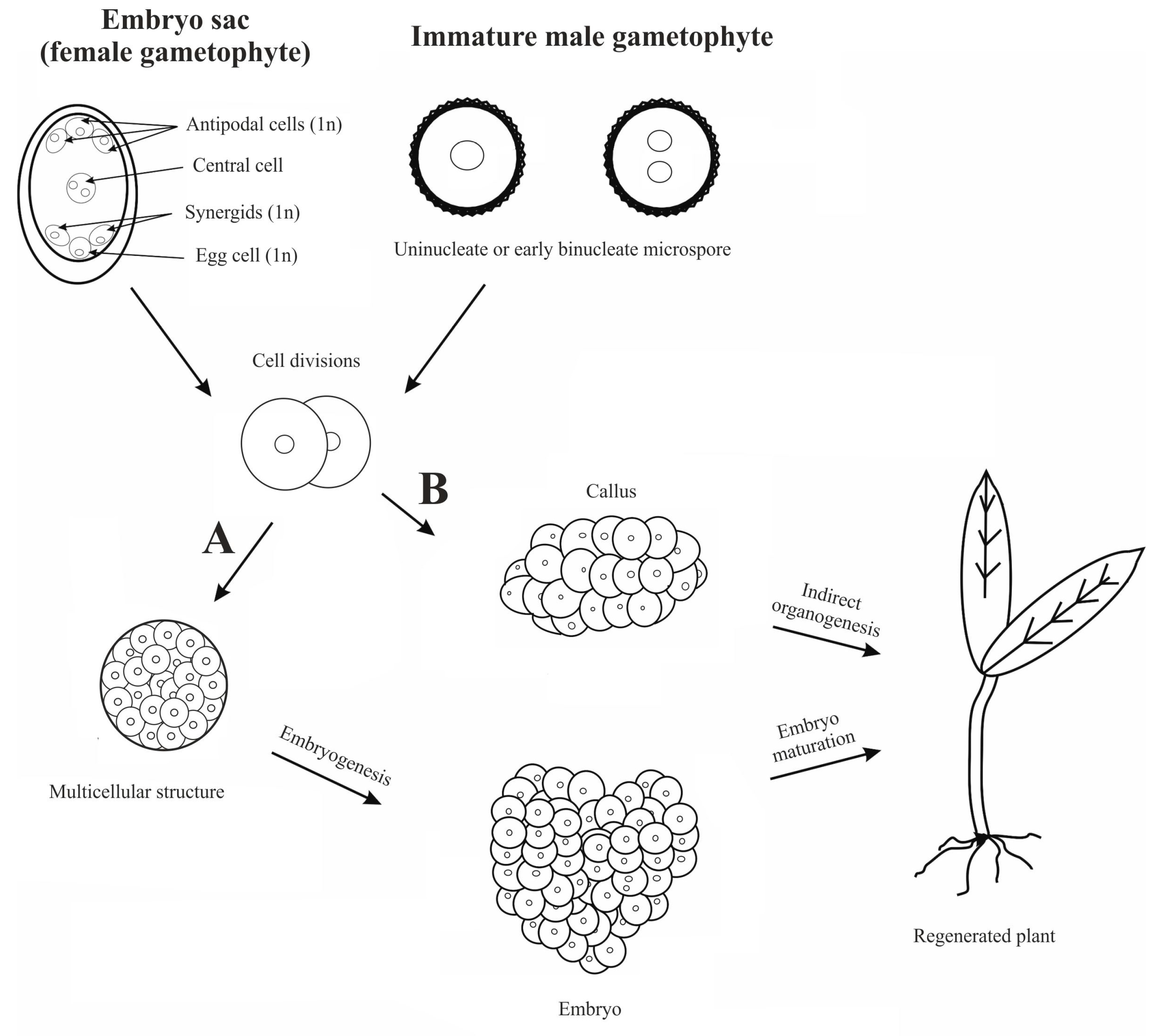
Figure 1. The main pathways for obtaining haploid plants: A—direct gametic embryogenesis; B—indirect pathway through the callus tissue developed from haploid male or female gametophyte cells.
Agriculture 14 01031 g001
Figure 2. Determination of the pollen developmental stages in the anthers after DAPI staining (AD) and microspore culture (EG) in V. faba. (A)—tetrads; (B)—microspores at the uninucleate stage; (C)—microspores at the late uninucleate and early binucleate stage; (D)—binucleate cells; (E,F) show the same field of view after FDA staining, the viable microspores are marked with red arrows; (G)—the results of viability in the cultured microspores after FDA and acetocarmine (AC). (The bars are means ± SEM. The means denoted with the same letters are not significantly different according to Tukey’s HSD test (p ≤ 0.05).
Agriculture 14 01031 g002
Figure 3. Induction of parthenogenesis in Vicia faba after distance pollination with pollen of Phaseolus vulgaris (AD) and Lathyrus odoratus (EH): (A)—fertile whorls morphology of P. vulgaris; arrows marked anthers; (B)—germination of P. vulgaris pollen grains on V. faba stigma; (C)—ovules of V. faba in ovary of flower pollinated with P. vulgaris pollen; (D)—V. faba ovaries cultured in vitro and isolated from flowers pollinated with P. vulgaris pollen; arrow points ovules inside the cultured ovary; (E)—fertile whorls morphology of L. odoratus; arrows marked anthers; (F)—germination of L. odoratus pollen grains on V. faba stigma; (G)—ovules of V. faba in ovary of flower pollinated with L. odoratus pollen; (H)—ovules of V. faba cultured in vitro and isolated from flowers pollinated with L. odoratus pollen.
Agriculture 14 01031 g003
Table 2. Reports on haploid research in Vicia faba L.
Method/
Technique
Material/Stage of DevelopmentConditions and TreatmentsDevelopmentRemarksReferences
Androgenesis/Anther cultureAnthers, tetrads,
uninucleate microspore, and binucleate pollen
Anther cultures kept at 28 °C and exposed to light (16 h photoperiod and 8 h darkness)CallusMultinuclear structures obtained from uninucleate and binucleate microspores—Kinetin had the highest influence on microspores[119]
Anthers
Stage not specified
UnknownCallusHaploid cells in anther-derived callus[120]
Anthers
isolated from different size flower buds
UnknownCallusHaploid and diploid anther-derived callus[121]
Anther
microspores in different developmental stages
No stresses applied, darkCallus
Embryo-like structures (heart)
Callus was obtained from the anthers containing microspores in the uninucleate stage
Embryo-like structures callused in time
No ploidy was analyzed
[122]
Table 3. Reports on haploid research in Lathyrus sativus L.
Method/
Technique
Material/Stage of DevelopmentConditions and TreatmentsDevelopmentRemarksReferences
Androgenesis/Isolated microspore cultureUninucleate microsporesFlower buds kept at 4 °C or 10 °C for 2, 4, 7, 14, 21 days, or 1 month;
osmotic stress provided by using mannitol or sucrose in various concentrations; Electric shock
Microspore-derived micro-calli
Regenerated plants
Plants obtained after cold stress stress (2 pcs from embryogenic callus and 1 pc from organogenic callus)[106]
Table 4. Reports on haploid research in Lens culinaris subsp. culinaris.
Method/
Technique
Material/Stage of DevelopmentConditions and TreatmentsDevelopmentRemarksReferences
Androgenesis/Anther cultureAnthers
Uninucleate microspore
Heat and cold stressesCallus
Proembryos
Anther derived calli with a few pro-embryos were obtained but no plants regenerated Applied stresses were not effective[147]
Androgenesis/Isolated microspore cultureNot specifiedFlower buds kept at 4 °C for 96 hCell divisions
8-cell structures
No embryos were regenerated[15]
Uninucleate microsporesFlower buds kept at 4 °C for 72 hCallusLS media with various concentrations of 2,4-D and BAP were used but hormonal treatment affected no significant differences[148]
Table 6. Reports on haploid research in genus Cicer arietinum L.
Method/
Technique
Material/Stage of DevelopmentConditions and TreatmentsDevelopmentRemarksReferences
Androgenesis/Anther cultureAnthers
Uninucleate and binucleate microspores
Anthers kept in the dark for 3 days at 25 ± °C, then incubated for 10 h in lightCallus
Embryoids
Various ploidy levels in callus: haploid (28%), diploid (38%), and aneuploid (38%)
Rhizogenesis from callus.
[176]
Greenish-white anthers containing tetrads and cells at the first mitosisFlower buds kept at 4 °C for 3 to 7 days.Callus
No shoot regeneration
Callus produced on media containing BAP + 2,4-D + NAA in different combinations. Unknown ploidy level.[177]
Anthers, uninucleate microsporesFloral buds kept at 4 °C 72 hCallus
Embryoids
Rhizogenesis from callus
Callus with different chromosome numbers.[178]
Anthers, uninucleate microsporesA total of 72 h cold pretreatment.Callus
Embryoids
Rhizogenesis from callus
Embryoids did not develop further.
Callus had no haploid cells.
[98]
Anthers, microspores at mid to late uninucleate stageCold pretreatment: flower buds kept at 4–5 °C
for 3 to 7 days.
Shoots regenerated from anther-derived callus,
callus-derived globular embryos
Unstated ploidy level of regenerated shoots.[179]
Not specifiedNot appliedCallus
Plant regeneration
Regenerated plants were diploid.[184,185]
Anthers, uninucleate microsporesCold pretreatment: buds kept in cold for 7 to 10 daysCallus
Organogenesis and embryogenesis
Callus obtained on MS media supplemented with 2,4-D and kinetin. Mature embryos obtained on modified Blayd’s media containing kinetin and 10% sucrose.[180]
Anthers, uninucleate microsporesCold pretreatment: buds kept at 4 °C for 3 to 4 days, electric shock, centrifugation, and high osmotic pressureEmbryos
Callus
Plant regeneration
The highest number of embryos per anther was obtained when stresses were applied combined (cold pretreatment + centrifugation + electroporation).[181]
Anthers, mid–late uninucleate microsporesCold pretreatment: buds kept at 4 °C for 2 to 4 days; stress induced by centrifugation and osmotic stressMulticellular structuresThe combination of stress factors resulted in a positive effect on the frequency of divisions.[182]
Anthers, uninucleate microsporesCold and heat pretreatment in different combinations: cultured anthers stored
at 4 °C for 4 and 7 days and then transferred to 30 °C for 10 days, 32° for 2 days, or 35 °C for 8 h
Callus
Embryos
Haploid and diploid plants regenerated from embryos
The highest percentage of regenerated plants from embryos was observed for anthers treated with a combination of cold and heat stress.[183]
Androgenesis/Isolated microspores cultureNot specified-CallusNo plants regenerated[184]
Microspores at the uninucleate stageHeat stress: flower buds kept at 32 °C for 16 hProembryo structuresNo plants regenerated[15]
Microspores at the uninucleate stageCold or heat pretreatment: flower buds kept at 4 °C for 24 h or at 32 °C for 12 hMulticellular structures
Embryos (globular)
No plants regenerated[185]
In vivo pollinationMale-sterile mutant used as a female parent and fertile variety as a male parent-Haploid plants obtained from seedsThe haploid plants constituted 34% of the plants obtained from seeds[16]
Wide crossingCicer aretinum and Cicer pinnatifidum Multicellular
microspores produced as a result of wide crossing
-[186]
Disclaimer/Publisher’s Note: The statements, opinions and data contained in all publications are solely those of the individual author(s) and contributor(s) and not of MDPI and/or the editor(s). MDPI and/or the editor(s) disclaim responsibility for any injury to people or property resulting from any ideas, methods, instructions or products referred to in the content.