Article

Physiological, Biochemical, and Ultrastructural Changes in Naturally Aged Sweet Corn Seeds

1
Southern Zhejiang Key Laboratory of Crop Breeding, Wenzhou Vocational College of Science and Technology, Wenzhou 325006, China
2
Guangdong Provincial Key Laboratory of Plant Molecular Breeding, College of Agriculture, South China Agricultural University, Guangzhou 510642, China
3
Sweet and Waxy corn Research Lab, Sorghum Research Institute, Shanxi Agricultural University, Jinzhong 030600, China
4
Crop Institute of Guangdong Provincial Agricultural Academic, Guangzhou 510640, China
*
Authors to whom correspondence should be addressed.
Agriculture 2024, 14(7), 1039; https://doi.org/10.3390/agriculture14071039
Submission received: 24 April 2024 / Revised: 25 June 2024 / Accepted: 26 June 2024 / Published: 28 June 2024
(This article belongs to the Section Seed Science and Technology)

Abstract

:
Due to low starch content and poor seed vigor, sweet corn seeds exhibit poor storage stability. Therefore, understanding the physiological and biochemical changes in seeds after natural aging is crucial for assessing seed status and extending the storage period. This study aims to investigate the physiological, biochemical, and ultrastructural changes in aged seeds of different genotypes. An eight-month natural aging experiment was conducted on 10 sweet corn inbred lines. The results showed an obvious decrease in germination potential, germination ratio, germination index, and vigor index after natural aging, and two inbred lines with stronger tolerance to natural aging were identified from the 10 inbred lines studied. In aged seeds, levels of gibberellin, abscisic acid, total protein, total starch, as well as activities of antioxidant enzymes, lipoxygenase, and amylase, and malondialdehyde (MDA) content, exhibited significant differences among inbred lines. Correlation analysis revealed a positive correlation among four seed vigor indices and a highly negative correlation between seed vigor indices and MDA content. Germination ratio, germination index, and vigor index displayed a highly negative correlation with lipoxygenase activity. Furthermore, starch granule decomposition was observed in the endosperm of low-vigor inbred lines, contrary to amylase activity. Thus, this study indicates variations in seed vigor, biochemical indicators, and the ultrastructure of aged sweet corn seeds among different genotypes. Both lower lipoxygenase activity and reduced MDA accumulation contribute to seed resistance to aging.

1. Introduction

Sweet corn (Zea mays L. Saccharata Sturt) is a type of maize characterized by its sweet taste, resulting from one or a few recessive mutations in starch synthesis genes, primarily intended for fresh consumption [1]. The sweet corn varieties in China are dominated by the sh2sh2 gene. The mature seeds of sweet corn have low starch content and poor storage resistance, which adversely affect seed germination and subsequent plant growth [2]. South China serves as the primary production region for sweet corn in China [3], characterized by long periods of high temperatures and heavy rainfall, leading to suboptimal seed storage conditions and rapid seed deterioration. Under normal natural conditions, seeds lose commodity value after about 6 months of storage, which has a great impact on sweet corn production.
Commercial sweet corn seed is generally produced in the autumn or winter in China, with the peak sales season for sweet corn seeds usually occurring from June to September. The storage period of seeds is approximately 6–10 months, during which the seeds may undergo deterioration. Even under optimal conditions, seed deterioration is unavoidable. Moisture content and temperature are the primary external factors influencing seed preservation [4]. Seed deterioration is characterized by irreversible metabolic and cellular changes, such as decreased antioxidant capacity, membrane damage, depletion of reserves, and genetic damage. The accumulation of reactive oxygen species (ROS) within the cells, along with a disruption in the balance between ROS production and elimination, is the primary cause of seed aging, leading to oxidative damage and loss of seed vigor [5,6].
ROS include singlet oxygen, superoxide, the hydroxyl radical, and hydrogen peroxide, and some ROS accumulate during the process of seed aging [7]. Reactive oxygen molecules are highly active and toxic, capable of causing irreversible damage to cell membranes, nucleic acids, proteins, carbohydrates, and lipids, and ultimately leading to seed inactivation [8,9]. It has been reported that lipid peroxidation is the key event in the aging process, while oxygen radical-mediated lipid peroxidation and membrane damage are considered the primary sources of damage during seed deterioration [10]. However, O2 contributed more to seed aging in the glassy cytoplasm of orthodox seeds [11].
Plants possess a multitude of enzymatic and non-enzymatic antioxidant defense systems to alleviate the damage caused by ROS, which work together to maintain a balance between ROS and antioxidants [12]. Key enzymes in the antioxidant system include superoxide dismutase (SOD), catalase (CAT), ascorbate peroxidase (APX), glutathione peroxidase (GPX), peroxidase (POD), peroxiredoxin (Prx), monodehydroascorbate reductase (MDHAR), dehydroascorbate reductase (DHAR), and glutathione reductase (GR) [13,14]. Additionally, non-enzymatic antioxidants like α-tocopherol (vitamin E), ascorbic acid (vitamin C), polyphenols, flavonoids, and carotenoids also play a crucial role in counteracting the cellular damage induced by ROS [12,15,16].
Membrane damage is considered a critical event during the seed deterioration process [17]. Lipid peroxidation leading to membrane injury involves the formation of hydrogen peroxide through the oxidation of unsaturated fatty acids [18], altering membrane permeability and disrupting membrane integrity. MDA is the primary byproduct of lipid peroxidation [10], which serves as a major biomarker of seed deterioration and represents one of the end products released during lipid peroxidation [19]. In aged soybean seeds, the levels of both H2O2 and MDA were elevated compared to the control [20].
The signaling cascades mediated by abscisic acid (ABA), auxin, and gibberellins (GAs) can influence the longevity of seeds. Most studies attribute a role to ABA-related mechanisms in promoting seed longevity [21,22,23], and this regulation is intricate. The biosynthesis of indole-3-acetic acid (IAA) may affect seed longevity, with reports indicating both positive and negative correlations between the expression of its biosynthetic genes and seed longevity [24,25]. Studies in Arabidopsis have discovered a positive involvement of GAs in seed longevity [26]. Conversely, research on tomatoes has revealed a negative impact of GA signaling pathways on seed longevity [27,28]. It is evident that the influence of plant hormones on seed storage is multifaceted.
The seeds of sweet corn often exhibit shrinking, accompanied by low grain weight and low seed vigor, and a few relevant studies have been published. Fan Longjiang and Yan Qizhuan [2] analyzed the physiological, genetic, and environmental factors of low seed vigor in sweet corn. Guan et al. [29] analyzed three supersweet corn varieties and found that seed dry weight had the greatest impact on seed vigor in sweet corn. With the extension of artificial aging time, the activities of dehydrogenase, peroxide, and seed vigor gradually decrease, whereas relative conductivity and MDA content progressively increase in sweet corn [30]. Wang et al. [31] conducted an analysis of the seed vigor and biochemical indicators in sweet corn inbred lines after artificial and natural aging. However, comprehensive evaluations regarding the characteristics of seed vigor and biochemical parameters after natural aging in sweet corn seeds are relatively scarce.
It is crucial to understand the physiological and biochemical changes in seeds during storage to ensure seed quality and prolong their lifespan. Currently, there is a lack of detailed reports on the changes during natural aging in sweet corn. This study aimed to evaluate the physiological and ultrastructural changes occurring in sweet corn seeds during natural storage, providing insights to optimize the storage conditions for sweet corn seeds.

2. Materials and Methods

2.1. Material

Ten sweet corn inbred lines (sh2sh2) with distinct genetic backgrounds were planted in the autumn of 2021 at the Teaching and Research Base of South China Agricultural University in Guangzhou, China. The harvested seeds were air-dried in sunlight for four days to remove the initial moisture. Subsequently, the seeds were dried in a seed dryer at 40 °C for 72 h until the seed moisture content decreased to below 13%. The seeds from each line were then divided into three portions. Seeds from each portion were taken for germination testing (0M), and the remaining seeds were packaged in polyethylene bags and stored at room temperature for natural aging in the storage room. The natural aging process commenced on 27 December 2021, and lasted for eight months (8M), with 0 months serving as the control. All seeds for germination tests and physiological and biochemical tests were taken from the three portions in each line.

2.2. Germination Assay

Germination tests were conducted on seeds of 0M and 8M following the method described by Zhu et al. [30]. The germination test was conducted using germination boxes, which spanned 24.5 cm in length, 14.0 cm in width, and 6 cm in height. A total of 25 seeds were placed in each box, and the experiment was repeated three times for statistical significance. Each box contained 25 seeds with three replications. River sand (60–80 mesh) was subjected to high-temperature sterilization at 120 °C for 2 h, followed by cooling for later use. The seeds were sown in germination boxes with a depth of 4 cm of moistened sand and covered with 2 cm of sand. The germination boxes were placed in a growth chamber (Ningbo Lai Fu, Ningbo, China) under a photoperiod of 16 h light and 8 h dark at 25 °C. The parameters for germination potential (GP), germination ratio (GR), germination index (GI), and vigor index (VI) were determined according to the method described by Guan et al. [29] GP represented the germination ratio on the fourth day after sowing. GR was calculated as the ratio of germinated seeds on the seventh day (n) to the total number of seeds (N), expressed as a formula: GR = (n/N) × 100%. The calculation of GI was formulated as: GI = ΣGt/Dt, where Dt is the germination time and Gt represents the number of seeds germinated on that day. The VI was calculated as VI = GI × SW, where SW indicates the fresh weight of seedlings on the eighth day.

2.3. Measurements of Physiological and Biochemical Indictors

The aged whole seeds of 0.1 to 0.3 g were initially washed with a low-temperature phosphate solution and subsequently dried on filter paper. The seeds were then weighed and deposited into a 5 mL centrifuge tube containing a ratio of 1:9 seed weight (g) to phosphate buffer (mL), amounting to 0.9–2.7 mL. The seeds were finely minced in an ice bath and subsequently pulverized by bead beating using a homogenizer operating at 10,000–15,000 r min−1. Following centrifugation at room temperature for 10–15 min at 3000 r min−1, the supernatant was collected. The activities of catalase (CAT), peroxidase (POD), superoxide dismutase (SOD), lipoxygenase (LOX), and amylase (AMS) were assessed using specific kits: plant catalase extraction kit (ZK-L0424), plant peroxidase extraction kit (ZK-L0223), plant superoxide dismutase extraction kit (ZK-L4276), plant LOX kit (ZK-P7487), and plant AMS kit (ZC-P6291). Furthermore, the contents of gibberellic acid (GA), abscisic acid (ABA), malondialdehyde (MDA), starch, and protein were determined using ELISA kits: plant GA ELISA kit (ZK-P6293), plant ABA ELISA kit, plant malondialdehyde ELISA kit (ZK-P7111), starch assay ELISA kit (ZIKER181), and BCA protein assay ELISA kit (ZIKER314), respectively, and all kits were purchased from Shenzhen Zike Biotechnology Co., Ltd., Shenzhen, China. All experimental procedures followed the instructions provided by the respective kit manufacturers to ensure the accuracy and reliability of the results.

2.4. Ultrastructure Observation

The ultrastructure observation of seeds through scanning electron microscopy was conducted following the method as described by Lu et al. [32]. The ultrastructural analysis was conducted on aged seeds of A51 and A89, which exhibit notable variations in their tolerance to seed storage. Take the middle part of the aged seeds and cut it into small pieces about 5 mm × 5 mm with a blade. The samples were rapidly immersed in a 2.5% glutaraldehyde solution, then subjected to vacuum infiltration to promote material sinking, and subsequently fixed at 4 °C for 24 h. The fixed samples underwent triple washing with phosphate buffer, followed by gradient dehydration using ethanol, and then a final dehydration step using a mixture of acetone and isoamyl acetate. Subsequently, pure isoamyl acetate was used for the ultimate dehydration process, after which the samples were dried at the critical point. The prepared samples were affixed to cover glasses and put into the vacuum spraying machine for spraying. The observation of starch particle decomposition in the two lines was conducted using scanning electron microscopy (SEM) (Zeiss, Oberkochen, Germany). Specifically, the starch particle decomposition at three locations—namely, the connection between endosperm and aleurone layer, the middle of endosperm, and the connection between endosperm and embryo—was examined at a magnification of 6500×.

2.5. Statistical Analysis

All experiments, including germination tests and physiological and biochemical tests, were performed with three biological replications. The descriptive statistics, variance, and correlation analysis of phenotypic data were conducted by Microsoft Excel 2010 and SAS 9.2 software. Normality of the data and homogeneity of variance were determined by the Shapiro–Wilk test and the hovtest, respectively. The fixed model was used for ANOVA analysis, and the multiple comparison method was Duncan’s test. Visualization of the correlation analysis matrix was performed using TBtools software 2.096 [33].

3. Results

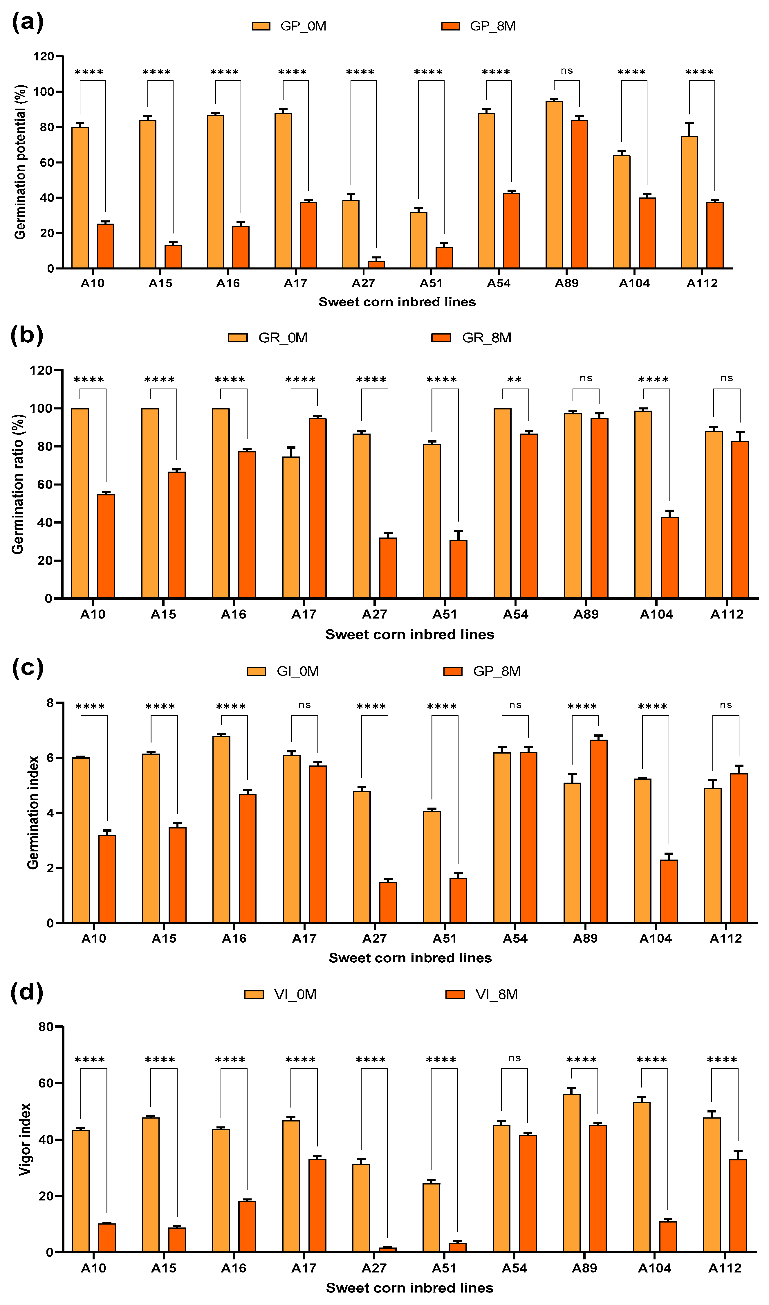
3.1. Comparison of Seed Viability between 0M and 8M

A two-way analysis of variance was conducted for natural aging treatments and inbred lines. As shown in Table 1, significant differences were observed among the four seed vigor-related indices in terms of aging treatments, inbred lines, and the interaction between natural aging and lines. The mean square for aging treatments was greater than that for the inbred lines.
Compared with 0M seeds, the GP, GR, GI, and VI of most inbred lines decreased after 8 months of natural aging (Figure 1). The inbred line A89 exhibited the highest GP in both 0M and 8M treatments. Several inbred lines had a mean germination ratio of 100%, namely A10, A15, A16, and A54 at the 0M treatment. Following natural aging treatment, the inbred lines A17 and A89 showed the highest germination ratios. In terms of germination index, A16 had the highest value at 0M treatment with a mean of 6.77, while A89 had the highest germination index with a mean of 6.65 after natural aging treatment. A89 displayed the highest vigor index in both the 0M and 8M treatments, with mean values of 56.12 and 45.22, respectively. When comparing the seed vigor indices before and after natural aging treatment, no differences were detected in GP and GI for A89. Similarly, for A112, no differences were found in GR and GI. A54 showed no differences in GI and VI, while A17 only exhibited no differences in GI. Upon comparison of the coefficient of variation pre- and post-natural aging treatment, an evident increase in the coefficient of variation was noted following the aging treatment. Considering all four seed vigor indicators, the more tolerant inbred lines to natural seed aging were identified as A89 and A54.

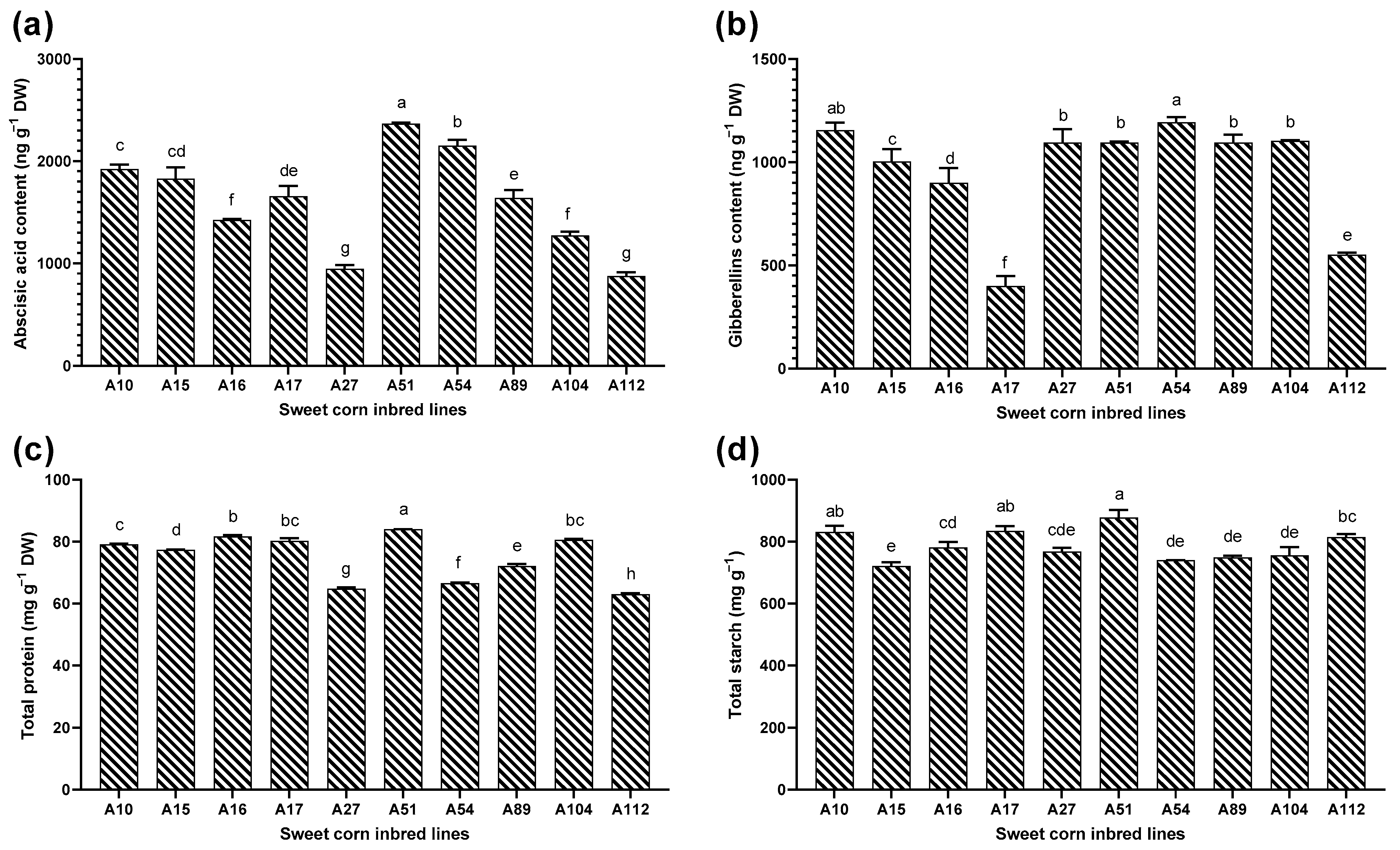
3.2. Evaluation of Phytohormones, Total Protein, and Starch Content

GA and ABA content were measured in the seeds of different inbred lines after natural aging for 8 months (Figure 2a,b). The results revealed variations in both GA and ABA content among 10 lines. The GA content showed an average value of 959.24 ng g−1 DW with a range of 399.88 to 1193.87 ng g−1 DW and a coefficient of variation of 28.14. The inbred line A54 exhibited the highest GA content, while the lowest was observed in A17. Regarding ABA content, the analysis indicated an average value of 1608.43 ng g−1 DW, with a range of 875.50 to 2365.66 ng g−1 DW and a coefficient of variation of 30.29. After 8 months of aging, the inbred line A51 showed the highest ABA content, whereas the lowest was found in A112.
Significant differences were observed among the inbred lines for total protein content and total starch content, as depicted in Figure 2c,d. The analysis of total protein content revealed an average of 74.92 mg g−1 DW among the 10 inbred lines, with a range of 62.99 to 83.98 mg g−1 DW and a coefficient of variation of 10.26. After 8 months of natural aging, A51 exhibited the highest total protein content, while A112 showed the lowest. The average value of total starch content was 787.11 mg g−1 DW and varied from 720.45 to 877.81 mg g−1 DW with a coefficient of variation of 6.38. A51 displayed the highest total starch content, whereas A15 exhibited the lowest.

3.3. Evaluation of MDA Content, LOX, and Antioxidant Enzyme Activity

The multiple comparison analysis of MDA content among 10 inbred lines showed significant differences, as depicted in Figure 3a. The analysis revealed an average MDA content of 51.78 nmol g−1 DW among 10 inbred lines, with a range of 26.69 to 72.00 mg g−1 DW and a coefficient of variation of 25.86. Line A27 exhibited the highest MDA content, while line A89 exhibited the lowest. On average, LOX activity was found to be 3334.65 U g−1 DW among 10 sweet corn lines, with a range spanning from 2280.00 to 4323.50 U g−1 DW and a coefficient of variation of 13.32 (Figure 3b). In terms of LOX activity, line A51 had the highest level, whereas line A54 had the lowest.
Ten sweet corn lines were compared for the activities of CAT, POD, and SOD in seeds after natural aging for 8 months (Figure 3c–e). The result shows significant differences in the three antioxidant enzymes among the 10 inbred lines. The average CAT activity was 242.58 U g−1 DW, ranging from 108.97 to 316.42 U g−1 DW, with a coefficient of variation of 27.12. Line A104 exhibited the highest CAT activity, while the lowest was observed in A89. The average POD activity was 2425.47 U g−1 DW, ranging from 1548.00 to 3567.56 U g−1 DW, with a coefficient of variation of 26.33. Line A17 showed the highest POD activity, whereas line A10 displayed the lowest. Additionally, the average SOD activity was 1709.30 U g−1 DW, varying from 1494.20 to 2225.72 U g−1 DW, with a coefficient of variation of 13.32. Line A16 exhibited the highest SOD activity, while the lowest was observed in line A27.

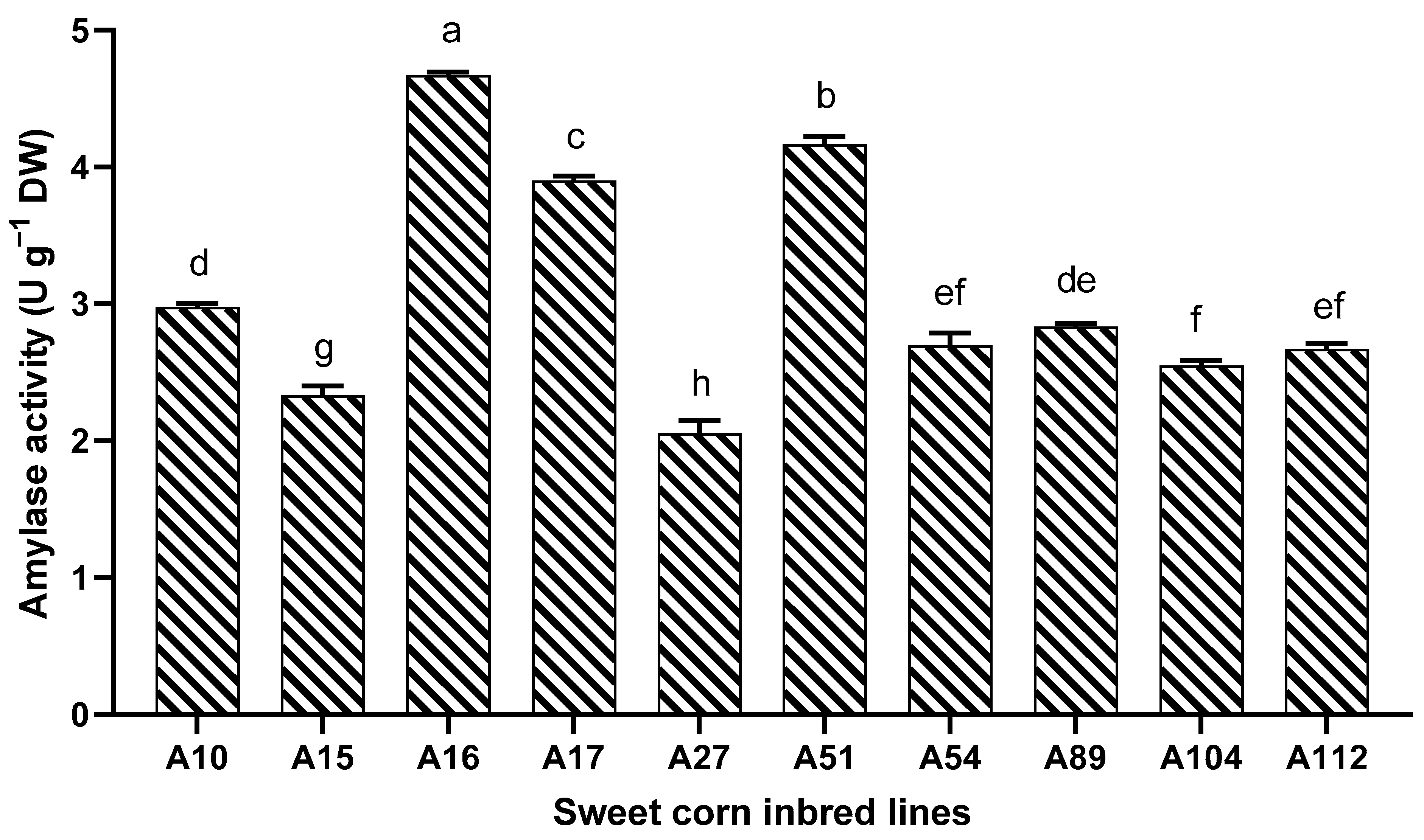
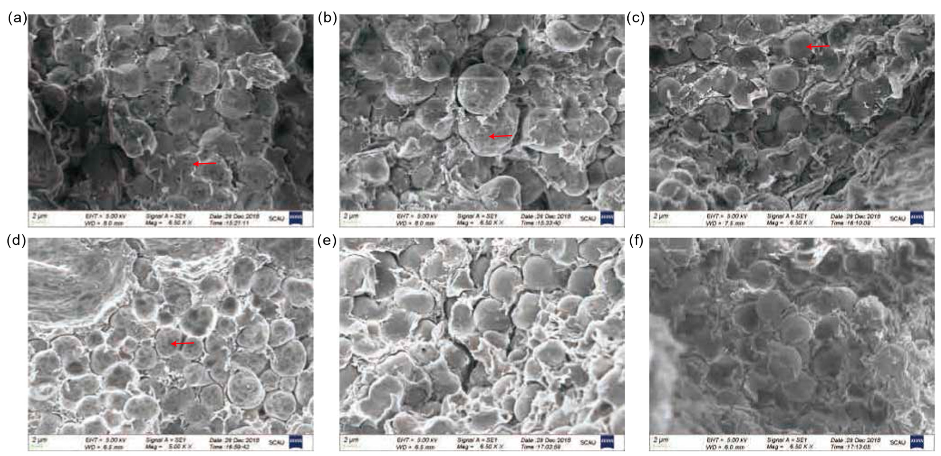
3.4. Evaluation of Amylase Activities and Ultrastructural Observations

Obvious differences were observed among ten lines for amylase activity, as depicted in Figure 4. The analysis of amylase activity indicated an average of 3.08 U g−1 DW, ranging from 2.05 to 4.67 U g−1 DW, with a coefficient of variation of 27.91. After 8 months of aging, A16 demonstrated the highest amylase activity, while A27 showed the lowest.
Ultrastructural observations were conducted using 8-month-aged seeds of the low vigor sweet corn inbred line A51 and the high-vigor sweet corn inbred line A89 by scanning electron microscopy. Under a magnification of 6500×, the ultrastructure of the starch grain was observed at three different locations, namely the center of the endosperm, the junction between the endosperm and aleurone layer, and the junction between the endosperm and the embryo. As shown in Figure 5, clear evidence of starch grain decomposition was observed in the junction between endosperm and aleurone layer, the center of endosperm, and the junction between endosperm and embryo for the low-vigor inbred line A51. However, for the high vigor inbred line A89, starch grain decomposition was only observed at the junction between the endosperm and aleurone layer. This result is consistent with the difference in amylase activity between A51 and A89.

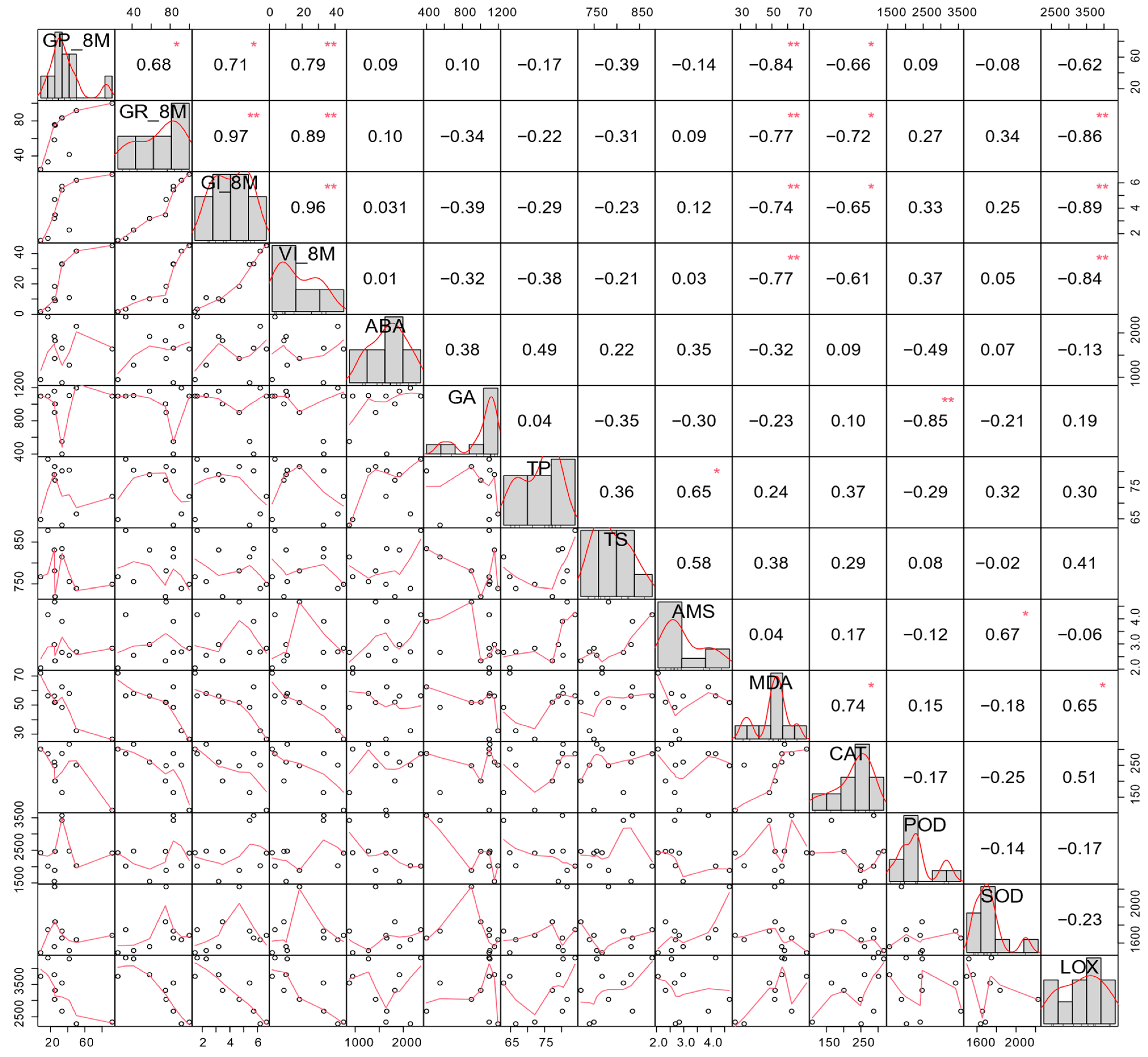
3.5. Correlation of Seed Vigor Indices and Physiological Indices in Naturally Aged Seeds

Correlative analyses were conducted on four seed vigor indices and ten biochemical indices of seeds from 10 inbred lines following natural aging over 8 months (Figure 6). Positive correlations were observed among the four seed vigor indices. The highest correlation coefficient was found between GR and GI, with a correlation coefficient of 0.97. A positive correlation was also detected between TP and AMS, with a correlation coefficient of 0.65. Furthermore, the four seed vigor indices GP, GR, GI, and VI were found to be negatively correlated with MDA content with respective correlation coefficients of −0.84, −0.77, −0.74, and −0.77, indicating that the content of MDA affects seed storage longevity. Additionally, GR, GI, and VI were each found to be negatively correlated with LOX activity, exhibiting respective coefficients of −0.86, −0.89, and −0.84. Notably, LOX activity showed a positive correlation with MDA content (coefficient: 0.65), indicating a direct relationship between LOX-catalyzed oxidation of polyunsaturated fatty acids and MDA generation. Our findings also revealed negative correlations between CAT and GP, GR, and GI, whereas a positive correlation with MDA was observed. Moreover, POD exhibited a highly negative correlation with GA content, while SOD showed a positive correlation with AMS.

4. Discussion

Natural aging is a direct method for studying seed storability; however, assessing seed vigor is time-consuming and challenging under natural storage conditions. As such, its application in seed storage research presents certain challenges [34]. In this study, we determined the biochemical indicators and ultrastructural changes in naturally aged seeds. Our findings demonstrate a negative correlation between seed vigor indicators and levels of MDA as well as LOX activity. This method authentically reflects the biochemical and ultrastructural changes in the naturally aged seeds, providing a new perspective for enhancing sweet corn seed storage techniques.
GP, GR, GI, and VI are important indicators reflecting seed vigor. Generally, with increased aging time, these indicators showed a decreasing trend. The degree of seed aging is influenced by both genetic and environmental factors [12]. In this study, significant differences were observed in the four seed vigor indicators among natural aging treatments, inbred lines, and the interaction between treatments and lines. In particular, A89 and A54 exhibited considerable natural aging tolerance, thereby suggesting their potential utility in improving seed aging tolerance in sweet corn.
Numerous studies have reported an increase in the content of MDA in aging seeds. MDA serves as a primary biomarker of oxidative damage and also represents one of the end products released during the process of lipid peroxidation [12]. Reports have highlighted that lipid peroxidation is a primary event during the seed aging process [10]. There are also reports suggesting that lipid peroxidation and free radical-mediated cell membrane damage are the main injuries during seed aging [35]. Seyyedi et al. [36] found that the accelerated aging of rapeseed seeds increased the percentage of saturated fatty acids while reducing the percentage of unsaturated fatty acids. Seed vigor-related traits were negatively correlated with unsaturated fatty acids in rapeseed [37]. Levels of several diacylglycerols and free fatty acids increased in Arabidopsis seeds [38]. Levels of oxidized lipids and related volatiles increase under all four storage conditions in rice [39]. The fatty acids of maize seeds are primarily in the embryo, characterized by low stearic acid content and high oleic acid content, which is sensitive to oxidation and tends to be unstable under high-temperature conditions [40]. However, the weight proportion of embryos within sweet corn kernels is higher, accompanied by a greater oil content when compared to flint corn. It was observed that the high oil content in hybrid maize seeds led to a decrease in germination percentage and seed vigor [41]. LOX specifically catalyzes the oxygenation reaction of the pentadiene structure of polyunsaturated fatty acids, generating hydrogen peroxide and forming MDA as its final product, which leads to cellular membrane damage. Previous studies have demonstrated that knocking out OsLOX10 in rice can increase seed longevity [42]. In this study, differences in LOX activity and MDA content were observed among 10 inbred lines (Figure 3a,b), both showing negative correlations with four seed vigor indicators (Figure 6). These results indicate that MDA content and LOX activities could be key physiological indexes to evaluate seed quality in storage.
During drying, the cytoplasm transforms into a glassy state, which exhibits slow molecular mobility and high molecular packing [43]. However, respiration and storage conditions with high temperature and humidity can increase the water content of the seeds and make the seed tissue out of its glassy state [44]. Alterations in antioxidant enzyme activities during seed aging have been identified across multiple crops. For instance, a decrease of 12%, 17%, and 20% in CAT, MDHAR, and APX activities, respectively, was observed in rice seeds aged for one month compared to non-aged seeds [13]. CAT and APX activities increased in oat seeds aged for 16 and 32 days as well [45]. And similar findings were also reported in sweet corn [46]. Therefore, it is hypothesized that although the enzymatic proteins in dry seeds cannot freely move, they remain active. Compared to wild-type seeds, the germination ratio increased in the tobacco seeds overexpressing the Cu/Zn-SOD gene after two years of dry storage at room temperature [47]. NADH activity may be a critical node in seed aging, characterized by a diminished ability to oxidize NADH, which subsequently reduces substrate oxidation and increases ROS accumulation in rice [48]. In this study, significant differences were found in the antioxidant enzyme activity and MDA content among different inbred lines (Figure 3a). A negative correlation was detected between MDA content and four seed vigor indices. However, no correlations were observed between the seed vigor indices and POD/SOD enzyme activity, possibly due to the limited material sampling in this study. Nevertheless, the reason for the negative correlation between CAT activity and seed vigor indices after the natural aging of sweet corn seeds requires further investigation.
Phytohormones play pivotal roles in seed maturation, germination, and post-germination growth in plants [23,49,50]. Seed vigor exhibited a negative correlation with ABA content under artificially accelerated aging, whereas ABA and GA content did not correlate with seed vigor under natural aging [31]. Consistently, no correlation between the ABA and GA contents and seed vigor indicators was detected in our study.
During the storage period, seeds remain metabolically active, leading to energy consumption driven by respiration [12,51]. Respiration during storage results in the degradation of sugars and proteins, causing a decline in sugar and protein content within seeds and subsequently reducing their vigor [52]. There was a negative correlation between seed vigor and carbonyl protein content in wheat [53]. In this study, differences in starch and protein content were detected in aged seeds (Figure 2a,b), with obvious starch granule breakdown observed in the weak seed vigor line (Figure 5); however, no correlations between seed vigor indicators and starch or protein content were identified.
In this study, we analyzed the seed vigor, physiological and biochemical parameters, and ultrastructure of sweet corn seeds that had undergone natural aging for 8 months. As a result, we identified two inbred lines with relatively strong resistance to natural aging. Significant differences in four seed vigor indicators were observed among sweet corn lines, with lower LOX activity and less MDA accumulation contributing to the seed aging tolerance. Our findings suggest that changes in seed lipids should be a key focus in the storage of sweet corn seeds.

Author Contributions

F.F. and G.Y. designed the experiments, and F.F. obtained funding for the research. R.Y. and G.Y. contributed to compiling and analyzing the data and wrote the manuscript. Y.D. and Y.L. conducted the statistical analysis. R.Y. and D.L. performed the experimental analyses. All authors have read and agreed to the published version of the manuscript.

Funding

This research was supported by the National Natural Science Foundation of China (31871713), the Guangzhou Science and Technology Plan (2024B03J1303), the Open Project of Key Laboratory of Crop Breeding in South Zhejiang Province (2023SZCB03), the Team Project of Guangdong Agricultural Department (2022Kj106, 2023Kj106), and the Xinzhou Applied Basic Research Project (202105).

Institutional Review Board Statement

Not applicable.

Data Availability Statement

The data presented in this study are available upon request from the corresponding author.

Acknowledgments

We would like to thank Wang Bo for English language editing.

Conflicts of Interest

The authors declare no conflicts of interest.

References

  1. Revilla, P.; Anibas, C.M.; Tracy, W.F. Sweet Corn Research around the World 2015–2020. Agronomy 2021, 11, 534. [Google Scholar] [CrossRef]
  2. Fan, L.; Yan, Q. Research Progress on the Reasons for Low Seed Vitality of Sweet Corn and Its Seed Treatment Techniques. Chin. Agric. Sci. Bull. 1996, 12, 24–26. [Google Scholar]
  3. Li, K.; Huang, C. Current Production Status, Problem and Countermeasure on Sweet Corn Industry in China. Sugar Crops China 2021, 43, 67–71. [Google Scholar] [CrossRef]
  4. Kibinza, S.; Vinel, D.; Côme, D.; Bailly, C.; Corbineau, F. Sunflower seed deterioration as related to moisture content during ageing, energy metabolism and active oxygen species scavenging. Physiol. Plant. 2006, 128, 496–506. [Google Scholar] [CrossRef]
  5. Morscher, F.; Kranner, I.; Arc, E.; Bailly, C.; Roach, T. Glutathione redox state, tocochromanols, fatty acids, antioxidant enzymes and protein carbonylation in sunflower seed embryos associated with after-ripening and ageing. Ann. Bot. 2015, 116, 669–678. [Google Scholar] [CrossRef]
  6. Sano, N.; Rajjou, L.; North, H.M.; Debeaujon, I.; Marion-Poll, A.; Seo, M. Staying Alive: Molecular Aspects of Seed Longevity. Plant Cell Physiol. 2016, 57, 660–674. [Google Scholar] [CrossRef] [PubMed]
  7. Ratajczak, E.; Malecka, A.; Bagniewska-Zadworna, A.; Kalemba, E.M. The production, localization and spreading of reactive oxygen species contributes to the low vitality of long-term stored common beech (Fagus sylvatica L.) seeds. J. Plant Physiol. 2015, 174, 147–156. [Google Scholar] [CrossRef]
  8. Gill, S.S.; Tuteja, N. Reactive oxygen species and antioxidant machinery in abiotic stress tolerance in crop plants. Plant Physiol. Biochem. 2010, 48, 909–930. [Google Scholar] [CrossRef]
  9. Sharma, P.; Jha, A.B.; Dubey, R.S.; Pessarakli, M. Reactive Oxygen Species, Oxidative Damage, and Antioxidative Defense Mechanism in Plants under Stressful Conditions. J. Bot. 2012, 2012, 217037. [Google Scholar] [CrossRef]
  10. Ebone, L.A.; Caverzan, A.; Chavarria, G. Physiologic alterations in orthodox seeds due to deterioration processes. Plant Physiol. Biochem. 2019, 145, 34–42. [Google Scholar] [CrossRef]
  11. Gerna, D.; Ballesteros, D.; Arc, E.; Stöggl, W.; Seal, C.E.; Marami-Zonouz, N.; Na, C.S.; Kranner, I.; Roach, T. Does oxygen affect ageing mechanisms of Pinus densiflora seeds? A matter of cytoplasmic physical state. J. Exp. Bot. 2022, 73, 2631–2649. [Google Scholar] [CrossRef] [PubMed]
  12. Zhang, K.L.; Zhang, Y.; Sun, J.; Meng, J.S.; Tao, J. Deterioration of orthodox seeds during ageing: Influencing factors, physiological alterations and the role of reactive oxygen species. Plant Physiol. Biochem. 2021, 158, 475–485. [Google Scholar] [CrossRef] [PubMed]
  13. Yin, G.; Xin, X.; Song, C.; Chen, X.; Zhang, J.; Wu, S.; Li, R.; Liu, X.; Lu, X. Activity levels and expression of antioxidant enzymes in the ascorbate-glutathione cycle in artificially aged rice seed. Plant Physiol. Biochem. 2014, 80, 1–9. [Google Scholar] [CrossRef] [PubMed]
  14. Mittler, R. ROS Are Good. Trends Plant Sci. 2017, 22, 11–19. [Google Scholar] [CrossRef] [PubMed]
  15. Waszczak, C.; Carmody, M.; Kangasjärvi, J. Reactive Oxygen Species in Plant Signaling. Annu. Rev. Plant Biol. 2018, 69, 209–236. [Google Scholar] [CrossRef] [PubMed]
  16. Malea, P.; Kokkinidi, D.; Kevrekidou, A.; Adamakis, I.S. The Enzymatic and Non-Enzymatic Antioxidant System Response of the Seagrass Cymodocea nodosa to Bisphenol-A Toxicity. Int. J. Mol. Sci. 2022, 23, 1348. [Google Scholar] [CrossRef] [PubMed]
  17. Ratajczak, E.; Malecka, A.; Ciereszko, I.; Staszak, A.M. Mitochondria Are Important Determinants of the Aging of Seeds. Int. J. Mol. Sci. 2019, 20, 1568. [Google Scholar] [CrossRef] [PubMed]
  18. Gaschler, M.M.; Stockwell, B.R. Lipid peroxidation in cell death. Biochem. Biophys. Res. Commun. 2017, 482, 419–425. [Google Scholar] [CrossRef]
  19. Tsikas, D. Assessment of lipid peroxidation by measuring malondialdehyde (MDA) and relatives in biological samples: Analytical and biological challenges. Anal. Biochem. 2017, 524, 13–30. [Google Scholar] [CrossRef]
  20. Min, C.W.; Lee, S.H.; Cheon, Y.E.; Han, W.Y.; Ko, J.M.; Kang, H.W.; Kim, Y.C.; Agrawal, G.K.; Rakwal, R.; Gupta, R.; et al. In-depth proteomic analysis of Glycine max seeds during controlled deterioration treatment reveals a shift in seed metabolism. J. Proteom. 2017, 169, 125–135. [Google Scholar] [CrossRef]
  21. Sugliani, M.; Rajjou, L.; Clerkx, E.J.; Koornneef, M.; Soppe, W.J. Natural modifiers of seed longevity in the Arabidopsis mutants abscisic acid insensitive3-5 (abi3-5) and leafy cotyledon1-3 (lec1-3). New Phytol. 2009, 184, 898–908. [Google Scholar] [CrossRef]
  22. Clerkx, E.J.M.; Blankestijn-De Vries, H.; Ruys, G.J.; Groot, S.P.C.; Koornneef, M. Genetic differences in seed longevity of various Arabidopsis mutants. Physiol. Plant. 2004, 121, 448–461. [Google Scholar] [CrossRef]
  23. Zheng, Q.; Teng, Z.; Zhang, J.; Ye, N. ABA Inhibits Rice Seed Aging by Reducing H2O2 Accumulation in the Radicle of Seeds. Plants 2024, 13, 809. [Google Scholar] [CrossRef]
  24. Pellizzaro, A.; Neveu, M.; Lalanne, D.; Ly Vu, B.; Kanno, Y.; Seo, M.; Leprince, O.; Buitink, J. A role for auxin signaling in the acquisition of longevity during seed maturation. New Phytol. 2020, 225, 284–296. [Google Scholar] [CrossRef]
  25. Yuan, Z.; Fan, K.; Wang, Y.; Tian, L.; Zhang, C.; Sun, W.; He, H.; Yu, S. OsGRETCHENHAGEN3-2 modulates rice seed storability via accumulation of abscisic acid and protective substances. Plant Physiol. 2021, 186, 469–482. [Google Scholar] [CrossRef] [PubMed]
  26. Bueso, E.; Munoz-Bertomeu, J.; Campos, F.; Brunaud, V.; Martinez, L.; Sayas, E.; Ballester, P.; Yenush, L.; Serrano, R. ARABIDOPSIS THALIANA HOMEOBOX25 uncovers a role for Gibberellins in seed longevity. Plant Physiol. 2014, 164, 999–1010. [Google Scholar] [CrossRef]
  27. Bizouerne, E.; Buitink, J.; Vu, B.L.; Vu, J.L.; Esteban, E.; Pasha, A.; Provart, N.; Verdier, J.; Leprince, O. Gene co-expression analysis of tomato seed maturation reveals tissue-specific regulatory networks and hubs associated with the acquisition of desiccation tolerance and seed vigour. BMC Plant Biol. 2021, 21, 124. [Google Scholar] [CrossRef] [PubMed]
  28. Livne, S.; Lor, V.S.; Nir, I.; Eliaz, N.; Aharoni, A.; Olszewski, N.E.; Eshed, Y.; Weiss, D. Uncovering DELLA-Independent Gibberellin Responses by Characterizing New Tomato procera Mutants. Plant Cell 2015, 27, 1579–1594. [Google Scholar] [CrossRef] [PubMed]
  29. Guan, Y.J.; Hu, J.; Wang, Z.F.; Zhu, S.J.; Wang, J.C.; Knapp, A. Time series regression analysis between changes in kernel size and seed vigor during developmental stage of sh2 sweet corn (Zea mays L.) seeds. Sci. Hortic. 2013, 154, 25–30. [Google Scholar] [CrossRef]
  30. Zhu, Y.; Xia, L.; Zhu, S.; Liu, J.; Yang, R.; Wang, Q.; Li, X.; Feng, F. Changes of vigor, physiological characteristics and genetic diversities of artificially aged sweet corn seeds. J. South China Agric. Univ. 2018, 39, 25–30. [Google Scholar] [CrossRef]
  31. Wang, B.; Yang, R.; Ji, Z.; Zhang, H.; Zheng, W.; Zhang, H.; Feng, F. Evaluation of Biochemical and Physiological Changes in Sweet Corn Seeds under Natural Aging and Artificial Accelerated Aging. Agronomy 2022, 12, 1028. [Google Scholar] [CrossRef]
  32. Yan, L.; Yi, Q.; Xiao-Min, Z.; Yi-Fang, C.; Li, W.; Yu-Ping, L. Optimization of water-rich starch sample preparation methods for scanning electron microscopy. Plant Sci. J. 2018, 36, 119–126. [Google Scholar] [CrossRef]
  33. Chen, C.; Chen, H.; Zhang, Y.; Thomas, H.R.; Frank, M.H.; He, Y.; Xia, R. TBtools: An Integrative Toolkit Developed for Interactive Analyses of Big Biological Data. Mol. Plant 2020, 13, 1194–1202. [Google Scholar] [CrossRef] [PubMed]
  34. Michalczyk, D.J.; Krupka, M.; Kamiński, J.; Wierzbicka, M.; Floryańska, S.; Kopeć, W.; Piotrowicz-Cieślak, A.I. Physiological and Biochemical Parameters of Field Bean (Vicia faba var. minor) Seeds Stored for 33 Years. Agriculture 2023, 13, 2012. [Google Scholar] [CrossRef]
  35. Fu, Y.-B.; Ahmed, Z.; Diederichsen, A. Towards a better monitoring of seed ageing under ex situ seed conservation. Conserv. Physiol. 2015, 3, cov026. [Google Scholar] [CrossRef] [PubMed]
  36. Seyyedi, S.M.; Tavakkol Afshari, R.; Daneshmandi, M.S. The relationships between fatty acids and heterotrophic seedling growth in winter canola cultivars during accelerated seed aging process. S. Afr. J. Bot. 2018, 119, 353–361. [Google Scholar] [CrossRef]
  37. Ding, Y.; Hou, D.; Yin, Y.; Chen, K.; He, J.; Yan, S.; Li, H.; Xiong, Y.; Zhou, W.; Li, M. Genetic dissection of Brassica napus seed vigor after aging. Theor. Appl. Genet. 2024, 137, 141. [Google Scholar] [CrossRef] [PubMed]
  38. Oenel, A.; Fekete, A.; Krischke, M.; Faul, S.C.; Gresser, G.; Havaux, M.; Mueller, M.J.; Berger, S. Enzymatic and Non-Enzymatic Mechanisms Contribute to Lipid Oxidation During Seed Aging. Plant Cell Physiol. 2017, 58, 925–933. [Google Scholar] [CrossRef]
  39. Prasad, C.T.M.; Kodde, J.; Angenent, G.C.; de Vos, R.C.H.; Diez-Simon, C.; Mumm, R.; Hay, F.R.; Siricharoen, S.; Yadava, D.K.; Groot, S.P.C. Experimental rice seed aging under elevated oxygen pressure: Methodology and mechanism. Front. Plant Sci. 2022, 13, 1050411. [Google Scholar] [CrossRef]
  40. Du, H.; Huang, M.; Hu, J.; Li, J. Modification of the fatty acid composition in Arabidopsis and maize seeds using a stearoyl-acyl carrier protein desaturase-1 (ZmSAD1) gene. BMC Plant Biol. 2016, 16, 137. [Google Scholar] [CrossRef]
  41. Huang, X.Y.; Liu, P.Q.; Peng, Y.Z.; Xiong, X.F.; Cai, J.G.; Lu, G.Y. Seed Vigor Comparisons of the High Oil Corn Hybrids and the Common Corn Hybrids Under Different Accelerated Aging Conditions. J. Maize Sci. 2003, 11, 36–38. [Google Scholar] [CrossRef]
  42. Wang, F.; Xu, H.; Zhang, L.; Shi, Y.; Song, Y.; Wang, X.; Cai, Q.; He, W.; Xie, H.; Zhang, J. The lipoxygenase OsLOX10 affects seed longevity and resistance to saline-alkaline stress during rice seedlings. Plant Mol. Biol. 2023, 111, 415–428. [Google Scholar] [CrossRef] [PubMed]
  43. Buitink, J.; Leprince, O. Glass formation in plant anhydrobiotes: Survival in the dry state. Cryobiology 2004, 48, 215–228. [Google Scholar] [CrossRef] [PubMed]
  44. Buitink, J.; Leprince, O. Intracellular glasses and seed survival in the dry state. C. R. Biol. 2008, 331, 788–795. [Google Scholar] [CrossRef]
  45. Cheng, H.; Ma, X.Q.; Jia, S.G.; Li, M.L.; Mao, P.S. Transcriptomic analysis reveals the changes of energy production and AsA-GSH cycle in oat embryos during seed ageing. Plant Physiol. Biochem. 2020, 153, 40–52. [Google Scholar] [CrossRef]
  46. Wang, B.; Yang, R.; Zhang, Z.; Huang, S.; Ji, Z.; Zheng, W.; Zhang, H.; Zhang, Y.; Feng, F. Integration of miRNA and mRNA analysis reveals the role of ribosome in to anti-artificial aging in sweetcorn. Int. J. Biol. Macromol. 2023, 240, 124434. [Google Scholar] [CrossRef] [PubMed]
  47. Lee, Y.P.; Baek, K.-H.; Lee, H.-S.; Kwak, S.-S.; Bang, J.-W.; Kwon, S.-Y. Tobacco seeds simultaneously over-expressing Cu/Zn-superoxide dismutase and ascorbate peroxidase display enhanced seed longevity and germination rates under stress conditions. J. Exp. Bot. 2010, 61, 2499–2506. [Google Scholar] [CrossRef] [PubMed]
  48. Chen, B.Y.; Yin, G.L.; Whelan, J.; Zhang, Z.S.; Xin, X.; He, J.J.; Chen, X.L.; Zhang, J.M.; Zhou, Y.C.; Lu, X.X. Composition of Mitochondrial Complex I during the Critical Node of Seed Aging in Oryza sativa. J. Plant Physiol. 2019, 236, 7–14. [Google Scholar] [CrossRef]
  49. Ali, F.; Qanmber, G.; Li, F.; Wang, Z. Updated role of ABA in seed maturation, dormancy, and germination. J. Adv. Res. 2022, 35, 199–214. [Google Scholar] [CrossRef]
  50. Kozaki, A.; Aoyanagi, T. Molecular Aspects of Seed Development Controlled by Gibberellins and Abscisic Acids. Int. J. Mol. Sci. 2022, 23, 1876. [Google Scholar] [CrossRef]
  51. Yin, G.K.; Whelan, J.; Wu, S.H.; Zhou, J.; Chen, B.Y.; Chen, X.L.; Zhang, J.M.; He, J.J.; Xin, X.; Lu, X.X. Comprehensive Mitochondrial Metabolic Shift during the Critical Node of Seed Ageing in Rice. PLoS ONE 2016, 11, e0148013. [Google Scholar] [CrossRef] [PubMed]
  52. Abbade, L.C.; Takaki, M. Biochemical and physiological changes of Tabebuia roseoalba (Ridl.) Sandwith (Bignoniaceae) seeds under storage. J. Seed Sci. 2014, 36, 100–107. [Google Scholar] [CrossRef]
  53. Li, B.B.; Zhang, S.B.; Lv, Y.Y.; Wei, S.; Hu, Y.S. Reactive oxygen species-induced protein carbonylation promotes deterioration of physiological activity of wheat seeds. PLoS ONE 2022, 17, e0263553. [Google Scholar] [CrossRef] [PubMed]
Figure 1. Multiple comparisons of four seed vigor indicators between non-aged (0M) and 8-month-aged (8M) seeds (a), Germination potential. (b), germination ratio. (c), germination index. (d), vigor index). Statistical differences are denoted as ns, **, and ****, representing p > 0.05, p < 0.01, and p < 0.0001, respectively. GP, GR, GI, and VI denote germination potential, germination ratio, germination index, and vigor index, respectively.
Agriculture 14 01039 g001
Figure 2. Comparison analysis of Gibberellins content (a), Abscisic acid (b), total protein (c), and total starch (d) contents among 10 sweet corn lines in 8-month-aged seeds. Different lowercase letters indicated differences among inbred lines (p < 0.05, Duncan’s test).
Agriculture 14 01039 g002
Figure 3. Multiple comparisons of MDA contents (a), LOX (b), CAT (c), POD (d), and SOD (e) activities among 10 sweet corn lines in 8-month-aged seeds. MDA, LOX, CAT, POD, and SOD indicate the malondialdehyde contents, lipoxygenase, catalase, peroxidase, and superoxide dismutase activities, respectively. Different lowercase letters indicated statistical differences among inbred lines (p < 0.05, Duncan’s test).
Agriculture 14 01039 g003
Figure 4. Comparison analysis of Amylase activities among 10 sweet corn lines in 8-month-aged seeds. Different lowercase letters indicated statistical differences among inbred lines (p < 0.05, Duncan’s test).
Agriculture 14 01039 g004
Figure 5. Ultrastructural observation of naturally aged seeds for 8 months in A51 and A89. (ac) represent the connection between endosperm and aleurone layer, the middle of the endosperm, and the connection between endosperm and embryo of line A51, respectively. (df) represent the connection between endosperm and aleurone layer, the middle of the endosperm, and the connection between endosperm and embryo of line A89, respectively. The red arrows show the breakdown of starch grains.
Agriculture 14 01039 g005
Figure 6. Correlation analysis of seed vigor indexes and physiological indexes; * and ** represent statistical correlation coefficients at the level of 0.05 and 0.01, respectively. The upper triangular displays the correlation coefficients and their significance, while the lower triangular exhibits scatter plots for each trait. GP, GR, GI, VI, ABA, GA, TP, TS, AMS, MDA, CAT, POD, SOD, and LOX indicate germination potential, germination ratio, germination index, vigor index, abscisic acid, gibberellins, total protein, total starch, amylase, malondialdehyde, catalase, peroxidase, superoxide dismutase, and lipoxygenase, respectively.
Agriculture 14 01039 g006
Table 1. Two-way analysis of variance for four seed vigor indexes.
Soruce of VariationGermination PotentialGermination RatioGermination IndexVigor Index
MSp ValueMSp ValueMSp ValueMSp Value
natural aging25,174.00<0.0111,985.00<0.0141.22<0.018141.00<0.01
lines2524.00<0.011397.00<0.0110.38<0.01813.60<0.01
natural aging × lines598.00<0.01792.50<0.012.98<0.01244.90<0.01
Disclaimer/Publisher’s Note: The statements, opinions and data contained in all publications are solely those of the individual author(s) and contributor(s) and not of MDPI and/or the editor(s). MDPI and/or the editor(s) disclaim responsibility for any injury to people or property resulting from any ideas, methods, instructions or products referred to in the content.

