Article

Fusarium and Hazelnut: A Story of Twists and Turns

by
Beata Zimowska
1,
Agnieszka Ludwiczuk
2,
Gelsomina Manganiello
3,
Krzysztof Wojtanowski
2,
Izabela Kot
1,
Alessia Staropoli
3,
Francesco Vinale
4 and
Rosario Nicoletti
3,5,*
1
Department of Plant Protection, University of Life Sciences, 20-400 Lublin, Poland
2
Department of Pharmacognosy with the Medicinal Plant Garden, Medical University of Lublin, 20-439 Lublin, Poland
3
Department of Agricultural Sciences, University of Naples ‘Federico II’, 80055 Portici, Italy
4
Department of Veterinary Medicine and Animal Production, University of Naples ‘Federico II’, 80138 Naples, Italy
5
Council for Agricultural Research and Economics, Research Center for Olive, Fruit and Citrus Crops, 81100 Caserta, Italy
*
Author to whom correspondence should be addressed.
Agriculture 2024, 14(7), 1080; https://doi.org/10.3390/agriculture14071080
Submission received: 5 April 2024 / Revised: 28 June 2024 / Accepted: 2 July 2024 / Published: 4 July 2024
(This article belongs to the Section Crop Protection, Diseases, Pests and Weeds)

Abstract

:
In recent years, the number of reports of Fusarium in association with hazelnut (Corylus avellana) has been increasing worldwide, related to both pathogenic aptitude and endophytic occurrence. However, the assessment of the real ecological role and relevance to plant health of these fungi has been impaired by uncertainty in species identification, deriving from both the evolving taxonomic structure of the genus and an inaccurate use of molecular markers. In this paper, the characterization of two hazelnut endophytic strains isolated in Poland is reported with reference to their secondary metabolite profiles and interactions with pests and pathogens. Our results are indicative of a possible role of these strains in defensive mutualism which could be related to the production of several bioactive compounds, especially cyclohexadepsipeptides of the enniatin family. At the same time, these biochemical properties create some concern for the possible mycotoxin contamination of hazelnut products.

1. Introduction

Within the several fungal diseases reported among the adversities of hazelnut (Corylus avellana L.), those affecting fruits are of major concern due to their implications for product storage and possible mycotoxin contamination. Indeed, many fungi may have an economic impact in terms of the quality of fresh and confectionery products, making it necessary to achieve adequate control in both pre- and post-harvest fruit management [1]. While species of Penicillium and Aspergillus basically have impacts during storage and processing, other fungi start their infections in the field; hence, they can be monitored and more timely managed. This is the case for species of Fusarium, which establish various kinds of ecological interactions with the host plants and their associated organisms and are notorious mycotoxin producers [2].
Despite their widespread occurrence, infections of hazelnut fruits by Fusarium have only started being documented quite recently; however, some reports limit identification to the genus level [3,4,5,6,7], which is not very informative in terms of the possible phytopathological or ecological significance. In other cases, identifications were performed at the species level, but these data are not always entirely reliable because of both the evolving taxonomic structuring of Fusarium, still based on several ‘species complexes’ awaiting to be resolved [8,9], and the need to use an appropriate set of DNA markers. In Chile, isolates from brown-grayish spots on the nuts were identified as F. sporotrichioides after sequencing the internal transcribed spacers of rDNA (ITS) [10]; although of widespread use in fungal taxonomy, this marker is notoriously insufficient to correctly identify Fusarium spp. [9,11]. In fact, an updated blast in GenBank of the published sequences of these strains showed that one of them (code MF629827) matches better with F. avenaceum and F. lateritium, leaning for a more heterogeneous species assortment. Two isolates from moldy fruits collected in the field in Oregon were classified as F. lateritium and F. culmorum through ITS and translational elongation factor (tef-1) sequencing [12], the latter marker being more reliable for species identification in Fusarium; however, an updated blast in GenBank showed that both isolates match better with F. lateritium and a more recently described species (F. citricola), which is sister to the F. tricinctum species complex (FTSC) [13]. Indeed, the discovery of new Fusarium species is ongoing, and recent findings call for taxonomic adjustments even within the F. lateritium species complex (FLSC) [14].
Fusarium lateritium had previously been reported as the agent of gray necrosis of hazelnuts (NGN) in Latium, central Italy [15]. In that case, species identification was based on a complete set of DNA markers, and a phylogenetic analysis documented a certain heterogeneity of the bulk of isolates ascribed to this species at that time, with a clear separation between European and non-European isolates, regardless of the host [16]. More recently, the examination of isolates from hazelnuts affected by NGN in the same area confirmed morphological similarity with F. lateritium; however, the draft genome sequencing of one of these isolates demonstrated its closer proximity to F. tricinctum [17], introducing uncertainty about the correct identification of both the hazelnut isolates and those with DNA sequences deposited in GenBank as F. lateritium, which have been used as references over time. On the other hand, the possible misidentification of strains classified as F. lateritium, but actually more closely related to F. tricinctum, had already been pointed out in an earlier phylogenetic study [18].
Data concerning endophytic occurrence of Fusarium in hazelnut are even more limited and refer to a few studies carried out in Iran and Turkey in which isolates from several plant parts were ascribed to the species F. equiseti, F. fujikuroi, F. graminearum, F. oxysporum, F. proliferatum, and F. tricinctum based on morphological features or ITS sequencing [19]. Nevertheless, Fusarium spp. have a widespread occurrence as endophytes in tree crops [20,21,22,23,24,25,26]; their association with plants could be attributable either to interception during the latency stage of the disease cycle or to a real endophytic settlement with implications in defensive mutualism against pests and pathogens. The latter role could be mediated by bioactive secondary metabolites, which might also lead to fruit contamination with mycotoxins.
Indeed, the accumulation of additional data from other geographical areas is appropriate for disentangling the taxonomic puzzle of Fusarium associates of C. avellana, as well as for increasing our knowledge on the real impact of these fungi on plant health and the safety of hazelnut products. Here, we report the identification of two endophytic strains collected from branches of asymptomatic hazelnut trees thriving in a forest area in south-eastern Poland, their metabolomic profiles, as well as effects against plant pathogenic fungi and a model insect.

2. Materials and Methods

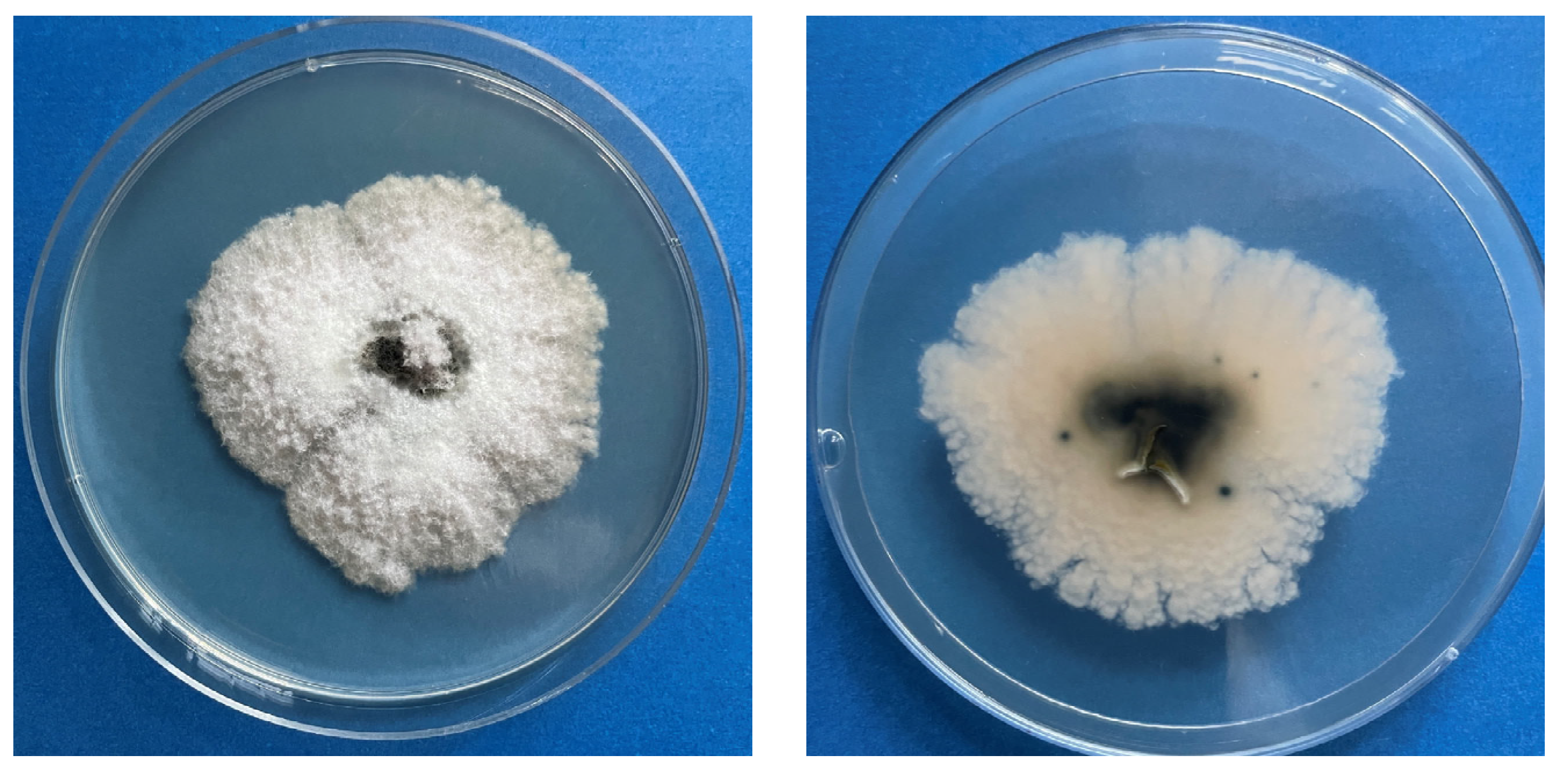
2.1. Isolation and Morphological Characterization

Within the cooperative research on endophytic fungi of C. avellana in progress at our laboratories [27], two Fusarium isolates (Hzn1 and Hzn5) were recovered from asymptomatic tissues of secondary branches of hazelnut plants collected in the Lublin voivodeship, south-eastern Poland (51.235598° N, 22.385680° E). Branch segments of about 4 cm in length were cut and surface-sterilized by consecutive immersions in 70% ethanol for 1 min, 3% sodium hypochlorite for 3 min, and 70% ethanol again for 1 min, and finally washed three times in sterile distilled water for 1 min. After removing bark with a sterile scalpel, the sterilized samples were cut into five pieces of 8 mm and aseptically transferred to Petri dishes containing potato dextrose agar (PDA: Difco, Detroit, MI, USA). The dishes were incubated at 25 ± 1 °C and the emerging endophytic fungi were transferred onto fresh PDA. From these plates, single-spore subcultures on PDA were prepared for each isolate prior to the identification procedure.
To investigate the morphological characteristics, the Fusarium strains were individually cultured on PDA and synthetic nutrient agar (SNA, made from ingredients in the laboratory) in darkness at 25 ± 1 °C. After 10 days, observations of phenotypes on PDA were carried out considering colony diameter, margins, pigmentation, and averse and reverse colors [28]. Micromorphological features concerning the formation of conidiogenous cells, conidia, and chlamydospores were inspected and photographed using a light microscope (Nikon Eclipse Ni-U, Tokyo, Japan).

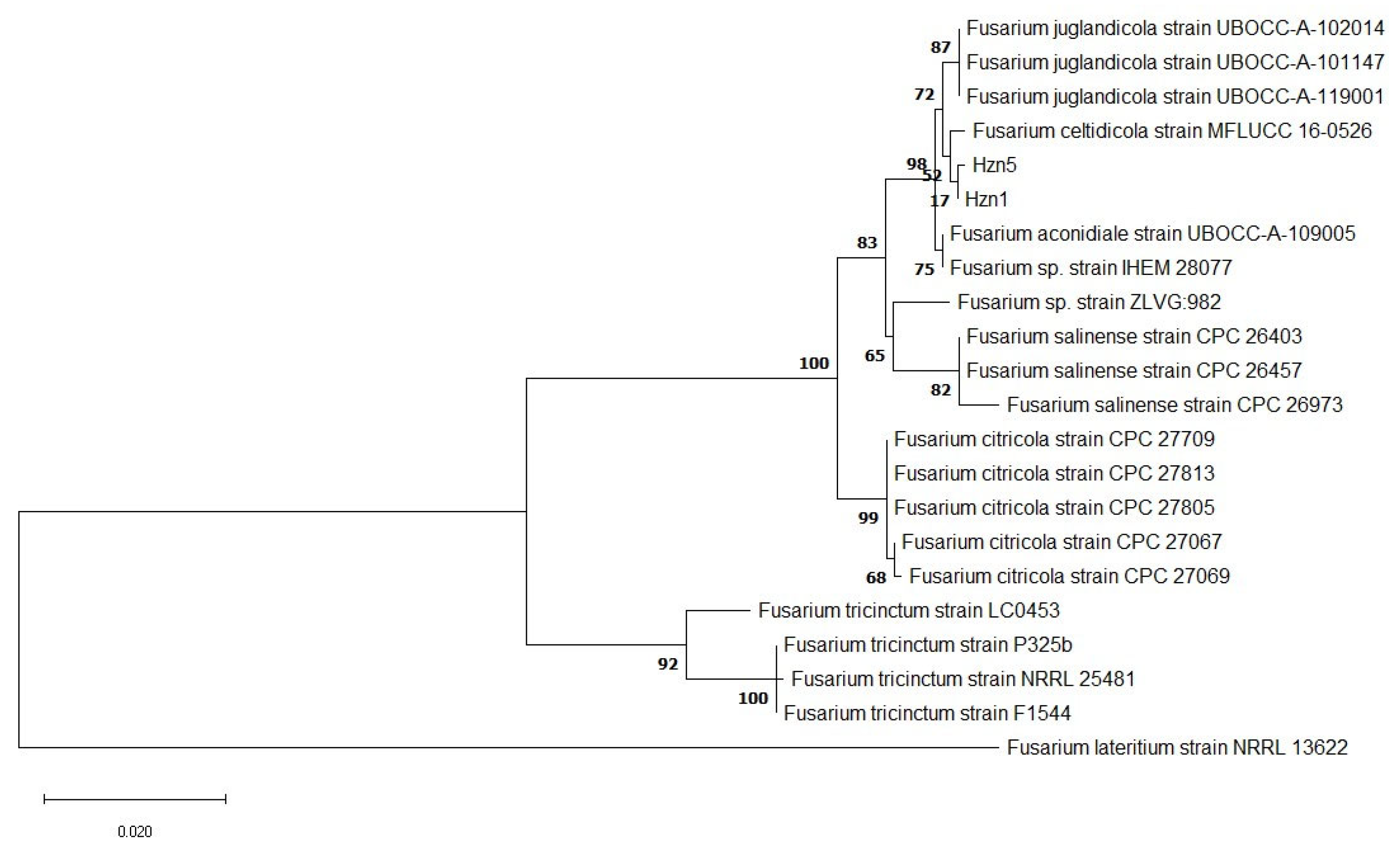
2.2. Biomolecular Markers and Phylogenetic Analysis

Morphological features were integrated with the sequencing of relevant DNA markers, namely ITS, tef-1, the RNA polymerase II (rpb2), and β-tubulin (tub) genes. One hundred milligrams of mycelium collected from PDA cultures of isolates Hzn1 and Hzn5 were ground in liquid nitrogen using a mortar and a pestle. The mycelial powder was transferred to sterile 2 mL tubes and DNA extraction was performed using the E.Z.N.A.® Plant & Fungal kit (Omega Bio-tek, Norcross, GA, USA), according to the manufacturer’s protocol. The extracted DNA was dissolved in 50 μL water (molecular biology grade), and its concentration and purity were checked through a DS-11 spectrophotometer (DeNovix Inc., Wilmington, DE, USA). PCR amplification was performed in 30 μL of DreamTaq Green PCR Master Mix (Thermo Scientific, Waltham, MA, USA) with 2 μL of genomic DNA and 1 μL of each primer (Table 1). The reaction parameters were initial denaturation at 95 °C for 5 min, followed by 35 cycles at 95 °C for 30 s, 58 °C for 30 s, and 72 °C for 45 s, with a final extension step at 72 °C for 10 min. PCR products were then run on 1.5% agarose gel stained with SYBR Safe DNA Gel Stain (Thermo Scientific) along with a 1 kb DNA ladder (A&A, Gdansk, Poland) to estimate the size of the amplified bands, and purified using magnetic beads (Quantabio, Beverly, MA, USA). Sanger sequencing of the purified PCR products was performed using the BigDye Terminator v3.1 cycle sequencing kit (Applied Biosystems, Waltham, MA, USA). Sequences were generated on an Applied Biosystems 3130XL Genetic Analyzer, optimized, and corrected manually when necessary. The obtained sequences were subjected to individual blast searches in GenBank for a preliminary identification based on sequence homology.
A phylogenetic analysis was carried out based on the concatenated rpb2 and tef-1 sequences, including reference strains of all of the species recognized within the Fusarium citricola species complex (FCCSC), four selected strains of F. tricinctum, and a strain of F. lateritium which was considered a genuine representative of this species in a recent comprehensive taxonomic study on Fusarium [35]. Moreover, two strains identified as Fusarium sp. were also included, since they both had rpb2 and tef-1 sequences deposited in GenBank and had high homology with our hazelnut isolates (Table 2). Individual gene sequences were validated through alignment using ClustalW 2.1 in MEGA X with the default alignment parameters [36] and checked manually. Sequences of rpb2 and tef-1 were then combined for multi-locus sequencing analysis (MLSA). Concatenated sequences were aligned and checked for alignment errors in the same manner as the individual gene sequences. The evolutionary history was inferred using the maximum likelihood method and Tamura-Nei model [37]. Initial trees for the heuristic search were obtained automatically by applying Neighbor-Joining and BioNJ algorithms to a matrix of pairwise distances estimated using the Tamura-Nei model and selecting the topology with the superior log likelihood value.

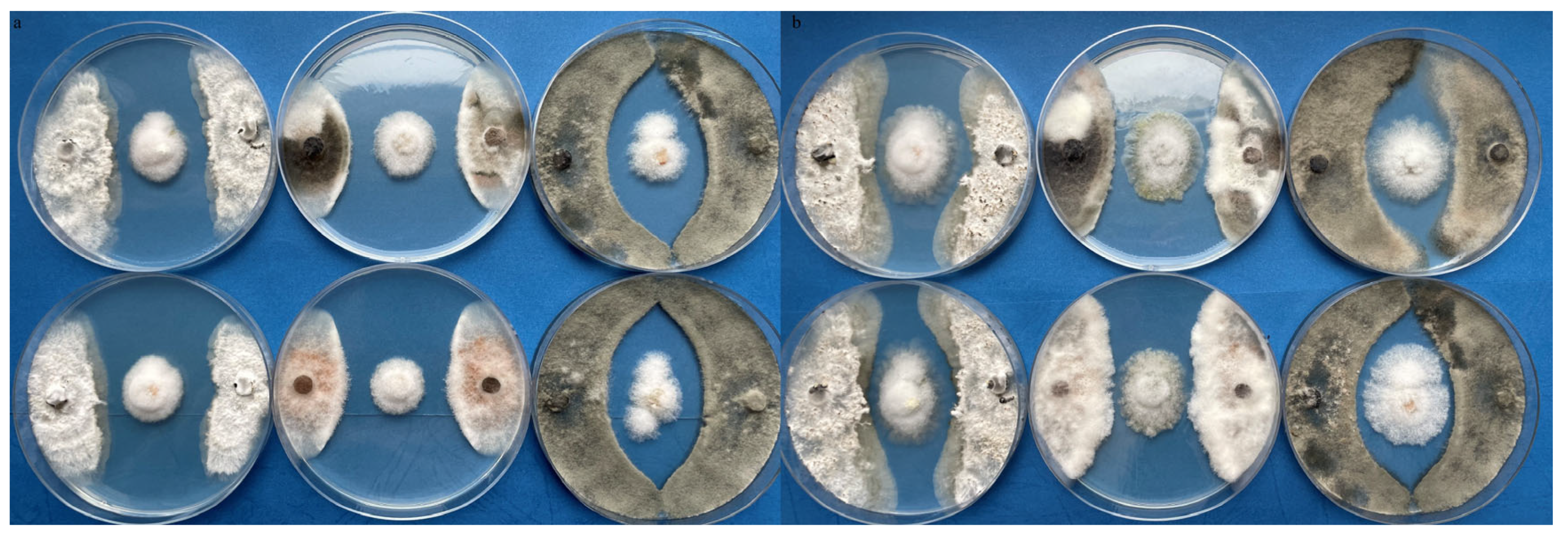
2.3. Antagonism against Plant Pathogenic Fungi

The interactions between the isolate Hzn5 and three strains of fungal pathogens which are also reported to infect hazelnut, namely Botrytis cinerea, Colletotrichum gloeosporioides, and Diaporthe eres, from the mycological collection of the Department of Plant Protection of the University of Life Sciences in Lublin, were examined in dual cultures. All of the fungi were preliminarily grown on PDA for 7 days at 25 ± 1 °C. Test plates were prepared on PDA by placing 5 mm diameter mycelial plugs of Hzn5 at the center and two mycelial plugs of each pathogen on the left and right sides at the same distance. The cultures were incubated at 25 ± 1 °C in darkness, and the inhibitory effects were visually recorded after 7 and 14 days.

2.4. Bioassays on Aphids

Parthenogenetic colonies of the bird cherry-oat aphid (Rhopalosiphum padi) are maintained at the Department of Plant Protection as a laboratory stock on plantlets of wheat (Triticum aestivum) in an environmental chamber at 25 ± 1 °C, with a 16:8 light–darkness photoperiod and 60 ± 5% relative humidity. Twenty nymphs from these colonies were placed onto new plantlets at the three-leaf stage. After the establishment of the new colonies, 5 mL of a conidial suspension of the isolate Hzn5 was applied using a hand sprayer. The conidial suspension was prepared by rinsing the surface of SNA cultures of Hzn5 with sterile distilled water mixed with 0.01% Tween 80, adjusting it to a density of 105 conidia mL−1 by using a hemocytometer. Control plants were treated at the same time with sterile distilled water mixed with 0.01% Tween 80. Both treatments were applied in triplicates. The number of adults and nymphs was counted daily to follow the colony development over 10 days after the treatment.
At the end of the experiment, 10 adult aphids were collected from the treated plants and plated on PDA after mild surface sterilization (70% ethanol for 1 min, followed by washing in sterile distilled water for 1 min). Moreover, fragments cut from both the leaves and stem under the sheaths were surface disinfected by dipping them in 3% sodium hypochlorite for 2 min, washed three times in sterile distilled water, and plated on PDA to check for possible colonization by Hzn5 of the inner tissues of the treated plants. The plates were kept in darkness at 25 ± 1 °C; on emergence, the fungal colonies were transferred to pure cultures for morphological identification.

2.5. Metabolomic Analysis

Following our previous experience concerning the differential production of secondary metabolites by Fusarium strains in different media [38], liquid cultures of isolates Hzn1 and Hzn5 were prepared on Czapek–Dox broth (CDB, Difco) and malt extract broth (MEB, Difco) in 500 mL Erlenmeyer flasks containing 250 mL of substrate. After two weeks of growth in darkness at 25 ± 1 °C, the cultures were filtered with 0.45 μm syringe filters, and the culture filtrates were extracted with an equal volume of chloroform in a separation funnel. The chloroform fractions were dried to remove residual water using anhydrous sodium sulfate, filtered, and concentrated under reduced pressure. They were then redissolved in methanol and qualitatively analyzed by a HPLC/ESI-QTOF-MS system in both positive and negative ion mode with a 6530B accurate-mass QTOF-MS mass spectrometer (Agilent Technologies, Santa Clara, CA, USA) with an ESI-Jet Stream ion source. The Agilent 1260 chromatograph was equipped with a DAD detector, autosampler, binary gradient pump, and column oven. The extract (10 μL) was injected and eluted using a mixture of water with 0.1% formic acid (solvent A) and acetonitrile with 0.1% formic acid (solvent B) as the mobile phase, with the following gradient: 0–45 min, 0–60% B; 45–46 min, 60–95% B; 46–55 min 95% B; the post-time was 10 min. The total time of analysis was 65 min, with a stable flow rate of 0.2 mL min−1. ESI-QToF-MS analysis was performed according to the following parameters of the ion source: dual-spray jet stream ESI; positive and negative ion mode; gas (N2) flow rate of 12 L min−1; nebulizer pressure of 35 psig; vaporizer temperature of 300 °C; m/z range of 100–1000 mass units, with acquisition Mode Auto MS/MS; collision-induced dissociation (CID) at 10 and 30 eV with an MS scan rate of one spectrum per s and two spectra per cycle; skimmer at 65 V; fragmenter at 140 V; octopole RF Peak of 750 V. The identification of compounds was based on both an in-house database and the MS-DIAL database (http://prime.psc.riken.jp/compms/msdial/main.html, accessed on 5 February 2024).
To check the possible release of secondary metabolites in plants treated with the conidial suspension of the isolate Hzn5, at the end of the experiment, the leaves and stems of wheat plants were cut into small pieces and an amount of 0.3 g was crushed in a mortar together with 3 mL chloroform as the extraction solvent. The extraction was repeated three times, and the crude extract was filtered through a Pasteur pipette packed with celite. The filtered extract was evaporated to afford a residue of 20 mg, which was redissolved in 1 mL methanol and analyzed by LC/MS following the above-described procedure.

3. Results

3.1. Identification and Phylogenetic Analysis

The colonies of isolates Hzn1 and Hzn5 were very similar in appearance: after a 10-day incubation, they reached a diameter of 45–47 mm on PDA and 78–85 mm on SNA. Aerial mycelium on PDA was abundant and dense, floccose to woolly, and white–cream with a crenate margin. The reverse was salmon-pink, darkening at the center, while no pigmentation was noticed in the medium (Figure 1). In older 30-day cultures on PDA, dark grey sclerotia-like structures were visible. On SNA, macroconidia were abundantly formed in sporodochia, from monophialidic conidiogenous cells, while microconidia were absent. Chlamydospores were formed quickly and abundant, mainly in chains, but also single or paired, smooth-walled, intercalary, and globose–subglobose to pyriform.
The obtained sequences of the DNA markers to be considered for biomolecular identification of isolates Hzn1 and Hzn5 were submitted to GenBank, with the following corresponding codes: Hzn1: OR178404 (ITS), PP738981 (rpb2), and PP788632 (tef-1); Hzn5: OR178403 (ITS), PP738982 (rpb2), PP788631 (tef-1), and PP791104 (Tub).
While confirming that isolates Hzn1 and Hzn5 are conspecific, the results of the blasts in GenBank displayed an inconsistency in their closest matches with the sequences available in this repository (Table 3), indicating uncertainty in species identification when merely based on DNA sequence homology. Therefore, a phylogenetic analysis was performed based on concatenated rpb2 and tef-1 sequences, which were considered more reliable markers for correct taxonomic identification with reference to the sequences of members of the FCCSC available in GenBank. This analysis unequivocally demonstrated that the two hazelnut endophytic isolates belong to this species complex, with a closer proximity to F. celtidicola (Figure 2). The additional strains included in the analysis because of high sequence homology with our isolates were also determined to belong to the FCCSC; in fact, the Belgian strain IHEM 28077 clustered together with the single available strain of F. aconidiale, while the Slovenian strain ZLVG.982 was positioned on an independent branch related to F. salinense.

3.2. Effects against Plant Pathogenic Fungi and Aphids

The growth of the tested plant pathogens was clearly affected in dual cultures with both Fusarium strains; the formation of inhibition zones, which persisted over two weeks, was indicative of the release of antibiotic products. The largest inhibition zone was observed in dual cultures with C. gloeosporioides (Figure 3). In the absence of contact between the opposing strains, no direct mycelial interactions could be observed.
An inhibitory effect was also evident in the development of colonies of R. padi on wheat plants. In fact, the number of adult aphids declined at a higher rate on plants treated with the conidial suspension, while the number of nymphs increased at a much lower rate compared to the control plants (Figure 4). Both of these trends are indicative of detrimental effects deriving from the inoculation of the isolate Hzn5, which could be dependent on either its entomopathogenic aptitude, or again the release of toxic products after its eventual settling in the plant tissues. As a matter of fact, the isolate Hzn5 could be re-isolated on PDA from the treated plants, as well as from all of the aphids sampled, demonstrating the ability of this fungus to establish itself as an endophyte in wheat plants and to infect R. padi. In the latter respect, it was not possible to assess whether the aphids used for re-isolation were infected during the treatment, or whether they assumed the fungus by sap sucking or through contact with other aphids. Of course, the real modalities have great relevance and deserve to be more deeply investigated in dedicated experiments.

3.3. Metabolomic Analysis

The conspecificity of isolates Hzn1 and Hzn5 was supported by the results of the LC-MS analysis of culture extracts, yielding very similar chromatographic profiles for both CDB and MEB culture extracts. As examined with reference to our in-house database and the MS-DIAL database, the monoisotopic mass data were clearly indicative of the presence of several typical Fusarium secondary metabolites (Table 4). In more detail, enniatins were consistently produced in both media; in fact, several members of this cyclohexadepsipeptide family were detectable in the analyzed culture extracts, with the notable exception of beauvericin. However, it is not possible to provide details on the identity for all of the analogues since some of them share the same molecular mass. The sesquiterpenoid culmorin was also detected in all of the analyzed samples, along with its precursor longiborneol. Likewise, the fusarielins, a series of polyketides with a decalin core, were also produced by both isolates; specifically, fusarielins A, B, F, and M were produced on both media, while other members of this family were detected only occasionally. Finally, chrysogine, a quinazolinone yellow pigment, was only detected in CDB cultures of both isolates.
The analysis of the extract obtained from wheat plants at the end of the assays carried out on aphids showed the presence of a peak at 51.02 min, corresponding to enniatin B (mass of 639.4094). None of the other identified compounds could be detected in the plant extract.

4. Discussion

Members of the FCCSC have been described since 2018; in particular, the founding species F. citricola was identified along with F. salinense from symptomatic tissues of several Citrus species sampled in southern Italy [13]. Afterwards, a single strain of F. celtidicola was recovered from a dead branch of the lote tree (Celtis australis) in Italy [39], while F. aconidiale and F. juglandicola were, respectively, identified from wheat and from walnut (Juglans regia) buds and fruits in France [40,41]. Both of the latter species have been more recently detected on larvae and inside galls of the cecidomyid midges Asphondylia echii and Lasioptera rubi in Slovakia [42], while F. juglandicola has been identified in leaves of mistletoe (Viscum album subsp. austriacum) in Poland [43]. However, as usually happens with recently characterized taxa, the FCCSC could be more widespread and include isolates previously identified with other names. This is more than a mere hypothesis, since our phylogenetic analysis demonstrated that two isolates provisionally classified as Fusarium sp. clearly belong to this species complex: one of them was identifiable as F. aconidiale, while the other, positioned next to F. salinense, could be representative of a new species. All members of the FCCSC were confirmed to be related to F. tricinctum, while a higher phylogenetic distance was found with F. lateritium, consistent with the inference of a recent study by Turco and coworkers [17] that hazelnut isolates ascribed to the latter species may have been misidentified. Moreover, considering the available GenBank sequences, close matches were found with isolates collected from dead branches of sycamore maple (Acer pseudoplatanus) in Slovenia [44], from olives in Greece [45], and from the boxelder maple (Acer negundo) in Pomerania, northern Poland [46], which were all identified as F. lateritium based on an incomplete set of markers. In particular, as compared with our Hzn5, the strain FI47S-18An from the latter source displayed 99.24% homology with tef-1 and 100% homology with tub sequences (Table 3); unfortunately, the rpb2 sequence of this strain is not available, preventing its inclusion in our phylogenetic analysis. Interestingly, the above points are indicative of the widespread distribution in central and southern Europe of species in the FCCSC, calling for a revision of the taxonomic identification of isolates from this geographical area which were ascribed to F. lateritium before 2018.
The laboratory assays displayed antibiosis against plant pathogenic fungi and entomopathogenic effects against aphids, which were consistent with the production of bioactive secondary metabolites by our isolates. Anti-insect assays were carried out on a model aphid species available at our laboratory, the results of which could be reasonably transposed at least to other hemipterans infesting hazelnut. Likewise, the plant pathogens used for the dual-culture assays are representative of several Ascomycetes classes, depicting a broad range of antifungal properties. Of course, the ecological conditions can be very variable due to the unpredictable influence of many abiotic and biotic factors; nevertheless, these biological properties may be relevant for conjecturing a symbiotic association with hazelnut contributing to defensive mutualism.
The metabolomic profiles of our isolates were consistent in comparison with one another on both substrates used. Due to their recent identification, no data are currently available on the secondary metabolite profile of species belonging to the FCCSC, while those of the closely related FTSC have been accurately examined [47,48]. Among the products which best characterize the latter species complex, no clues could be detected for moniliformin, aurofusarin, chlamydosporol, acuminatopyrone, 2-amino-14,16-dimethyloctadecan-3-ol, and fungerin, while clear indications were obtained for the presence of enniatins. Recently, concern has been raised regarding food contamination by these products due to their cytotoxicity observed in several mammalian cell lines and possible synergic effects with other mycotoxins [49,50,51,52]. So far, the ability to produce enniatins has been reported in many Fusarium species of phytopathological relevance, upon both direct detection [53,54,55,56,57] and identification of biosynthetic genes [58,59,60]. A recent review mentions 28 species from 7 species complexes: in particular, F. acuminatum, F. arthrosporioides, F. avenaceum, F. torulosum, and F. tricinctum from the FTSC; F. compactum, F. kyushuense, F. poae, F. sambucinum, and F. venenatum from the F. sambucinum s.c.; F. sporotrichioides from the F. sporotrichioides s.c.; F. acutatum, F. ananatum, F. andiyazi, F. antophilum, F. concentricum, F. fujikuroi, F. lactis, F. nygamai, F. proliferatum, F. ramigenum, F. subglutinans, F. temperatum, F. thapsinum, and F. verticillioides from the F. fujikuroi s.c.; F. equiseti from the F. incarnatum-equiseti s.c.; F. oxysporum from the F. oxysporum s.c.; and F. lateritium from the FLSC [61]. Besides plant pathogens, the production of enniatins has been repeatedly reported in endophytic isolates, belonging to species such as F. acuminatum [62,63], F. dimerum [64], F. tricinctum, and related species in the FTSC [56,57,65,66,67], and to the F. redolens s.c. [68]. Moreover, these compounds have been characterized for their insecticidal activity for a long time [69,70], as well as for their antifungal and antibacterial properties [57,71,72], implying a possible role in defensive mutualism of plant endophytic strains. Notably, the detection of enniatin B in extracts from wheat plants treated with the Hzn5 conidial suspension is indicative that at least this compound can be produced in vivo and eventually exert its bioactivity against plant pests and pathogens.
First identified as a secondary metabolite of Penicillium chrysogenum [73], chrysogine is also widespread in Fusarium. In fact, this compound has been reported from F. sambucinum [74], F. culmorum, F. equiseti [75], F. langsethiae, F. sporotrichioides [76], F. avenaceum [77], F. graminearum, F. pseudograminearum [78], F. tricinctum [79], F. cerealis [80], and F. oxysporum [81], and more recently from an endophytic F. multiceps [82]. Moreover, the presence of the putative gene cluster for the biosynthesis of chrysogine has been documented in the new species F. indicum [83].
Conversely, fusarielins have been reported from a restricted range of Fusarium species so far. In fact, an investigation involving 42 strains from 18 species showed that these compounds could only be detected in cultures of F. tricinctum (fusarielin A) and F. graminearum (fusarielins F, G, and H) [84]. Besides fusarielin A, the production of fusarielins B, J, K, and L was later documented in endophytic isolates of F. tricinctum [57,85], while fusarielins M and N have been identified as products of marine strains of F. graminearum [86,87]; a few more reports refer to Fusarium isolates of unidentified species [88,89,90]. Hence, our isolates seem to combine the biosynthetic abilities of both the known producer species. In terms of biological properties, fusarielins A and E were first reported for their antifungal activities [88,89], while their antibacterial properties were documented for other fusarielins [57,90], leaning toward an involvement of these compounds in the ecological interactions with other species which are part of the plant microbiome. Interestingly, genes for the biosynthesis of fusarielins were downregulated in the interaction of F. graminearum with the mycoparasite Clonostachys rosea [91]. Moreover, fusarielins have been reported to have antiproliferative [92] and estrogenic activities [93], calling for a more accurate assessment of their effects on human health.
First isolated from F. culmorum [94], culmorin is biosynthesized through the hydroxylation of longiborneol [95]. The latter compound, also produced by our isolates, is a known metabolite of F. tricinctum [48], while culmorin has never been reported from isolates of this species, despite a couple of biosynthetic genes being found in its genome [96]. Culmorin is regarded as an emerging mycotoxin, also produced by F. graminearum, F. crookwellense, and F. venenatum [97,98] where it co-occurs with trichothecenes, somehow influencing their toxicological properties [96,99]. However, no known trichothecenes could be identified in the secondary metabolite profiles of our strains. Similarly to fusarielins, moderate antifungal activity has been documented for culmorin [100], along with low insecticidal effects [101].
In addition to enniatins, it is relevant to consider that chrysogine [102,103], culmorin [97], and fusarielin A [104] have all been reported as contaminants in foodstuffs, calling for a more accurate assessment of their possible effects on human health, alone and in association with other co-occurring mycotoxins.

5. Conclusions

Within the context of uncertainty regarding the identification and the ecological role of Fusarium strains associated with hazelnut, two endophytic isolates collected in Poland were determined to belong to the FCCSC. This finding introduces the opportunity for a comparison with the hazelnut pathogenic strains previously identified as F. lateritium, also with the aim to assess whether the pathogenic aptitude on fruits is secondary to a more general endophytic habit.
Furthermore, this study represents the first metabolomic characterization of a member of the FCCSC, disclosing relatedness with the biosynthetic capabilities of species in the FTSC. The identified compounds, especially members of the enniatin family, may have implications in the defensive mutualism established in hazelnut plants by endophytic strains possessing these biosynthetic abilities; however, the infection of kernels progressing along with endophytic development may also affect the quality and safety of both fresh and processed hazelnut products.

Author Contributions

Conceptualization, B.Z. and R.N.; methodology, B.Z., A.L., G.M., K.W., I.K., A.S. and F.V.; validation, A.L., G.M. and F.V.; formal analysis, G.M., K.W., I.K. and A.S.; resources, B.Z., A.L., F.V. and R.N.; writing—original draft preparation, B.Z., A.L., G.M., F.V. and R.N.; writing—review and editing, B.Z., G.M. and R.N.; supervision, B.Z., F.V. and R.N.; funding acquisition, B.Z., F.V. and R.N. All authors have read and agreed to the published version of the manuscript.

Funding

This study was carried out within the Agritech National Research Center and received funding from European Union Next-Generation EU (Piano Nazionale di Ripresa e Resilienza (PNRR)—Missione 4 Componente 2, Investimento 1.4—D.D. 1032 17 June 2022, CN00000022). This manuscript reflects only the authors’ views and opinions; neither the European Union nor the European Commission can be considered responsible for them. G.M. acknowledges funding from PON R&I 2014–2020 (FSE REACT-EU) DM 1062 Azione IV.6 (CUP: E65F21003080003).

Institutional Review Board Statement

Not applicable.

Data Availability Statement

The sequences of DNA markers generated in this study have been deposited in the GenBank repository (www.ncbi.nlm.nih.gov/genbank/ accessed on 2 May 2024).

Acknowledgments

The authors gratefully acknowledge the contribution of Simon Gibbons (Natural and Medical Sciences Research Center, University of Nizwa, Oman) for the revision of the English style of the final version of the manuscript.

Conflicts of Interest

The authors declare no conflicts of interest.

References

  1. Salvatore, M.M.; Andolfi, A.; Nicoletti, R. Mycotoxin contamination in hazelnut: Current status, analytical strategies, and future prospects. Toxins 2023, 15, 99. [Google Scholar] [CrossRef]
  2. Munkvold, G.P.; Proctor, R.H.; Moretti, A. Mycotoxin production in Fusarium according to contemporary species concepts. Ann. Rev. Phytopathol. 2021, 59, 373–402. [Google Scholar] [CrossRef] [PubMed]
  3. Xu, Y.; Bianchini, A.; Hanna, M.A. Evaluation of mold and mycotoxin contaminations in hybrid hazelnuts grown in Nebraska. J. Food Process. Technol. 2011, 2, 119. [Google Scholar] [CrossRef]
  4. Sezer, A.; Dolar, F.S. Hazelnut kernel defects and associated fungi in three provinces in Turkey. In Proceedings of the 7th International Scientific Agriculture Symposium “Agrosym 2016”, Jahorina, Bosnia and Herzegovina, 6–9 October 2016; pp. 1312–1318. [Google Scholar]
  5. Battilani, P.; Chiusa, G.; Arciuolo, R.; Somenzi, M.; Fontana, M.; Castello, G.; Spigolon, N. Diaporthe as the main cause of hazelnut defects in the Caucasus region. Phytopathol. Mediterr. 2018, 54, 241–252. [Google Scholar]
  6. Arciuolo, R.; Santos, C.; Soares, C.; Castello, G.; Spigolon, N.; Chiusa, G.; Lima, N.; Battilani, P. Molecular characterization of Diaporthe species associated with hazelnut defects. Front. Plant Sci. 2020, 11, 611655. [Google Scholar] [CrossRef] [PubMed]
  7. Arciuolo, R.; Chiusa, G.; Castello, G.; Camardo Leggieri, M.; Spigolon, N.; Battilani, P. Diaporthe spp. is confirmed as the main fungus associated with defective Turkish hazelnuts. Plant Health Progr. 2022, 23, 440–448. [Google Scholar] [CrossRef]
  8. Lombard, L.; Sandoval-Denis, M.; Cai, L.; Crous, P.W. Changing the game: Resolving systematic issues in key Fusarium species complexes. Persoonia 2019, 43, i–ii. [Google Scholar] [CrossRef] [PubMed]
  9. Wang, M.M.; Crous, P.W.; Sandoval-Denis, M.; Han, S.L.; Liu, F.; Liang, J.M.; Duan, W.J.; Cai, L. Fusarium and allied genera from China: Species diversity and distribution. Persoonia 2022, 48, 1–53. [Google Scholar] [CrossRef] [PubMed]
  10. Duran, P.; Barra, P.J.; de la Luz Mora, M.; Morina, F.; Viscardi, S.; Meriño-Gergichevich, C. First report of fungal complex causing grey necrosis of hazelnut in Chile. New Dis. Rep. 2020, 42, 7. [Google Scholar] [CrossRef]
  11. Torres-Cruz, T.J.; Whitaker, B.K.; Proctor, R.H.; Broders, K.; Laraba, I.; Kim, H.S.; Brown, D.W.; O’Donnell, K.; Estrada-Rodriguez, T.L.; Lee, Y.H.; et al. FUSARIUM-ID v. 3.0: An updated, downloadable resource for Fusarium species identification. Plant Dis. 2022, 106, 1610–1616. [Google Scholar] [CrossRef]
  12. Pscheidt, J.W.; Heckert, S.; Wiseman, M.; Jones, L. Fungi associated with and influence of moisture on development of kernel mold of hazelnut. Plant Dis. 2019, 103, 922–928. [Google Scholar] [CrossRef] [PubMed]
  13. Sandoval-Denis, M.; Guarnaccia, V.; Polizzi, G.; Crous, P.W. Symptomatic Citrus trees reveal a new pathogenic lineage in Fusarium and two new Neocosmospora species. Persoonia 2018, 40, 1–25. [Google Scholar] [CrossRef] [PubMed]
  14. Dias Cavalcanti, A.; da Silva Santos, A.C.; de Oliveira Ferro, L.; Bezerra, J.D.; Souza-Motta, C.M.; Magalhães, O.M.C. Fusarium massalimae sp. nov. (F. lateritium species complex) occurs endophytically in leaves of Handroanthus chrysotrichus. Mycol. Progr. 2020, 19, 1133–1142. [Google Scholar] [CrossRef]
  15. Santori, A.; Vitale, S.; Luongo, L.; Belisario, A. First report of Fusarium lateritium as the agent of nut gray necrosis on hazelnut in Italy. Plant Dis. 2010, 94, 484. [Google Scholar] [CrossRef] [PubMed]
  16. Vitale, S.; Santori, A.; Wajnberg, E.; Castagnone-Sereno, P.; Luongo, L.; Belisario, A. Morphological and molecular analysis of Fusarium lateritium, the cause of gray necrosis of hazelnut fruit in Italy. Phytopathology 2011, 101, 679–686. [Google Scholar] [CrossRef] [PubMed]
  17. Turco, S.; Grottoli, A.; Drais, M.I.; De Spirito, C.; Faino, L.; Reverberi, M.; Cristofori, V.; Mazzaglia, A. Draft genome sequence of a new Fusarium isolate belonging to Fusarium tricinctum species complex collected from hazelnut in central Italy. Front. Plant Sci. 2021, 12, 788584. [Google Scholar] [CrossRef]
  18. Watanabe, M.; Yonezawa, T.; Lee, K.I.; Kumagai, S.; Sugita-Konishi, Y.; Goto, K.; Hara-Kudo, Y. Molecular phylogeny of the higher and lower taxonomy of the Fusarium genus and differences in the evolutionary histories of multiple genes. BMC Evol. Biol. 2011, 11, 322. [Google Scholar] [CrossRef]
  19. Nicoletti, R.; Zimowska, B. Endophytic fungi of hazelnut (Corylus avellana). Plant Prot. Sci. 2023, 59, 107–123. [Google Scholar] [CrossRef]
  20. Nicoletti, R. Endophytic fungi of citrus plants. Agriculture 2019, 9, 247. [Google Scholar] [CrossRef]
  21. Ren, F.; Dong, W.; Sun, H.; Yan, D.H. Endophytic mycobiota of jingbai pear trees in north China. Forests 2019, 10, 260. [Google Scholar] [CrossRef]
  22. Ren, F.; Dong, W.; Yan, D.H. Organs, cultivars, soil, and fruit properties affect structure of endophytic mycobiota of pinggu peach trees. Microorganisms 2019, 7, 322. [Google Scholar] [CrossRef] [PubMed]
  23. Nicoletti, R.; Di Vaio, C.; Cirillo, C. Endophytic fungi of olive tree. Microorganisms 2020, 8, 1321. [Google Scholar] [CrossRef] [PubMed]
  24. Nicoletti, R.; Beccaro, G.L.; Sekara, A.; Cirillo, C.; Di Vaio, C. Endophytic fungi and ecological fitness of chestnuts. Plants 2021, 10, 542. [Google Scholar] [CrossRef] [PubMed]
  25. Nekrasov, E.V.; Shumilova, L.P.; Gomzhina, M.M.; Aleksandrova, A.V.; Kokaeva, L.Y.; Pavlova, L.M. Diversity of endophytic fungi in annual shoots of Prunus mandshurica (Rosaceae) in the South of Amur region, Russia. Diversity 2022, 14, 1124. [Google Scholar] [CrossRef]
  26. Yabaneri, C.; Sevim, A. Endophytic fungi from the common walnut and their in vitro antagonistic activity against Ophiognomonia leptostyla. Biologia 2023, 78, 361–371. [Google Scholar] [CrossRef]
  27. Zimowska, B.; Nicoletti, R. Arcopilus aureus: A valuable endophytic associate of hazelnut. Acta Agrobot. 2023, 76, 175998. [Google Scholar] [CrossRef]
  28. Leslie, J.F.; Summerell, B.A. The Fusarium Laboratory Manual; Wiley: Hoboken, NJ, USA, 2008. [Google Scholar]
  29. Gardes, M.; Bruns, T.D. ITS primers with enhanced specificity for Basidiomycetes: Application to identification of mycorrhizae and rusts. Mol. Ecol. 1993, 2, 113–118. [Google Scholar] [CrossRef] [PubMed]
  30. White, T.J.; Bruns, T.D.; Lee, S.; Taylor, J. Amplification and direct sequencing of fungal ribosomal RNA genes for phylogenetics. In PCR Protocols, a Guide to Methods and Applications; Innis, M.A., Gelfand, D.H., Sninsky, J.J., White, T.J., Eds.; Academic Press: San Diego, CA, USA, 1990; pp. 315–322. [Google Scholar]
  31. Matheny, P.B. Improving phylogenetic inference of mushrooms with RPB1and RPB2 nucleotide sequences (Inocybe; Agaricales). Mol. Phylogen. Evol. 2005, 35, 1–20. [Google Scholar] [CrossRef] [PubMed]
  32. Stielow, J.B.; Lévesque, C.A.; Seifert, K.A.; Meyer, W.; Irinyi, L.; Smits, D.; Renfurm, R.; Verkley, G.J.M.; Groenewald, M.; Chaduli, D.; et al. One fungus, which genes? Development and assessment of universal primers for potential secondary fungal DNA barcodes. Persoonia 2015, 35, 242–263. [Google Scholar] [CrossRef]
  33. O’Donnell, K.; Cigelnik, E. Two different intragenomic rDNA ITS2 types within a monophyletic lineage of the fungus Fusarium are nonorthologous. Mol. Phylogenet. Evol. 1997, 7, 103–116. [Google Scholar] [CrossRef]
  34. Glass, N.L.; Donaldson, G.C. Development of primer sets designed for use with the PCR to amplify conserved genes from filamentous Ascomycetes. Appl. Environ. Microbiol. 1995, 61, 1323–1330. [Google Scholar] [CrossRef] [PubMed]
  35. Crous, P.W.; Lombard, L.; Sandoval-Denis, M.; Seifert, K.A.; Schroers, H.J.; Chaverri, P.; Gené, J.; Guarro, J.; Hirooka, Y.; Bensch, K.; et al. Fusarium: More than a node or a foot-shaped basal cell. Stud. Mycol. 2021, 98, 100116. [Google Scholar] [CrossRef] [PubMed]
  36. Kumar, S.; Stecher, G.; Li, M.; Knyaz, C.; Tamura, K. MEGA X: Molecular evolutionary genetics analysis across computing platforms. Mol. Biol. Evol. 2018, 35, 1547–1549. [Google Scholar] [CrossRef] [PubMed]
  37. Tamura, K.; Nei, M. Estimation of the number of nucleotide substitutions in the control region of mitochondrial DNA in humans and chimpanzees. Mol. Biol. Evol. 1993, 10, 512–526. [Google Scholar] [PubMed]
  38. Manganiello, G.; Marra, R.; Staropoli, A.; Lombardi, N.; Vinale, F.; Nicoletti, R. The shifting mycotoxin profiles of endophytic Fusarium strains: A case study. Agriculture 2019, 9, 143. [Google Scholar] [CrossRef]
  39. Shang, Q.J.; Phookamsak, R.; Camporesi, E.; Khan, S.; Lumyong, S.; Hyde, K.D. The holomorph of Fusarium celtidicola sp. nov. from Celtis australis. Phytotaxa 2018, 361, 251–265. [Google Scholar] [CrossRef]
  40. Lombard, L.; Crous, P.W.; Cobo-Diaz, J.F.; Le Floch, G.; Nodet, P. Fungal Planet 1282. Persoonia 2021, 46, 520–521. [Google Scholar]
  41. Lombard, L.; Crous, P.W.; Weill, A.; Le Floch, G.; Nodet, P. Fungal Planet 1283. Persoonia 2021, 46, 522–523. [Google Scholar]
  42. Pyszko, P.; Šigutová, H.; Kolařík, M.; Kostovčík, M.; Ševčík, J.; Šigut, M.; Višňovská, D.; Drozd, P. Mycobiomes of two distinct clades of ambrosia gall midges (Diptera: Cecidomyiidae) are species-specific in larvae but similar in nutritive mycelia. Microbiol. Spectr. 2024, 12, e02830-23. [Google Scholar] [CrossRef]
  43. Jankowiak, R.; Bilański, P.; Zając, J.; Jobczyk, A.; Taerum, S.J. The culturable leaf mycobiome of Viscum album subsp. austriacum. For. Pathol. 2023, 53, e12821. [Google Scholar] [CrossRef]
  44. Brglez, A.; Piškur, B.; Ogris, N. Eutypella parasitica and other frequently isolated fungi in wood of dead branches of young sycamore maple (Acer pseudoplatanus) in Slovenia. Forests 2020, 11, 467. [Google Scholar] [CrossRef]
  45. Markakis, E.A.; Roditakis, E.N.; Kalantzakis, G.S.; Chatzaki, A.; Soultatos, S.K.; Stavrakaki, M.; Tavlaki, G.I.; Koubouris, G.C.; Bagkis, N.; Goumas, D.E. Characterization of fungi associated with olive fruit rot and olive oil degradation in Crete, southern Greece. Plant Dis. 2021, 105, 3623–3635. [Google Scholar] [CrossRef] [PubMed]
  46. Patejuk, K.; Baturo-Cieśniewska, A.; Najberek, K.; Pusz, W. First report of Fusarium lateritium causing shoot dieback of Acer negundo in Europe. Plant Dis. 2022, 106, 1519. [Google Scholar] [CrossRef] [PubMed]
  47. Laraba, I.; Busman, M.; Geiser, D.M.; O’Donnell, K. Phylogenetic diversity and mycotoxin potential of emergent phytopathogens within the Fusarium tricinctum species complex. Phytopathology 2022, 112, 1284–1298. [Google Scholar] [CrossRef] [PubMed]
  48. Senatore, M.T.; Ward, T.J.; Cappelletti, E.; Beccari, G.; McCormick, S.P.; Busman, M.; Laraba, I.; O’Donnell, K.; Prodi, A. Species diversity and mycotoxin production by members of the Fusarium tricinctum species complex associated with Fusarium head blight of wheat and barley in Italy. Int. J. Food Microbiol. 2021, 358, 109298. [Google Scholar] [CrossRef] [PubMed]
  49. Prosperini, A.; Berrada, H.; Ruiz, M.J.; Caloni, F.; Coccini, T.; Spicer, L.J.; Perego, M.C.; Lafranconi, A. A review of the mycotoxin enniatin B. Front. Public Health 2017, 5, 304. [Google Scholar] [CrossRef]
  50. Urbaniak, M.; Waśkiewicz, A.; Stępień, Ł. Fusarium cyclodepsipeptide mycotoxins: Chemistry, biosynthesis, and occurrence. Toxins 2020, 12, 765. [Google Scholar] [CrossRef] [PubMed]
  51. De Felice, B.; Spicer, L.J.; Caloni, F. Enniatin B1: Emerging mycotoxin and emerging issues. Toxins 2023, 15, 383. [Google Scholar] [CrossRef] [PubMed]
  52. Valenti, I.; Tini, F.; Sevarika, M.; Agazzi, A.; Beccari, G.; Bellezza, I.; Ederli, L.; Grottelli, S.; Pasquali, M.; Romani, R.; et al. Impact of enniatin and deoxynivalenol co-occurrence on plant, microbial, insect, animal and human systems: Current knowledge and future perspectives. Toxins 2023, 15, 271. [Google Scholar] [CrossRef]
  53. Herrmann, M.; Zocher, R.; Haese, A. Enniatin production by Fusarium strains and its effect on potato tuber tissue. Appl. Environ. Microbiol. 1996, 62, 393–398. [Google Scholar] [CrossRef]
  54. Fanelli, F.; Ferracane, R.; Ritieni, A.; Logrieco, A.F.; Mulè, G. Transcriptional regulation of enniatins production by Fusarium avenaceum. J. Appl. Microbiol. 2014, 116, 390–399. [Google Scholar] [CrossRef] [PubMed]
  55. Liuzzi, V.C.; Mirabelli, V.; Cimmarusti, M.T.; Haidukowski, M.; Leslie, J.F.; Logrieco, A.F.; Caliandro, R.; Fanelli, F.; Mulè, G. Enniatin and beauvericin biosynthesis in Fusarium species: Production profiles and structural determinant prediction. Toxins 2017, 9, 45. [Google Scholar] [CrossRef] [PubMed]
  56. Zaher, A.M.; Makboul, M.A.; Moharram, A.M.; Tekwani, B.L.; Calderón, A.I. A new enniatin antibiotic from the endophyte Fusarium tricinctum Corda. J. Antib. 2015, 68, 197–200. [Google Scholar] [CrossRef] [PubMed]
  57. Zhang, J.; Liu, D.; Wang, H.; Liu, T.; Xin, Z. Fusartricin, a sesquiterpenoid ether produced by an endophytic fungus Fusarium tricinctum Salicorn 19. Eur. Food Res. Technol. 2015, 240, 805–814. [Google Scholar] [CrossRef]
  58. Zocher, R.; Keller, U.; Kleinkauf, H. Enniatin synthetase, a novel type of multifunctional enzyme catalyzing depsipeptide synthesis in Fusarium oxysporum. Biochemistry 1982, 21, 43–48. [Google Scholar] [CrossRef] [PubMed]
  59. Haese, A.; Schubert, M.; Herrmann, M.; Zocher, R. Molecular characterization of the enniatin synthetase gene encoding a multifunctional enzyme catalysing N-methyldepsipeptide formation in Fusarium scirpi. Mol. Microbiol. 1993, 7, 905–914. [Google Scholar] [CrossRef]
  60. Nicholson, P.; Simpson, D.R.; Wilson, A.H.; Chandler, E.; Thomsett, M. Detection and differentiation of trichothecene and enniatin-producing Fusarium species on small-grain cereals. Eur. J. Plant Pathol. 2004, 110, 503–514. [Google Scholar] [CrossRef]
  61. Gautier, C.; Pinson-Gadais, L.; Richard-Forget, F. Fusarium mycotoxins enniatins: An updated review of their occurrence, the producing Fusarium species, and the abiotic determinants of their accumulation in crop harvests. J. Agric. Food Chem. 2020, 68, 4788–4798. [Google Scholar] [CrossRef]
  62. Bashyal, B.P.; Faeth, S.H.; Gunatilaka, A.L. 13α–Hydroxylucilactaene and other metabolites of an endophytic strain of Fusarium acuminatum. Nat. Prod. Commun. 2007, 2, 547–550. [Google Scholar] [CrossRef]
  63. Clark, T.N.; Carroll, M.; Ellsworth, K.; Guerrette, R.; Robichaud, G.A.; Johnson, J.A.; Gray, C.A. Antibiotic mycotoxins from an endophytic Fusarium acuminatum isolated from the medicinal plant Geum macophyllum. Nat. Prod. Commun. 2018, 13, 1301–1304. [Google Scholar]
  64. Firakova, S.; Šturdíková, M.; Liptaj, T.; Prónayová, N.; Bezáková, L.; Proksa, B. Enniatins produced by Fusarium dimerum, an endophytic fungal strain. Die Pharmazie 2008, 63, 539–541. [Google Scholar] [PubMed]
  65. Wätjen, W.; Debbab, A.; Hohlfeld, A.; Chovolou, Y.; Kampkötter, A.; Edrada, R.A.; Ebel, R.; Hakiki, A.; Mosaddak, M.; Totzke, F.; et al. Enniatins A1, B and B1 from an endophytic strain of Fusarium tricinctum induce apoptotic cell death in H4IIE hepatoma cells accompanied by inhibition of ERK phosphorylation. Mol. Nutr. Food Res. 2009, 53, 431–440. [Google Scholar] [CrossRef] [PubMed]
  66. Ma, J.T.; Dong, X.Y.; Li, Z.H.; Yan, H.; He, J.; Liu, J.K.; Feng, T. Antibacterial metabolites from kiwi endophytic fungus Fusarium tricinctum, a potential biocontrol strain for kiwi canker disease. J. Agric. Food Chem. 2023, 71, 7679–7688. [Google Scholar] [CrossRef] [PubMed]
  67. He, T.; Li, X.; Iacovelli, R.; Hackl, T.; Haslinger, K. Genomic and metabolomic analysis of the endophytic fungus Fusarium sp. VM-40 isolated from the medicinal plant Vinca minor. J. Fungi 2023, 9, 704. [Google Scholar] [CrossRef] [PubMed]
  68. Shi, S.; Li, Y.; Ming, Y.; Li, C.; Li, Z.; Chen, J.; Luo, M. Biological activity and chemical composition of the endophytic fungus Fusarium sp. TP-G1 obtained from the root of Dendrobium officinale Kimura et Migo. Rec. Nat. Prod. 2018, 12, 549–556. [Google Scholar] [CrossRef]
  69. Grove, J.F.; Pople, M. The insecticidal activity of beauvericin and the enniatin complex. Mycopathologia 1980, 70, 103–105. [Google Scholar] [CrossRef]
  70. Strongman, D.B.; Strunz, G.M.; Giguère, P.; Yu, C.-M.; Calhoun, L. Enniatins from Fusarium avenaceum isolated from balsam fir foliage and their toxicity to spruce budworm larvae, Choristoneura fumiferana (Clem.) (Lepidoptera: Tortricidae). J. Chem. Ecol. 1988, 14, 753–764. [Google Scholar] [CrossRef] [PubMed]
  71. Meca, G.; Soriano, J.M.; Gaspari, A.; Ritieni, A.; Moretti, A.; Mañes, J. Antifungal effects of the bioactive compounds enniatins A, A1, B, B1. Toxicon 2010, 56, 480–485. [Google Scholar] [CrossRef]
  72. Meca, G.; Sospedra, I.; Valero, M.A.; Mañes, J.; Font, G.; Ruiz, M.J. Antibacterial activity of the enniatin B, produced by Fusarium tricinctum in liquid culture, and cytotoxic effects on Caco-2 cells. Toxicol. Mech. Methods 2011, 21, 503–512. [Google Scholar] [CrossRef]
  73. Hikino, H.; Nabetani, S.; Takemoto, T. Structure and biosynthesis of chrysogine, a metabolite of Penicillium chrysogenum. Yakugaku Zasshi 1973, 93, 619–623. [Google Scholar] [CrossRef]
  74. Niederer, D.; Tamm, C.; Zürcher, W. Nitrogen containing metabolites of Fusarium sambucinum. Tetr. Lett. 1992, 33, 3997–4000. [Google Scholar] [CrossRef]
  75. Hestbjerg, H.; Nielsen, K.F.; Thrane, U.; Elmholt, S. Production of trichothecenes and other secondary metabolites by Fusarium culmorum and Fusarium equiseti on common laboratory media and a soil organic matter agar: An ecological interpretation. J. Agric. Food Chem. 2002, 50, 7593–7599. [Google Scholar] [CrossRef] [PubMed]
  76. Thrane, U.; Adler, A.; Clasen, P.E.; Galvano, F.; Langseth, W.; Lew, H.; Logrieco, A.; Nielsen, K.F.; Ritieni, A. Diversity in metabolite production by Fusarium langsethiae, Fusarium poae, and Fusarium sporotrichioides. Int. J. Food Microbiol. 2004, 95, 257–266. [Google Scholar] [CrossRef] [PubMed]
  77. Sørensen, J.L.; Phipps, R.K.; Nielsen, K.F.; Schroers, H.J.; Frank, J.; Thrane, U. Analysis of Fusarium avenaceum metabolites produced during wet apple core rot. J. Agric. Food Chem. 2009, 57, 1632–1639. [Google Scholar] [CrossRef] [PubMed]
  78. Wollenberg, R.D.; Saei, W.; Westphal, K.R.; Klitgaard, C.S.; Nielsen, K.L.; Lysøe, E.; Gardiner, D.M.; Wimmer, R.; Sondergaard, T.E.; Sørensen, J.L. Chrysogine biosynthesis is mediated by a two-module nonribosomal peptide synthetase. J. Nat. Prod. 2017, 80, 2131–2135. [Google Scholar] [CrossRef]
  79. Beccari, G.; Senatore, M.T.; Tini, F.; Sulyok, M.; Covarelli, L. Fungal community, Fusarium head blight complex and secondary metabolites associated with malting barley grains harvested in Umbria, central Italy. Int. J. Food Microbiol. 2018, 273, 33–42. [Google Scholar] [CrossRef]
  80. Palacios, S.A.; Del Canto, A.; Erazo, J.; Torres, A.M. Fusarium cerealis causing Fusarium head blight of durum wheat and its associated mycotoxins. Int. J. Food Microbiol. 2021, 346, 109161. [Google Scholar] [CrossRef] [PubMed]
  81. Ayada, H.; Dhioui, B.; Mazouz, H.; El Harrak, A.; Jaiti, F.; Ouhmidou, B.; Diouri, M.; Moumni, M. In silico comparative genomic analysis unravels a new candidate protein arsenal specifically associated with Fusarium oxysporum f. sp. albedinis pathogenesis. Sci. Rep. 2022, 12, 19098. [Google Scholar] [CrossRef]
  82. Shah, S.P.; Chunduri, J.R. Genome-wide analysis and in silico screening of secondary metabolite potential of endophytic fungi Fusarium multiceps BPAL1 obtained in Mumbai, India. Egypt. J. Basic Appl. Sci. 2023, 10, 812–823. [Google Scholar] [CrossRef]
  83. Rana, S.; Singh, S.K. Insights into the genomic architecture of a newly discovered endophytic Fusarium species belonging to the Fusarium concolor complex from India. Front. Microbiol. 2023, 14, 1266620. [Google Scholar] [CrossRef]
  84. Sørensen, J.L.; Akk, E.; Thrane, U.; Giese, H.; Sondergaard, T.E. Production of fusarielins by Fusarium. Int. J. Food Microbiol. 2013, 160, 206–211. [Google Scholar] [CrossRef]
  85. Hemphill, C.F.P.; Sureechatchaiyan, P.; Kassack, M.U.; Orfali, R.S.; Lin, W.; Daletos, G.; Proksch, P. OSMAC approach leads to new fusarielin metabolites from Fusarium tricinctum. J. Antib. 2017, 70, 726–732. [Google Scholar] [CrossRef]
  86. Chen, D.; Liu, L.; Lu, Y.; Chen, S. Identification of fusarielin M as a novel inhibitor of Mycobacterium tuberculosis protein tyrosine phosphatase B (MptpB). Bioorg. Chem. 2021, 106, 104495. [Google Scholar] [CrossRef] [PubMed]
  87. Zaman, K.A.U.; Sarotti, A.M.; Wu, X.; DeVine, L.; Cao, S. Polyketides, diketopiperazines and an isochromanone from the marine-derived fungal strain Fusarium graminearum FM1010 from Hawaii. Phytochemistry 2022, 198, 113138. [Google Scholar] [CrossRef]
  88. Kobayashi, H.; Sunaga, R.; Furihata, K.; Morisaki, N.; Iwasaki, S. Isolation and structures of an antifungal antibiotic, fusarielin A, and related compounds produced by a Fusarium sp. J. Antib. 1995, 48, 42–52. [Google Scholar] [CrossRef] [PubMed]
  89. Gai, Y.; Zhao, L.L.; Hu, C.Q.; Zhang, H.P. Fusarielin E, a new antifungal antibiotic from Fusarium sp. Chin. Chem. Lett. 2007, 18, 954–956. [Google Scholar] [CrossRef]
  90. Tchoukoua, A.; Hasegawa, R.; Hendracipta, K.A.; Sato, S.; Koseki, T.; Shiono, Y. Structure elucidation of new fusarielins from Fusarium sp. and their antimicrobial activity. Magn. Reson. Chem. 2018, 56, 32–36. [Google Scholar] [CrossRef] [PubMed]
  91. Demissie, Z.A.; Witte, T.; Robinson, K.A.; Sproule, A.; Foote, S.J.; Johnston, A.; Harris, L.J.; Overy, D.P.; Loewen, M.C. Transcriptomic and exometabolomic profiling reveals antagonistic and defensive modes of Clonostachys rosea action against Fusarium graminearum. Mol. Plant-Microbe Interact. 2020, 33, 842–858. [Google Scholar] [CrossRef] [PubMed]
  92. Fujimoto, H.; Aoyama, H.; Noguchi-Yachide, T.; Hashimoto, Y.; Kobayashi, H. Fusarielin A as an anti-angiogenic and anti-proliferative agent: Basic biological characterization. Chem. Pharm. Bull. 2008, 56, 298–304. [Google Scholar] [CrossRef] [PubMed]
  93. Sondergaard, T.E.; Klitgaard, L.G.; Purup, S.; Kobayashi, H.; Giese, H.; Sørensen, J.L. Estrogenic effects of fusarielins in human breast cancer cell lines. Toxicol. Lett. 2012, 214, 259–262. [Google Scholar] [CrossRef] [PubMed]
  94. Ashley, J.N.; Hobbs, B.C.; Raistrick, H. Studies in the biochemistry of micro-organisms. LIII. The crystalline colouring matters of Fusarium culmorum (WG Smith) Sacc and related forms. Biochem. J. 1937, 31, 385–397. [Google Scholar] [CrossRef] [PubMed]
  95. Bahadoor, A.; Schneiderman, D.; Gemmill, L.; Bosnich, W.; Blackwell, B.; Melanson, J.E.; McRae, G.; Harris, L.J. Hydroxylation of longiborneol by a Clm2-encoded CYP450 monooxygenase to produce culmorin in Fusarium graminearum. J. Nat. Prod. 2016, 79, 81–88. [Google Scholar] [CrossRef] [PubMed]
  96. Wipfler, R.; McCormick, S.P.; Proctor, R.H.; Teresi, J.M.; Hao, G.; Ward, T.J.; Alexander, N.; Vaughan, M.M. Synergistic phytotoxic effects of culmorin and trichothecene mycotoxins. Toxins 2019, 11, 555. [Google Scholar] [CrossRef] [PubMed]
  97. Pedersen, P.B.; Miller, J.D. The fungal metabolite culmorin and related compounds. Nat. Toxins 1999, 7, 305–309. [Google Scholar] [CrossRef]
  98. Tittlemier, S.A.; Blagden, R.; Chan, J.; Roscoe, M.; Pleskach, K. A multi-year survey of mycotoxins and ergosterol in Canadian oats. Mycotoxin Res. 2020, 36, 103–114. [Google Scholar] [CrossRef] [PubMed]
  99. Weber, J.; Vaclavikova, M.; Wiesenberger, G.; Haider, M.; Hametner, C.; Fröhlich, J.; Berthiller, F.; Adam, G.; Mikula, H.; Fruhmann, P. Chemical synthesis of culmorin metabolites and their biologic role in culmorin and acetyl-culmorin treated wheat cells. Org. Biomol. Chem. 2018, 16, 2043–2048. [Google Scholar] [CrossRef] [PubMed]
  100. Strongman, D.B.; Miller, J.D.; Calhoun, L.; Findlay, J.A.; Whitney, N.J. The biochemical basis for interference competition among some lignicolous marine fungi. Bot. Mar. 1987, 30, 21–26. [Google Scholar] [CrossRef]
  101. Dowd, P.F.; Miller, J.D.; Greenhalgh, R. Toxicity and interactions of some Fusarium graminearum metabolites to caterpillars. Mycologia 1989, 81, 646–650. [Google Scholar] [CrossRef]
  102. Spanic, V.; Katanic, Z.; Sulyok, M.; Krska, R.; Puskas, K.; Vida, G.; Drezner, G.; Šarkanj, B. Multiple fungal metabolites including mycotoxins in naturally infected and Fusarium-inoculated wheat samples. Microorganisms 2020, 8, 578. [Google Scholar] [CrossRef]
  103. Spanic, V.; Maricevic, M.; Ikic, I.; Sulyok, M.; Sarcevic, H. Three-year survey of Fusarium multi-metabolites/mycotoxins contamination in wheat samples in potentially epidemic FHB conditions. Agronomy 2023, 13, 805. [Google Scholar] [CrossRef]
  104. Lehner, S.M.; Neumann, N.K.N.; Sulyok, M.; Lemmens, M.; Krska, R.; Schuhmacher, R. Evaluation of LC-high-resolution FT-Orbitrap MS for the quantification of selected mycotoxins and the simultaneous screening of fungal metabolites in food. Food Addit. Contam. Part A 2011, 28, 1457–1468. [Google Scholar] [CrossRef] [PubMed]
Figure 1. PDA culture of isolate Hzn5 (averse at (left), reverse at (right)).
Agriculture 14 01080 g001
Figure 2. Phylogenetic analysis inferred through the maximum likelihood method and Tamura-Nei method, employing concatenated sequences of RNA polymerase II (rpb2) and translational elongation factor (tef-1) housekeeping genes. The tree with the highest log likelihood (2918.13), displaying relationships of hazelnut endophytic isolates with species in the FCCSC, F. tricinctum and F. lateritium, is reported. The percentage of trees in which the associated taxa clustered together is shown next to the branches. This analysis involved 22 nucleotide sequences. The codon positions included were 1st + 2nd + 3rd + Noncoding. There were a total of 1144 positions in the final dataset. Evolutionary analyses were conducted in MEGA X.
Agriculture 14 01080 g002
Figure 3. Inhibitory effects of isolate Hzn5 against strains of Diaporthe eres (left), Colletotrichum gloeosporioides (center), and Botrytis cinerea (right) after one (a) and two (b) weeks of co-culture on PDA.
Agriculture 14 01080 g003
Figure 4. Number of adults (bold line) and nymphs (dashed line) in colonies of Rhopalosiphum padi treated (T) or not (C) with conidial suspension of isolate Hzn5.
Agriculture 14 01080 g004
Table 1. Primers used for Hzn1 and Hzn5 housekeeping gene amplifications.
PrimerSequence 3′–5′Reference
ITS1-FCTTGGTCATTTAGAGGAAGTAA[29]
ITS4TCCTCCGCTTATTGATATGC[30]
bRPB2-6FTGGGGYATGGTNTGYCCYGC[31]
bRPB2-7RGAYTGRTTRTGRTCRGGGAAVGG
EF1-1018FGAYTTCATCAAGAACATGAT[32]
EF1-1620RGACGTTGAADCCRACRTTGTC
T1AACATGCGTGAGATTGTAAGT[33]
Bt2bACCCTCAGTGTAGTGACCCTTGGC[34]
Table 2. DNA sequences of reference strains used in phylogenetic analysis.
IsolateSpeciesOrigintef-1rpb2
UBOCC-A-109005F. aconidialeTriticum aestivum, FranceMZ078246MZ078218
MFLUCC 16-0526F. celtidicolaCeltis australis, ItalyON745620ON759296
CPC 27067F. citricolaCitrus limon, ItalyLT746194LT746307
CPC 27069F. citricolaCitrus sinensis, ItalyLT746195LT746308
CPC 27709F. citricolaC. sinensis, ItalyLT746196LT746309
CPC 27805F. citricolaCitrus reticulata, ItalyLT746197LT746310
CPC 27813F. citricolaC. reticulata, ItalyLT746198LT746311
UBOCC-A-101147F. juglandicolaJuglans regia, FranceMZ078244MZ078216
UBOCC-A-102014F. juglandicolaJ. regia, FranceMZ078245MZ078217
UBOCC-A-119001F. juglandicolaJ. regia, FranceMZ078243MZ078215
NRRL 13622F. lateritiumUlmus americana, USAAY707173JX171571
CPC 26403F. salinenseC. sinensis, ItalyLT746191LT746304
CPC 26457F. salinenseC. sinensis, ItalyLT746192LT746305
CPC 26973F. salinenseC. sinensis, ItalyLT746193LT746306
F1544F. tricinctumTriticum turgidum, ItalyOL964791OL658768
LC0453F. tricinctumHosta sp., ChinaMW620151MW474676
NRRL 25481F. tricinctumT. aestivum, GermanyOL772833MH582357
P325bF. tricinctumT. turgidum, ItalyOL658799OL658796
IHEM 28077Fusarium sp.bat, BelgiumOU641411OU641410
ZLVG.982Fusarium sp.Pinus sylvestris, SloveniaOR105858OR098304
Table 3. Results of GenBank blasts of DNA sequences of isolates Hzn1 and Hzn5.
ITStef-1rpb2Tub
Hzn1OP699807
F. juglandicola
id 99.82, qc 99%
OP715604
F. juglandicola
MZ191070
F. lateritium *
id 99.24, qc 99%
ON759296
F. celtidicola
id 99.73, qc 99%
Hzn5OP699807
F. juglandicola
id 99.82, qc 99%
OP715604
F. juglandicola
MZ191070
F. lateritium *
id 99.24, qc 99%
ON759296
F. celtidicola
OL690434
F. juglandicola
MZ078218
F. aconidiale
id 99.86, qc 99%
MZ191071
F. lateritium *
id 100, qc 96%
id: % identity; qc: query cover; * species identification of these strains is questionable.
Table 4. Secondary metabolites identified in culture filtrates of isolates Hzn1 and Hzn5.
CompoundFormulaMonoisotopic Mass (MW)Hzn1Hzn5
CDBMEBCDBMEB
Enniatin A/C/F *C36H63N3O9681.456430++++
Enniatin A1/E/G/I/O1/O2/O3 *C35H61N3O9667.440780++++
Enniatin BC33H57N3O9639.409480++++
Enniatin B1/B4/D/H *C34H59N3O9653.425130++++
Enniatin B2/B3/J2/J3/K1 *C32H55N3O9625.393830++++
Enniatin J1C31H53N3O9611.378180++++
Enniatin L/P1 *C34H59N3O10669.420045++++
Enniatin P2C33H57N3O10655.404395++++
ChrysogineC10H10N2O2190.074227++
Fusarielin AC25H38O4402.277009++++
Fusarielin BC25H40O5420.287574+++
Fusarielin D/G *C25H36O4400.261359+
Fusarielin EC25H39ClO4438.253687+
Fusarielin FC25H36O5416.256274+++
Fusarielin MC25H36O3384.266445++++
CulmorinC15H26O2238.193280++++
LongiborneolC15H26O222.198365++++
* Metabolites with same MW and formula.
Disclaimer/Publisher’s Note: The statements, opinions and data contained in all publications are solely those of the individual author(s) and contributor(s) and not of MDPI and/or the editor(s). MDPI and/or the editor(s) disclaim responsibility for any injury to people or property resulting from any ideas, methods, instructions or products referred to in the content.

