Article

Identification of Fusarium spp. Associated with Chickpea Root Rot in Montana

by
Swarnalatha Moparthi
1,2,
Oscar Perez-Hernandez
3,
Mary Eileen Burrows
1,
Michael J. Bradshaw
2,*,
Collins Bugingo
4,
Monica Brelsford
1 and
Kevin McPhee
1,*
1
Department of Plant Sciences & Plant Pathology, Montana State University, Bozeman, MT 59717, USA
2
Department of Entomology and Plant Pathology, North Carolina State University, Raleigh, NC 27695, USA
3
School of Agricultural Sciences, Northwest Missouri State University, Maryville, MO 64468, USA
4
Department of Crop and Soil Sciences, Oregon State University, Corvallis, OR 97331, USA
*
Authors to whom correspondence should be addressed.
Agriculture 2024, 14(7), 974; https://doi.org/10.3390/agriculture14070974
Submission received: 20 April 2024 / Revised: 13 June 2024 / Accepted: 18 June 2024 / Published: 21 June 2024
(This article belongs to the Section Crop Protection, Diseases, Pests and Weeds)

Abstract

:
Root rot caused by Fusarium spp. is a significant issue in the chickpea-growing regions of Montana. The specific Fusarium species responsible for the disease and their prevalence remain uncertain. A survey was conducted in 2020 and 2021 to identify Montana’s Fusarium species associated with chickpea. Four hundred and twenty-six Fusarium isolates were recovered from symptomatic chickpea roots across ten counties in the state. Isolates were identified by comparing translation elongation factor 1-α (TEF1-α) sequences in the FUSARIUM-ID database. Among the recovered isolates, Fusarium oxysporum was the most prevalent species (33%), followed by F. acuminatum (21%), F. avenaceum (15%), F. redolens (14%), F. culmorum (6%), F. sporotrichioides (6%), Neocosmospora solani (6%), F. equiseti (2%), F. torulosum (0.9%), F. gamsii (0.8%), F. proliferatum (0.2%), F. pseudograminearum (0.2%), and F. brachygibbosum (0.1%). The aggressiveness of a subset of 51 isolates representing various Fusarium spp. was tested on chickpea cv. ‘CDC Frontier’. A non-parametric variance analysis conducted on disease severity ranks indicated that F. avenaceum isolates were highly aggressive. This study reports for the first time that F. gamsii, F. proliferatum and F. brachygibbosum are causal agents of root rot in chickpea in the United States. This knowledge is invaluable for making informed decisions regarding crop rotation, disease management, and developing resistant chickpea varieties against economically significant Fusarium pathogens.

1. Introduction

The chickpea (Cicer arietinum L.), belonging to the Fabaceae family, is the only cultivated species within the Cicer genus, believed to have emerged from its wild counterpart, Cicer reticulatum [1]. The southeastern region of Türkiye bordering Syria is considered the place of origin for chickpeas due to the presence of their wild relatives in that region [2]. As per the Food and Agriculture Organization (FAO), around 80% of chickpea production occurs in Asia, with the leading producers globally being India, Australia, and Türkiye [3].
Chickpea cultivation has grown significantly since its introduction into the United States of America (USA) in the early 1980s. The USA is the tenth-largest producer among the top chickpea-producing nations [3,4]. Previously, chickpea production was mainly located in California and the Pacific Northwest (west coast regions of Washington and Oregon states) regions of the USA [4]. In recent years, chickpea cultivation has expanded across various states in the USA, including Montana, Idaho, North Dakota, Nebraska, Colorado, and South Dakota [5]. Additionally, research is being conducted to establish this crop as a winter cash crop in South Carolina [6].
Chickpeas are incorporated into crop rotations with wheat and barley. It has shown strong integration as a significant crop in the U.S. High Plains regions [7]. Chickpeas exhibit remarkable diversity, encompassing desi (microsperma), small kabuli, and large kabuli (macrosperma) varieties distinguished by their seed coat color and size. While desi-type chickpea production prevails worldwide, kabuli types dominate in the USA and Canada [8]. Chickpeas are an excellent source of plant-based protein, making them particularly valuable in vegetarian and vegan diets [9]. Historical records indicate that chickpea was used for culinary and medicinal purposes in Roman, Indian, and medieval European countries [2]. Chickpea crops are more tolerant to drought conditions than other pulse crops, making them suitable for regions with irregular rainfall [10]. Cultivating chickpeas provides benefits, including enhancing soil health and nutrient remineralization through substantial foliage shedding [11].
With its challenging but suitable climate, Montana has emerged as the leading producer of chickpeas in the USA. In 2020 alone, 102,000 acres were planted, with 99,200 acres harvested [12]. Despite the economic importance of chickpea cultivation in Montana, this pulse crop faces distinctive challenges arising from biotic factors [13]. The prevalence of soil-borne pathogens, particularly Fusarium spp., is a significant hurdle that contributes to yield-limiting factors in cropping systems worldwide [14].
Diseases associated with Fusarium species, such as root rot and Fusarium wilt, significantly impact chickpea yields globally [15,16]. Fusarium species are among the pathogens causing damping-off of chickpea seedlings. Fusarium oxysporum f. sp. ciceri is identified as the principal cause of chickpea wilt across various production regions. [16,17]. While Fusarium wilt in chickpea is well documented, there is still a noticeable gap in the research concerning Fusarium root rot pathogens in chickpea. A recent study by Zhou et al. [18] highlighted that symptoms such as yellowing of leaves, drooping, and the development of brown to black decay in the lower taproot, along with deterioration in the cortical area, are linked to Fusarium root rot.
Several Fusarium species have been implicated in the disease complex of pulse crops in Montana [19]. Despite these findings, there remains a lack of thorough exploration into the specific species and range of Fusarium spp. responsible for root rot in the chickpea-growing regions of Montana. This knowledge gap is of critical concern as it hinders the development of targeted management strategies for mitigating the impact of these pathogens on chickpea yields. This current study addresses this significant research void, spanning the 2020 and 2021 growing seasons. Knowing the specific Fusarium species prevalent in Montana is essential for implementing effective and sustainable disease management strategies.
The primary objectives of this research were to identify the population of Fusarium species isolated from chickpea during the field survey and to determine the aggressiveness of the recovered Fusarium isolates on chickpea. By addressing these objectives, this study aims to provide critical insights that can guide breeding efforts and offer practical crop rotation recommendations to enhance the sustainability of chickpea cultivation in Montana.

2. Materials and Methods

2.1. Field Sampling and Recovery of Isolates

Surveys were carried out in 2020 and 2021 from conventional chickpea fields across ten counties in Montana (Supplementary Table S1). Chickpea plants (one desi-type chickpea field and the remaining kabuli chickpea fields) with wilting symptoms were sampled in each field. Plants were collected following a W pattern across the disease foci, and 571 samples were collected. The specific details regarding the growth stage, chickpea cultivar type, and the specific organs sampled are comprehensively detailed in Supplementary Table S1. The diseased plants were placed into plastic bags, sealed, stored in coolers (with ice packs in them), and transported to the laboratory. In the laboratory, the roots underwent rinsing with tap water and surface sterilization using a 1% solution of sodium hypochlorite (NaOCl) for two minutes. After being rinsed twice with sterile water, they were kept on a sterile blotting paper within a laminar flow hood. Using a sterile scalpel, the crown tissue section was excised from the margin of healthy and symptomatic tissue and placed on one-quarter strength acid potato dextrose agar (PDA; Alpha Biosciences Inc., Baltimore, MD, USA) and incubated for three to five days at 22 °C. The cultures were pure, derived from the hyphal tip extracted from the colony margins, and transferred twice onto plates containing one-quarter strength PDA (contains 1/4th concentration of potato extract and dextrose that is supplemented with granulated agar; PDA powder (9.75 g), granulated agar (11.25 g), and 1 mL of 90% lactic acid were added to 1 L of deionized water). Quarter-strength agar was used to encourage spore production in Fusarium species, as regular PDA is high in nutrients. Microscopic examinations involved placing a culture plate inverted under the microscope, or conidia, on a slide with a drop of distilled water. The observations were conducted using a Nikon Eclipse E400 microscope (Nikon Inc., Melville, NY, USA).

2.2. Genomic DNA Isolation and Amplification

Of the 426 putative Fusarium pure cultures obtained from field surveys, a representative set of (n = 126) isolates was selected based on their distinct morphological features for molecular analysis. The decision to narrow the focus was predicated on the predominant manifestation of uniform colony morphology across most isolates (Supplementary Figure S1). The mycelia from these cultures were collected by scraping a 1–2 cm2 area on agar plates using a sterile toothpick and transferring it into a screwcap extraction tube (two milliliters). Nucleic acid isolation was performed using the PrepMan Ultra kit (Applied Biosystems, Waltham, MA, USA). The isolated deoxyribonucleic acid (DNA) concentration was subsequently measured using a Nanodrop 2000c spectrophotometer at 260 nm (Thermo Fisher Scientific, Waltham, MA, USA). The DNA in 1.5 ml tubes was stored at −20 °C until needed. PCR amplifications were performed using Bio-Rad Laboratories thermal cycler (Hercules, CA, USA) with primer pairs as described in Moparthi et al. [19] to amplify a segment of the translation elongation factor 1-alpha (TEF1-α) gene region. Each PCR reaction was carried out in a 25 µL mixture containing 12.5 µL of Dream Taq Green PCR Master Mix (Thermo Fisher Scientific, Waltham, MA, USA), 0.5 µL of each 10 µM forward and reverse primer, 9.5 µL of nuclease-free water, and 2 µL of DNA with a template concentration of 25 ng/µL. The PCR conditions followed those described in Moparthi et al. [18]. The molecular identification of other fungal taxa that were recovered from the chickpea crown tissue was confirmed either by amplifying the internal transcribed spacer (ITS) region [20] or cytochrome c oxidase subunit I (COI) region by Robideau et al. [21] if the colony morphology (white cottony growth) resembled oomycete on the aPDA.

2.3. Amplicon Sequencing

The PCR products were directly purified through alcohol precipitation and then sequenced bidirectionally using the same primer pairs utilized for amplification, employing Sanger sequencing at McLab DNA sequencing. The assembly of DNA sequences was performed using Sequencher™ 3.0 (Applied Biosystems), after which the edited sequences were compared to Fusarium sequences available in both GenBank and the FUSARIUM-ID database. v.3.0. [22].

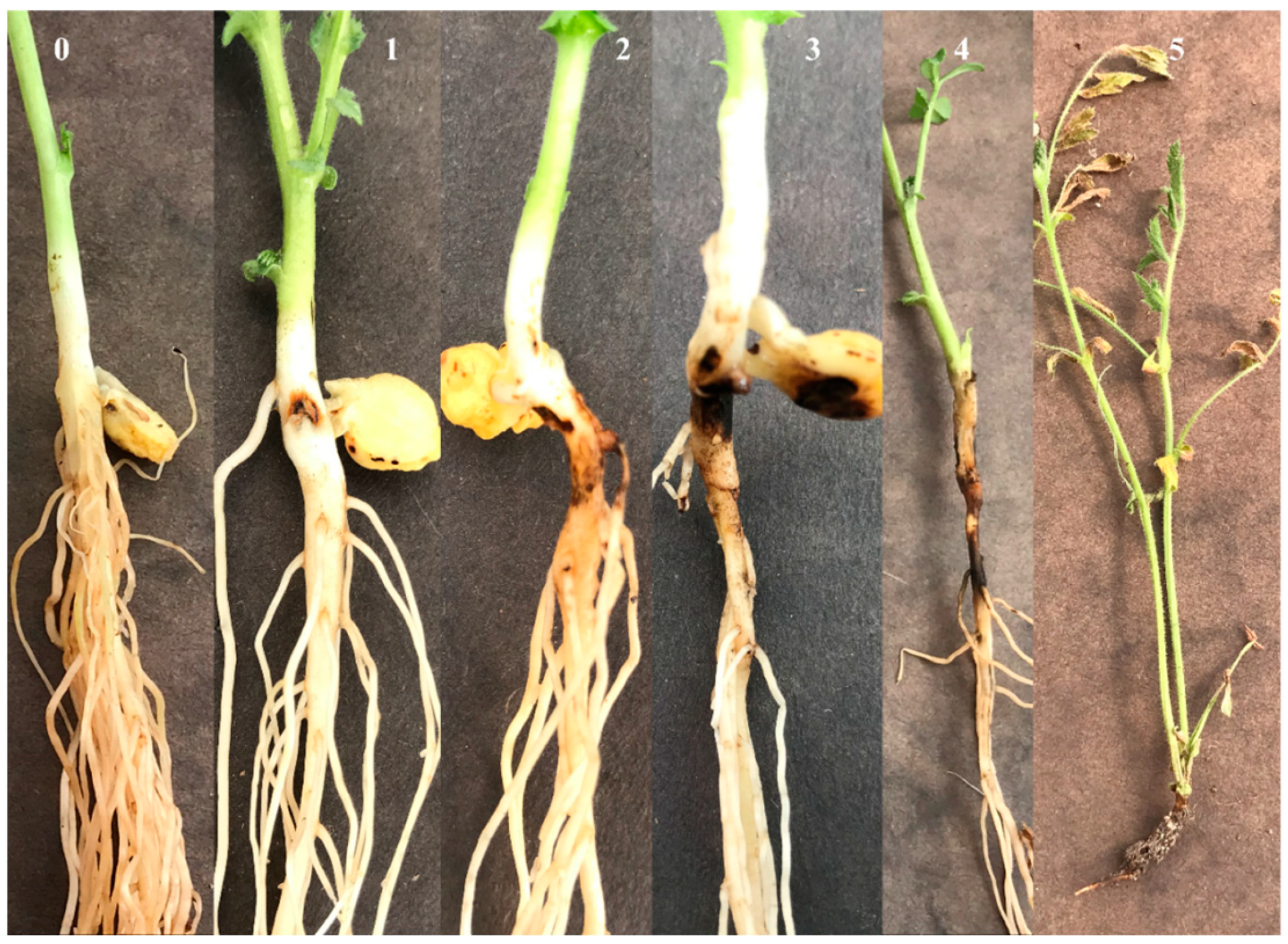
2.4. Aggressiveness Tests in the Greenhouse

A subset of 51 isolates was chosen from a pool of 426 isolates, which constituted 13 Fusarium species: Fusarium avenaceum, F. oxysporum, F. accuminatum, F. culmorum, F. torulosum, F. gamsii (a newly identified lineage within the F. tricinctum species complex), F. sporotrichioides, Neocosmospora solani (genus changed from Fusarium to Neocosmospora based on Crous et al. [23]), F. redolens, F. pseudograminearum, F. equiseti, F. proliferatum, and F. brachygibbosum. In a greenhouse experiment, the aggressiveness of the isolates was evaluated using the chickpea cultivar ‘CDC Frontier’. This cultivar (Kabuli-type chickpea) is commonly grown in a state characterized by pinnate leaves. Seeds were sourced from the Montana State seed lab, with a pre-experiment germination rate of 90%. Plastic cone-tainers (measuring 3.8 cm in diameter and 21 cm in depth; Ray Leach container, Tangent, OR, USA) were treated with a 10% sodium hypochlorite solution and subsequently rinsed. The growth medium comprised a mixture of peat and Sunshine Mix 1 (Sun Gro Horticulture Inc., Bellevue, WA, USA) at a 1:1 ratio. Cone-tainers were filled with the growth medium up to 5 cm below the cone-tainer’s top, and a single seed was placed on the surface of the medium.
All the Fusarium isolates were cultured on one-quarter strength PDA to prepare inoculum plugs for greenhouse aggressiveness tests. Agar plugs were cut with a 4.5 mm diameter cork borer from the colony margins of 7-day-old cultures. Two agar plugs were placed on either side of the seed (sandwich-style) and covered with Sunshine mix (the soil used was not sterilized). Agar plugs without fungus served as the negative control. Each treatment included ten replicates, with each replicate containing a single plant. The treatments were arranged in a randomized, complete block design. The entire experiment was repeated once to verify the results. Plants were watered daily and grown for 21 days on greenhouse benches without any nutritional supplements, maintained at 22 °C during the day and 18 °C at night, with 16:8 h light/dark cycles and a relative humidity of 75%. The chickpea seedlings were harvested 21 days post-inoculation, and the roots were cleaned using low-pressure running water. Subsequently, the plants were placed on sterilized paper towels for root disease severity assessment, employing a visual scale of 0–5 adapted from Grunwald et al. [24]. The grading system is outlined as follows: 0 indicates the absence of disease symptoms; 1 represents minor lesions on the hypocotyl; 2 represents lesions merging around epicotyls and hypocotyls; 3 denotes lesions extending into the root system with initial root tip infection; 4 indicates infection of the epicotyl, hypocotyl, and a significant portion of the root system; and 5 indicates complete infection of the entire root system (refer to Figure 1). To adhere to Koch’s postulates, each pathogenic species was isolated again from the harvested plant roots, and these cultures were then compared with known cultures of the respective species.

2.5. Data Analysis

The data were analyzed using SAS version 9.4 (SAS Institute, Cary, NC, USA). Given that Fusarium root rot ratings were recorded on an ordinal scale ranging from 0 to 5, non-parametric tests outlined by Shah and Madden [25] were employed to compute the median, mean rank ( R ¯ i j ), and relative treatment effect ( p ^ i j ) alongside 95% confidence intervals for the severity of root rot caused by each Fusarium spp. isolate. PROC RANK was utilized for determining the median and mean rank, while PROC MIXED was employed to calculate the relative treatment effects ( p ^ i j ), with confidence intervals (CIs) generated using a SAS macro OWL, as detailed by Konietschke et al. [26]. The dataset included variables such as subject (a unique identifier for each experimental unit), isolate, experiment, and severity rating. The relative treatment effect depicted the severity of the disease caused by each isolate on the tested chickpea plants.

2.6. Phylogenetic Analysis

A phylogenetic tree was constructed from the EF 1-α generated in the current study. Available Ex-type sequences of the taxa of interest were determined by Crous et al. [23] and included in the analysis. If no type sequences were available for a given taxon, GenBank sequences from NRRL and/or CBS were included. Sequences underwent alignment and editing using MUSCLE within MEGA11: Molecular Evolutionary Genetics Analysis Version 11 [27]. For phylogenetic analyses, a GTR+G+I evolutionary model was employed, chosen for its inclusivity, covering all other evolutionary models [28]. Bayesian analysis for phylogeny inference was conducted on combined loci using a Yule tree prior [29] and a strict molecular clock in BEAST version 1.10.4 [30], running a single MCMC chain for 10^7 steps with a burn-in of 10%. Posterior probabilities were computed from the remaining 9000 sampled trees. TreeAnnotator version 1.10.4 (part of the BEAST package) was utilized to generate a maximum-clade credibility tree. Stationarity was confirmed through multiple runs, ensuring convergence. The resulting tree was visualized using FigTree ver. 1.3.1 [31]. Maximum likelihood analysis was performed using raxmlGUI [32] under default settings with a GTR+G+I evolutionary model, and bootstrap analyses were executed with 1000 replications [33].

3. Results

3.1. Identification and Prevalence of Fusarium Species

Fusarium was the predominant fungal taxa (95%) that was associated with chickpea root rot, followed by Rhizoctonia species (4%) and Pythium species (1%). Other fungal taxa recovered from these surveys include Diaporthe gulyae, Calonectria montana, Pyrenophora teres, Stromatinia narcissi, Trichoderma spp., and Epicoccum layuesnse. The chickpea field surveys yielded a collection of 426 Fusarium isolates, resulting in the identification of 13 distinct Fusarium morphospecies characterized by their colony morphology. (Supplementary Figure S1) and microscopic observations of the hyphae and/or conidia (Supplementary Figure S2). Colony appearance within a species varied depending on the quarter strength PDA. Isolates within the same species displayed differences in color and hyphal morphology (for example, some F. avenacuem isolates formed fluffy aerial hyphae, whereas a few other isolates formed flat on the aPDA). Some isolates formed conidia within 3–4 days of culture, whereas some formed very few. This taxonomic delineation was further corroborated through comparative analyses of TEF1-α with reference sequences sourced from GenBank and FUSARIUM-ID databases, resulting in congruent identifications across these platforms (Supplementary Table S2). Fusarium oxysporum was the most abundant (33%) species among all the isolates; the other species recovered were F. acuminatum (21%), F. avenaceum (15%), F. redolens (14%), N. solani (6%), F. equiseti (2%), F. sporotrichioides (6%), F. pseudograminearum (0.2%), F. culmorum (0.8%), F. proliferatum (0.2%), F. torulosum (0.9%), F. gamsii (0.8%), and F. brachygibbosum (0.1%).

3.2. Aggressiveness Assessment Tests

Aggressiveness tests revealed significant variation in disease severity and aggressiveness among the tested Fusarium isolates. The mock-inoculated control plants were free of disease symptoms. In contrast, the inoculated plants displayed varying intensities of dark brown to black lesions, with consistent symptoms observed across the tested chickpea cultivar. These symptoms remained consistent regardless of the Fusarium isolates utilized in the study, all of which induced lesions in the cotyledon region. Non-parametric analysis of the marginal effects on the severity ranks of disease values revealed variations in aggressiveness among the Fusarium isolates, significantly differing from the non-inoculated control plants. Fusarium isolates were categorized into three groups based on their levels of aggressiveness, as outlined in Moparthi et al. [19]. Isolates inducing severe root rot with mean ranks ( R ¯ i j ) ranging from 901 to 1100 were classified as highly aggressive; those with mean ranks between 700 and 900 were considered moderately aggressive; isolates with mean ranks between 501 and 700 were regarded as less aggressive; and isolates with mean ranks below 500 were deemed weakly aggressive. Fusarium avenaceum isolates exhibited notable aggressiveness when assessed on the chickpea cultivar ‘CDC Frontier’ under controlled greenhouse conditions. The observed aggressiveness ranged from high to moderate, with severe stunting or complete plant death occurring within two weeks post-inoculation. Among the tested F. avenaceum isolates, four out of five (Fav1, Fav3, Fav4, and Fav5) demonstrated particularly high levels of aggressiveness, eliciting pronounced pathological responses in the host cultivar. Notably, isolate Fav2 displayed a moderately aggressive phenotype.
Furthermore, two isolates of N. solani (Fso17 and Fso18) and solitary isolates of F. redolens (Fre11) and F. oxysporum (Fox22) exhibited moderate levels of aggressiveness in the experimental setup. Conversely, the remaining isolates were characterized by comparatively lower levels of aggressiveness. Detailed records of the pathogen aggressiveness trials, including specific isolate designations and corresponding aggressiveness ratings, are shown in Table 1.

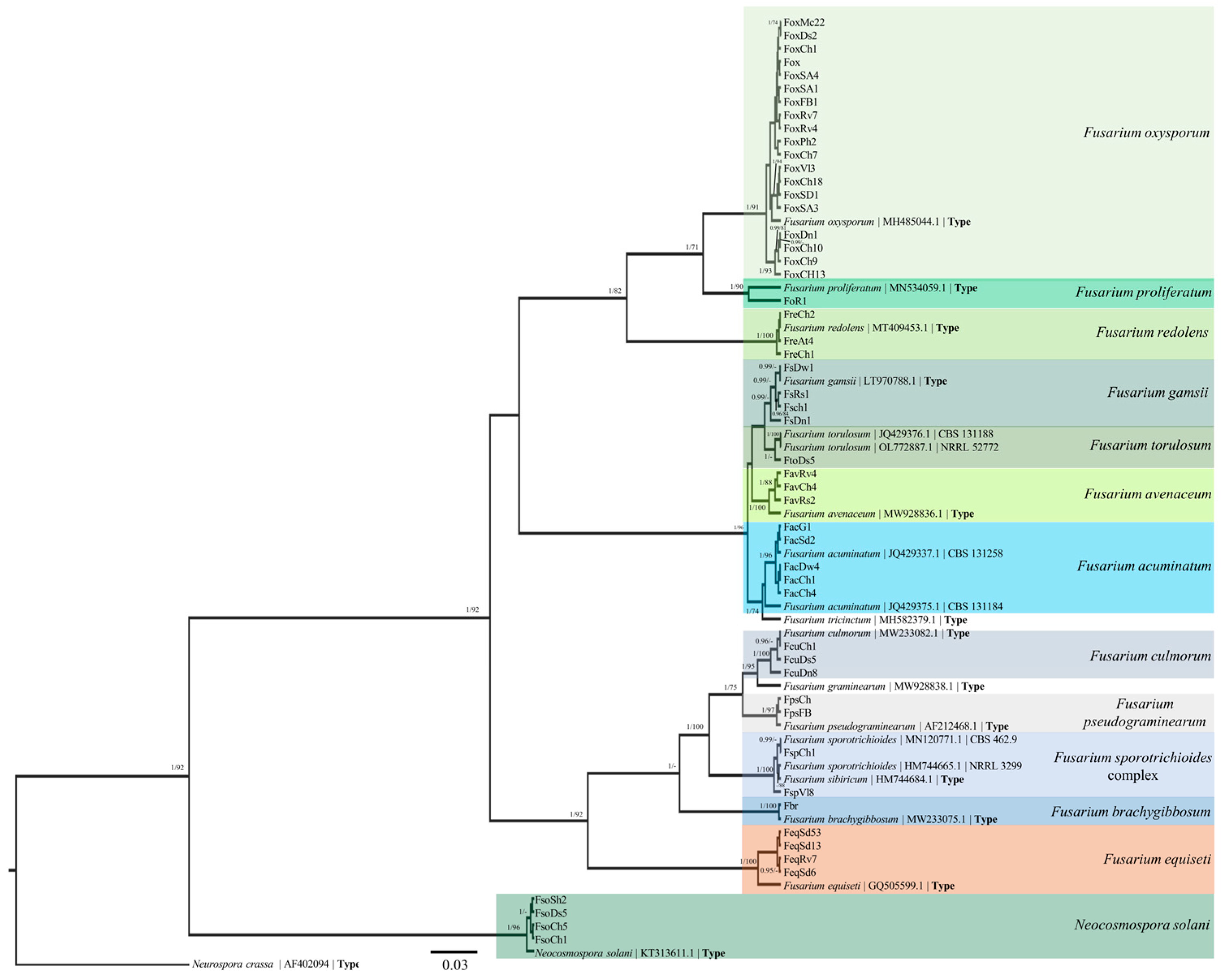
3.3. TEF-1α Sequences and Phylogenetic Analysis

The TEF-1α translation sequences spanned from 700 to 900 base pairs. The accession numbers for these sequences were acquired from GenBank and are provided in Supplementary Tables S1 and S2. The phylogenetic analyses assessed each distinct isolate genotype for representative purposes. In total, 70 Fusarium isolates underwent analysis, with 52 of these isolates sequenced specifically for the current study. Using Bayesian analyses of the TEF 1-α sequences, a maximum-clade credibility tree was constructed. Posterior probabilities equal to or exceeding 90% are shown, followed by bootstrap values exceeding 70% for the maximum likelihood (ML) analyses performed. The depicted maximum-clade credibility tree is shown in Figure 2.

4. Discussion

The objective of this study was to gain insights into the prevalence and diversity of Fusarium species in chickpea cultivation in Montana. The survey was conducted in conventionally grown chickpea fields in the state. Knowledge of major pathogens is crucial for crop rotation and disease management decisions. It also assists crop breeders in selecting and developing disease-resistant cultivars. The current study identified 13 Fusarium species through phylogenetic analysis of symptomatic chickpea roots. This is the first survey of Fusarium species on chickpea in Montana, and it revealed the predominant pathogen among other soil-borne pathogens. This finding is particularly interesting, as foliage infections could lead to possible blossom infections and ultimately spread to seeds. Fusarium was a frequently isolated group of fungi from pulse seeds, including chickpea seeds [19]. Further studies are needed to explore whether the same species are involved in root and foliage infections.
The morphological identification of Fusarium species poses significant challenges [22]. This study observed variation in colony morphology among the isolates within the same species (Supplementary Figure S1). This observation agrees with previous research [34] and emphasizes the complexity of Fusarium species identification and the importance of comprehensive approaches combining morphological and molecular data.
The gene TEF1-α is phylogenetically informative at the species level [35]. Sequence data from TEF1-α alone can offer reasonably accurate placement of unknown isolates within a species complex, achievable through nucleotide BLAST queries of databases like FUSARIUM-ID and/or Fusarium MLST or phylogenetic analysis [36]. Our work focused on TEF1-α loci due to the high sequence quality and the greater availability of sequences from this locus in databases such as FUSARIUM-ID and based on our previous success of species differentiation. Utilizing TEF1-α alongside RPB1 and RPB2 sequences often results in more robust species-level identification. While these three genetic loci have been used in numerous studies for Fusarium species identification, it is worth noting that RPB1 and RPB2 do not possess the same level of taxonomic coverage within the Fusarium genus as TEF1-α does [22].
A recent study from Alberta, Canada, identified five Fusarium species from chickpea [18]. These researchers identified F. redolens as the predominant species, followed by F. culmorum, F. sporotrichioides, F. oxysproum, and F. equiseti. In the current study, F. oxysporum was the predominant species on chickpeas. A field survey in North Dakota showed that F. oxysporum and F. avenaceum were the predominant species in field peas [37]. Field surveys from the lentil-growing regions in North Dakota also identified F. oxysporum as the most abundant species [38], indicating the widespread nature of this species in the pulse-growing regions of the Great Plains.
In this study, F. acuminatum was the second most frequently identified species after F. oxysporum, and earlier, this species was reported from chickpea roots and seeds in Australia [39]. A study from Alberta, Canada, showed F. acuminatum as the predominant species associated with canola [40] and the second most abundant species associated with soybean in South Dakota [41]. This survey revealed F. avenaceum as one of the most frequently recovered species after F. acuminatum on chickpea roots. F. avenaceum was reportedly part of the chickpea root rot complex in Canada, Australia, and Poland [42,43,44]. Fusarium redolens and N. solani were also frequently recovered from the symptomatic chickpea roots in Montana. Fusarium redolens was also isolated from chickpeas in Lebanon, Morocco, Spain, Tunisia, Iran, and Pakistan, suggesting it is a typical pathogen associated with root rot in chickpea-growing regions [45,46,47]. Westerlund et al. [48] first reported N. solani causing root rot in chickpeas from California. This study shows that it is prevalent in all counties in Montana. F. proliferatum, F. pseudograminearum, F. culmorum, F. gamsii, and F. torulosum were less prevalent. A common cereal pathogen, F. pseudograminearum, was recovered from a few samples; this could be due to the rotation of cereal crops (wheat and barley) with pulses. Previous studies also showed that F. pseudograminearum was frequently isolated from chickpea roots [49,50]. F. brachygibbosum was the least frequently recovered species and was only recovered from one site. A few recent reports [51,52,53] of this pathogen from various crops indicate that it has a wide host range.
Fusarium equiseti is a member of the Fusarium incarnatum-equiseti complex (FIESC). This species complex includes 33 phylogenetic species found in various habitats and hosts around the world [54]. Although it appeared infrequently (2%) in our study, it has been documented in chickpeas from different global regions. Recent research in Canada, Morocco, and Iran has demonstrated that this species causes wilt and root rot in chickpeas [18,55,56]. The presence of F. equiseti in multiple geographic areas underscores its potential significance in chickpea pathology.
Aggressiveness tests carried out in the greenhouse showed that F. avenaceum was highly aggressive on the chickpea cv. ‘CDC Frontier’. This is consistent with a previous study by Moparthi et al. [19] in which F. avenaceum isolates collected from symptomatic dry pea caused higher severity in several hosts tested, including chickpea. Research conducted in North Dakota on field peas revealed that F. avenaceum was the most aggressive species among the nine tested Fusarium species [37]. Two isolates of N. solani and one isolate of F. redolens were found to be moderately aggressive, aligning with previous findings on chickpeas [19]. Despite F. oxysporum being the predominant species, only one isolate exhibited moderate aggressiveness on chickpea, consistent with results from a Canadian study on the same cultivar [18]. This study did not identify the forma specialis of the various F. oxysporum isolates. F. acuminatum was classified as less aggressive, along with F. brachygibbosum and one isolate of F. pseudograminearum. Notably, this is the first report of F. brachygibbosum causing root rot in chickpeas in the USA and globally. Additionally, F. acuminatum, F. gamsii, F. equiseti, F. torulosum, F. culmorum, F. sporotrichioides, and F. proliferatum were identified as weakly aggressive, consistent with previous findings on the same cultivar. Contrarily, Zhou et al. [18] reported that F. culmorum and F. sporotrichioides isolates caused severe root rot on chickpea cv. ‘CDC Frontier’. This discrepancy could be attributed to differences in the genetic variation of the isolates used in each study, significantly affecting pathogenicity. For F. equiseti, our results concur with those of Zhou et al. [18]. Fusarium proliferatum has been reported to cause vascular wilt in chickpeas from Cuba [57]. A recent Canadian study by Yu et al. [58] showed that canola and leguminous crops were highly susceptible to F. proliferatum. At the same time, barley and wheat were partially susceptible, indicating a broad host range for this species.
This variation in the aggressiveness of the same Fusarium species among various studies underscores the complex nature of Fusarium isolates and suggests potential differences in pathogenicity among Fusarium species. Understanding these differences is vital for effective disease management in chickpea cultivation, emphasizing the importance of targeted control measures and ongoing research to elucidate the mechanisms underlying Fusarium species aggressiveness. As mentioned by other researchers [59], we also believe that further research is essential to determine the influence of environmental factors on the aggressiveness of individual Fusarium species and their interactions with each other and with the host plant.
Given that F. avenaceum has a broad host range and can infect small grain crops such as barley, wheat, and canola [19,40], alternative non-host crops for this aggressive species must be incorporated into crop rotation programs. Previous studies [50,51,52,53,54,55,56,57,58,59,60,61,62] highlighted the need for identifying sources of resistance to F. oxysporum f. sp. ciceri. The results from this study indicate the need for breeding resistant varieties against highly aggressive isolates of F. avenaceum in chickpeas. A prior study [63] offered insights into yield losses and disease management strategies for Fusarium wilt caused by F. oxysporum f. sp. ciceri. Further research is required to estimate the yield losses caused by F. avenaceum and other cultural practices to reduce the impact of this aggressive pathogen. One limitation of this study is that it does not include organic chickpea fields in the survey; future surveys in the organic chickpea field would help understand the pathogen diversity in both settings. Numerous isolates of Trichoderma spp. and a few isolates of Epicoccum spp. were also recovered. It would be interesting to check whether biological controls can suppress soil-borne pathogens and explore their combined effect on crop yield.

5. Conclusions

This is the first study on chickpea Fusarium root rot conducted over extensive field sampling in Montana. Through two-year field surveys, 13 Fusarium species were found to be associated with chickpea root rot; F. oxysporum was the predominant species among all the species. Greenhouse assessment tests indicated that F. avenaceum was the most aggressive species. This study is the first to report F. gamsii, F. proliferatum, and F. brachygibbosum as causal agents of chickpea root rot in the USA. Future research should aim to test the aggressiveness of various Fusarium species on different chickpea cultivars to identify resistant varieties, conduct genomic studies to understand the genetic basis of pathogenicity, and explore how environmental conditions affect disease development.

Supplementary Materials

The following supporting information can be downloaded at https://www.mdpi.com/article/10.3390/agriculture14070974/s1, Table S1: Fusarium species, location, and identification by translation elongation factor 1-α (EF1-α) sequencing. Table S2: Details on Fusarium sequences of current study and their percent identity with GenBank and FUSARIUM- ID sequences. Figure S1: Fusarium isolates and their colony morphology on PDA after seven days of incubation at 22 °C. Fusarium avenaceum (A-C color variation), F. gamsii (D), F. acuminatum (E), F. oxysporum (F & G color variation), F. pseudograminearum (H), F. torulosum (I), F. culmorum (J), F. equiseti (K), F. sporotrichiooides (L), F. proliferatum (M), F. solani (N), F. redolens (O), and F. brachygibbosum (P). Figure S2: Fusarium spore morphology. Fusarium avenaceum (A), F. gamsii (B), F. acuminatum (C), F. oxysporum (D), F. pseudograminearum (E), F. torulosum (F), F. culmorum (G), F. equiseti (H), F. sporotrichioides (I), F. proliferatum (J), F. solani (K), F. redolens (L), and F. brachygibbosum (M).

Author Contributions

Conceptualization, S.M. and K.M.; methodology, S.M., M.E.B., M.B., K.M.; M.J.B. and C.B.; formal analysis, S.M. and O.P.-H.; investigation, S.M. and M.B.; resources, S.M., K.M. and M.E.B.; data curation, S.M.; writing—original draft preparation, S.M.; supervision, M.E.B. and K.M.; project administration, S.M.; funding acquisition, S.M., K.M. and M.E.B. All authors have read and agreed to the published version of the manuscript.

Funding

This work was funded by the Montana Department of Agriculture- Specialty Crop Block Grant (21SC07007).

Institutional Review Board Statement

Not applicable.

Data Availability Statement

The data presented in this study are available on request from the first author.

Acknowledgments

We thank David Geiser for his valuable feedback on our manuscript, checking our sequences, and offering valuable suggestions. We also thank Rebecca King for assisting in the field surveys and Sahasra Lavu for her assistance with lab work. Additionally, we are grateful to Mike Munster for his pre-submission revision of the manuscript. Finally, we thank the reviewers for their time and helpful suggestions to make the manuscript better.

Conflicts of Interest

The authors declare no conflicts of interest.

References

  1. Taylor, P.W.; Ford, R. Chickpea. In Pulses, Sugar and Tuber Crops; Springer: Berlin/Heidelberg, Germany, 2007; pp. 109–121. [Google Scholar]
  2. Van der Maessen, L.J.G. Cicer L., a Monograph of the Genus, with Special Reference to the Chickpea (Cicer arietinum L.), Its Ecology and Cultivation; Wageningen University and Research: Wageningen, The Netherlands, 1972. [Google Scholar]
  3. FAOSTAT. Food and Agriculture Organization Statistical Database. Available online: http://www.fao.org/faostat/en/#data/QC (accessed on 29 May 2004).
  4. Dugan, F.M.; Lupien, S.L.; Hu, J. Fungal plant pathogens associated with emerging crops in North America: A challenge for plant health professionals. Plant Health Prog. 2017, 18, 221–229. [Google Scholar] [CrossRef]
  5. USA Dry Pea, Lentil, and Chickpea Production. Available online: https://agresearch.montana.edu/wtarc/producerinfo/agronomy-nutrient-management/Pulses/USADryPeaCouncil%20FactSheet.pdf (accessed on 28 May 2024).
  6. Attaway, D. Clemson Scientist Discover Plant Genes to Help Grow S.C. Economy. Available online: https://news.clemson.edu/clemson-scientists-discover-plant-genes-to-help-boost-s-c-economy/ (accessed on 23 May 2024).
  7. Miller, P.R.; Holmes, J.A. Cropping sequences effects of four broadleaf crops on four cereal crops in the northern Great Plains. Agron. J. 2005, 97, 189–200. [Google Scholar] [CrossRef]
  8. Muehlbauer, F.J.; Sarker, A. Economic importance of chickpea: Production, value, and world trade. In The Chickpea Genome; Springer: Cham, Switzerland, 2017; pp. 5–12. [Google Scholar] [CrossRef]
  9. Boukid, F. Chickpea (Cicer arietinum L.) protein as a prospective plant- based ingredient: A review. Inter. J. Food Sci. Technol. 2021, 56, 5435–5444. [Google Scholar] [CrossRef]
  10. Karalija, E.; Vergata, C.; Basso, M.F.; Negussu, M.; Zaccai, M.; Grossi-de-Sa, M.F.; Martinelli, F. Chickpeas’ tolerance of drought and heat: Current knowledge and next steps. Agronomy 2022, 12, 2248. [Google Scholar] [CrossRef]
  11. Singh, R.; Pratap, T.; Singh, D.; Singh, G.; Singh, A.K. Effect of phosphorus, sulphur and biofertilizers on growth attributes and yield of chickpea (Cicer arietinum L.). J. Pharmacogn. Phytochem. 2018, 7, 3871–3875. [Google Scholar]
  12. United States Department of Agriculture National Agricultural Statistic Service [USDA NASS]. Statistics by Subject Results. 2020. Available online: https://www.nass.usda.gov/Statistics_by_State/Montana/Publications/Special_Interest_Reports/agfacts.pdf (accessed on 23 April 2020).
  13. Zhou, Y. Chickpea Cultivar Evaluation and Intercropping for Disease Management and Yield. Master’s Dissertation, Montana State University, Bozeman, MT, USA, 2022. [Google Scholar]
  14. Bahr, L.; Castelli, M.V.; Barolo, M.I.; Mostacero, N.R.; Tosello, M.E.; López, S.N. Ascochyta blight: Isolation, characterization, and development of a rapid method to detect inhibitors of the chickpea fungal pathogen Ascochyta rabiei. Fungal Biol. 2016, 120, 424–432. [Google Scholar] [CrossRef] [PubMed]
  15. Gaur, P.M.; Jukanti, A.K.; Varshney, R.K. Impact of genomic technologies on chickpea breeding strategies. Agronomy 2012, 2, 199–221. [Google Scholar] [CrossRef]
  16. Choudhary, A.K.; Kumar, S.; Patil, B.S.; Sharma, M.; Kemal, S.; Ontagodi, T.P.; Datta, S.; Patil, P.; Chaturvedi, S.K.; Sultana, R.; et al. Narrowing yield gaps through genetic improvement for Fusarium wilt resistance in three pulse crops of the semi-arid tropics. SABRAO J. Breed. Genet. 2013, 45, 341–370. Available online: http://oar.icrisat.org/id/eprint/1079 (accessed on 27 May 2024).
  17. Halila, I.; Rubio, J.; Millán, T.; Gil, J.; Kharrat, M.; Marrakchi, M. Resistance in chickpea (Cicer arietinum) to Fusarium wilt race ‘0’. Plant Breed. 2010, 129, 563–566. [Google Scholar] [CrossRef]
  18. Zhou, Q.; Yang, Y.; Wang, Y.; Jones, C.; Feindel, D.; Harding, M.; Feng, J. Phylogenetic, phenotypic and host range characterization of five Fusarium species isolated from chickpea in Alberta, Canada. Can. J. Plant Pathol. 2021, 43, 651–657. [Google Scholar] [CrossRef]
  19. Moparthi, S.; Burrows, M.; Mgbechi-Ezeri, J.; Agindotan, B. Fusarium spp. associated with root rot of pulse crops and their cross pathogenicity to cereal crops in Montana. Plant Dis. 2020, 105, 548–557. [Google Scholar] [CrossRef] [PubMed]
  20. White, T.J.; Bruns, T.D.; Lee, S.B.; Taylor, J.W. Amplificaiton and direct sequencing of fungal ribosomal RNA genes for phylogenetics. In PCR Protocols, Guide to Methods and Applications; Innis, M.A., Gelfand, D.H., Sninsky, J.J., White, T.J., Eds.; Academic Press: San Diego, CA, USA, 1990; pp. 315–322. [Google Scholar]
  21. Robideau, G.P.; De Cock, A.W.; Coffey, M.D.; Voglmayr, H.; Brouwer, H.; Bala, K.; Citty, D.; Desaulniers, N.; Eggerston, Q.; Gachon, C.M. DNA barcoding of oomycetes with cytochrome c oxidase subunit I and internal transcribed spcer. Mol. Ecol. Resour. 2011, 11, 1002–1011. [Google Scholar] [CrossRef] [PubMed]
  22. Torres-Cruz, T.J.; Whitaker, B.K.; Proctor, R.H.; Broders, K.; Laraba, I.; Kim, H.S.; Brown, D.W.; O’Donnell, K.; Estrada-Rodríguez, T.L.; Lee, Y.H.; et al. FUSARIUM-ID v. 3.0: An updated, downloadable resource for Fusarium species identification. Plant Dis. 2022, 106, 1610–1616. [Google Scholar] [CrossRef] [PubMed]
  23. Crous, P.W.; Lombard, L.; Sandoval-Denis, M.; Seifert, K.A.; Schroers, H.J.; Chaverri, P.; Gené, J.; Guarro, J.; Hirooka, Y.; Bensch, K.; et al. Fusarium: More than a node or a foot-shaped basal cell. Stud. Mycol. 2021, 98, 100116. [Google Scholar] [CrossRef] [PubMed]
  24. Grünwald, N.J.; Coffman, V.A.; Kraft, J.M. Sources of partial resistance to Fusarium root rot in the Pisum core collection. Plant Dis. 2003, 87, 1197–1200. [Google Scholar] [CrossRef] [PubMed]
  25. Shah, D.A.; Madden, L.V. Nonparametric analysis of ordinal data in designed factorial experiments. Phytopathology 2004, 94, 33–43. [Google Scholar] [CrossRef] [PubMed]
  26. Konietschke, F.; Hothorn, L.A.; Brunner, E. Rank-based multiple test procedures and simultaneous confidence intervals. Electron. J. Stat. 2012, 6, 738–759. [Google Scholar] [CrossRef]
  27. Tamura, K.; Stecher, G.; Kumar, S. MEGA11: Molecular evolutionary genetics analysis version 11. Mol. Biol. Evol. 2021, 38, 3022–3027. [Google Scholar] [CrossRef] [PubMed]
  28. Abadi, S.; Azouri, D.; Mayrose, I.; Pupko, T. Model selection may not be a mandatory step for phylogeny reconstruction. Nat. Commun. 2019, 10, 934. [Google Scholar] [CrossRef]
  29. Gernhard, T. The conditioned reconstructed process. J. Theoretical. Biol. 2008, 253, 769–778. [Google Scholar] [CrossRef]
  30. Suchard, M.A.; Lemey, P.; Baele, G.; Ayres, D.L.; Drummond, A.J.; Rambaut, A. Bayesian phylogenetic and phylodynamic data integration using BEAST 1.10. Virus Evol. 2018, 4, Vey016. [Google Scholar] [CrossRef] [PubMed]
  31. Rambaut, A. Fig Tree Ver. 1.3.1. 2009. Available online: http://tree.bio.ed.ac.uk/software/figtree (accessed on 27 May 2024).
  32. Silvestro, D.; Michalak, I. RaxmlGUI: A graphical front-end for RAxML. Org. Divers. Evol. 2011, 12, 335–337. [Google Scholar] [CrossRef]
  33. Felsenstein, J. Confidence limits on phylogenies: An approach using the bootstrap. Evolution 1985, 39, 783–791. [Google Scholar] [CrossRef] [PubMed]
  34. O’Donnell, K.; Whitaker, B.K.; Laraba, I.; Proctor, R.H.; Brown, D.W.; Broders, K.; Kim, H.S.; McCormick, S.P.; Busman, M.; Aoki, T.; et al. DNA sequence-based identification of Fusarium: A work in progress. Plant Dis. 2022, 106, 1597–1609. [Google Scholar] [CrossRef] [PubMed]
  35. Geiser, D.M.; del Mar Jiménez-Gasco, M.; Kang, S.; Makalowska, I.; Veeraraghavan, N.; Ward, T.J.; Zhang, N.; Kuldau, G.A.; O’Donnell, K. FUSARIUM-ID v. 1.0: A DNA sequence database for identifying Fusarium. Eur. J. Plant Pathol. 2004, 11, 473–479. [Google Scholar] [CrossRef]
  36. O’Donnell, K.; Ward, T.J.; Robert, V.A.; Crous, P.W.; Geiser, D.M.; Kang, S. DNA sequence-based identification of Fusarium: Current status and future directions. Phytoparasitica 2015, 43, 583–595. [Google Scholar] [CrossRef]
  37. Chittem, K.; Mathew, F.M.; Gregoire, M.; Lamppa, R.S.; Chang, Y.W.; Markell, S.G.; Bradley, C.A.; Barasubiye, T.; Goswami, R.S. Identification and characterization of Fusarium spp. associated with root rots of field pea in North Dakota. Eur. J. Plant Pathol. 2015, 143, 641–649. [Google Scholar] [CrossRef]
  38. Zitnick-Anderson, K.; Gargouri Jbir, T.; Carlson, A.; Postovit, S.; Pasche, J.; Kalil, A. Fusarium species associated with root rot of lentil (Lens culinaris) in North Dakota. Plant Health Prog. 2021, 22, 524–528. [Google Scholar] [CrossRef]
  39. Bretag, T.W.; Mebalds, M.I. Pathogenicity of fungi isolated from Cicer arietinum (chickpea) grown in northwestern Victoria. Aust. J. Exp. Agric. 1987, 27, 141–148. [Google Scholar] [CrossRef]
  40. Chen, Y.; Zhou, Q.; Strelkov, S.E.; Hwang, S.F. Genetic diversity and aggressiveness of Fusarium spp. isolated from canola in Alberta, Canada. Plant Dis. 2014, 98, 727–738. [Google Scholar] [CrossRef]
  41. Okello, P.N.; Petrovic, K.; Singh, A.K.; Kontz, B.; Mathew, F.M. Characterization of species of Fusarium causing root rot of soybean (Glycine max L.) in South Dakota, USA. Canada. Can. J. Plant Pathol. 2020, 42, 560–571. [Google Scholar] [CrossRef]
  42. Cother, E.J. Identification and control of root-rot fungi in Cicer arietinum (chickpea). Plant Dis. Rep. 1977, 61, 736–740. [Google Scholar]
  43. Cruz, A.F.; Hamel, C.; Yang, C.; Matsubara, T.; Gan, Y.; Singh, A.K.; Kuwada, K.; Ishii, T. Phytochemicals to suppress Fusarium head blight in wheat–chickpea rotation. Phytochemistry 2012, 78, 72–80. [Google Scholar] [CrossRef] [PubMed]
  44. Mazur, S.; Nawrocki, J.; Kucmierz, J. Fungal diseases of chickpea (Cicer arietinum L.) cultivated in the south region of Poland. Plant Prot. Sci. 2002, 38, 332–335. [Google Scholar] [CrossRef]
  45. Jiménez-Fernández, D.; Navas-Cortés, J.A.; Montes-Borrego, M.; Jiménez-Díaz, R.M.; Landa, B.B. Molecular and pathogenic characterization of Fusarium redolens, a new causal agent of Fusarium yellows in chickpea. Plant Dis. 2011, 95, 860–870. [Google Scholar] [CrossRef] [PubMed]
  46. Bouhadida, M.; Jendoubi, W.; Gargouri, S.; Beji, M.; Kharrat, M.; Chen, W. First report of Fusarium redolens causing Fusarium yellowing and wilt of chickpea in Tunisia. Plant Dis. 2017, 101, 1038. [Google Scholar] [CrossRef]
  47. Saeedi, S.; Jamali, S. Molecular characterization and distribution of Fusarium isolates from uncultivated soils and chickpea plants in Iran with special reference to Fusarium redolens. J. Plant Pathol. 2021, 103, 167–183. [Google Scholar] [CrossRef]
  48. Westerlund, F.V.; Campbell, R.N.; Kimble, K.A. Fungal root rots and wilt of chickpea in California. Phytopathology 1974, 64, 432–436. [Google Scholar]
  49. Dugan, F.M.; Lupien, S.L.; Hernandez-Bello, M.; Peever, T.L.; Chen, W. Fungi resident in chickpea debris and their suppression of growth and reproduction of Didymella rabiei under laboratory conditions. J. Phytopathol. 2005, 153, 431–439. [Google Scholar] [CrossRef]
  50. Kirkegaard, J.A.; Simpfendorfer, S.; Holland, J.; Bambach, R.; Moore, K.J.; Rebetzke, G.J. Effect of previous crops on crown rot and yield of durum and bread wheat in northern NSW. Aust. J. Agric Res. 2004, 55, 321–334. [Google Scholar] [CrossRef]
  51. Ali, H.Z.; Hameed, M.S.; Abdulrahman, A.A.; Saood, H.M. First report on Fusarium brachygibbosum isolate FIR 16_ITS isolated from Iraqi wheat plant. J. Ecol. Eng. 2020, 21, 81–86. [Google Scholar] [CrossRef] [PubMed]
  52. Namsi, A.; Rabaaoui, A.; Masiello, M.; Moretti, A.; Othmani, A.; Gargouri, S.; Gdoura, R.; Werbrouck, S.P. First report of leaf wilt caused by Fusarium proliferatum and F. brachygibbosum on Date Palm (Phoenix dactylifera) in Tunisia. Plant Dis. 2021, 105, 1217. [Google Scholar] [CrossRef] [PubMed]
  53. Qiu, R.; Li, J.; Zheng, W.M.; Su, X.H.; Xing, G.Z.; Li, S.J.; Zhang, Z.Y.; Li, C.J.; Wang, J.; Chen, Y.G.; et al. First report of root rot of tobacco caused by Fusarium brachygibbosum in China. Plant Dis. 2021, 105, 4170. [Google Scholar] [CrossRef] [PubMed]
  54. Xia, J.W.; Sandoval-Denis, M.; Crous, P.W.; Zhang, X.G.; Lombard, L. Numbers to names-restyling the Fusarium incarnatum-equiseti species complex. Persoonia. Mol. Phylo. Evol. Fung. 2019, 43, 186–221. [Google Scholar] [CrossRef] [PubMed]
  55. Adnani, M.; El Hazzat, N.; Msairi, S.; El Alaoui, M.A.; Mouden, N.; Selmaoui, K.; Benkirane, R.; Ouazzani Touhami, A.; Douira, A. Exploring the efficacy of a Trichoderma asperellum-based seed treatment for controlling Fusarium equiseti in chickpea. Egypt. J. Biol. Pest Co. 2024, 34, 7. [Google Scholar] [CrossRef]
  56. Younesi, H.; Darvishnia, M.; Bazgir, E.; Chehri, K. Morphological, molecular and pathogenic characterization of Fusarium spp. associated with chickpea wilt in western Iran. J. Plant Prot. Res. 2021, 61, 402–413. [Google Scholar]
  57. Duarte-Leal, Y.; Martínez-Coca, B.; Echevarria-Hernández, A.; do Carmo de Souza, E.S.; Miller, R.N.G.; Café-Filho, A.C. First report of vascular wilt caused by Fusarium proliferatum on chickpea in Cuba. N. Dis. Rep. 2020, 41, 32. [Google Scholar] [CrossRef]
  58. Yu, H.; Hwang, S.F.; Strelkov, S.E. The Host Range of Fusarium proliferatum in Western Canada. Pathogens 2024, 13, 407. [Google Scholar] [CrossRef] [PubMed]
  59. Arias, M.M.D.; Leandro, L.F.; Munkvold, G.P. Aggressiveness of Fusarium species and impact of root infection on growth and yield of soybeans. Phytopathology 2013, 103, 822–832. [Google Scholar] [CrossRef]
  60. Jha, U.C.; Bohra, A.; Pandey, S.; Parida, S.K. Breeding, genetics, and genomics approaches for improving Fusarium wilt resistance in major grain legumes. Front. Gen. 2020, 11, 1001. [Google Scholar] [CrossRef]
  61. Mannur, D.M.; Babbar, A.; Thudi, M.; Sabbavarapu, M.M.; Roorkiwal, M.; Yeri, S.B.; Bansal, V.P.; Jayalakshmi, S.K.; Singh Yadav, S.; Rathore, A.; et al. Super Annigeri 1 and improved JG 74: Two Fusarium wilt-resistant introgression lines developed using marker-assisted backcrossing approach in chickpea (Cicer arietinum L.). Molec. Breed. 2019, 39, 2. [Google Scholar] [CrossRef] [PubMed]
  62. Sharma, M.; Ghosh, R.; Tarafdar, A.; Rathore, A.; Chobe, D.R.; Kumar, A.V.; Gaur, P.M.; Samineni, S.; Gupta, O.; Singh, N.P.; et al. Exploring the genetic cipher of chickpea (Cicer arietinum L.) through identification and multi-environment validation of resistant sources against Fusarium wilt (Fusarium oxysporum f. sp. ciceris). Front. Sustain. Food Syst. 2019, 3, 78. [Google Scholar] [CrossRef]
  63. Navas-Cortés, J.A.; Hau, B.; Jiménez-Díaz, R.M. Yield loss in chickpeas in relation to development of Fusarium wilt epidemics. Phytopathology 2000, 90, 1269–1278. [Google Scholar] [CrossRef] [PubMed]
Figure 1. Disease assessment scale for scoring the root rot in chickpea cv. ‘CDC Frontier’; where 0 = no disease symptoms; 1 = slight hypocotyl lesions; 2 = lesions coalescing around epicotyls and hypocotyls; 3 = lesions starting to spread into the root system with root tips starting to infect; 4 = epicotyl, hypocotyl, and most of the root system infected; and 5 = entire root system infected.
Agriculture 14 00974 g001
Figure 2. Bayesian maximum-clade credibility tree of the TEF 1-α region of select Fusarium taxa evaluated in the current study. Posterior probabilities ≥ 90 are displayed, followed by bootstrap values greater than 70% for the maximum likelihood (ML) analyses conducted. Type = sequence was generated from a type specimen.
Agriculture 14 00974 g002
Table 1. The severity of root rot caused by Fusarium isolates in chickpea cultivar ‘CDC Frontier’ was evaluated on a scale of 0–5, and the median, mean rank ( R ¯ i j ), and relative treatment effect ( p ^ i j ), were computed alongside 95% confidence intervals (CIs).
95% CI for p ^ i j
Fusarium spp.IsolateMDR y R ¯ i j  z p ^ i j LowerUpper
F. avenaceumFav 151001.70.960.960.97
F. avenaceumFav 23793.50.760.670.86
F. avenaceumFav 35976.20.940.910.97
F. avenaceumFav 44976.70.940.920.95
F. avenaceumFav 551006.10.970.960.97
F. accuminatumFac 61362.00.350.250.44
F. accuminatumFac 71330.70.320.240.40
F. accuminatumFac 81417.40.400.300.50
F. accuminatumFac 92544.20.520.420.62
F. accuminatumFac 102488.30.470.360.58
F. redolensFre 112724.60.700.620.77
F. redolensFre 122591.00.570.460.67
F. redolensFre 132527.20.510.380.63
F. redolensFre 142551.70.530.420.64
F. redolensFre 152630.60.610.510.70
N. solaniFso 161370.30.360.270.45
N. solaniFso 172736.10.710.630.79
N. solaniFso 183802.00.770.720.82
N. solaniFso 191465.70.450.360.54
N. solaniFso 201386.50.370.290.46
F. brachygibbosumFbr 212695.10.670.640.70
F. oxysporumFox 222716.30.690.610.77
F. oxysporumFox 232553.20.530.450.61
F. oxysporumFox 242513.60.490.410.58
F. oxysporumFox 251426.10.410.320.50
F. oxysporumFox 261283.50.270.230.32
F. equisetiFeq 271276.00.260.250.28
F. equisetiFeq 281315.60.300.250.35
F. equisetiFeq 291335.40.320.260.38
F. equisetiFeq 301370.30.360.270.45
F. torulosumFto 311276.00.260.250.28
F. torulosumFto 321283.50.270.230.32
F. torulosumFto 332551.40.530.420.64
F. torulosumFto 342556.50.530.440.63
F. gamsiiFtr 350141.00.140.080.19
F. gamsiiFtr 361465.70.450.360.54
F. gamsiiFtr 371276.00.260.250.28
F. gamsiiFtr 381401.30.390.280.49
F. culmorumFcu 392647.10.620.560.68
F. culmorumFcu 401.5497.10.480.380.58
F. culmorumFcu 411488.80.470.360.57
F. pseudograminearumFps 422622.30.600.500.69
F. pseudograminearumFps 431.5473.30.450.360.55
F. pseudograminearumFps 441.5502.70.480.370.60
F. sporotrichioidesFsp 452638.90.610.530.70
F. sporotrichioidesFsp 462607.60.580.500.67
F. sporotrichioidesFsp 471417.90.400.310.50
F. sporotrichioidesFsp 482604.70.580.480.68
F. sporotrichioidesFsp 492584.50.560.480.64
F. proliferatumFpr 501310.90.300.230.37
F. proliferatumFpr 512521.10.500.410.59
control 030.50.030.020.03
y = Median root rot rating for two trials on a scale of 0–5. z  R ¯ i j = mean rank in root rot severity ratings for the Fusarium isolates was determined following the method outlined by Shah and Madden (2004), where a higher rank signifies a more aggressive isolate inducing root rot. The relative treatment effect ( p ^ i j ) represents the impact of the isolate group on root rot. Additionally, 95% confidence intervals (CIs) were calculated using the approach detailed in Konietschke et al. [26].
Disclaimer/Publisher’s Note: The statements, opinions and data contained in all publications are solely those of the individual author(s) and contributor(s) and not of MDPI and/or the editor(s). MDPI and/or the editor(s) disclaim responsibility for any injury to people or property resulting from any ideas, methods, instructions or products referred to in the content.

