Article

The Antifungal and Inhibitory Effects of Massoia Essential Oil and C10 Massoia Lactone on Mycotoxin Production in Fusarium graminearum KACC 41047

1
Department of Integrative Biology, Kyungpook National University, Daegu 41566, Republic of Korea
2
Department of Applied Biosciences, Kyungpook National University, Daegu 41566, Republic of Korea
*
Author to whom correspondence should be addressed.
Agriculture 2024, 14(8), 1216; https://doi.org/10.3390/agriculture14081216
Submission received: 5 June 2024 / Revised: 22 July 2024 / Accepted: 22 July 2024 / Published: 24 July 2024
(This article belongs to the Section Crop Protection, Diseases, Pests and Weeds)

Abstract

:
In wheat and barley, Fusarium head blight is mainly caused by Fusarium graminearum, and its control is based on the agricultural practices of not leaving crop residues in the field, growing phytopathogenic fungi-resistant varieties, biological control, and chemical treatment, including using fungicides. Here, we investigated the antifungal and antimycotoxigenic activities of Massoia essential oil (MEO) and C10 Massoia lactone (C10) on Fusarium graminearum KACC 41047. Because DMSO, which was used as a solvent in this study, exhibited antifungal activity at 5% in a fungal growth medium, it was used in the antifungal and antimycotoxigenic experiments at 0.05%. Three assays were used to investigate the antifungal activities of MEO and C10, which exhibited potent antifungal activity in the agar dilution assay, with complete fungal growth inhibition at 100 mg/L. At 5–50 mg/L, MEO and C10 suppressed deoxynivalenol and 15-acetyl-deoxynivalenol production by >50% by downregulating the Tri10 gene, which expresses trichodiene synthase. MEO and C10 might be potent antifungal agents for F. graminearum control with less toxicological concerns because they are GRAS chemicals.

1. Introduction

Fusarium head blight (FHB), also known as scab, is mainly caused by infection with Fusarium graminearum in temperate and subtropical areas [1], and it mainly affects wheat, barley, oats, and triticale [2,3]. FHB-infected crops exhibit angiosperm wilting, rotting roots and stems, leaf spots, red mold disease in cereals, and postharvest rot [4]. FHB is also known as wheat red mold, and its 1993 outbreaks in cereals in Minnesota, North Dakota, South Dakota, and Manitoba caused economic losses of about 1 billion dollars [5]. Moreover, in the USA, in the 2015–2016 seasons, FHB caused 1.2 billion dollars in economic losses [6]. In 2002, FHB was reported to affect 59% of wheat farms in the southern provinces of South Korea, and its outbreaks vary across years because of weather conditions during the heading and flowering stages of growth [7]. FHB is, therefore, considered an emerging threat to Korean wheat production [8].
Several control strategies have been used for proper FHB management, including agricultural practices, chemical fungicides, host resistance, and biological control. Agricultural practices, such as rotating wheat, barley, and maize with soybean [9] and growing fungal-resistant crop cultivars [10], are reported to successfully mitigate FHB incidence and manage crop residues [3,11]. Treating amended wheat field soils with the fertilizer, calcium silicate can also help mitigate FHB spread [12].
Chemical control strategies are the tools most available to farmers. In the USA, the common fungicides are azole-type chemicals, which target fungal ergosterol biosynthesis by inhibiting cytochrome P450 sterol 14α-demethylase, therefore damaging fungal cell membranes [13,14]. However, intensive azole fungicide use leads to F. graminearum resistance. Indeed, when compared with isolates collected before the year 2000, metconazole and tebuconazole lost efficacy against F. graminearum when compared with those collected in the 2000–2014 seasons [13], highlighting the need for new F. graminearum-suppressing chemicals for use in wheat cultivation. In this regard, in South Korea, the fungicidal activity of diphenyleneiodonium chloride has been examined against several phytopathogenic fungi, and it has been shown to inhibit F. graminearum fungal spore germination by >70% when compared with the control group [15]. Some novel natural compounds, such as essential oils (EOs), which are reported to have strong antifungal activity and are easily obtainable from plant sources, have also been suggested as antifungal agents for use in agricultural fields [16]. Six EOs from Syzygium aromaticum, Origanum vulgare, Thymus vulgaris, Hyssopus officinalis, Ocimum basilicum, and Myristica fragrans exhibited a strong inhibitory effect (by about 40%) against F. graminearum CCM 8244 mycelial growth at a concentration of 100 mg/L [16].
F. graminearum produces the well-known vicious mycotoxins, nivalenol, deoxynivalenol (DON), and T-2 [16,17,18]. In Italy, an analysis of 141 durum wheat field samples collected in three years detected DON at an average of 240 µg/kg. However, although Central and Northern Italy samples were contaminated with DON, Southern Italy samples had T-2 contamination, indicating that DON and T-2 incidence can vary across geographical regions and years [19]. In South Korea, Fusarium asiaticum, another Fusarium species, produces the nivalenol toxin and causes cereal head blight [17]. In agricultural commodities and foods, the maximum DON residue level varies across countries, and in unprocessed cereals in the EU and South Korea, it is <1.25 and <1 mg/kg, respectively [20,21].
Here, we investigated the antifungal activity of Massoia essential oil (MEO) and its major component, C10 Massoia lactone (C10), against F. graminearum. The antifungal and antiaflatoxigenic activities of these natural products have been previously investigated against Aspergillus flavus (A. flavus, ATCC22546) [22]. With these previous findings, this study tried to confirm that the growth of F. graminearum is controlled by the addition of MEO and C10, and the production of two mycotoxins such as DON and 15-acetyl DON, is inhibited in the MEO- and C10-treated F. graminearum. The analysis methods used were disc diffusion, agar dilution, and dry weight assays. The inhibitory effects of MEO and C10 on mycotoxin (DON and 15-Acetyl DON) production were assessed using Liquid Chromatography–Tandem Mass Spectrometry (LC–MS/MS). Real-time quantitative PCR (RT-qPCR) revealed that reduced mycotoxin production correlated with decreased expression of mycotoxin production-related genes.

2. Materials and Methods

2.1. Chemicals

MEO was purchased from Escentials of Australia (Noosaville, QLD, Australia). C10 (6-Pentyl-5,6-dihydropyran-2-one, purity: ≥95%), azoxystrobin, and dimethyl sulfoxide (DMSO) were purchased from Sigma-Aldrich (St. Louis, MO, USA). Deoxynivalenol (DON) and 15-acetyldeoxynivalenol (15-AcDON) standards were purchased from Romer Labs (Getzersdorf, Austria) and Sigma-Aldrich, respectively.

2.2. Fungal Strain and Culture Conditions

F. graminearum KACC 41047 was obtained from the Korean Agricultural Culture Collection (KACC, Wanju, Republic of Korea). Potato dextrose agar (PDA, Difco, Sparks, MD, USA) was used for subculture and agar dilution, as well as paper disc assays. Glucose–Yeast Extract–Peptone (GYEP) medium was used for liquid culture. F. graminearum was cultured at 25 ± 1 °C. Spores were collected and suspended in 0.1% Tween-80 and quantified using a hemocytometer.

2.3. Disc Diffusion Assay

A spore suspension of 5 × 10⁶ spores/mL was uniformly spread on the PDA medium, following which 6 mm paper discs were placed on the surface. MEO and C10 (in dimethyl sulfoxide [DMSO]) were applied to the paper discs at various concentrations (0.5–25 mg/mL), whereas the negative control discs were treated with DMSO only. The fungicide azoxystrobin was used as the positive control at a concentration of 0.5 mg/mL. After incubation for four days at 25 °C in the dark, antifungal activity was examined by measuring the diameter of the inhibition zones surrounding the paper discs. Each experiment was conducted in triplicate.

2.4. Agar Dilution Assay

PDA (25 mL) was autoclaved at 121 °C for 15 min. After cooling to 45 °C, chemicals were added to the PDA medium. The effects of DMSO on F. graminearum were assessed at concentrations of 0, 0.05, 0.1, 0.5, 1, and 5%. To assess their antifungal activities, MEO and C10 were dissolved in 0.05% or 1% DMSO at concentrations of 0, 10, 50, 100, 250, and 500 mg/L. The negative control group received 0.05% or 1% DMSO, whereas the positive group was treated with azoxystrobin at 50 and 100 mg/L. The 6 mm paper discs were inoculated with a spore suspension (5 × 10⁶ spores/mL) and then placed at the center of the chemical-treated PDA medium in Petri dishes. Petri dishes were sealed with parafilm and then incubated at 25 ± 1 °C for seven days, with daily mycelial growth measurements. All experiments were performed in triplicate.

2.5. Mycelial Growth Assay

An F. graminearum spore suspension (107 spores/mL) and chemicals were added to 25 mL of sterile GYEP. The impact of DMSO on F. graminearum was examined at DMSO concentrations ranging from 0–5%. To assess their antifungal activity, MEO and C10 were dissolved in 0.05 or 1% DMSO at concentrations ranging from 0–100 mg/L. After F. graminearum culture for 10 days at 25 °C with shaking at 120 rpm, the mycelia were collected onto filter paper (Whatman No. 2) and dried at 50 °C for 24 h, followed by fungal weight measurement.

2.6. Mycotoxin Analysis Using LC–MS/MS

The fungal biomass-containing liquid medium was filtered through a 70 μm cell strainer to remove mycelia. Next, 16 mL of the liquid medium was mixed (shaking) with 84 mL of acetonitrile for five minutes. The mixture was then filtered through a filter paper (Whatman No. 2) and subsequently cleaned up by passing the solution through a Bond Elut Mycotoxin cartridge (Agilent Technologies, Santa Clara, CA, USA). SPE cartridges were first preconditioned with 2 mL of acetonitrile, after which a 10 mL aliquot was passed through. A rotary evaporator was then used to evaporate the eluate at 50 °C, and its product was redissolved in 1 mL of acetonitrile/water (80:20, v/v). Finally, the extracted sample was filtered through a 0.22 μm nylon syringe filter before LC–MS/MS analysis (Agilent Technologies, Santa Clara, CA, USA). An Eclipse Plus C18 RRHD column (1.8 μm, 2.1 × 50 mm) with a guard column (Eclipse Plus C18, 2.1 × 5 mm, 1.8 μm) was used for analyte separation. The mobile phase was water and acetonitrile containing 0.1% formic acid and 5 mM ammonium formate (Table S1). An electrospray ionization source was used in positive mode with multiple reaction monitoring (Table S2). Method validation was conducted to optimize the LC-MS/MS method for the analysis of DON and 15-AcDON. The estimated performance characteristics were linearity, limit of detection (LOD), limit of quantification (LOQ), and recovery (Table S3).

2.7. RNA Extraction and RT-qPCR

The mycelia of F. graminearum cultured in the GYEP medium were harvested and filtered through a 70 μm cell strainer. Next, using liquid nitrogen, they were ground into a fine powder, followed by total RNA extraction using TRIzol reagent as per the manufacturer’s protocol. RNA quality and concentration were determined using agarose gel electrophoresis and absorbance reading at 260/280 nm on a μDrop™ plate system (Thermo Fisher Scientific, Waltham, MA, USA), respectively. RNA concentration was normalized to 50 ng/μL. Complementary DNA was synthesized using a Maxima First Strand cDNA Synthesis Kit with dsDNase (Thermo Fisher Scientific Inc., Waltham, MA, USA). RT-qPCR analysis was performed using a Luna Universal qPCR Master Mix (New England BioLabs Inc., Ipswich, MA, USA) on a Rotor-Gene Q system (Qiagen, Dusseldorf, Germany) using the following program: 40 cycles of denaturation at 95 °C for 30 s, annealing at 60 °C for 20 s, and elongation at 72 °C for 30 s. The primers used to amplify genes associated with deoxynivalenol and ergosterol biosynthesis are shown in Table S4. Gene expression data were analyzed using the 2−ΔΔCT method using GAPDH as the reference gene. Each sample was analyzed in triplicate.

2.8. Statistical Analyses

Statistical analysis was performed using one-way analysis of variance. Data analyses were performed on SPSS version 26.0. p < 0.05 indicated significant differences in control vs. treated groups.

3. Results

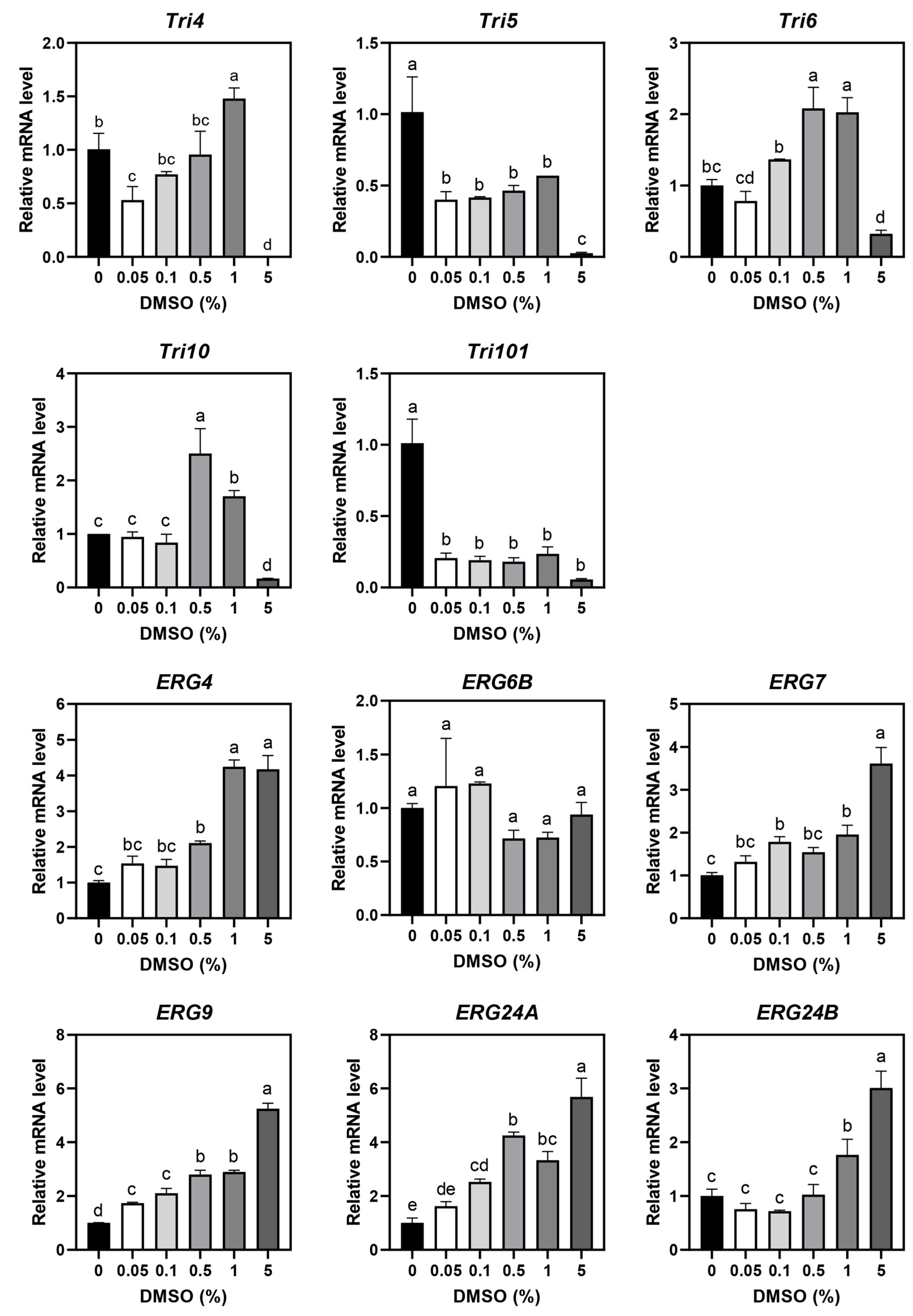
3.1. DMSO Effects on F. graminearum

DMSO induced a dose-dependent change in the color of F. graminearum colonies (Figure S1a). Treating the liquid medium with DMSO caused the fungus to change from yellowish to red, as it lost color. Moreover, when compared with the control, in the presence of the highest DMSO concentration (5%), the spores did not colonize properly and grew in a slushy mess (Figure S1a). In DMSO (5%)-containing agar medium, reddish fungus turned yellow–white (Figure S1a). These data show that DMSO affected the decolorization reaction, as well as F. graminearum growth and mycotoxin production. Although the dry weight of the fungus growing in a liquid medium did not vary significantly across DMSO concentrations (Figure S1b), in agar medium, plates containing 5% DMSO had a 40% reduction in mycelia (Figure S1c). DON and 15-acetyldeoxynivalenol (15-AcDON) concentrations decreased dose-dependently, and notably, they were below the limit of quantification at 0.5% DMSO (Figure S1d,e).
RT-qPCR analysis of the expression levels of DON- and ergosterol biosynthesis-related genes revealed that Tri5 and Tri101, which encode trichodiene synthase and trichothecene 3-O-acetyltransferase, respectively, were downregulated by 0.05% DMSO (Figure 1). Tri4, encoding cytochrome P450 monooxygenase, along with Tri6 and Tri10, which encode trichothecene biosynthesis transcription regulators, were downregulated at a concentration of 5%. However, the genes related to ergosterol biosynthesis, except ERG6B, were dose-dependently upregulated. Therefore, to minimize the contribution of DMSO to the antifungal effects of MEO and C10, they were dissolved in 0.05% DMSO, which was the least influential.

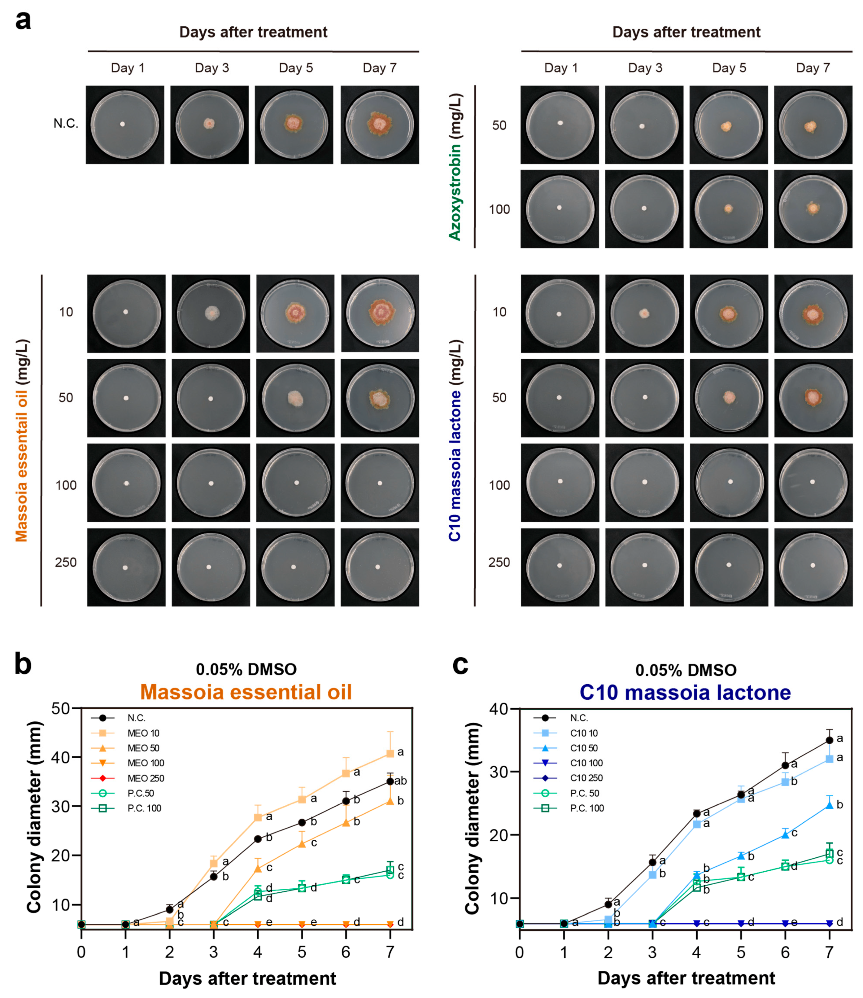
3.2. The Antifungal Activities of MEO and C10 against F. graminearum

The antifungal activities of MEO and C10 were assessed using the agar dilution, disc diffusion, and mycelial growth assays. In the disc diffusion assay, MEO and C10 exhibited antifungal activity on day 2, starting at the concentration of 2.5 mg/mL (Figure 2). However, over time, the fungus gradually covered the inhibitory zone. The antifungal effect was effective on day 3 and day 4 at concentrations starting at 10 mg/mL and 25 mg/mL, respectively. The positive control, azoxystrobin, inhibited fungal growth, causing it to grow sparsely. However, it did not completely inhibit the fungus to form a distinct inhibition zone. As a result, the inhibition zone diameter was relatively small, around 10 mm, and from the third day onwards, the inhibition zone was covered again by the fungus.
In the agar dilution assay, MEO and C10 exhibited potent antifungal effects at 50 mg/L (Figure 3), and they completely inhibited F. graminearum growth at concentrations of >100 mg/L up to seven days after treatment. However, when compared with 50 mg/mL, azoxystrobin did not exhibit significantly greater antifungal activity at 100 mg/mL, and it did not completely inhibit F. graminearum.
Notably, when compared with the disc diffusion assay, the effective concentration range determined through the agar dilution assay was 100 times lower. In the mycelial growth assay, F. graminearum exhibited similar growth in the presence of up to 25 mg/L, but its growth declined in the presence of ≥50 mg/L and was completely inhibited at 100 mg/L (Figure 4a–c).
The three methods showed that to exhibit noticeable antifungal effects, MEO and C10 need to reach a specific concentration and that they can completely inhibit F. graminearum at concentrations lower than those required by azoxystrobin, an antifungal agent in current use. Moreover, although azoxystrobin exhibits a gradual antifungal effect, it does not completely inhibit F. graminearum at the effective concentrations of MEO and C10.
Analysis of the antifungal effects of MEO and C10 in 1% DMSO (Figures S2 and S3) revealed that although their antifungal activities in solid cultures were similar to their effect in 0.05% DMSO (Figure S2), in liquid culture, they completely inhibited F. graminearum at 50 mg/L, which is lower than their effective concentration in 0.05% DMSO (Figure S3). This indicates that DMSO and MEO have synergistic effects that result in enhanced antifungal efficacy.

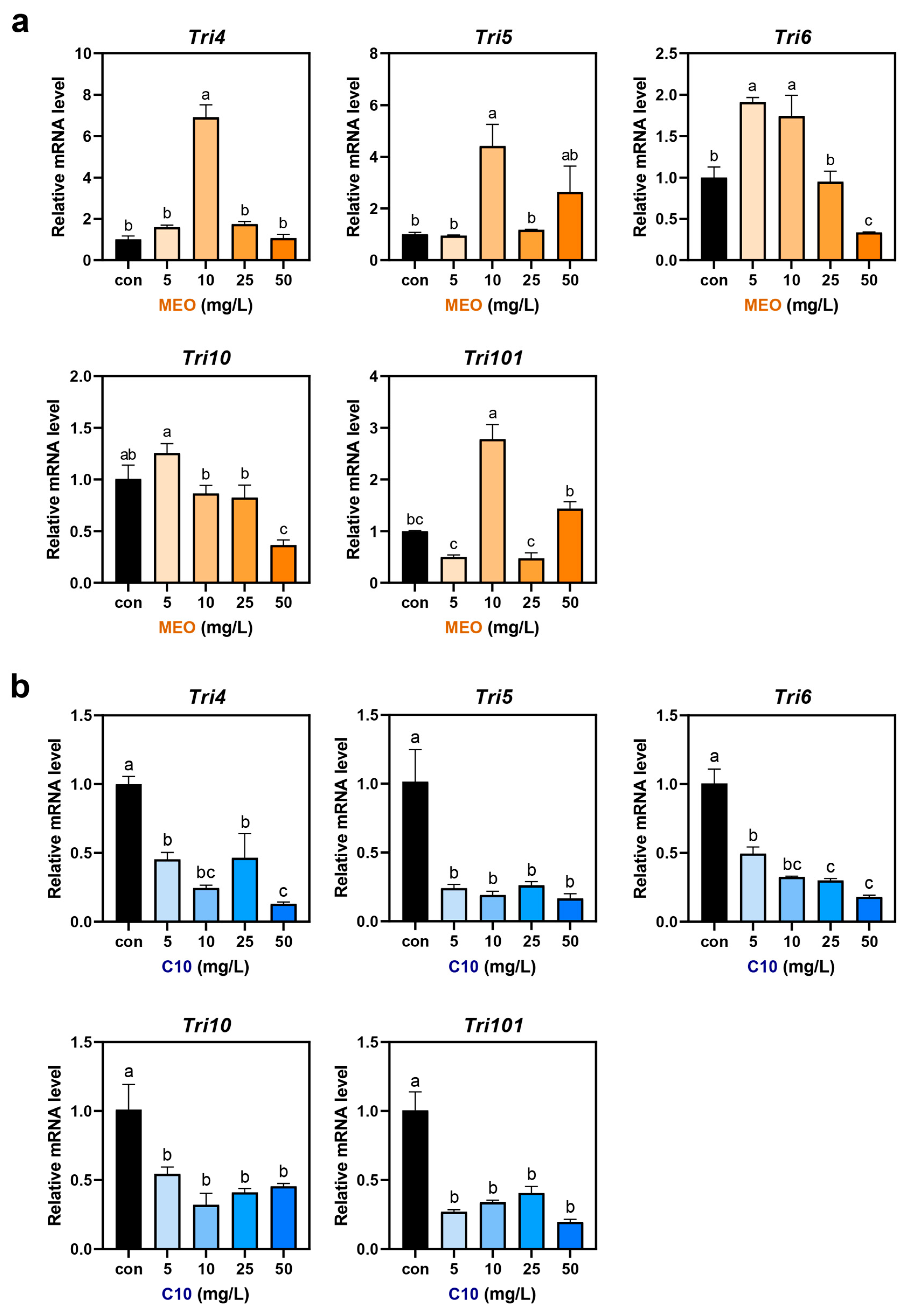
3.3. Mycotoxin Production and Gene Expression in MEO- and C10-Treated F. graminearum

Analysis of the levels of DON and its precursor, 15-AcDON, in F. graminearum growth medium following treatment with MEO and C10 revealed that the lowest MEO concentration, 5 mg/L, inhibited DON and 15-AcDON production most significantly (Figure 4d). However, increasing MEO concentration was associated with rising mycotoxin levels, although all MEO treatment groups had lower mycotoxin levels than the control group. C10 exhibited the most significant mycotoxin production inhibition at 10 mg/L, with a similar trend to MEO (Figure 4e).
The expression of genes associated with DON and ergosterol biosynthesis was analyzed using RT-qPCR (Figure 5 and Figure 6). In the MEO treatment group, the trichothecene biosynthesis transcription regulators, Tri6 and Tri10, were downregulated at 50 mg/L, while Tri4, Tri5, and Tri101 were upregulated at 10 mg/L and 50 mg/L (Figure 5a). In the C10 treatment group, genes associated with DON synthesis were consistently downregulated across all treatments (Figure 5b). ERG4, which encodes sterol C-24 reductase that catalyzes the final step of the ergosterol biosynthesis pathway for the conversion of ergosta-5, 7, 22, 24(28)-tetraenol to ergosterol, was downregulated at 25 mg/L and 50 mg/L of MEO (Figure 6a). ERG6B, ERG7, ERG9, ERG24A, and ERG24B, which are involved in the upstream steps of ERG4 in the ergosterol biosynthesis pathway, were upregulated by 3- to 20-fold at 50 mg/L of MEO. At all C10 concentrations, the genes related to ergosterol biosynthesis, except ERG6B, were downregulated (Figure 6b).

4. Discussion

4.1. DMSO as an Antifungal Agent Solvent

In this study, to assess the antifungal activity of MEO and C10 against F. graminearum, they were solubilized in DMSO, which is often used as a solvent when testing the biological activities of water-insoluble compounds [23]. Moreover, the natural products MEO and C10 might mediate their biological activities by reducing the production of F. graminearum mycotoxins, such as DON, nivalenol, and T-2. Therefore, it was crucial to understand DMSO’s antifungal and antimycotoxigenic effects before using it as a solvent.
DMSO is reported to cause oxidative stress in Saccharomyces cerevisiae [24], and when combined with itraconazole, it forms a pore on the lipid membrane [25]. DMSO can also enhance cell membrane permeability by binding to the plasma membrane [23]. Through these additive effects, DMSO is reported to influence minimum inhibitory concentration (MIC) values, especially MIC-2, as substantial inhibition of fungal cell growth in comparison to the control group by two doubling dilutions [23], indicating that during in vitro or in vivo antifungal tests, in test media, DMSO should be used at a concentration that does not suppress the targeted fungal growth. Similarly, analysis of the effects of 0.125–10% DMSO against dermatophyte growth using the agar diffusion method found that it completely inhibited the three tested dermatophyte isolates and that it had a dose-dependent inhibitory effect at 1.25–5% [26]. Here, the mycelial growth assay revealed that DMSO had an inhibitory effect on F. graminearum growth after reaching the concentration of 1% (Figure S1a,b) and that in the agar dilution assay, colony diameter was significantly reduced by 5% DMSO (Figure S1c). These results indicate that in F. graminearum growth medium, DMSO can be used at <1%.
Because DMSO affects transcription and secondary metabolism in A. flavus [27], it is necessary to measure the levels of secondary metabolites, like the target mycotoxins, DON, and 15-AcDON, in DMSO-containing fungal growth medium. A previous study found that a high DMSO concentration (282 mM) in the medium decreased the levels of 91% of secondary metabolite cluster genes in A. flavus, including aflatoxins- and cyclopiazonic acid-producing genes [27]. Additionally, in A. flavus, at 35 mM and 292 mM, DMSO inhibited aflatoxin B1 production by 63.5% and 100%, respectively [27]. Similarly, here, 0.05–5% DMSO significantly reduced DON and 15-AcDON levels (Figure S1d,e). Moreover, analysis of DON-producing genes in F. graminearum revealed that Tri5 and Tri101 were significantly downregulated across this range of DMSO treatments (Figure 1). These results indicate that DMSO use should be carefully considered when assessing antimycotoxigenic activity in F. graminearum.

4.2. MEO and C10 as Antifungal Agents against F. graminearum

For the control of F. graminearum, various EOs have been shown to have antifungal and antimycotoxigenic activity, with thyme’s MIC EO (ThEO) being the lowest (11.25 mg/L), while ginger, turmeric, and rosemary EOs had MICs of 364, 366, and 11,580 mg/L, respectively [28]. The minimum fungicidal concentrations of the four EOs had similar values, and ThEO contained 41% thymol and 24% p-cymene, and these major monoterpenes contributed to ThEO’s MIC and minimum fungicidal concentration values against F. graminearum [28]. Here, we used three fungicidal assays to assess MEO’s antifungal activity against F. graminearum (Figure 2, Figure 3 and Figure 4). Disc diffusion assays are not suitable for measuring the antifungal activity of volatile compounds because they escape into the air fast [22]. Indeed, when compared with agar dilution assays, disc diffusion assays are reported to be 50-fold less sensitive [22]. Similarly, in this study, using disc diffusion assays, the effect of MEO against F. graminearum was assessed at 0–25 mg/mL (Figure 2), while in agar dilution assays, MEO was used at 0–250 mg/L (Figure 3), representing a 100-fold difference between the assays.
Interestingly, MEO exhibits different antifungal activities depending on fungal species, and at 1 mg/mL, it achieved complete growth inhibition against A. flavus ATCC 22546 [22]. However, in this study, MEO completely inhibited F. graminearum at 100 mg/L, meaning 10-fold susceptibility to MEO in F. graminearum rather than A. flavus. In comparison, against F. graminearum, ThEO was about 10-fold stronger than MEO, and it was stronger than ginger, turmeric, and rosemary EOs [28]. Regarding mycelial growth inhibition, MEO (100 mg/L) was associated with marked decreases in the dry weights of A. flavus and F. graminearum, indicating that its antifungal activities vary with the assays used [22]. These results indicate that formulating MEO as an antifungal agent might be important to show its optimal field biological activities.
Recently, mycelial growth inhibition assays were used to determine the antifungal activities of 12 monoterpenes against the plant pathogenic fungi, Rhizoctonia solani, Fusarium oxysporum, Penicillium digitatum and Aspergillus niger [29]. Of the monoterpenes, which were tested on the four fungi at 20.14–50.35 mg/L, thymol was the most active since ThEO, which contains 40% thymol, had the highest antifungal activity against F. graminearum [28,29]. Against F. graminearum, thymol is reported to inhibit ergosterol biosynthesis via induction of the cell membrane through lipid peroxidation [30]. Here, the antifungal activity of C10 lactone, MEO’s primary compound, against F. graminearum was similar to that of MEO (Figure 2, Figure 3 and Figure 4).
MEO’s C10 lactone content is reported to range from about 45% to 80% [22,31]. In this study, because MEO was used, as previously reported by Lee et al. [22], it contained 45% C10 lactone. At all tested concentrations, MEO and C10 suppressed the production of DON and 15-AcDON. The inhibitory effects of flusilazole on the expression of trichothecene biosynthesis genes in F. graminearum and F. culmorum were studied, and flusilazole is one of the EBIs [32]. Treatment of flusilazole on these two phytopathogenic fungi showed up-regulation of Tri5 and Tri6 genes in F. graminnearum, whereas it did not change the expressions of Tri5, Tri6, and Tri12 genes in F. culmorum [32]. However, antimycotoxigenic effects by DMSO and C10 showed similar patterns on DON-producing gene expression, while MEO showed up- and down-regulation on five tested gene expressions because MEO consists of various natural products, even if it contains high levels of C10 up to 40% of the total [22]. Therefore, C10 lactone dramatically reduced DON and 15-DON production in F. graminearum via significant down-regulation of DON-producing gene expression of Tri4, Tri5, Tri6, Tri10, and Tri101 genes in this study.
Ergosterol (ER) biosynthesis in fungi has been well documented, and it is formed via an acetate pathway through lanosterol [33]. Its intermediates are known as zymosterol, fecosterol, episterol, ergosta-5,7,24(28)-trienol, and ergosta-5,7,22,24(28)-tetranol [33]. Ergosterol biosynthesis inhibitors (EBIs) are intensively used to control various fungi, including Fusarium sp. in agricultural industries, and EBIs inhibit cytochrome P450 sterol 14α-demethylase, which plays an important role in ergosterol formation [34]. Recently, the erg6 gene, the sterol C-24 methyltransferase expressing gene, was repressed in Aspergillus fumigatus, and erg6-repressed strains showed wild-type susceptibility towards EBIs [35]. With these results, each enzyme in ergosterol biosynthesis is essential for fungal life as well as forming ergosterol.
In this study, DMSO and MEO elevated ergosterol-producing gene expression, especially ERG6B, ERG24A, and ERG24B, were highly up-regulated by greater than 10-fold by MEO treatments in this study. However, ERG4 was significantly downregulated in MEO-treated F. graminearum at 25 and 50 mg/L concentrations. Similar results were found with itraconazole-treated Candida albicans, which is a typical EBI agent [36]. Itraconazole-treated C. albicans strains elevated the expressions of ERG6, ERG1, ERG3, ERG4, ERG10, ERG9, ERG26, and ERG25, which are involved in the ergosterol biosynthesis [36]. However, C10 lactone suppressed all tested ergosterol-producing gene expression, except for ERG6B gene expression at the concentration of 50 mg/L in this study. With these results, MEO and C10 lactone are considered an inhibitor of ERG4 gene expression in F. graminearum, and C10 lactone generally possesses stronger inhibitory effects on ergosterol-producing gene expression than MEO because MEO consists of other constituents as a mixture.
Natural products may play an important role in the list of active ingredients for F. graminearum control [37]. γ-Oryzanol, neem seed phenolic fraction, and rice bran extracts are reported to exhibit antifungal activity against F. graminearum growth at MIC50 concentrations of 0.9, 0.032, and 0.037 g/kg, respectively. Piper sarmentosum extract contains 17 anti-F. graminearum natural products, including salsolinol, 4-oxoproline, propylnorapomorphine, salicylic acid, oroxylin A, and octadec-9-ynoic acid [38]. Moreover, ferulic acid, a potent antifungal product that is abundant in Ferula communis [39], exhibits antifungal activity against F. graminearum at EC50 and EC90 values of 20 mg/L and 10 mg/L, respectively, via hyphae cell membrane impairment [40]. Taken together, natural products, including MEO and C10 lactone, which are generally safe for humans and the environment, can be potent antifungal candidates.

5. Conclusions

F. graminearum, which causes FHB, has spread quickly across Korean wheat fields because of climate change. However, FHB control using fungicides has not been successful because of fungal resistance caused by intensive and repetitive fungicide use. MEO and C10 lactone, which are recognized as GRAS compounds, exhibit potent antifungal and antimycotoxigenic activities against F. graminearum. Within this study, MEO and C10 at 100 mg/L showed potent antifungal activities against F. graminearum, and they inhibited the production of DON and 15-AcDON at concentrations ranging from 5 to 50 mg/L. This decreased production of mycotoxins was associated with the down-regulation of trichodiene-producing genes in MEO- and C10-treated F. graminearum. Therefore, they can be used to control F. graminearum in combination with or without currently used fungicides or in combination with other natural products, like thymol and neem seed extracts, which may delay resistance emergence. However, before their application, more toxicological data are needed, which can be obtained through various experiments, including acute and chronic toxicity studies in rodents, acute toxicity studies on beneficial insects like honeybees, and environmental toxicology analysis using zebrafish and earthworms [41].

Supplementary Materials

The following supporting information can be downloaded at: https://www.mdpi.com/article/10.3390/agriculture14081216/s1, Figure S1. The effects of dimethyl sulfoxide (DMSO) on depigmentation, growth, and mycotoxin production of Fusarium graminearum, with concentrations ranging from 0% to 5%. (a) Pictures of F. graminearum after treatment with DMSO in liquid medium or agar medium at 10 days. (b) Dry weight of F. graminearum treated with DMSO in liquid medium. (c) Colony diameter of F. graminearum treated with DMSO in agar medium. (d,e), The effect of DMSO on the production of deoxynivalenol and 15-acetyldeoxynivalenol. All experiments were conducted in triplicates. Data analysis was performed using one-way ANOVA, followed by post-hoc Tukey’s test. Lowercase letters denote significant differences among groups: a > b > c > d (p < 0.05). (**: p < 0.01, ns: not significant). Figure S2. Antifungal activities of massoia essential oil (MEO) and C10 massoia lactone (C10) against Fusariunm gramineaum using agar dilution assay, with concentrations ranging from 10 to 500 mg/L over a 7-day incubation period. (a) Pictures of antifungal activities of MEO or C10 over the course of the incubation time (day). (b,c) Colony diameter (mm) after treatment with MEO or C10. N.C., negative control (DMSO (1%)); P.C., positive control (azoxystrobin 50 and 100 mg/L). All experiments were conducted in triplicates. Data analysis was performed using one-way ANOVA, followed by post-hoc Tukey’s test. Lowercase letters denote significant differences among groups: a > b > c > d > e (p < 0.05). Figure S3. Antifungal activities of massoia essential oil (MEO) and C10 massoia lactone (C10) against Fusariunm gramineaum using mycelium growth assay. (a) Pictures of antifungal activities of MEO and C10 in liquid medium. (b,c) Dry weight of F. graminearum after treatment of MEO or C10. DMSO (1%) is used as solvent control. All experiments were conducted in triplicates. Data analysis was performed using one-way ANOVA, followed by post-hoc Tukey’s test. (**, p < 0.01; ***, p < 0.001; ****, p < 0.0001). Table S1. HPLC gradient conditions for mycotoxin analysis. Table S2. LC-MS/MS multiple reaction monitoring (MRM) conditions. Table S3. Linearity data, LOD, LOQ and recovery (%) for tested compounds by LC-MS/MS. Table S4. List of primers used for amplification of genes involved in deoxynivalenol and ergosterol biosynthesis by RT-qPCR [42,43,44,45].

Author Contributions

Conceptualization, S.-E.L.; methodology, J.L.; software, J.L.; validation, J.L. and S.-E.L.; formal analysis, J.L. and S.-E.L.; investigation, J.L. and S.-E.L.; data curation, J.L. and S.-E.L.; writing—original draft preparation, J.L. and S.-E.L.; writing—review and editing, S.-E.L.; visualization, J.L. and S.-E.L.; supervision, S.-E.L.; funding acquisition, S.-E.L. All authors have read and agreed to the published version of the manuscript.

Funding

This research was supported by Biological Materials Specialized Graduate Program through the Korea Environmental Industry & Technology Institute (KEITI), funded by the Ministry of Environment (MOE); this research was supported by the Korea Basic Science Institute (National Research Facilities and Equipment Center) grant funded by the Ministry of Education (2021R1A6C101A416), and Quantitative PCR was performed at KNU NGS center (Daegu, Republic of Korea).

Institutional Review Board Statement

Not applicable.

Data Availability Statement

The data presented in this study are available on request from the corresponding author.

Conflicts of Interest

The authors declare no conflicts of interest.

References

  1. Bai, G.; Shaner, G. Scab of wheat; prospects of control. Plant Dis. 1994, 78, 760–766. [Google Scholar]
  2. Xu, X.; Nicholson, P. Community ecology of fungal pathogens causing wheat head blight. Annu. Rev. Phytopathol. 2009, 47, 83–103. [Google Scholar] [CrossRef] [PubMed]
  3. McMullen, M.; Bergstrom, G.; De Wolf, E.; Dill-Macky, R.; Hershman, D.; Shaner, G.; Van Scanford, D. A unified effort to fight an enemy of wheat and barley: Fusarium head blight. Plant Dis. 2012, 96, 1712–1728. [Google Scholar] [CrossRef] [PubMed]
  4. Singh, J.; Chhabra, B.; Raza, A.; Yang, S.H.; Sandhu, K.S. Important wheat diseases in the US and their management in the 21st century. Front. Plant Sci. 2023, 13, 1010191. [Google Scholar] [CrossRef] [PubMed]
  5. McMullen, M.; Jones, R.; Gallenberg, D. Scab of wheat and barley: A re-emerging disease of devastating impact. Plant Dis. 1997, 81, 1340–1348. [Google Scholar] [CrossRef] [PubMed]
  6. Wilson, W.; Dahl, B.; Nganje, W. Economic costs of fusarium head blight, scab and deoxynivalenol. World Mycotoxin J. 2018, 11, 291–302. [Google Scholar] [CrossRef]
  7. Shim, H.K.; Gang, I.J. Development of monitoring and management technology for Fusarium head blight. Natl. Inst. Crop Sci. 2018, 2, 1593–1666. [Google Scholar]
  8. Jung, J.Y.; Kim, J.H.; Baek, M.; Cho, C.; Cho, J.; Kim, J.; Pavan, W.; Kim, K.H. Adapting to the projected epidemics of Fusarium head blight of wheat in Korea under climate changes scenarios. Front. Plant Dis. 2022, 13, 1040752. [Google Scholar] [CrossRef] [PubMed]
  9. Shah, L.; Ali, A.; Yahya, M.; Zhu, Y.; Wang, S.; Si, H.; Rahman, H.; Ma, C. Integrated control of Fusarium head blight and deoxynivalenol mycotoxin in wheat. Plant Pathol. 2018, 67, 532–548. [Google Scholar] [CrossRef]
  10. Moonjely, S.; Ebert, M.; Paton-Glassbrook, D.; Noel, Z.A.; Roze, L.; Shay, R.; Watkins, T.; Trail, F. Update on the state of research to manage Fusarium head blight. Fungal Genet. Biol. 2023, 169, 103829. [Google Scholar] [CrossRef]
  11. Blandino, M.; Haidukowski, M.; Pascale, M.; Plizzari, L.; Scudellari, D.; Reyneri, A. Integrated strategies for the control of Fusarium head blight and deoxynivalenol contamination in winter wheat. Field Crop. Res. 2012, 133, 139–149. [Google Scholar] [CrossRef]
  12. Pazdiora, P.C.; da Rosa Dorneles, K.; Morello, T.N.; Nicholson, P.; Dallagnol, L.J. Silicon soil amendment as a complement to manage tan spot and Fusarium head blight in wheat. Agron. Sustain. Dev. 2021, 41, 21. [Google Scholar] [CrossRef]
  13. Anderson, N.R.; Freije, A.N.; Bergstrom, G.C.; Bradley, C.A.; Cowger, C.; Faske, T.; Hollier, C.; Kleczewski, N.; Padgett, G.B.; Paul, P.; et al. Sensitivity of Fusarium graminearum to metconazole and tebuconazole fungicides before and after widespread use in wheat in the United States. Plant Health Prog. 2020, 21, 85–90. [Google Scholar] [CrossRef]
  14. Paul, P.A.; Bradley, C.A.; Madden, L.V.; Dalla Lana, F.; Bergstrom, G.C.; Dill-Macky, R.; Esker, P.D.; Wise, K.A.; McMullen, M.; Grybauskas, A.; et al. Meta-analysis of the effects of QoI and DMI fungicide combinations on Fusarium head blight and deoxynivalenol in wheat. Plant Dis. 2018, 102, 2602–2615. [Google Scholar] [CrossRef]
  15. Jung, B.; Li, T.; Ji, S.; Lee, J. Efficacy of diphenyleneiodonium chloride (DPIC) against diverse plant pathogens. Mycobiology 2019, 47, 105–111. [Google Scholar] [CrossRef]
  16. Harčárová, M.; Čonková, E.; Proškovcová, M.; Váczi, P.; Marcinčáková, D.; Bujňák, L. Comparison of antifungal activity of selected essential oils against Fusarium graminearum in vitro. Ann. Agric. Environ. Med. 2021, 28, 414–418. [Google Scholar] [CrossRef] [PubMed]
  17. Jang, J.Y.; Baek, S.G.; Choi, J.H.; Kim, S.; Kim, J.; Kim, D.W.; Yun, S.H.; Lee, T. Characterization of nivalenol-producing Fusarium asiaticum that causes cereal head blight in Korea. Plant Pathol. J. 2019, 35, 543–552. [Google Scholar] [CrossRef]
  18. Sydenham, E.W.; Thiel, P.G.; Marasas, W.F.O.; Nieuwenhuis, J.J. Occurrence of deoxynivalenol and nivalenol in Fusarium graminearum infected undergrade wheat in South Africa. J. Agric. Food Chem. 1989, 37, 921–926. [Google Scholar] [CrossRef]
  19. Haidukowski, M.; Somma, S.; Ghionna, V.; Cimmarusti, M.T.; Masiello, M.; Logrieco, A.F.; Moretti, A. Deoxynivalenol and T-2 toxin as major concerns in durum wheat from Italy. Toxins 2022, 14, 627. [Google Scholar] [CrossRef]
  20. EU (European Union). Maximum Levels for Certain Contamination in Foodstuffs as Regards Fusarium Toxins in Maize and Maize Products. Commission Regulation (EC) No. 1126/2007. Available online: https://eur-lex.europa.eu/legal-content/EN/ALL/?uri=CELEX%3A32007R1126 (accessed on 22 May 2024).
  21. Lee, T.; Baek, S.G.; Kim, S.; Paek, J.S.; Choi, J.; Choi, J.H.; Jang, Y.J.; Kim, J. Trends in mycotoxin contamination of cereals and cereal products in Korea. Res. Plant Dis. 2022, 28, 179–194. [Google Scholar] [CrossRef]
  22. Lee, Y.; Park, S.J.; Kim, K.; Kim, T.O.; Lee, S.E. Antifungal and antiaflatoxigenic activities of massoia oil and C10 massoia lactone against aflatoxin-producing Aspergillus flavus. Toxins 2023, 15, 571. [Google Scholar] [CrossRef] [PubMed]
  23. Hazen, K.C. Influence of DMSO on antifungal activity during susceptibility testing in vitro. Diagn. Microbiol. Infect. Dis. 2013, 75, 60–63. [Google Scholar] [CrossRef] [PubMed]
  24. Sadowska-Bartosz, I.; Pączka, A.; Mołon, M.; Bartosz, G. Dimethyl sulfoxide induces oxidative stress in the yeast Saccharomyces cerevisiae. FEMS Yeast Res. 2013, 13, 820–830. [Google Scholar] [CrossRef] [PubMed]
  25. Kasparyan, G.; Poojari, C.; Rog, T.; Hub, J.S. Cooperative effects of an antifungal moiety and DMSO on pore formation over lipid membranes revealed by free energy calculations. J. Phys. Chem. B 2020, 124, 8811–8821. [Google Scholar] [CrossRef] [PubMed]
  26. Randhawa, M.A. The effect of dimethyl sulfoxide (DMSO) on the growth of dermatophytes. Jap. J. Med. Mycol. 2006, 47, 313–318. [Google Scholar] [CrossRef] [PubMed]
  27. Costes, L.H.; Lippi, Y.; Naylies, C.; Jamin, E.L.; Genthon, C.; Bailly, S.; Oswald, I.P.; Bailly, J.-D.; Puel, O. The solvent dimethyl sulfoxide affects physiology, transcriptome and secondary Metabolism of Aspergillus flavus. J. Fungi 2021, 7, 1055. [Google Scholar] [CrossRef]
  28. Romoli, J.C.Z.; Silva, M.V.; Pante, G.C.; Hoeltgebaum, D.; Castro, J.C.; da Rocha, G.H.O.; Capoci, I.R.G.; Nerilo, S.B.; Mossini, S.A.G.; da Gloria, E.M.; et al. Anti-mycotoxigenic and antifungal activity of ginger, turmeric, thyme and rosemary essential oils in deoxynivalenol (DON) and zearalenone (ZEA) producing Fusarium graminearum. Food Addit. Contam. A 2022, 39, 362–372. [Google Scholar] [CrossRef] [PubMed]
  29. Maeri, G.I.K.; Abdel Rasoul, M.A.; Abdelgaleil, S.A.M. Comparative antifungal activities and biochemical effects of monoterpenes on plant pathogenic fungi. Pestic. Biochem. Physiol. 2012, 103, 56–61. [Google Scholar] [CrossRef]
  30. Gao, T.; Zhou, H.; Zhou, W.; Hu, L.; Chen, J.; Shi, Z. The fungicidal activity of thymol against F. graminearum via inducing lipid peroxidation and disrupting ergosterol biosynthesis. Molecules 2016, 21, 770. [Google Scholar] [CrossRef]
  31. Seo, S.M.; Lee, J.W.; Shin, J.; Tak, J.H.; Hyun, J.; Park, I.K. Development of cellulose nanocrystal-stabilized pickering emulsions of massoia and nutmeg essential oils for the control of Aedes abbopictus. Sci. Rep. 2022, 11, 12038. [Google Scholar] [CrossRef]
  32. Popiel, D.; Dawidziuk, A.; Koczyk, G.; Mackowiak, A.; Marcinkowska, K. Multiple facets of response to fungicides—The influence of azole treatment on expression of key mycotoxin biosynthetic genes and candidate resistance factors in the control of resistant Fusarium strains. Eur. J. Plant Pathol. 2017, 147, 773–785. [Google Scholar] [CrossRef]
  33. Dupont, S.; Lemetais, G.; Ferreira, T.; Cayot, P.; Gervais, P.; Beney, L. Ergosterol biosynthesis: A fungal pathway for life on land? Evolution 2012, 66, 2961–2968. [Google Scholar] [CrossRef]
  34. Fan, J.; Urban, M.; Parker, J.E.; Brewer, H.C.; Kelly, S.L.; Hammond-Kosack, K.E.; Fraaije, B.A.; Liu, X.; Cools, H.J. Characterization of the sterol 14a-demethylase of Fusarium graminearum identifies a novel genus-specific CYP51 function. New Phytol. 2013, 198, 821–835. [Google Scholar] [CrossRef] [PubMed]
  35. Xie, J.; Rybak, J.M.; Martin-Vincente, A.; Guruceaga, X.; Thorn, H.I.; Nywening, A.V.; Ge, W.; Parker, J.E.; Kelly, S.L.; Rogers, P.D.; et al. The sterol C-24 methyltranferase encoding gene, erg6, is essential for viability of Aspergillus species. Nat. Commun. 2024, 15, 4261. [Google Scholar] [CrossRef]
  36. De Backer, M.D.; Ilyina, T.; Ma, X.J.; Vandoninck, S.; Luyten, W.H.M.; Vanden Bossche, H. Genomic profiling of the response of Candida albicans to itraconazole treatment using a DNA microarray. Antimicrob. Agents Chemother. 2001, 45, 1660–1670. [Google Scholar] [CrossRef]
  37. Heidtmann-Bemvenuti, R.; Tralamazza, S.M.; Ferreira, C.F.J.; Correa, B.; Badiale-Furlong, E. Effect of natural compounds on Fusarium graminearum complex. J. Sci. Food Agric. 2016, 96, 3998–4008. [Google Scholar] [CrossRef]
  38. Zhou, L.; Zhou, H.; Hou, G.; Ji, F.; Wang, D. Antifungal activity and metabolomics analysis of Piper sarmentosum extracts against Fusarium graminearum. J. Appl. Microbiol. 2023, 134, lxad019. [Google Scholar] [CrossRef] [PubMed]
  39. Kim, J.K.; Park, S.U. A recent overview on the biological and pharmacological activities of ferulic acid. EXCLI J. 2019, 18, 132–138. [Google Scholar] [PubMed]
  40. Yan, H.; Meng, X.; Lin, X.; Duan, N.; Wang, Z.; Wu, S. Antifungal activity and inhibitory mechanisms of ferulic acid against the growth of Fusarium graminearum. Food Biosci. 2023, 52, 102414. [Google Scholar] [CrossRef]
  41. Lee, Y.; Kim, C.; Kim, T.O.; Lee, S.E. Developmental toxicity of C10 massoia lactone, the main constituent of Cryptorya massoia, on zebrafish (Danio rerio) embryos. Appl. Sci. 2024, 14, 538. [Google Scholar] [CrossRef]
  42. Montibus, M.; Ducos, C.; Bonnin-Verdal, M.-N.; Bormann, J.; Ponts, N. The bZIP Transcription Factor Fgap1 Mediates Oxidative Stress Response and Trichothecene Biosynthesis but Not Virulence in Fusarium graminearum. PLoS ONE 2013, 8, e83377. [Google Scholar]
  43. Boutigny, A.L.; Barreau, C.; Atanasova-Penichon, V.; Verdal-Bonnin, M.N.; Pinson-Gadais, L.; Richard-Forget, F. Ferulic acid, an efficient inhibitor of type B trichothecene biosynthesis and Tri gene expression in Fusarium liquid cultures. Mycol. Res. 2009, 113, 746–753. [Google Scholar]
  44. Wang, L.Q.; Wu, K.T.; Yang, P.; Hou, F.; Rajput, S.A.; Qi, D.S.; Wang, S. Transcriptomics reveals the effect of thymol on the growth and toxin production of Fusarium graminearum. Toxins 2022, 14, 142. [Google Scholar]
  45. Liu, X.; Jiang, J.; Yin, Y.; Ma, Z. Involvement of FgERG4 in ergosterol biosynthesis, vegetative differentiation and virulence in Fusarium graminearum. Mol. Plant Pathol. 2013, 14, 71–83. [Google Scholar]
Figure 1. The gene expression level of Fusarium graminearum treated with dimethyl sulfoxide (DMSO), with concentrations ranging from 0% to 5%. All experiments were conducted in triplicates. Data analysis was performed using one-way ANOVA, followed by post-hoc Tukey’s test. Lowercase letters denote significant differences among groups: a > b > c > d > e (p < 0.05). Tri4: cytochrome P450 monooxygenase, Tri5: trichodiene synthase, Tri6: trichothecene biosynthesis transcription regulator 6, Tri10: trichothecene biosynthesis transcription regulator 10, Tri101: trichothecene 3-O-acetyltransferase, ERG4: sterol C-24 reductase, ERG6B: sterol 24-C-methyltransferase, ERG7: lanosterol synthase, ERG9: squalene synthase, ERG24A: sterol C-14 reductase, ERG24B: sterol C-14 reductase.
Agriculture 14 01216 g001
Figure 2. Antifungal activities of massoia essential oil (MEO) and C10 massoia lactone (C10) against Fusarium graminearum using disc diffusion assay, with concentrations ranging from 0.5 to 25 mg/mL over a 4-day incubation period at 25 °C. (a,b) Pictures of antifungal activities of MEO or C10 over the course of the incubation time (day). (c,d) Inhibition zone diameter (mm) of F. graminearum after treatment with MEO or C10 on days 2, 3, and 4. N.C., negative control (DMSO); P.C., positive control (azoxystrobin 0.5 mg/mL). All experiments were conducted in triplicates. Data analysis was performed using one-way ANOVA, followed by post-hoc Tukey’s test. Lowercase letters denote significant differences among groups: a > b > c > d > e (p < 0.05).
Agriculture 14 01216 g002
Figure 3. Antifungal activities of massoia essential oil (MEO) and C10 massoia lactone (C10) against Fusarium graminearum using agar dilution assay, with concentrations ranging from 10 to 250 mg/L over a 7-day incubation period at 25 °C. (a) Pictures of antifungal activities of MEO or C10 over the course of the incubation time (day). (b,c) Colony diameter (mm) after treatment with MEO or C10. N.C., negative control (DMSO [0.05%]); P.C., positive control (azoxystrobin 50 and 100 mg/L). All experiments were conducted in triplicates. Data analysis was performed using one-way ANOVA, followed by post-hoc Tukey’s test. Lowercase letters denote significant differences among groups: a > b > c > d > e (p < 0.05).
Agriculture 14 01216 g003
Figure 4. Antifungal and anti-mycotoxigenic activities of massoia essential oil (MEO) and C10 massoia lactone (C10) against Fusarium graminearum. (a) Pictures of antifungal activities of MEO and C10 in GYEP medium at 10 days. (b,c) Dry weight of F. graminearum after treatment of MEO or C10. (dg) The effect of MEO or C10 on the production of deoxynivalenol and 15-acetyldeoxynivalenol. DMSO (0.05%) is used as solvent control. All experiments were conducted in triplicates. Data analysis was performed using one-way ANOVA, followed by post-hoc Tukey’s test. Lowercase letters denote significant differences among groups: a > b > c > d (p < 0.05). (**, p < 0.01; ***, p < 0.001; ****, p < 0.0001).
Agriculture 14 01216 g004
Figure 5. Expression levels of deoxynivalenol synthesis-related genes in Fusarium graminearum treated with (a) Massoia essential oil (MEO) and (b) C10 massoia lactone (C10). DMSO (0.05%) is used as solvent control. All experiments were conducted in triplicates. Data analysis was performed using one-way ANOVA, followed by post-hoc Tukey’s test. Lowercase letters denote significant differences among groups: a > b > c (p < 0.05). Tri4: cytochrome P450 monooxygenase, Tri5: trichodiene synthase, Tri6: trichothecene biosynthesis transcription regulator 6, Tri10: trichothecene biosynthesis transcription regulator 10, Tri101: trichothecene 3-O-acetyltransferase.
Agriculture 14 01216 g005
Figure 6. Expression levels of ergosterol synthesis-related genes in Fusarium graminearum treated with (a) Massoia essential oil (MEO) and (b) C10 massoia lactone (C10). DMSO (0.05%) is used as solvent control. All experiments were conducted in triplicates. Data analysis was performed using one-way ANOVA, followed by post-hoc Tukey’s test. Lowercase letters denote significant differences among groups: a > b > c > d (p < 0.05). ERG4: sterol C-24 reductase, ERG6B: sterol 24-C-methyltransferase, ERG7: lanosterol synthase, ERG9: squalene synthase, ERG24A: sterol C-14 reductase, ERG24B: sterol C-14 reductase.
Agriculture 14 01216 g006
Disclaimer/Publisher’s Note: The statements, opinions and data contained in all publications are solely those of the individual author(s) and contributor(s) and not of MDPI and/or the editor(s). MDPI and/or the editor(s) disclaim responsibility for any injury to people or property resulting from any ideas, methods, instructions or products referred to in the content.

