Article

Wolbachia Transinfection and Effect on the Biological Traits of Matsumuratettix hiroglyphicus (Matsumura), the Leafhopper Vector of Sugarcane White Leaf Disease

by
Kamonrat Suwanchaisri
1,
Jariya Roddee
2 and
Jureemart Wangkeeree
3,*
1
Department of Biotechnology, Faculty of Science and Technology, Thammasat University, Rangsit Campus, Pathum Thani 12120, Thailand
2
School of Crop Production Technology, Institute of Agricultural Technology, Suranaree University of Technology, Nakhon Ratchasima 30000, Thailand
3
Department of Agricultural Technology, Faculty of Science and Technology, Thammasat University, Rangsit Campus, Pathum Thani 12120, Thailand
*
Author to whom correspondence should be addressed.
Agriculture 2024, 14(8), 1236; https://doi.org/10.3390/agriculture14081236
Submission received: 4 June 2024 / Revised: 22 July 2024 / Accepted: 23 July 2024 / Published: 26 July 2024
(This article belongs to the Section Crop Protection, Diseases, Pests and Weeds)

Abstract

:
The bacterial genus Wolbachia induces reproductive abnormalities in its insect host, including cytoplasmic incompatibility (CI), which causes embryonic death in the crossing of infected males and uninfected females. Hence, Wolbachia-based strategies are employed to control insect pests. However, Wolbachia does not naturally infect Matsumuratettix hiroglyphicus (Matsumura), the main vector of the phytoplasma causing the sugarcane white leaf (SCWL) disease. In this study, the wYfla Wolbachia strain, which induces strong CI in its original host, was microinjected into nymphs of M. hiroglyphicus. Molecular detection revealed that Wolbachia was successfully transinfected into the recipient host, with an infection frequency of 55–80% in up to eight generations after transinfection. Wolbachia exhibited no significant detrimental effects on the developmental time of the immature stages, adult emergences, and female longevity, whereas the lifespan of transinfected males was decreased. Reciprocal crossing revealed that Wolbachia infection did not affect the number of eggs laid per female. However, the hatching rate produced by the pairs between the transinfected males and naturally uninfected females significantly decreased. The evidence of Wolbachia transmitted through the generations tested and partial CI occurrence in transinfected M. hiroglyphicus highlights the possibility of the future development of Wolbachia-based strategies for controlling the vector of SCWL.

1. Introduction

Wolbachia-based strategies are currently accepted as a novel and ecologically friendly alternative method for controlling mosquito-borne diseases that afflict humans [1,2,3], and it has been employed in practical field release [4,5,6]. Recently, this method has been developed to control insect pests of agricultural economic crops [7,8]. Wolbachia is a maternally inherited endosymbiont belonging to Class Alphaproteobacteria within the Order Rickettsiales and is the most abundant endosymbiont that naturally occurs in various invertebrate species; in particular, approximately 50% of all known insect species are estimated to be infected [9,10].
Wolbachia infection influences the reproductive traits and fitness of their hosts; for instance, it induces reproductive abnormalities, including parthenogenesis, feminization, male killing, and cytoplasmic incompatibility (CI) [11,12,13,14]. Moreover, Wolbachia elicits deleterious effects on the life history and reproductive performance [15]. In contrast, it has been reported that Wolbachia infection can also increase the fecundity of its host [16]. Owing to its ability to induce CI, Wolbachia is being used as a biological agent to control mosquito vectors. CI occurs when Wolbachia-infected males mate with uninfected females or are infected with different Wolbachia strains, resulting in the failure of the embryonic stage to further develop [17,18]. It is an essential phenotype induced by Wolbachia that can suppress the population of mosquitoes [19]. For example, Culex quinquefasciatus transinfected with a single or superinfected Wolbachia strain (wAlbB or wPipwAlbA) exhibited complete CI or 100% embryonic death [20]. Despite the abovementioned effects, some Wolbachia strains reduce the transmission of insect-borne diseases by inhibiting the infection and replication of pathogens [21].
Sugarcane white leaf (SCWL) is the most devastating disease of sugarcane, resulting in annual economic losses of up to USD 20 million per year in the sugar industry of Southeast Asian countries [22,23]. The disease is caused by “Candidatus Phytoplasma sacchari” [24], which mainly spreads through the plants’ propagating materials and is transmitted by two sap-sucking leafhoppers: Matsumuratettix hiroglyphicus (Matsumura) and Yamatotettix flavovittatus (Matsumura) (Suborder Auchenorrhyncha, Family Cicadellidae) [25]. M. hiroglyphicus is considered the main vector of the pathogen because it is more abundant than Y. flavovittatus; moreover, the peak abundance of M. hiroglyphicus coincides with the early stage after sugarcane planting [26,27]. Currently, there are no commercial sugarcane cultivars resistant to phytoplasma diseases; therefore, an effective means to limit the disease distribution requires several methods. Healthy propagation materials for new plantings could be an effective means of controlling the SCWL disease [28]. Effective sustainable approaches have also focused on controlling leafhopper vectors, and insecticide treatment has been the primary method for reducing their population density [29]. However, owing to growing concerns about the risks posed by insecticides to the environment and human health, alternative control strategies must be developed.
The environmental friendliness, sustainability, and cost-effectiveness can be considered advantages of Wolbachia-based strategies compared with traditional insecticides [30]. Previous research has found that Wolbachia (wYfla strain) naturally infects Y. flavovittatus leafhoppers, inducing strong CI and resulting in 100% embryonic death. In addition, it is completely transmitted from the mother generation to the progeny in the Y. flavovittatus [31]. Owing to the ability of the wYfla strain to induce CI, Wolbachia-based pest control strategies are gaining attention as an alternative method of suppressing the population of leafhopper vectors. However, the prevalence of Wolbachia infection has not been detected in the main vector of SCWL, M. hiroglyphicus.
The first step towards developing Wolbachia-based strategies to control M. hiroglyphicus population requires the development of a Wolbachia-infected lineage through transinfection and characterization its phenotypic effects. Hence, in this study, M. hiroglyphicus was transinfected with the wYfla Wolbachia strain through nymphal microinjection. The dynamic infection and distribution of Wolbachia in the transinfected M. hiroglyphicus samples were examined. The effects of Wolbachia transinfection on the biological traits and CI induction were determined. Our findings will contribute to the development of Wolbachia-based strategies to control more insect pests such as those that carry phytoplasma-vector-borne diseases, particularly the main leafhopper vector of SCWL.

2. Materials and Methods

2.1. Collection and Mass Rearing of the Leafhoppers

A natural population of Wolbachia-infected Y. flavovittatus leafhoppers served as the source donor, while uninfected M. hiroglyphicus leafhoppers were used as recipient hosts for transinfection. Samples of these two leafhopper species were collected from sugarcane fields located in Udon Thani province, northeastern Thailand (17°06′ N, 102°79′ E). The captured samples were transported to the laboratory for mass rearing. The leafhopper colonies were maintained in rearing cages containing a potted sugarcane plant; there were 10 males and 10 females per cage. The rearing cages were kept in the laboratory under the following controlled conditions: 30 ± 2 °C temperature, 70 ± 5% relative humidity, and 14 h/10 h (light/dark) photoperiod. After producing a new generation, the field populations were collected for DNA extraction, and the infection status of Wolbachia was determined. The offspring were transferred to new sugarcane plant cages (20 leafhoppers per cage), allowed to produce consecutive generations, and reared for further use in subsequent experiments.

2.2. Preparation of Wolbachia Source

The Wolbachia source for transinfection was prepared via embryo homogenization. One-day-old eggs of Y. flavovittatus, which served as the source donor, were gently collected from sugarcane leaves using a fine paintbrush and then transferred to a 1.5 L tube. Forty eggs were pooled and washed with distilled water and transferred to a new tube, wherein 40 µL of phosphate-buffered saline (PBS) buffer (130 mM NaCl, 7 mM Na2HPO4 · 2H2O, 3 mM NaHPO4 · 2H2O, pH 7.0) was added. The pooled eggs were homogenized using a tight-fitting B-type pestle. The suspension was centrifuged at 300× g for 5 min to remove large debris. The supernatant was transferred to a new tube and centrifuged at 10,000× g for 10 min; then, the supernatant was removed, and the pellet of Wolbachia cells was resuspended in 20 μL of homogenizing buffer. The resulting suspension was either used for fresh transinfection or maintained at 25 °C until further use for microinjection (<3 h).

2.3. Microinjection for Wolbachia Transinfection

A needle for microinjection was prepared by pulling out a 3.5 L glass capillary tube (outer and inner diameters: 1.1 and 0.53 mm, respectively; Drummond Scientific, Broomall, PA, USA) with a Narishige needle puller (PC-100; Narishige, Tokyo, Japan), with the following puller settings: single-stage pull mode; heating value of 60 °C; pulling force adjusted by weight (2 type light = 46.65 g); and time of 31.2 s. The sharp tip was cut manually with a razor to make an angled point with a diameter of approximately 8 μm. The microneedle was connected to a Nanoject II injector apparatus (Drummond Scientific, Broomall, PA, USA), which was set to a slow speed, with an injection volume of 46 nL. The Wolbachia cell suspension was introduced into the microneedle using a micropipette with a long tip.
Freshly emerging third-instar nymphs of M. hiroglyphicus were used as recipient hosts. Prior to microinjection, the leafhoppers were anesthetized by placing them on a frozen surface and immobilized by subjecting them to a cold environment throughout the procedure. The microinjections were performed in the laboratory conditions as mentioned above. With the aid of a stereomicroscope, the microneedle was carefully inserted into the intersegmental region between the thorax and abdomen. After the microinjection, the revived nymphs of M. hiroglyphicus were gently transferred to a clip cage containing sugarcane leaves as a food source. The Wolbachia-injected nymphs that recovered from the injury and survived after 24 h were continuously reared until adult emergence, while those that died before 24 h were discarded. Only females that emerged from the transinfected nymphs were collected and mated with wild males to establish an isofemale line.

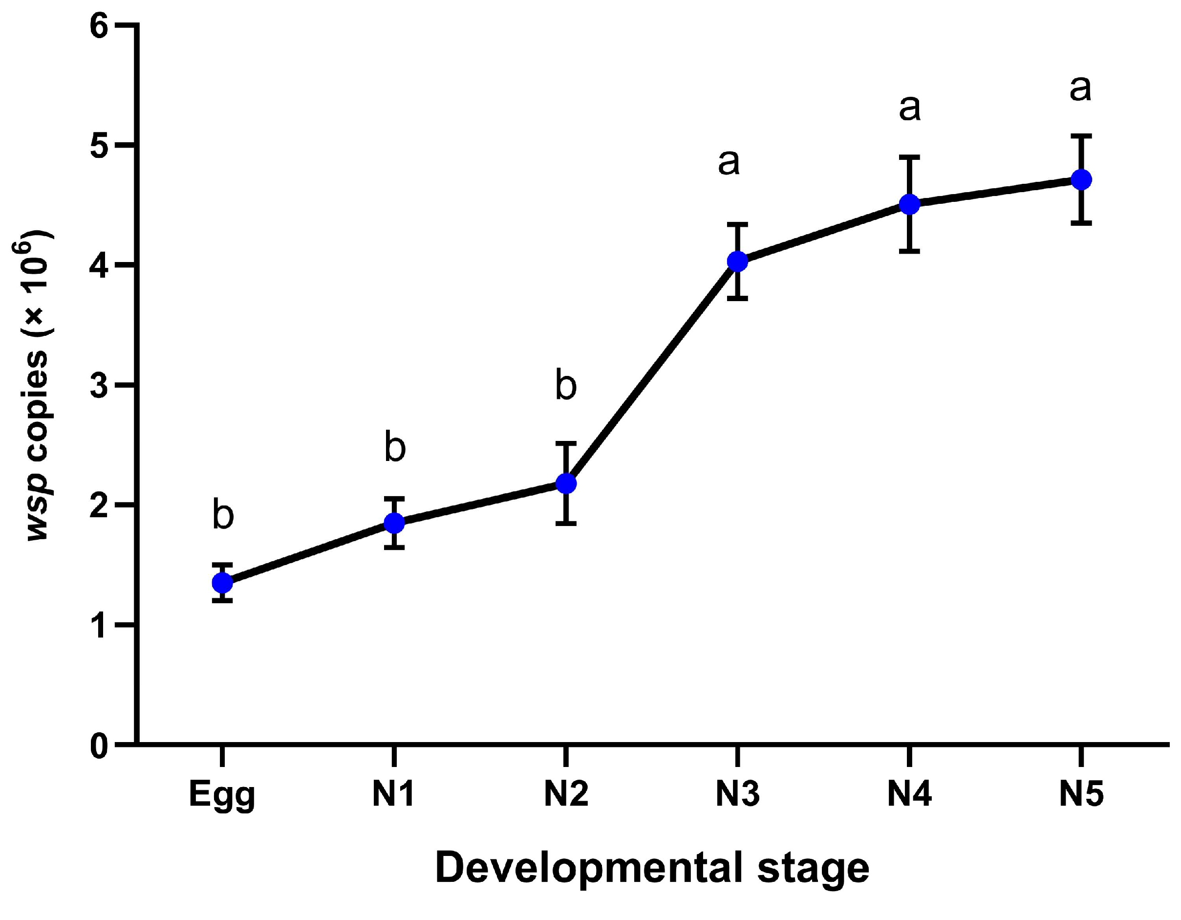
2.4. Wolbachia Infection Dynamics

To establish a transinfected population, the virgin females derived from the Wolbachia-injected nymphs (G0) were collected and individuals were paired with virgin wild males (uninfected). The mating pairs were confined in cages containing sugarcane leaves for oviposition (G1 eggs) under laboratory conditions. After laying their eggs, their progenies (G1) were retained, the parents were removed, and the females were subjected to PCR to test for Wolbachia infection. The G0 parents were discarded along with their progeny when females tested negative for Wolbachia. In contrast, the offspring (G1) of the infected mothers were reared until adult emergence. Fifty female progenies (G1) were randomly selected from the infected mothers and formed a single pair with virgin wild males to produce the G2 generation. The infected progenies were obtained by performing the same procedure until the G8 generation was established.
The G1 generation was used to examine the infection frequency at different developmental stages. The transinfected females (G0) that were paired with wild males were allowed to produce their offspring (G1). The immature developmental stages of the G1 generation were retained and collected after Wolbachia was detected in their mothers. Fifty individual samples were selected from five Wolbachia-infected mothers in the G0 generation. DNA was extracted from the individual samples to determine Wolbachia prevalence via PCR, and the infection frequencies were calculated (n = 10 in each of the five replicates). Representative samples were selected for quantification using real-time PCR.
The infection frequency was examined from the G1 up to the G8 generation. For each generation, 50 individual offspring (1:1 sex ratio) were selected from five Wolbachia-infected mothers from the preceding generation (randomly selected among infected individuals). DNA was extracted from the individual samples to determine Wolbachia prevalence through PCR, and the infection frequencies were calculated (n = 10 in each of the five replicates). Representative samples were selected for quantification using real-time PCR.

2.5. DNA Extraction and PCR Amplification

Before DNA extraction, the specimens were surface-sterilized with 75% ethanol for 3 min and washed thrice with deionized water for 1 min each time. DNA from individual samples was extracted using a phenol-chloroform method as previously described [31]. Briefly, individual leafhoppers were homogenized in 80 μL of lysis buffer (200 mM Tris pH 8.0, 250 mM NaCl, 25 mM EDTA, 0.5% SDS, 0.1 mg/mL proteinase K; Invitrogen, Carlsbad, CA, USA) After incubation at 37 °C for 24 h, DNA was extracted with phenol:chloroform:isoamyl alcohol (25:24:1), followed by 5 min of centrifugation at 10,000× g at 4 °C. The supernatant was transferred to a new tube, and chloroform:isoamyl alcohol (24:1) was added. Samples were centrifuged again for 5 min, and the DNA was precipitated with 3 M sodium acetate and isopropanol. The resulting pellet was washed with 70% absolute ethanol, air dried, and resuspended in TE buffer (10 mM Tris, 1 mM EDTA), before being stored at −20 °C until further use.
The Wolbachia infection status was confirmed by performing PCR using specific primers for the wsp surface protein marker. The 610-bp wsp gene was generated using the forward primer 81F (5′-TGGTCCAATAAGTGATGAAGAAAC-3′) and the reverse primer 961R (5′-AAAAATTAAACGCTACTCCA-3′) [32]. Each PCR mixture consisted of 1× reaction buffer, 2.5 mM MgCl2, 0.5 µM primers, 0.2 mM dNTPs, 1 U Taq DNA polymerase (Invitrogen, Carlsbad, CA, USA), 2 µL of template DNA (50 ng), and double-distilled water to obtain the final volume of 25 µL. Reactions with a template of the genomic DNA of Wolbachia’s original host Y. flavovittatus and double-distilled water were used as positive and negative control, respectively. The optimized cycling conditions were as follows: initial denaturation (94 °C, 5 min); 30 cycles of denaturation at 95 °C (1 min), annealing at 55 °C (1 min), extension at 72 °C (1 min); and final extension at 72 °C (10 min). To determine the length of the amplified DNA fragments, the amplicons were analyzed through electrophoresis on 1% agarose gels stained with the SYBR Safe DNA Gel Stain (Invitrogen, Carlsbad, CA, USA) and then visualized using a UV transilluminator (Major Science, Saratoga, CA, USA.).

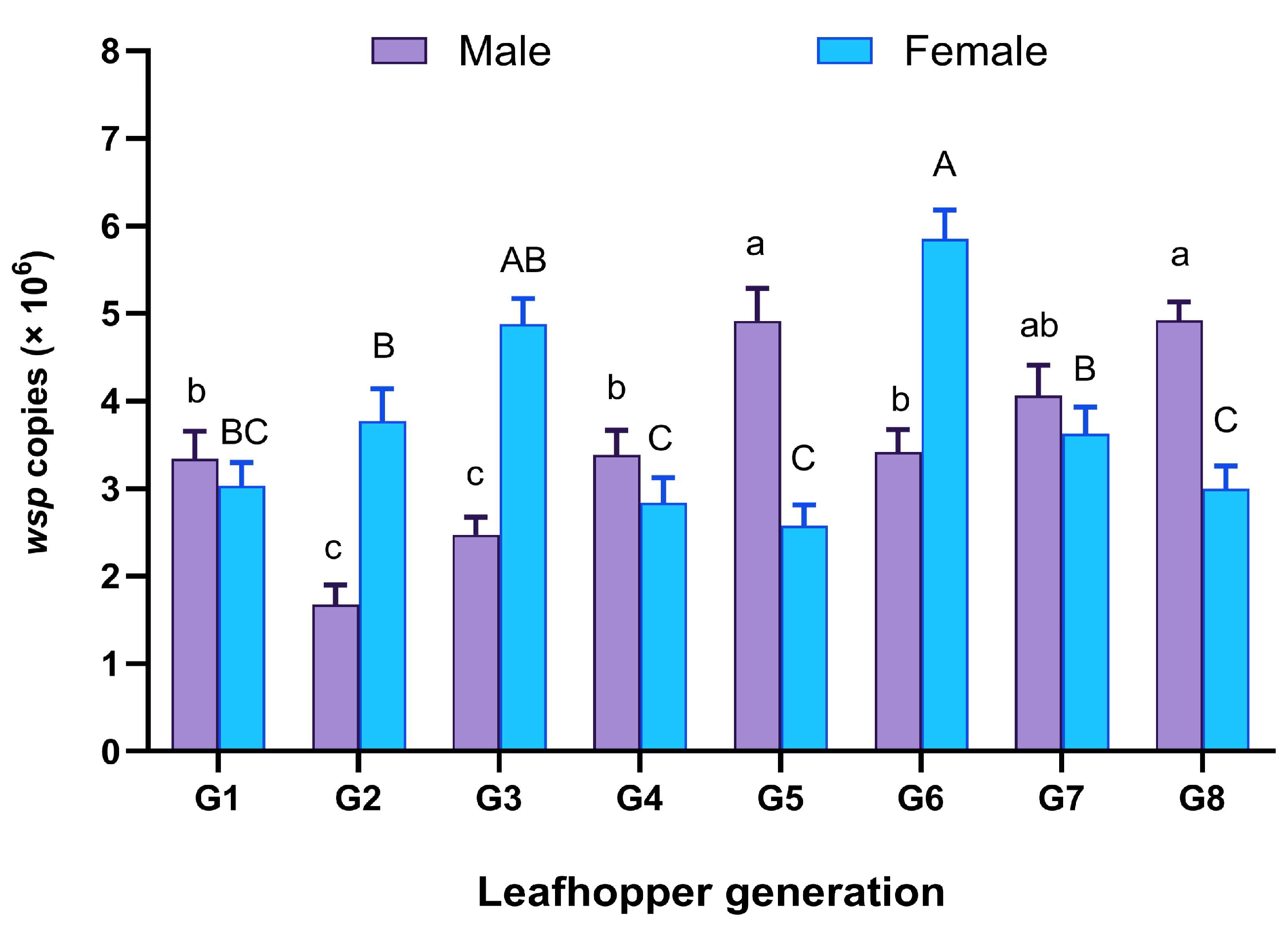
2.6. Wolbachia Quantification

To construct the standard curve, PCR was performed to amplify 198 p wsp gene fragments from Wolbachia-infected Y. flavittatus. Specific forward primer wYfla–F (5′-GGTGTTGGTGCAGCGTATGT-3′) and reverse primer wYfla–R (5′-TCCGCCATCATCTT TAGCTGT-3′) were used. The amplicons were cloned into a 3956-bp pCR4-TOPO® TA plasmid vector (TOPO–TA Cloning kit; Life Technologies, Carlsbad, CA, USA), according to the manufacturer’s protocol. Recombinant plasmids were linearized with the PstI restriction enzyme (Life Technologies, Carlsbad, CA, USA). The standard curve was generated using five serial dilutions (107–103 copies) of the linearized plasmid. The copy numbers of the wsp fragments in the initial concentrations were calculated using the calculator at http://www.scienceprimer.com/copy-number-calculator-for-realtime-pcr (accessed on 1 June 2023).
Prior to quantification analysis, the concentration of genomic DNA in the samples was measured using a spectrophotometer and diluted to 50 ng/μL. Quantitative PCR (qPCR) was performed using the Applied Biosystems StepOnePlus Real-time PCR system (Applied Biosystems, Foster City, CA, USA). The total volume of the reaction mixture was 20 µL, consisting of 10 μL of SYBR Green Master Mix (Applied Biosystems, Foster City, CA, USA), 1 μL (final 50 ng) template DNA, and 0.5 μM of primers (wYfla–F and wYfla–R). The qPCR cycles were conducted at 95 °C for 5 min, followed by 30 cycles at 95 °C for 45 s, 55 °C for 30 s, and 60 °C for 30 s. The samples were measured in triplicate to ensure accuracy. Negative and positive reaction mixtures included in all amplifications. The titers of the wsp gene fragment in the unknown samples were calculated by comparing their cycle threshold values with those produced by the serial dilutions of the standards. The qPCR efficacy was >90% and R2 was >99% for all assays. A melting curve analysis was performed at 60–95 °C; one correct peak was produced for all tests.

2.7. Wolbachia Distribution in Dissected Organs

Wolbachia infection was detected in various dissected tissues of M. hiroglyphicus. Transinfected adults were obtained from the G2 generation. Five-day-old adults were collected and surface-sterilized with 75% ethanol for 3 min; afterward, they were washed thrice with sterile water (1 min each time). Under a stereomicroscope, the head and thorax of the samples were cut using fine forceps. The remaining organs, including the midgut, fat body, ovaries, and testes, were dissected and collected from the abdomen. DNA was extracted from each organ, and Wolbachia infection was determined via PCR, as described above.

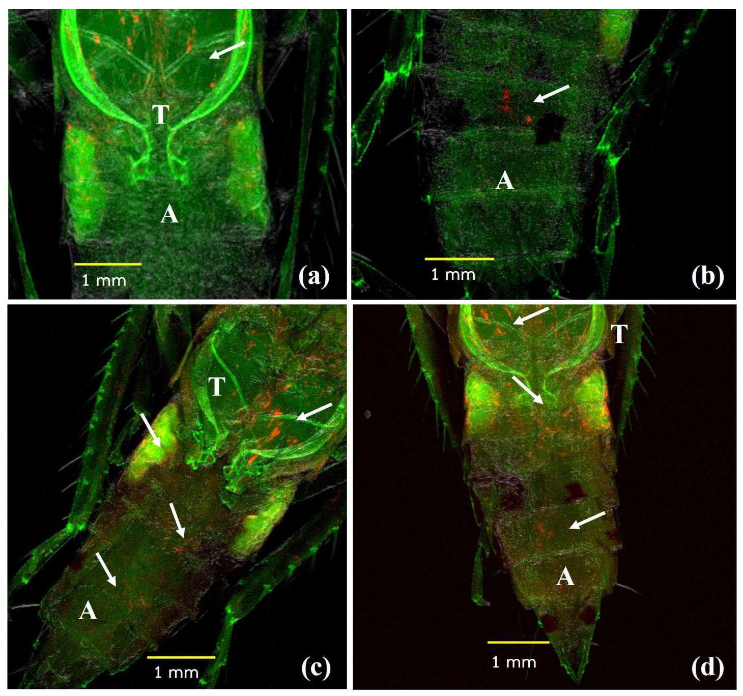
2.8. Fluorescence In Situ Hybridization (FISH)

FISH was performed to determine the distribution of Wolbachia in the transinfected nymphs, adults, and dissected ovaries and testis of the M. hiroglyphicus adults derived from the transinfected G2 generation. Whole-mount samples were analyzed using previously reported methods [33], with slight modifications. The specimens were collected and immersed in Carnoy’s solution (mixture of chloroform: ethanol: glacial acetic acid at 6:3:1 ratio) at room temperature (28–35 °C) for 24 h. Subsequently, the samples were washed thrice in 80% ethanol (10 min) and decolorized in 6% (v/v) hydrogen peroxide in 95% ethanol at 4 °C for 5 days to stop autofluorescence. Prior to hybridization, the preserved leafhoppers were immersed twice in 95% ethanol and PBS with 0.2% Tween-20 for 10 min each immersion. Then, the samples were hydrated by immersing them thrice in hybridization buffer (20 mM Tris-HCl (pH 8), 0.9 M NaCl, 0.01% (v/v) SDS, and 30% (v/v) formamide) for 10 min each immersion. Subsequently, the samples were hybridized for 24 h in buffer containing a 100 nM probe and then incubated at 46 °C in the dark. The nonspecific probes were removed by washing the samples thrice with PBST for 10 min each wash; then, the samples were mounted with ProLong antifade solution (Invitrogen, Carlsbad, CA, USA) and observed under a confocal laser-scanning microscope (FV1000; Olympus, Tokyo, Japan). The hybridization probe was W2 (5′-CTTCTGTGAGTACCGTCATTATC-3′); it targeted the 319–336 positions of the 16S rRNA gene of Wolbachia [33], which was 5′-labeled with Quasar 670. The reactions without probes and the uninfected leafhoppers were used as negative controls.

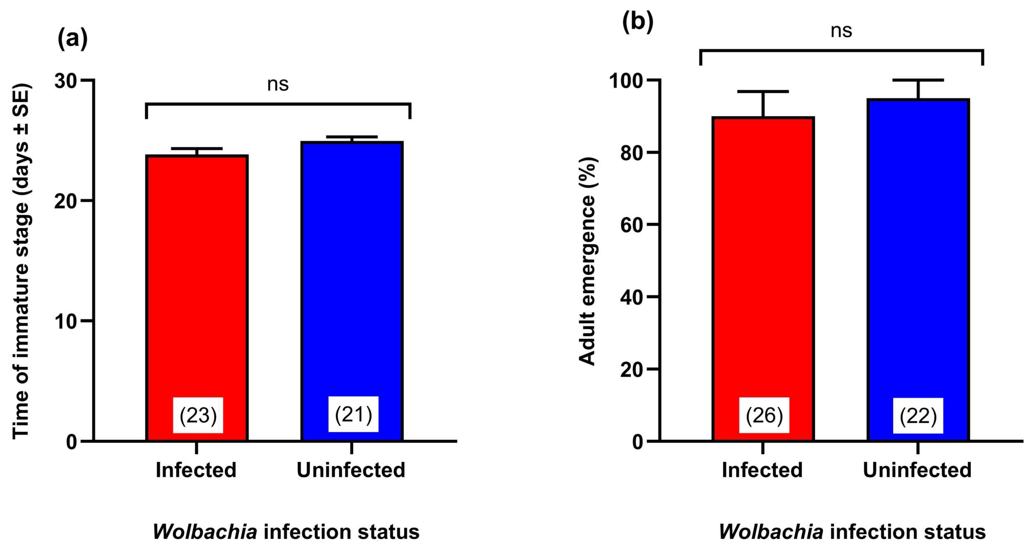
2.9. Determining the Effect of Wolbachia Transinfection on Development Time and Adult Emergence

Notably, due to unpredictability of whether Wolbachia was transmitted to the following generations, the G2 generation was used to investigate the fitness parameters. The virgin females of the G1 generation were obtained from infected mothers and were paired with uninfected wild males. The mating pairs were confined in cages containing sugarcane leaves for oviposition (G2 eggs) under laboratory conditions. A total of 50 eggs derived from 5 infected mothers were collected, and the developmental time until adult emergence was investigated. The number of emerging adults was counted, and the developmental duration of the immature stages (egg through the final nymphal instar) was recorded. After adult emergence, the Wolbachia infection status of the samples was determined, and the samples that tested negative for infection were discarded from the calculations. The same procedure was performed on the natural population (uninfected) leafhoppers.

2.10. Effect of Wolbachia Transinfection on Adult Survival and Longevity

To determine the effect of Wolbachia infection on the adult survival and longevity of M. hiroglyphicus, samples derived from the G2 generation were used. Fresh adult emergence was immediately determined by sex, and the adults were confined in a clip cage with sugarcane leaves. Mortality was recorded daily for each adult (n = 50 for males, n = 50 for females). The Wolbachia infection status of the samples was confirmed after they had died, and the transinfected samples were discarded from the calculation when the PCR result was negative. The survival of the natural population (uninfected) was assessed using the same procedure.

2.11. Crossing Experiment

Crossing experiments were performed to examine the effect of Wolbachia infection on female fecundity and confirm CI induction in the transinfected M. hiroglyphicus. Virgin adults derived from the transinfected G2 generation were used for infected leafhoppers, and the natural populations were used as the uninfected lineage. After the sexes were determined, 1-day-old leafhoppers were collected and used for the crossing experiment. Four types of crosses between virgin individuals were established, including naturally uninfected males and females (Un ♂ × Un ♀; n = 20), transinfected males and naturally uninfected females (In ♂ × Un ♀; n = 19), naturally uninfected males and transinfected females (Un ♂ × In ♀; n = 21), and transinfected males and transinfected females (In ♂ × In ♀; n = 20).
The individual mating pairs were confined in a clip cage with sugarcane leaves and the females were allowed to oviposit. The number of eggs laid per individual was recorded until the egg-laying female died. Hatched eggs were monitored to determine the hatch proportions for each single pair. The females that did not lay eggs or died within 10 days were excluded from the analysis. Wolbachia infection status was confirmed in transinfected leafhoppers, and in PCR-negative cases, mating pairs were discarded from the experiment.

2.12. Statistical Analysis

The data obtained (except for the survival rate) from the experiments were checked for normality using the Kolmogorov–Smirnov test prior to performing statistical analysis. The Wolbachia infection frequency, number of copies of wsp from Wolbachia, number of eggs laid, and egg hatchability were calculated. Statistically significant differences were determined by performing a one-way analysis of variance, followed by Tukey’s HSD test to compare the means. The developmental time, percentage of adult emergence, and longevity were compared by performing the t-test. Statistical analyses were performed using IBM SPSS Statistics for Windows (version 22.0; IBM Corp., Armonk, NY, USA). Differences in adult survival between the Wolbachia-infected and uninfected leafhopper samples were tested using the log-rank (Mantel–Cox) test in Graph Pad Prism 9 software.

3. Results

3.1. Wolbachia Transinfection

Of the 200 third-instar M. hiroglyphicus nymphs that were injected with the wYfla Wolbachia strain, only a 30–44% (n = 75) survival rate after 24 h was recorded in four experiments. After adult emergence, the sexes of the adults were determined, and male leafhoppers were discarded. Female leafhoppers (G0, n = 26) were mated with uninfected wild males to establish an isofemale line (Table 1). Wolbachia infection was detected in 61.54% of the isofemales or 8.00% of all injected nymphs (n = 16). The offspring (G1) from the infected mothers were maintained, and the females were collected and paired with wild males to produce the subsequent generation. Transinfected progeny were produced using the same procedure until the G8 generation was established.

3.2. Wolbachia Infection Frequency in the Transinfected M. hiroglyphicus Samples

There was no significant difference in Wolbachia infection frequency among the different developmental stages in the G1 generation (p = 0.325). The percentage of infected eggs was 62.00%, while that of the first–fifth nymphal instars was 58.00–70.00% (Table 2). A significant difference in the titer of wsp gene copies among the immature stages was observed (p = 0.016). The egg samples had the lowest titer of wsp copies (1.35 × 10⁶ copies); the titer of wsp copies gradually increased from the first to the later nymphal stages. The highest copy number (4.71 × 10⁶) was observed in the fifth nymphal instar (Figure 1).
Wolbachia infection was detected in 5-day-old adult progeny from the infected mothers in G1–G8 generations, and the percentage of infection was calculated as the proportion of the infected samples (Table 3). A significant difference in Wolbachia infection frequency was observed among the different generations (p = 0.027). The highest infection rate was detected in the G3 generation (80.00%), while the lowest was found in the G7 generation (55.00%). Relative stability in infection frequency was not observed, suggesting that Wolbachia could be transmitted from the parental generation to their progeny; however, Wolbachia was only partially transmitted from the mothers to their offspring.
The results of the quantitative analysis revealed a significant difference in Wolbachia titer among the generations tested (male, p = 0.018; female, p = 0.031). Moreover, the change in Wolbachia titers from the G1 to G8 generation in males and females was dissimilar. For the male leafhoppers, the titer of wsp gene copies decreased to its lowest level in the G2 generation (1.67 × 10⁶ copies); however, it was higher in the G5 generation (4.91 × 10⁶ copies). Subsequently, the titer decreased in the G6 generation, then increased again and was higher in the G8 generation (4.92 × 10⁶ copies). For the female leafhoppers, the titer of wsp gene copies increased from the G1 (3.03 × 10⁶ copies) to G3 generation (4.88 × 10⁶ copies). However, it was lower in the G4 and G5 generations; specifically, the G5 generation had the lowest titer of wsp gene copies (2.57 × 10⁶ copies). The titer of wsp gene copies also varied from the G6 to G8 generation, ranging from 2.99 to 5.85 × 10⁶ copies (Figure 2).

3.3. Distribution of Wolbachia in the Transinfected M. hiroglyphicus Samples

PCR results revealed that Wolbachia was present in 100% (50 out of 50), 40% (20 out of 50), 50% (25 out of 50), and 80% (40 out of 50) of the thoracic muscle, ovaries, testis, and fat body, respectively, of the transinfected (G2) M. hiroglyphicus adults. However, this prevalence was not observed in the head, and gut samples. A more detailed localization of Wolbachia in the transinfected M. hiroglyphicus samples was revealed using FISH. For the nymphal stages, the specific signals of Wolbachia were widely distributed in the somatic tissues, including the thoracic and abdominal areas (Figure 3a,b). The specific signals were abundant in the adult stage and were distributed in the thorax and abdominal areas of the males and females (Figure 3c,d). The specific signals were also detected in the ovaries, including nurse cells (Figure 4a), and were concentrated around the apices of fully grown oocytes (Figure 4b,c). In addition, specific fluorescence signals were scattered in the testes of M. hiroglyphicus (Figure 4d–f). However, fluorescence intensity differed between individuals, as a very weak signal (Figure 4d) and a higher intensity were detected (Figure 4e,f).
By subjecting the dissected organs to PCR and FISH, it was confirmed that Wolbachia was successfully transinfected to the M. hiroglyphicus samples. This result indicates that Wolbachia can be transmitted from the parental generation to the offspring via the egg cytoplasm. The no-probe control reaction and RNase digestion before hybridization revealed no specific signals.

3.4. Effect of Wolbachia Transinfection on the Development Time of the Immature Stages

There was no significant difference in the developmental time of the immature stages (egg stage to the final nymphal instar) derived from the transinfected G2 generation and the natural uninfected lineage (p = 0.067); the transinfected and uninfected samples required 23.82 and 24.95 days, respectively, to reach the adult stage (Figure 5a). Moreover, there was no significant difference in the percentage of adult emergence (p = 0.087), with an emergence rate of 80–90% (Figure 5b). Of note, no significant difference was found in these two parameters between the uninfected lineages from natural and transinfected leafhoppers (Table S1).

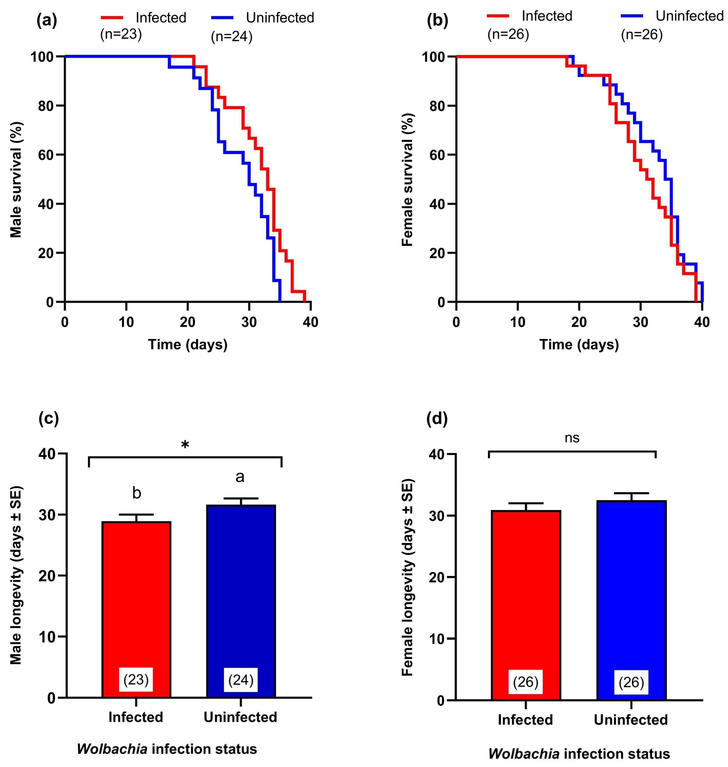
3.5. Effect of Wolbachia Transinfection on Adult Survival

The comparison of the survival rate of the adults derived from the transinfected G2 generation with that of the natural uninfected lineage revealed a significant reduction in the survival rate of male transinfected leafhoppers (log-rank statistic = 4.71, p = 0.03). The average male longevity of the transinfected and uninfected samples was approximately 28.91 and 31.62 days, respectively (Figure 6a,b). However, there was no statistical difference in female longevity (log-rank test statistic = 1.49, p = 0.22) between the transinfected (31.53 days) and uninfected (33.15 days) samples (Figure 6c,d). Of note, no significant difference was found in adult longevity between the uninfected lineages from natural and transinfected leafhoppers (Table S1).

3.6. Effect of Wolbachia Infection on Fecundity and CI Expression

The results of the crossing experiments revealed that the highest number of eggs laid per female (37.20 eggs/female) was recorded during the mating between naturally uninfected males and females (Un ♂ × Un ♀). However, there were no statistically significant differences in fecundity among those four crossing types (p = 0.52) (Figure 7a).
However, a statistically significant difference was detected in hatching rates (p < 0.001); the lowest hatching rate (28.16%) was recorded when transinfected males were crossed with naturally uninfected females (In ♂ × Un ♀). There were no statistically significant differences in hatching rate among the other crossing types. The crossing of naturally uninfected males and naturally uninfected females (Un ♂ × Un ♀), naturally uninfected males and transinfected females (Un ♂ × In ♀), and transinfected males and transinfected females (In ♂ × In ♀) showed hatching rates of 77.65, 76.02, and 78.04%, respectively (Figure 7b).

4. Discussion

The Wolbachia strain can be artificially transferred to a new insect host through microinjection (transinfection), which has been used to develop Wolbachia-based strategies for controlling disease vectors and agricultural pests [34]. In this study, we artificially transferred the wYfla Wolbachia strain into M. hiroglyphicus, the vector of SCWL. Several studies have reported that embryonic microinjection is the primary technique used to generate Wolbachia-infected insect lineage [35,36]. We attempted to use using M. hiroglyphicus eggs as the recipients; however, the hatching rate of the injected eggs was extremely low (<5%). This may be because the physiology of M. hiroglyphicus eggs is unsuitable for needle penetration. The appropriate developmental stage for microinjection may depend on the insect species [34]. Based on the higher survival rate compared to injected eggs, nymphal microinjection has been suggested as the suitable method for Wolbachia transinfection for M. hiroglyphicus. Likewise, successfully established Wolbachia colonies were obtained when microinjection was conducted at other developmental stages. For example, Kawai et al. [37] showed that Wolbachia transinfection in brown planthopper (Nilaparvata lugens) was successfully achieved with nymphal microinjection. In whitefly (Bemisia tabaci), the microinjection of fourth-instar nymphs (pseudopupas) generated a lineage of Wolbachia-infected whitefly [38].
The molecular detection results validated the presence of Wolbachia in the transinfected M. hiroglyphicus samples in the G0 generation, and Wolbachia infection was monitored from the G1 to G8 generations. Positive infection suggests that Wolbachia can be transmitted from the transinfected G0 to the succeeding generations. However, consistent infection among generations was not found; i.e., highest in G3 and lowest in G7 generation. A possible cause for this discrepancy might be the selection methods, as offspring were randomly selected from infected mothers. This observation was similar to that of previous studies on B. tabaci transinfected with the wMel Wolbachia strain, in which the maternal transmission efficiency of Wolbachia exhibited a fluctuating trend over 12 generations [39]. In contrast, Zhong and Li [40] reported that B. tabaci samples transinfected with the wSguBJ Wolbachia strain had a low maternal transmission rate in the early generation (G1–G3) and then it increased steadily after the G4 generation and plateaued after the G6 generation (70–80%). However, a stable complete infection (100%) has been reported in the transinfection of N. lugens with the wStri Wolbachia strain [36].
Transmission through the maternal line is an important parameter for the successful maintenance of Wolbachia in the insect host population [41]. However, the maternal transmission rate is often imperfect in nature, and efficiency may reflect Wolbachia tissue tropism, as it has a particularly strong affinity to ovarian tissue [42]. However, the pattern of transmission rate within one species varies; for example, a nearly complete vertical transmission was reported in cabbage fly (Delia radicum) [43], but it was 33–66% in the parasitoid wasp (Hyposoter horticola) [44]. We assume that there was a variation in the maternal transmission rate of Wolbachia among the different generations of the transinfected M. hiroglyphicus samples. Moreover, fluctuations in the number of Wolbachia were found in different generations and between sexes. For insects, fluctuation in Wolbachia infection is related to tissue tropism, immune system, and gene expression, which regulates Wolbachia proliferation [45]. The differences between sexes could be explained by those mechanisms that drive appropriate ranges of infection density; however, they appear to be highly dependent on the Wolbachia–host and sex-specific interactions. However, the underlying mechanisms that regulate the number of Wolbachia in transinfected M. hiroglyphicus require further research.
Results of PCR detection and FISH revealed that Wolbachia was present in somatic tissues throughout the body of the transinfected M. hiroglyphicus samples. Likewise, transinfected buffalo flies (Haematobia irritans exigua) had Wolbachia in their head, thorax, midgut, and fat bodies [46]. Wolbachia may have adapted to recipient tissues by suppressing the immune system [47]. However, the precise mechanism of the immune response requires further research to elucidate how the wYfla strain infects M. hiroglyphicus. In addition, Wolbachia was prevalent in the reproductive tissues, such as the ovaries and testes. The distribution of Wolbachia has significant impacts on its role or ability to affect leafhoppers. Prevalence in the ovaries suggest that Wolbachia can be inherited by the offspring. This evidence also supports the vertical transmission of Wolbachia from the recipient G0 generation to multiple subsequent generations after transinfection, as described above. Prevalence in the testes was related to CI expression, which is induced in Wolbachia-infected males (discussed below).
In this study, the wYfla Wolbachia strain isolated from Y. hiroglyphicus eggs was used for transinfection. However, the wYfla strain expressed different phenotypes, had no significant effects on female lifespan, and only induced partial CI in the transinfected M. hiroglyphicus samples. Some previous studies have shown that Wolbachia strains do not express the original phenotype or induce a novel phenotype after transinfection into new hosts [48,49]. Moreover, reproductive manipulation was not observed in Drosophila melanogaster and D. simulans transinfected with the male-killing Wolbachia strain from D. innubila [50]. This suggests that phenotypic differences affect Wolbachia transinfection; those authors proposed that if Wolbachia expresses the same phenotype in both native and recipient hosts, the Wolbachia strain can determine the phenotype. In contrast, the expression of a novel (or no) original phenotype reveals the importance of the host species and Wolbachia strain interactions in the context of the co-evolution of the host and Wolbachia. Indeed, multiple recent Wolbachia-induced cases have shown identical phenotypes in both donor and recipient hosts. For instance, the wMelPop Wolbachia strain causes CI and reduces the adult lifespan in its native host D. melanogaster; these effects were also observed in the transinfected host Aedes aegypti [51].
Regarding the biological traits of the transinfected M.hiroglyphicus samples, Wolbachia infection had no significant effect on the examined fitness parameters (except the lifespan of male leafhoppers). Similarly, a difference in the lifespan of male and female B. tabaci specimens transinfected with the wSguBJ Wolbachia strain was observed [40]. However, the lifespan of both male and female A. aegypti specimens transinfected with the wPip Wolbachia strain was significantly shortened [35]. A possible mechanism behind the shortened lifespan of Wolbachia-infected insect hosts is that Wolbachia infection induces the upregulation of the innate immune system of the host, which may contribute to the life-shortening phenotype [52]. Although the transinfected M. hiroglyphicus females were not negatively affected by Wolbachia, we hypothesized that Wolbachia maintains its population in females for propagation so that it can be promoted and transmitted to the next generation via the egg cytoplasm. Notably, the mechanism responsible for the longevity of transinfected M. hiroglyphicus requires further investigation. Moreover, the results revealed that Wolbachia transinfection had no significant detrimental effects on the fecundity of the female M. hiroglyphicus samples. Previous studies on the effect of Wolbachia on the fecundity of transinfected females reported various results, depending on the species of recipient hosts and Wolbachia strain. A consistent result has been reported for the transinfection of D. melanogaster with the wCcep Wolbachia strain, which had no effect on the number of eggs laid per female [53]. However, the transinfection of buffalo flies H. irritans exigua with the wAlbB, wMelPop, and wMel strains resulted in a significantly lower number of eggs compared with uninfected females [46].
CI is the most common reproductive abnormality induced by Wolbachia in its insect hosts [54]. Our results revealed that 28% of the eggs resulting from incompatible crossing tests were developed, compared with the normal hatch rate expected (>75%). This finding indicates that transinfection with the wYfla Wolbachia strain could induce partial CI, in which the egg hatching rate was lower than that of the original host (100% embryonic death). This CI level was unexpected, and the transinfected M. hiroglyphicus samples showed a weaker CI than that of the native host. Similarly, the transinfection of N. lugens with the wStr Wolbachia strain resulted in the development of approximately 51.47% of eggs [37]. Not surprisingly, CI strength varies from relatively weak to 100% embryonic death, and the range of CI results from several factors, such as host genotype and age, Wolbachia strain, and Wolbachia density [55]. However, the CI level in a new recipient host varies among transinfected insect hosts. The mosquito vector C. quinquefasciatus transinfected with a single or superinfected Wolbachia strain (wAlbB or wPipwAlbA) also exhibited complete CI [20]. Similarly, the N. lugens samples transinfected with the wStri Wolbachia strain showed high CI levels [36]. A possible explanation for the occurrence of incomplete CI (some eggs hatching) could be related to the mechanisms that govern CI expression. Its occurrence following the cross between infected males and uninfected females could result from incomplete modification of the sperm in Wolbachia-infected males or incomplete rescue of the modified sperm in infected females [56].
There are also some limitations in this study. First, the evidence for the establishment of Wolbachia in the transinfected M. hiroglyphicus samples was obtained from PCR and FISH results. Further studies should investigate the mechanism that induces an immune response by invading the introduced Wolbachia strain in transinfected M. hiroglyphicus. Second, the data on the effects of Wolbachia transinfection on the biological traits and crossing experiments were obtained from representative leafhoppers in the G2 generation. More accurate results can be achieved by examining the other generations. Additionally, further studies should elucidate the mechanism of the phenotypic effect induced by Wolbachia, that is, shortening the lifespan of males and inducing partial CI in incompatible crossings.

5. Conclusions

The wYfla Wolbachia strain isolated from Y. flavovittatus was artificially transferred into M. hiroglyphicus leafhoppers through nymphal microinjection. It can be established and transmitted across eight generations, with an infection rate of 55–80%. Wolbachia transinfection had no detrimental effects on the general biological traits, except for the shortened lifespan of the transinfected males. However, partial CI induction was observed, as evidenced by more than half of the undeveloped eggs obtained from incompatible crossings. This partial reproductive incompatibility may have an impact on altering the population of M. hiroglyphicus. To our knowledge, this is the first study to report a successful transinfection of Wolbachia in the leafhopper vector of phytoplasma disease. Further studies on the Wolbachia-based biocontrol method for leafhopper vectors are warranted. Moreover, additional research is required to evaluate the effect of Wolbachia transinfection on the capacity of M. hiroglyphicus leafhoppers to transmit phytoplasma pathogens. In addition, artificial transfer with other virulent or desirable strains of Wolbachia must be demonstrated in M. hiroglyphicus leafhoppers, which may improve CI levels.

Supplementary Materials

The following supporting information can be downloaded at: https://www.mdpi.com/article/10.3390/agriculture14081236/s1, Table S1: Effect of Wolbachia infection on fitness traits of of Matsumuratettix hiroglyphicus samples. Those collected from transinfected G2 generation (Wolbachia-infected and uninfected) and natural uninfected lineage.

Author Contributions

Conceptualization, K.S., J.R. and J.W.; methodology, K.S.; formal analysis; K.S. and J.W.; writing—original draft preparation K.S.; writing—review and editing; K.S., J.R. and J.W.; funding acquisition, J.W. All authors have read and agreed to the published version of the manuscript.

Funding

This study was supported by the Thailand Science Research and Innovation Fundamental Fund fiscal year 2023 (Grant number TUFF 17/2566), a Ph.D. scholarship from Thammasat University, 1/2021, and the Research Promotion Fund for International and Educational Excellence, 9/2564.

Institutional Review Board Statement

The experiments on leafhopper vectors using in this study were carried out under the guideline of animal use procedure, which was reviewed and approved by the Animal Care and Use Committee of Thammasat University (Protocol number 023/2562, date of approval 12 February 2021).

Data Availability Statement

All available data are contained within the article.

Acknowledgments

The authors would like to thank the Faculty of Science and Technology and Faculty of Medicine, Thammasat University, for providing research facilities.

Conflicts of Interest

The authors declare no conflicts of interest.

References

  1. Hoffmann, A.A.; Ross, P.A.; Rašić, G. Wolbachia strains for disease control: Ecological and evolutionary considerations. Evol. Appl. 2015, 8, 751–768. [Google Scholar] [CrossRef] [PubMed]
  2. O’Neill, S.L. The use of Wolbachia by the world mosquito program to interrupt transmission of Aedes aegypti transmitted viruses. Adv. Exp. Med. Biol. 2018, 1062, 355–360. [Google Scholar] [PubMed]
  3. Cardona-Salgado, D.; Campo-Duarte, D.E.; Sepulveda-Salcedo, L.S.; Vasilieva, O. Wolbachia-based biocontrol for dengue reduction using dynamic optimization approach. Appl. Math. Modell. 2020, 82, 125–149. [Google Scholar] [CrossRef]
  4. Carrington, L.B.; Tran, B.C.N.; Le, N.T.H.; Luong, T.T.H.; Nguyen, T.T.; Nguyen, P.T.; Nguyen, C.V.V.; Nguyen, H.T.C.; Vu, T.T.; Thi Vo, L.; et al. Field- and clinically derived estimates of Wolbachia-mediated blocking of dengue virus transmission potential in Aedes aegypti mosquitoes. Proc. Natl. Acad. Sci. USA 2018, 115, 361–366. [Google Scholar] [CrossRef] [PubMed]
  5. Ross, P.A.; Turelli, M.; Hoffmann, A.A. Evolutionary ecology of Wolbachia releases for disease control. Annu. Rev. Genet. 2019, 53, 93–116. [Google Scholar] [CrossRef] [PubMed]
  6. Yen, P.S.; Failloux, A.B. A review: Wolbachia-based population replacement for mosquito control shares common points with genetically modified control approaches. Pathogens 2020, 9, 404. [Google Scholar] [CrossRef]
  7. Guo, Y.; Shao, J.; Wu, Y.; Li, Y. Using Wolbachia to control rice planthopper populations: Progress and challenges. Front. Microbiol. 2023, 14, 1244239. [Google Scholar] [CrossRef] [PubMed]
  8. Gong, J.T.; Li, T.P.; Wang, M.K.; Hong, X.Y. Wolbachia-based strategies for control of agricultural pests. Curr. Opin. Insect Sci. 2023, 57, 101039. [Google Scholar] [CrossRef] [PubMed]
  9. Zug, R.; Hammerstein, P. Still a host of hosts for Wolbachia: Analysis of recent data suggests that 40% of terrestrial arthropod species are infected. PLoS ONE 2012, 7, e38544. [Google Scholar] [CrossRef] [PubMed]
  10. Kaur, R.; Shropshire, J.D.; Cross, K.L.; Leigh, B.; Mansueto, A.J.; Stewart, V.; Bordenstein, S.R.; Bordenstein, S.R. Living in the endosymbiotic world of Wolbachia: A centennial review. Cell Host Microbe 2021, 29, 879–893. [Google Scholar] [CrossRef] [PubMed]
  11. Asgharian, H.; Chang, P.L.; Mazzoglio, P.J.; Negri, I. Wolbachia is not all about sex: Male feminizing Wolbachia alters the leafhopper Zyginidia pullula transcriptome in a mainly sex-independent manner. Front. Microbiol. 2014, 5, 430. [Google Scholar] [CrossRef] [PubMed]
  12. He, L.F.; Feng, D.D.; Li, P.; Zhou, Z.S.; Xu, Z.F. Reproductive modes and daily fecundity of Aenasius bambawalei (Hymenoptera: Encyrtidae), a parasitoid of Phenacoccus solenopsis (Hemiptera: Pseudococcidae). Fla Entomol. 2015, 98, 358–360. [Google Scholar] [CrossRef]
  13. LePage, D.P.; Metcalf, J.A.; Bordenstein, S.R.; On, J.; Perlmutter, J.I.; Shropshire, J.D.; Layton, E.M.; Funkhouser-Jones, L.J.; Beckmann, J.F.; Bordenstein, S.R. Prophage WO genes recapitulate and enhance Wolbachia-induced cytoplasmic incompatibility. Nature 2017, 543, 243–247. [Google Scholar] [CrossRef] [PubMed]
  14. Harumoto, T.; Fukatsu, T.; Lemaitre, B. Common and unique strategies of male killing evolved in two distinct Drosophila symbionts. Proc. Biol. Sci. 2018, 285, 20172167. [Google Scholar]
  15. Sumida, Y.; Katsuki, M.; Okada, K.; Okayama, K.; Lewis, Z. Wolbachia induces costs to life-history and reproductive traits in the moth, Ephestia kuehniella. J. Stored Prod. Res. 2017, 71, 93–98. [Google Scholar] [CrossRef]
  16. Guo, Y.; Hoffmann, A.A.; Xu, Q.; Mo, W.; Huang, J.; Gong, T.; Ju, F.; Hong, Y. Vertical transmission of Wolbachia is associated with host vitellogenin in Laodelphax striatellus. Front. Microbiol. 2018, 9, 2016. [Google Scholar] [CrossRef]
  17. Werren, J.H.; Baldo, L.; Clark, M.E. Wolbachia: Master manipulators of invertebrate biology. Nat. Rev. Microbiol. 2008, 6, 741–751. [Google Scholar] [CrossRef] [PubMed]
  18. Zug, R.; Hammerstein, P. Bad guys turned nice? A critical assessment of Wolbachia mutualisms in arthropod hosts. Biol. Rev. Camb. Philos. Soc. 2015, 90, 89–111. [Google Scholar] [CrossRef]
  19. Caragata, E.P.; Dutra, H.L.C.; Sucupira, P.H.F.; Ferreira, A.G.A.; Moreira, L.A. Wolbachia as translational science: Controlling mosquito-borne pathogens. Trends Parasitol. 2021, 37, 1050–1067. [Google Scholar] [CrossRef] [PubMed]
  20. Ant, T.H.; Herd, C.; Louis, F.; Failloux, A.B.; Sinkins, S.P. Wolbachia transinfections in Culex quinquefasciatus generate cytoplasmic incompatibility. Insect Mol. Biol. 2020, 29, 1–8. [Google Scholar] [CrossRef] [PubMed]
  21. Lu, P.; Sun, Q.; Fu, P.; Li, K.; Liang, X.; Xi, Z. Wolbachia inhibits binding of Dengue and Zika viruses to mosquito cells. Front. Microbiol. 2020, 11, 1750. [Google Scholar] [CrossRef] [PubMed]
  22. Narmilan, A.; Gonzalez, F.; Salgadoe, A.S.A.; Powell, K. Detection of white leaf disease in sugarcane using machine learning techniques over UAV multispectral images. Drones 2020, 6, 230. [Google Scholar] [CrossRef]
  23. Donnua, S.; Moonjuntha, P.; Tiwari, A.K. Diversity, distribution, and status of phytoplasma diseases in Thailand. In Diversity, Distribution, and Current Status, 1st ed.; Tiwari, A.K., Caglayan, K., Al-Sadi, A.M., Azadvar, M., Abeysinghe, S., Eds.; Academic Press: Cambridge, MA, USA, 2023; Volume 1, pp. 31–38. [Google Scholar]
  24. Kirdat, K.; Tiwarekar, B.; Thorat, V.; Sathe, S.; Shouche, Y.; Yadav, A. Candidatus Phytoplasma sacchari’, a novel taxon—associated with Sugarcane Grassy Shoot (SCGS) disease. Int. J. Syst. Evol. Microbiol. 2021, 71, 004591. [Google Scholar] [CrossRef] [PubMed]
  25. Wangkeeree, J.; Tewaruxsa, P.; Hanboonsong, Y. New bacterium symbiont in the bacteriome of the leafhopper Yamatotettix flavovittatus Matsumura. J. Asia-Pasific Entomol. 2019, 22, 889–896. [Google Scholar] [CrossRef]
  26. Thein, M.M.; Jamjanya, T.; Kobori, Y.; Hanboonsong, Y. Dispersal of the leafhoppers Matsumuratettix hiroglyphicus and Yamatotettix flavovittatus (Homoptera: Cicadellidae), vectors of sugarcane white leaf disease. Appl. Entomol. Zool. 2012, 47, 255–262. [Google Scholar] [CrossRef]
  27. Roddee, J.; Kobori, Y.; Hanboonsong, Y. Multiplication and distribution of sugarcane white leaf Phytoplasma transmitted by the leafhopper, Matsumuratettix hiroglyphicus (Matsumura) (Hemiptera: Cicadellidae), in infected sugarcane. Sugar Tech 2018, 20, 445–453. [Google Scholar] [CrossRef]
  28. Sukcharoenvipharat, W.; Aumarrom, T.; Usaborisut, P.; Jantra, C.; Abdullakasim, W. Design and development of a hot water treatment device for small-scale sugarcane farmers. Sugar Tech 2024, 26, 234–241. [Google Scholar] [CrossRef]
  29. Hanboonsong, Y.; Kobori, Y. Effects of selected insecticides on Matsumuratettix hiroglyphicus (Matsumura), a vector of sugarcane white leaf disease, and on two natural enemies of the sugarcane stem borer in sugarcane fields. Sugar Tech 2017, 19, 573–578. [Google Scholar] [CrossRef]
  30. Minwuyelet, A.; Petronio, G.P.; Yewhalaw, D.; Sciarretta, A.; Magnifico, I.; Nicolosi, D.; Marco, R.D.; Atenafu, G. Symbiotic Wolbachia in mosquitoes and its role in reducing the transmission of mosquito-borne diseases: Updates and prospects. Front. Microbiol. 2023, 14, 1267832. [Google Scholar] [CrossRef] [PubMed]
  31. Wangkeeree, J.; Suwanchaisri, K.; Roddee, J.; Hanboonsong, Y. Effect of Wolbachia infection states on the life history and reproductive traits of the leafhopper Yamatotettix flavovittatus Matsumura. J. Invertebr. Pathol. 2020, 177, 107490. [Google Scholar] [CrossRef] [PubMed]
  32. Zhou, W.; Rousset, F.; O’Neil, S.L. Phylogeny and PCR-based classification of Wolbachia strains using wsp gene sequences. Proc. Biol. Sci. 1998, 265, 509–515. [Google Scholar] [CrossRef] [PubMed]
  33. Shi, P.; He, Z.; Li, S.; An, X.; Lv, N.; Ghanim, M.S.; Cuthbertson, A.G.; Ren, X.; Qiu, L. Wolbachia has two different localization patterns in whitefly Bemisia tabaci AsiaII7 species. PLoS ONE 2016, 11, e0162558. [Google Scholar] [CrossRef]
  34. Hughes, G.L.; Rasgon, J.L. Transinfection: A method to investigate Wolbachia-host interactions and control arthropod-borne disease. Insect Mol. Biol. 2014, 23, 141–151. [Google Scholar] [CrossRef] [PubMed]
  35. Fraser, J.E.; De Bruyne, J.T.; Iturbe-Ormaetxe, I.; Stepnell, J.; Burns, R.L.; Flores, H.A.; O’Neill, S.L. Novel Wolbachia-transinfected Aedes aegypti mosquitoes possess diverse fitness and vector competence phenotypes. PLoS Pathog. 2017, 13, e1006751. [Google Scholar] [CrossRef] [PubMed]
  36. Gong, J.T.; Li, Y.; Li, T.P.; Liang, Y.; Hu, L.; Zhang, D.; Zhou, C.Y.; Yang, C.; Zhang, X.; Zha, S.S.; et al. Stable introduction of plant-virus-inhibiting Wolbachia into planthoppers for rice protection. Curr. Biol. 2020, 30, 4837–4845. [Google Scholar] [CrossRef] [PubMed]
  37. Kawai, S.; Matsumoto, Y.; Gotoh, T.; Noda, H. Transinfection of Wolbachia in planthoppers: Nymphal injection of cultured Wolbachia and infection dynamics. Environ. Entomol. 2009, 38, 1626–1633. [Google Scholar] [CrossRef] [PubMed]
  38. Li, J.; Dong, B.; Zhong, Y.; Li, Z.X. Transinfected Wolbachia strains induce a complex of cytoplasmic incompatibility phenotypes: Roles of CI factor genes. Environ. Microbiol. Rep. 2023, 15, 370–382. [Google Scholar] [CrossRef] [PubMed]
  39. Zhou, X.F.; Li, Z.X. Establishment of the cytoplasmic incompatibility-inducing Wolbachia strain wMel in an important agricultural pest insect. Sci. Rep. 2016, 6, 39200. [Google Scholar] [CrossRef] [PubMed]
  40. Zhong, Y.; Li, Z.X. Bidirectional cytoplasmic incompatibility induced by cross-order transfection of Wolbachia: Implications for control of the host population. Microb. Ecol. 2014, 68, 463–471. [Google Scholar] [CrossRef] [PubMed]
  41. Hoffmann, A.A.; Turelli, M.; Harshman, L.G. Factors affecting the distribution of cytoplasmic incompatibility in Drosophila simulans. Genetics 1990, 126, 933–948. [Google Scholar] [CrossRef] [PubMed]
  42. Toomey, M.E.; Panaram, K.; Fast, E.M.; Beatty, C.; Frydman, H.M. Evolutionarily conserved Wolbachia-encoded factors control pattern of stem-cell niche tropism in Drosophila ovaries and favor infection. Proc. Natl. Acad. Sci. USA 2013, 110, 10788–10793. [Google Scholar] [CrossRef] [PubMed]
  43. Lopez, V.; Cortesero, A.M.; Poinsot, D. Influence of the symbiont Wolbachia on life history traits of the cabbage root fly (Delia radicum). J. Invertebr. Pathol. 2018, 158, 24–31. [Google Scholar] [CrossRef] [PubMed]
  44. Duplouy, A.; Couchoux, C.; Hanski, I.; van Nouhuys, S. Wolbachia infection in a natural parasitoid wasp population. PLoS ONE 2015, 10, e0134843. [Google Scholar] [CrossRef] [PubMed]
  45. López-Madrigal, S.; Duarte, E.H. Titer regulation in arthropod-Wolbachia symbioses. FEMS Microbiol. Lett. 2019, 366, fnz232. [Google Scholar] [CrossRef] [PubMed]
  46. Madhav, M.; Brown, G.; Morgan, J.A.T.; Asgari, S.; McGraw, E.A.; James, P. Transinfection of buffalo flies (Haematobia irritans exigua) with Wolbachia and effect on host biology. Parasit. Vectors 2020, 13, 296. [Google Scholar] [CrossRef] [PubMed]
  47. Hu, H.Y.; Li, Z.X. A novel Wolbachia strain from the rice moth Corcyra cephalonica induces reproductive incompatibility in the whitefly Bemisia tabaci: Sequence typing combined with phenotypic evidence. Environ. Microbiol. Rep. 2015, 7, 508–515. [Google Scholar] [CrossRef] [PubMed]
  48. Sasaki, T.; Massaki, N.; Kubo, T. Wolbachia variant that induces two distinct reproductive phenotypes in different hosts. Heredity 2005, 95, 389–393. [Google Scholar] [CrossRef] [PubMed]
  49. Jaenike, J. Spontaneous emergence of a new Wolbachia phenotype. Evolution 2007, 61, 2244–2252. [Google Scholar] [CrossRef] [PubMed]
  50. Veneti, Z.; Zabalou, S.; Papafotiou, G.; Paraskevopoulos, C.; Pattas, S.; Livadaras, I.; Markakis, G.; Herren, J.K.; Jaenike, J.; Bourtziz, K. Loss of reproductive parasitism following transfer of male-killing Wolbachia to Drosophila melanogaster and Drosophila simulans. Heredity 2012, 109, 306–312. [Google Scholar] [CrossRef]
  51. McMeniman, C.J.; Lane, R.V.; Cass, B.N.; Fong, A.W.; Sidhu, M.; Wang, Y.F.; O’Neill, S.L. Stable introduction of a life-shortening Wolbachia infection into the mosquito Aedes aegypti. Science 2009, 323, 141–144. [Google Scholar] [CrossRef] [PubMed]
  52. Kambris, Z.; Cook, P.E.; Phuc, H.K.; Sinkins, S.P. Immune activation by life-shortening Wolbachia and reduced filarial competence in mosquitoes. Science 2009, 326, 134–136. [Google Scholar] [CrossRef] [PubMed]
  53. Liu, X.C.; Li, Y.R.; Dong, B.; Li, Z.X. The intruding Wolbachia strain from the moth fails to establish itself in the fruit fly due to immune and exclusion reactions. Curr. Microbiol. 2020, 77, 2441–2448. [Google Scholar] [CrossRef] [PubMed]
  54. Turelli, M.; Katznelson, A.; Ginsberg, P.S. Why Wolbachia-induced cytoplasmic incompatibility is so common. Proc. Natl. Acad. Sci. USA 2022, 19, e2211637119. [Google Scholar] [CrossRef] [PubMed]
  55. Shropshire, J.D.; Hamant, E.; Cooper, B.S. Male age and Wolbachia dynamics: Investigating how fast and why bacterial densities and cytoplasmic incompatibility strengths vary. mBio 2021, 12, e0299821. [Google Scholar] [CrossRef] [PubMed]
  56. Bordenstein, S.R.; Werren, J.H. Bidirectional incompatibility among divergent Wolbachia and incompatibility level differences among closely related Wolbachia in Nasonia. Heredity 2007, 99, 278–287. [Google Scholar] [CrossRef] [PubMed]
Figure 1. Wolbachia titers in the immature developmental stages of the transinfected (G1) Matsumuratettix hiroglyphicus samples. N1–N5 refers to the first–fifth nymphal instars. The values represent the mean ± standard error of wsp gene copies (n = 6). Different lowercase letters indicate significant differences, as determined using Tukey’s HSD test (p = 0.016).
Agriculture 14 01236 g001
Figure 2. Wolbachia titers in the transinfected Matsumuratettix hiroglyphicus samples from the G1–G8 generations. The values represent the mean ± standard error of the number of wsp gene copies (n = 6). Different letters (lowercase and uppercase letters for males and females, respectively) indicate significant differences within the same sex, as determined using Tukey’s HSD test (male, p = 0.018; female, p = 0.031).
Agriculture 14 01236 g002
Figure 3. Fluorescence in situ hybridization of Wolbachia-specific probe (red signals) in the transinfected Matsumuratettix hiroglyphicus samples. (ad) Wolbachia signals are distributed through the somatic tissues (green: host, red: Wolbachia). (a,b) Thoracic and abdominal regions of the transinfected nymph and abdominal region of the transinfected nymph. (c,d) Thoracic and abdominal regions of the transinfected females and males, respectively. Scale bar = 1 mm, T: thorax, A: abdomen.
Agriculture 14 01236 g003
Figure 4. Fluorescence in situ hybridization of Wolbachia-specific probe (red signals) in the ovary of a transinfected Matsumuratettix hiroglyphicus sample. Wolbachia signals are distributed in the (a) the nurse cells and (b) full-grown oocytes; (c) magnification of the labeled area (asterisk) of channel (b) and (df) testis. OC: oocyte, NC: nurse cell.
Agriculture 14 01236 g004
Figure 5. Effect of Wolbachia infection on the developmental time and adult emergence. (a) Developmental time of the immature stages (in days, mean ± standard error) and (b) percentage adult emergence (±standard error) of transinfected and natural uninfected lineages of Matsumuratettix hiroglyphicus samples. ns; no significant difference (developmental time of immature stages, p = 0.067; percentage of adult emergence; p = 0.087). Number of individual samples indicated in parentheses.
Agriculture 14 01236 g005
Figure 6. Effect of Wolbachia infection on adult survival and longevity. (a) Percent male survival and (b) male longevity (in days, mean ± standard error) of transinfected and natural uninfected lineages of Matsumuratettix hiroglyphicus samples. (c) Percent female survival and (d) female longevity (in days, mean ± standard error) of transinfected and natural uninfected lineages of Matsumuratettix hiroglyphicus samples. Different lowercase letters indicate significant differences, as determined using Tukey’s HSD test (p = 0.023). Asterisks indicate significant difference at * p < 0.05, ns; no significant difference. Number of individual samples indicated in parentheses.
Agriculture 14 01236 g006
Figure 7. Different crossing types between transinfected and naturally uninfected Matsumuratettix hiroglyphicus samples. (a) Number of eggs laid per female and (b) hatching rate in the crossing of Un♂ × Un♀: naturally uninfected male × naturally uninfected female; In♂ × Un♀: transinfected male × naturally uninfected female; Un♂ × In♀: naturally uninfected male × transinfected female; and In♂ × In♀: transinfected male × transinfected female. Data are presented as the mean ± standard error, and the points are shown for each cross. Different lowercase letters indicate significant differences, as determined using Tukey’s HSD test (p < 0.001), Asterisks indicate significant difference at ** p < 0.001, ns; no significant difference. Number of individual pairs indicated in parentheses.
Agriculture 14 01236 g007
Table 1. Percentage of nymph survival, female emergence, and Wolbachia infection status among transinfected Matsumuratettix hiroglyphicus leafhoppers.
ExperimentNo. of Injected Nymphs/
No. of Surviving Nymphs (%)
No. of Surviving Nymphs/
No. of Females Emerged (G0) (%)
No. of G0 Female/No. of Wolbachia-Infected (%) No. of Injected Nymphs/
/No. of Wolbachia-Infected Female (%)
150/15 (30.00%)15/5 (33.33%)5/4 (80.00%)50/4 (8.00%)
250/21 (42.00%)21/8 (38.09%)8/4 (50.00%)50/4 (8.00%)
350/17 (34.00%)17/6 (35.29%)6/3 (50.00%)50/3 (8.00%)
450/22 (44.00%)22/7 (31.82%)7/5 (71.43%)50/5 (10.00%)
Total200/75 (37.50%)75/26 (34.67%)26/16 (61.54%)200/16 (8.00%)
G0 represents the female leafhoppers that emerged after nymphal microinjection.
Table 2. Detection of Wolbachia in different developmental stages of transinfected (G1) Matsumuratettix hiroglyphicus samples.
Developmental Stage (G1)Infection Frequency of Wolbachia (% ± Standard Error) *
Egg62.00 ± 3.74
1st nymph60.00 ± 4.47
2nd nymph70.00 ± 7.07
3rd nymph58.00 ± 4.90
4th nymph68.00 ± 3.74
5th nymph64.00 ± 5.10
* Values are presented as the mean ± standard error of Wolbachia infection frequencies in the samples (n = 50) derived from infected mothers. No statistically significant difference was found among the immature stages, as determined using Tukey’s HSD test (p = 0.325).
Table 3. Wolbachia infection frequency in the transinfected Matsumuratettix hiroglyphicus samples from the G1–G8 generations.
GenerationWolbachia Infection Frequency (% ± Standard Error) *
G164.00 ± 2.44 ab
G272.50 ± 4.28 a
G380.00 ± 8.16 a
G467.50 ± 4.28 ab
G572.50 ± 5.63 a
G662.50 ± 2.24 ab
G755.00 ± 5.77 b
G862.50 ± 2.24 ab
* Values are presented as the mean ± standard error of Wolbachia infection frequencies in the leafhopper samples (n = 50) derived from infected mothers. Different letters indicate significant differences, as determined using Tukey’s HSD test (p = 0.027).
Disclaimer/Publisher’s Note: The statements, opinions and data contained in all publications are solely those of the individual author(s) and contributor(s) and not of MDPI and/or the editor(s). MDPI and/or the editor(s) disclaim responsibility for any injury to people or property resulting from any ideas, methods, instructions or products referred to in the content.

