Article

Daidzein Promotes the Proliferation of Porcine Mammary Epithelial Cells Through the mTOR Signaling Pathway

1
College of Animal Science and Technology, Henan University of Animal Husbandry and Economy, No. 6 North Longzihu Road, Zhengdong New District, Zhengzhou 450046, China
2
Henan Swine Biobreeding Research Institute, No. 6 North Longzihu Road, Zhengdong New District, Zhengzhou 450046, China
3
College of Animal Science and Technology, Henan Agricultural University, No. 15 Longzi Lake University Campus, Zhengzhou 450046, China
4
School of Life Science and Engineering, Southwest University of Science and Technology, Mianyang 621010, China
*
Authors to whom correspondence should be addressed.
These authors contributed equally to this work.
Agriculture 2025, 15(9), 930; https://doi.org/10.3390/agriculture15090930
Submission received: 25 March 2025 / Revised: 21 April 2025 / Accepted: 22 April 2025 / Published: 24 April 2025
(This article belongs to the Section Farm Animal Production)

Abstract

:
The purpose of this study was to examine the effect of daidzein on the proliferation of porcine mammary epithelial cells (PMECs) and elucidate the underlying molecular mechanisms. PMECs were treated with varying daidzein or rapamycin levels, and then cell proliferation and mTOR pathway protein expression were detected. When the concentration of daidzein added was in the range of 0–80 μM, cell proliferation was significantly promoted (p < 0.05). These results were in agreement with those obtained using the 5-ethynyl-2′-deoxyuridine (EdU) assay. Daidzein administration at 20 and 40 μM concentrations triggered significant activation of the mTOR signaling cascade and enhanced expression of downstream cell-cycle-regulatory proteins (cyclin D1) (p < 0.05). Moreover, exposure to 40 μM daidzein attenuated apoptotic signaling, as evidenced by reduced levels of Bax protein and cleaved caspase-3 (p < 0.05). These effects were reversed when rapamycin was used to inhibit the mTOR pathway. In conclusion, our findings suggest that daidzein activates PMEC proliferation via the mTOR pathway. The present work not only characterizes new functional properties of daidzein but also establishes mechanistic evidence supporting its role in augmenting sow lactation efficiency.

1. Introduction

Genetic selection advances, improved management practices, and optimized nutrition have collectively contributed to an unprecedented increase in sow litter size [1,2]. An increasing number of weaned piglets implies that sows must produce more milk to meet the nutritional demands of the piglets [3]. The milk production capacity of sows does not match their litter sizes, resulting in insufficient milk secretion, which restricts the rapid growth of piglets [4]. The average milk production by modern high-yielding sows is 10–12 L per day. For the growth potential of piglets to reach the levels achieved when using artificial milk, milk production by sows should be at least 14 L per day [5]. Insufficient milk production leads to a 15–20% reduction in the weaning weight of piglets and a significant increase in their mortality rate during the lactation period [2].
Sow lactation is an extremely complex and sophisticated process. Once lactation is initiated, the metabolic pattern of the body of the sow undergoes significant remodeling [6]. Sows increase the mobilization of their internal reserves and also actively absorb the nutrients provided by the feed to meet the needs of lactation [6]. At the same time, the mammary gland transforms into an active immune organ. Through synergistic action with the body’s immune cells, a large number of immunoglobulins (IgG, IgA, and IgM) enter the milk to provide immune protection for newborn piglets [7]. The yield and composition of sow milk are directly regulated by the proliferation status and functional activity of porcine mammary epithelial cells (PMECs) [8,9]. During lactation in sows, prolactin continuously stimulates the growth of mammary gland cells and drives milk production. Previous studies have shown that prolactin-mediated lactation physiology is precisely regulated by the mTOR-mediated signaling cascade [10]. At the core of this pathway, the mTOR protein integrates the nutritional status, growth factors, and energy signals inside and outside the cell to regulate biological processes such as protein synthesis, cell-cycle progression, and cell metabolism [11]. Therefore, stimulation of the mTOR-mediated transduction cascade is important for promoting PMECs’ proliferation, improving milk yields, and improving the growth performance of piglets.
Nutrient requirements and distributions change rapidly during PMEC proliferation, significantly affecting sows’ productivity [12]. Consequently, dietary feeding strategies are crucial for enhancing mammary gland development in sows [13,14]. Soy isoflavones are primarily derived from leguminous plants [15] and can be digested by animals and absorbed into their bloodstream [16], playing a significant role in alleviating inflammation [17] and oxidative stress [18], as well as promoting cell growth [19]. Daidzein and genistein represent two predominant bioactive constituents within the soy isoflavone complex [20,21]. Emerging evidence indicates that daidzein acts as a mitogenic agent that stimulates the growth of rat mammary epithelial cells [22]. Additionally, studies have revealed that low concentrations of daidzein trigger the synthesis and phosphorylation of mTOR in bovine mammary epithelial cells [23]. However, whether it stimulates PMEC proliferation via the mTOR signaling pathway has not yet been reported. Based on previous findings, it has been postulated that daidzein may potentiate PMEC proliferation by stimulating the phosphoinositide-3 kinase (PI3K)/AKT–mTOR signaling cascade. By conducting systematic in vitro experiments, we aimed to investigate the effects of daidzein on PMEC proliferation and elucidate its regulatory mechanisms in cell proliferation.

2. Materials and Methods

2.1. Cells and Chemicals

The PMECs were derived from 8-month-old Duroc Landrace Large White (DLY) gilts, and the isolation method was taken from a previously described method [24]. Reagents related to the basal culture medium were purchased from Invitrogen (Beijing, China). Plastic culture plates and centrifuge tubes were purchased from Corning, Inc. (New York, NY, USA). Beyotime Biotechnology (Shanghai, China) supplied most of the experimental kits, including the Cell Counting Kit-8 (CCK-8) viability assay, bicinchoninic acid (BCA) protein quantification kit, 5-ethynyl-2′-deoxyuridine (EdU) proliferation detection kit, Hoechst 33,342, and standardized cell lysis reagents. All Western blotting reagents, including rapid gel preparation kits, electrophoresis running buffer, transfer buffer, and washing solutions, were purchased from Applygen Technologies, Inc. (Beijing, China). Critical reagents, including daidzein, rapamycin (mTOR inhibitor), insulin, epidermal growth factor, and hydrocortisone, were sourced from Sigma-Aldrich (St. Louis, MI, USA). Primary antibodies targeting key mTOR pathway components (mTOR/p-mTOR, 4EBP1/p-4EBP1, P70/p-P70) and cell-cycle regulators (P21, P27, cyclin D1) were sourced from Proteintech (Proteintech Group, Wuhan, China). Primary antibodies targeting PI3K-AKT signaling and apoptosis regulators (Bax/Bcl2/caspase-3) were obtained from Abcam (Cambridge, UK), with β-actin antibodies and secondary antibodies acquired from AmyJet Scientific (Wuhan, China).

2.2. Cell Culture and Treatment

The cultivated PMECs were cultured in a 10 cm diameter culture dish with growth medium. The culture medium formulation was prepared according to our previously published methodology [25]. The cells were maintained under standard physiological conditions (37 °C, 5% CO2, humidified atmosphere), with medium renewal at 48 h intervals. When cellular proliferation achieved 90% monolayer coverage, they were digested with 1 mL of 0.25% trypsin–EDTA and subsequently replated in 96-well or 6-well plates for continued proliferation. To determine the appropriate concentrations of daidzein and rapamycin, different concentrations were tested (daidzein final concentrations of 0, 20, 40, 60, 80, or 100 μM; rapamycin final concentrations of 0, 5, 10, 20, 40, or 80 nM; or co-treatment with appropriate concentrations of daidzein and rapamycin).

2.3. Cell Viability Assay Using CCK-8

Cell suspensions (6.0 × 10³ cells in 200 μL of medium) were dispensed into 96-well plates. When the cells reached a certain density, they were incubated with daidzein or rapamycin for 12 h or 24 h. The daidzein and rapamycin administration doses were established through the concentration optimization protocol detailed in Section 2.2 (n = 10 per group). Following treatment, 20 μL of CCK-8 solution was aliquoted per well, and the cultures were maintained at 37 °C for 2 h prior to measurement using a microplate reader (Thermo Fisher Scientific, Carlsbad, CA, USA) at 450 nm (OD 450). Based on these results, we selected the most suitable concentrations of daidzein and rapamycin for subsequent co-treatment experiments.

2.4. Cell Viability Assay Using EdU

PMEC proliferation was determined using the EdU Cell Proliferation Assay Kit (Beyotime, Shanghai, China). Cell suspensions (6.0 × 10³ cells in 200 μL of medium) were dispensed into 96-well plates. Subsequently, the cells were treated according to an established protocol. For the treatment concentrations of daidzein or rapamycin, please refer to Section 2.2 (n = 6 per group). After the cell treatment for 24 h, the culture medium received an addition of 100 μL of EdU solution, after which the cells were subjected to a 2 h incubation process. Following dual phosphate-buffered saline (PBS) washes, the cells were fixed (4% paraformaldehyde, 15 min) and permeabilized (0.3% Triton X-100, 10 min) for subsequent analyses. After two additional washes with PBS, 100 μL of the click reaction mixture was added, and then the cells were incubated for 30 min. After this, 100 μL of Hoechst 33,342 was added to the sample, and the sample went through another 10 min of incubation. The fluorescence intensity of the cells in each treatment group was observed using an inverted fluorescence microscope (NIS-Elements; Nikon, Tokyo, Japan).

2.5. Western Blot Analysis

Cell cultures were subjected to a daidzein concentration gradient (based on Section 2.2) for 24 h (n = 3 per group). In another experiment, PMECs were cultured for 24 h in a medium containing 0 μM daidzein, 40 μM daidzein, or 40 μM daidzein + 40 nM rapamycin (n = 3 per group). Cells were collected using cell lysis buffer, and the supernatant was obtained by centrifugation (12,000× g, 15 min, 4 °C) for immunoblotting analysis. The total protein content was quantified after centrifugation, and the protein concentration in the supernatant was determined using a BCA protein assay kit, in accordance with the instruction manual. Subsequently, protein aliquots (20 μg/lane) were resolved using discontinuous SDS-PAGE (10% separating gel, 4% stacking gel) and electroblotted onto PVDF membranes (0.45 μm pore size; obtained from Beyotime, Beijing, China) under wet transfer conditions. The blots were sectioned before hybridization with antibodies during the blotting process, and sectioning was determined based on the size of the target protein. Following blocking with the Western Quick Block Kit (Beyotime, Beijing, China), the PVDF membranes were incubated with primary antibodies overnight at 4 °C, followed by 1 h of room-temperature (25 °C) incubation with species-matched secondary antibodies. Following triple TBST washes (5 min each), immunoreactive bands were developed with ECL Prime substrate and captured using a Tanon Gel Imaging System (Shanghai, China). Band densitometry was performed using Image J software (https://imagej.net/, accessed on 1 April 2025), with β-actin as the loading control.

2.6. Statistical Analysis

The experimental results were subjected to a comprehensive analysis using GraphPad Prism 8.0 (GraphPad Software, San Diego, CA, USA) or SPSS 26.0 software (IBM, Armonk, NY, USA), depending on the specific requirements of the analysis. Unless otherwise explicitly indicated, all data are presented as the standard error of the mean. For comparisons between different groups, a one-way analysis of variance was used, followed by Tukey’s post hoc test to determine the significance of the differences. Statistical significance was set at p < 0.05.

3. Results

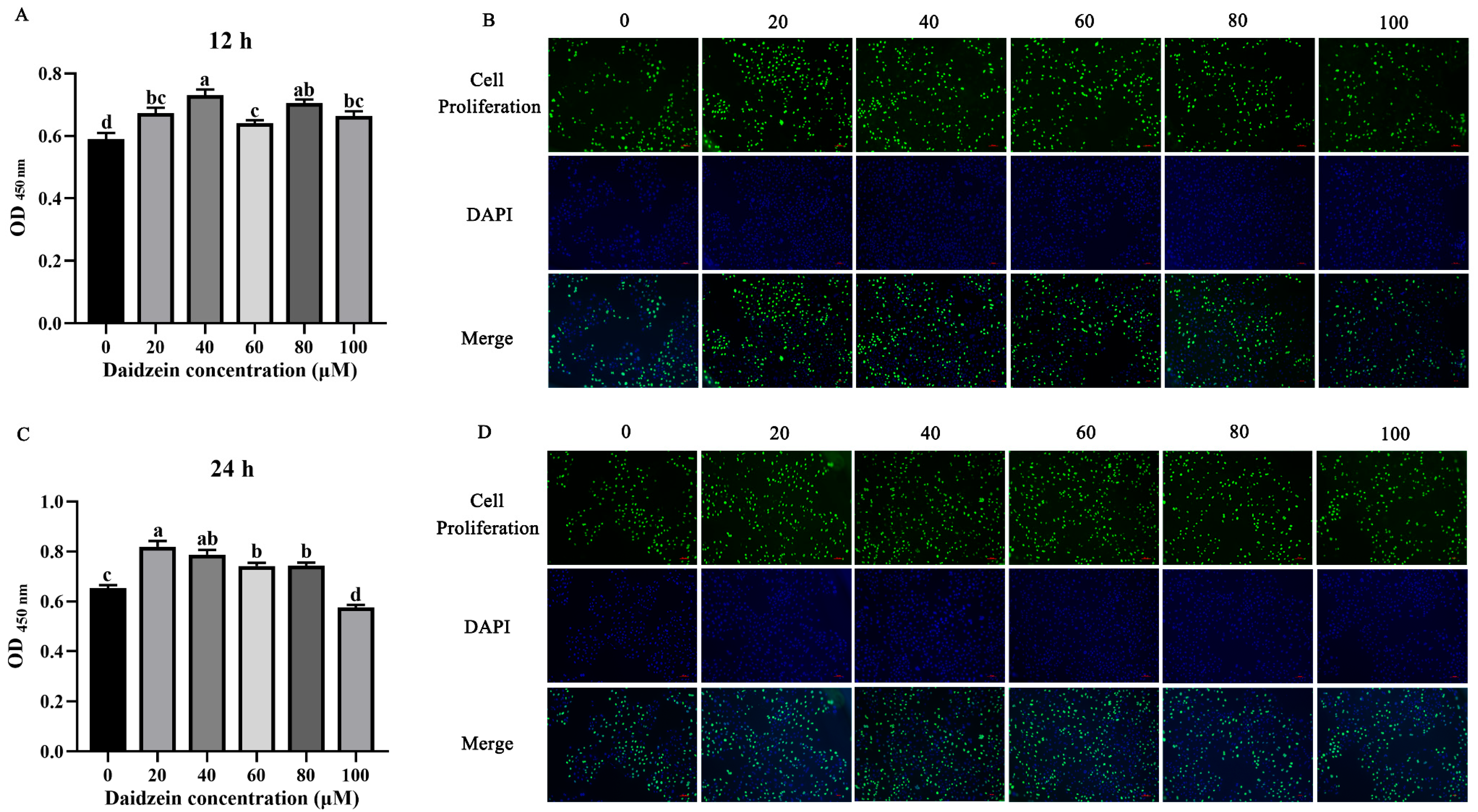
3.1. Effect of Daidzein Supplementation on PMEC Proliferation

The cell viability of PMECs was significantly enhanced by daidzein at concentrations ranging from 20 to 100 μM (p < 0.05) at 12 h, with the highest cell viability observed with 40 μM, in accordance with the EdU results (Figure 1A). After incubation with daidzein for 24 h, concentrations from 20 to 80 μM significantly promoted cell viability (p < 0.05), and the most prominent effect was achieved with 20 or 40 μM daidzein (Figure 1C). However, daidzein at 100 μM had an adverse effect on PMEC proliferation, consistent with the EdU results (Figure 1B,D). These results indicate that daidzein influences PMEC proliferation.

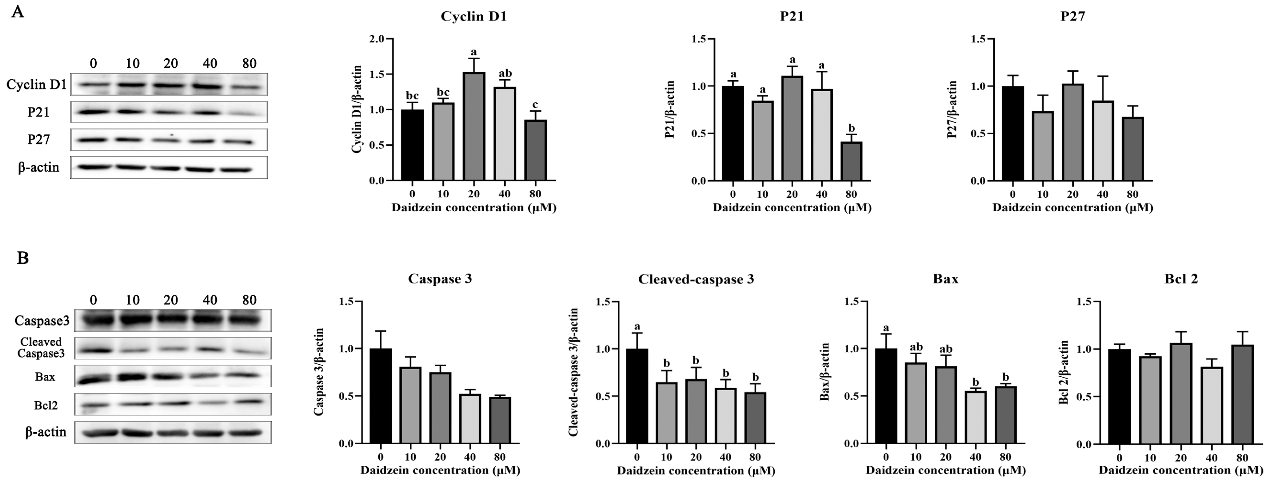
3.2. Effects of Daidzein Supplementation on the PMEC Cycle and Apoptosis

PMEC proliferation is mainly affected by the expression of cyclins or apoptosis-associated proteins. Therefore, we examined the expression levels of such proteins. Daidzein at 20 μM significantly increased the expression of cyclin D1 (p < 0.05) compared with the 0 μM group. At 80 μM, daidzein supplementation significantly decreased the expression of the P21 protein (p < 0.05) (Figure 2A). However, the expression of P27 did not show any significant differences with different concentrations of daidzein (p > 0.05). When compared with that in the 0 μM group, the addition of daidzein significantly reduced the expression level of the cell-apoptosis-related protein cleaved caspase-3 (p < 0.05). Meanwhile, the addition of 40 and 80 μM daidzein significantly decreased the protein expression level of Bax (p < 0.05) (Figure 2B). No significant discrepancies in the expression levels of Bcl2 and caspase-3 were identified between the groups (p > 0.05) (Figure 2B). Thus, daidzein enhanced cellular expansion through coordinated modulation of cyclin-dependent kinases and apoptosis-regulatory effectors.

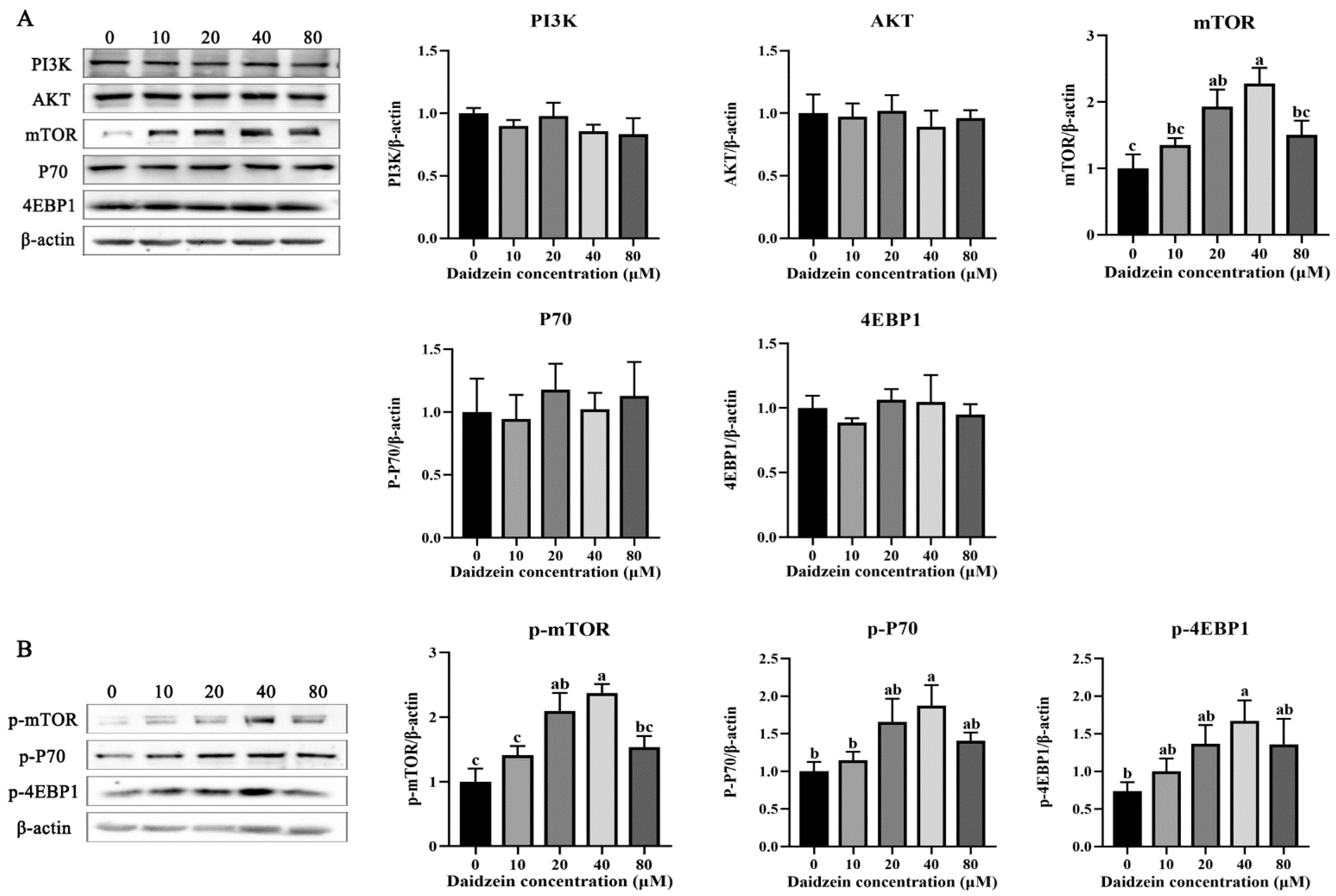
3.3. Effects of Daidzein Supplementation on PI3K/AKT/mTOR Signaling

We further investigated how daidzein modulates the expression of proteins involved in cell-cycle regulation. After treating PMECs with 0–80 μM daidzein, no significant differences were detected in the protein expression levels of PI3K and AKT among all groups (p > 0.05) (Figure 3A). When the PMECs were treated with 0–40 μM daidzein, the levels of mTOR and p-mTOR increased in a log-dose-dependent manner, and specifically, 20 and 40 μM daidzein significantly enhanced the expression levels of mTOR and p-mTOR (p < 0.05) (Figure 3A,B). However, a downward trend was observed in the 80 μM group. Although no statistically significant differences were observed in the expression of P70 and 4EBP1, 40 μM daidzein significantly increased the expression of p-P70 and p-4EBP1 (p < 0.05) (Figure 3B). Therefore, the proliferative effect of daidzein on PMECs could be mediated by mTOR signaling activation.

3.4. Effects of Rapamycin Supplementation on Cell Viability

After treating the PMECs with various concentrations of rapamycin for 24 h, a significant reduction in cell viability was observed. When treated with 40 nM rapamycin, cell survival was significantly inhibited (p < 0.05) (Figure 4A), consistent with the results of the EdU assay (Figure 4B). When the rapamycin dose exceeded 80 nM, cell proliferation was significantly inhibited, resulting in the death of many cells. Consequently, rapamycin at 40 nM was used for subsequent experimental studies. When the cells were treated with 40 nM rapamycin and 40 μM daidzein, it was found that the cell-proliferation-promoting effect of daidzein would be eliminated by the addition of rapamycin (Figure 4C).

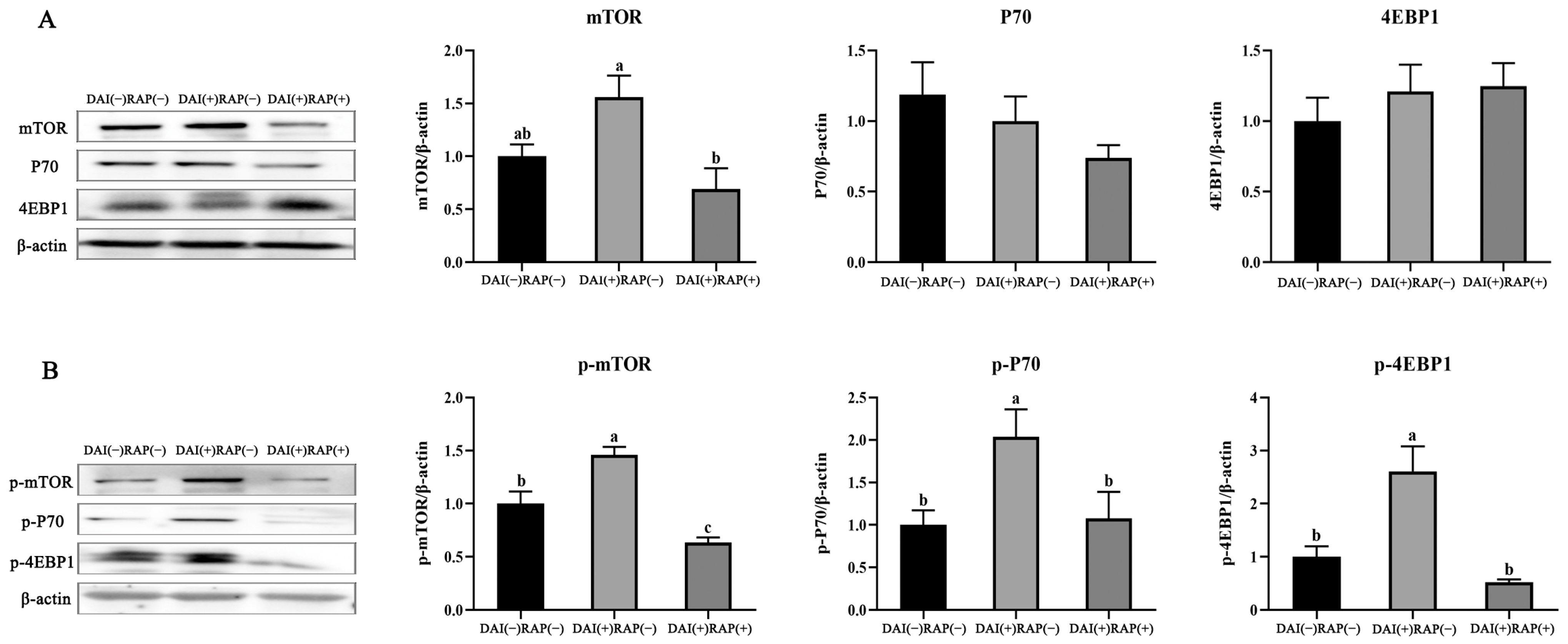
3.5. Effects of Daidzein and Rapamycin on mTOR Signaling and Downstream Proteins

Similar to the outcomes of earlier studies, daidzein (40 μM) significantly enhanced cell proliferation and upregulated the phosphorylation of mTOR (p < 0.05) (Figure 5A). After co-treatment with 40 nM rapamycin, daidzein-induced mTOR protein expression and the phosphorylation of mTOR, P70, and 4EBP1 were decreased (p < 0.05) (Figure 5B). Moreover, the changes in cyclin D1, P21, Bax, and cleaved caspase-3 levels were abolished (p < 0.05) (Figure 6A,B). These results imply a critical involvement of the mTOR pathway in mediating daidzein’s proliferative effects.

4. Discussion

Lactation performance in sows critically determines neonatal piglet survival and growth outcomes. During the lactation period of sows, the mammary alveoli and ductal system continuously proliferate with the secretion of prolactin. The lactation process in sows involves an active uptake of circulating nutrients (glucose, amino acids, and lipids) by mammary cells for the subsequent synthesis of milk constituents. Through a series of complex biochemical reactions within the cells, lactose, milk proteins, milk fats, and other milk components are synthesized, and they are then secreted into the alveolar lumen [7]. Therefore, the number of proliferating PMECs plays a decisive role in milk production by sows. Daidzein is the principal component of soy isoflavones, has extensive nutritional value, and is primarily extracted from soybean plants [26]. Previous studies have confirmed that daidzein exhibits species-specific bioactivity, promoting mammary cell proliferation in rats, and boosting hens’ productivity via antioxidant mechanisms [22,27,28]; however, the effects of daidzein on the proliferation of PMECs, along with their mechanistic basis, remain unelucidated. Through in vitro experiments, we observed that daidzein stimulated the mTOR signaling pathway and promoted PMEC proliferation. Previous studies have demonstrated that daidzein promotes cell proliferation in humans, mice, and cattle [29,30,31]. Our results are consistent with those of previous studies, showing that supplementation with intermediate concentrations of daidzein significantly improved PMECs’ viability in a dose-dependent manner, suggesting that this compound enhanced cell viability. These findings provide a basis for our subsequent in-depth in vitro study of daidzein-mediated regulation of PMECs’ physiological functions.
After cells are stimulated by effective nutritional signals, PI3K is activated to produce PIP3, which, in turn, activates AKT. AKT promotes the expression of cyclins by activating mTOR and inhibits GSK-3β to reduce cell-cycle inhibitors, thereby driving the cells from the G1 phase into the S phase [32,33]. However, our data revealed no significant alterations in PI3K/AKT protein levels following daidzein exposure. Beyond the PI3K/AKT axis, mTOR pathway activation may be regulated through alternative routes, including the Ras-MAPK signaling cascade, or post-translational modifications such as ubiquitination and acetylation [34]. Consequently, the precise molecular route through which daidzein activates mTOR signaling remains an open research question. mTOR complex activation is also modulated by various signals, including growth factors, amino acids, energy status, and cellular stress [35]. For example, mTORC2 mainly responds to growth factors and promotes cell-cycle entry, cell survival, actin cytoskeletal polarization, and anabolic output [36]. Meanwhile, mTORC1 is central to the regulation of these processes and controls cell growth by stimulating translation and ribosome biogenesis and modulating autophagy [37]. The substrates of mTORC1 include eIF4E-binding protein 1 and ribosomal protein S6 kinase (p70S6K) [35]. mTORC1 primarily promotes protein synthesis by phosphorylating p70S6K1 and 4EBP1, which regulate cap-dependent translation initiation and elongation processes, respectively [38]. In bovine mammary epithelial systems, the 20 μM dose of daidzein effectively stimulates the mTOR signaling axis, leading to subsequent cell-cycle modulation [19]. This corresponds to the results of previous research, indicating that the appropriate concentration of daidzein can activate the mTOR signaling pathway. To determine whether daidzein improves cell proliferation through mTOR activation, rapamycin was utilized as a targeted mTOR complex suppressor in this study. The results showed that the addition of rapamycin abolished the activation effect of daidzein on the mTOR signaling pathway. These observations suggested that daidzein influences cell proliferation via mTOR activation.
Cell proliferation is regulated by both positive and negative cell-cycle modulators. Studies have shown that phosphorylation of the mTOR downstream protein P70 promotes ribosome biogenesis and protein synthesis, which are beneficial for the expression of positive regulators of cell-cycle-related proteins [35]. This transition drives cell-cycle advancement through the G1/S checkpoint, ultimately enhancing the proliferative capacity [39]. Cyclin-dependent kinases (CDKs) are key factors involved in the formation of CDK/cyclin complexes such as CDK4/cyclin D1, whereas CDK inhibitors such as p21 and p27 act as negative regulators by blocking the formation of CDK/cyclin complexes, thus serving as brakes for cell-cycle progression [40]. Previous studies have indicated that daidzein regulates the expression of cyclins and related functional genes in sow mammary glands [31]. Our results are consistent with these findings, showing that an appropriate concentration of daidzein inhibits p21 protein expression and promotes cyclin D1 protein expression. This may be related to the fact that activated mTOR inhibits the regulatory function of the FoxO transcription factor, thereby alleviating p21’s inhibitory impact on the cell cycle, and promoting cell-cycle progression and cell proliferation [41].
The cellular response to apoptosis is regulated by the Bcl2 protein family, which includes the death agonist Bax and antagonist Bcl2. Bax is a pro-apoptotic member of the Bcl2 family and a target of TP53 [42]. Bax forms a heterodimer with Bcl2, and Bcl2 inhibits its activity [43]. Our research has shown that 40 μM daidzein exerts cytoprotective activity by differentially regulating apoptotic markers: enhancing anti-apoptotic Bcl2 expression while concurrently reducing pro-apoptotic Bax levels, indicating its potential to safeguard mammary epithelial cell viability. This finding is consistent with the conclusions of previous studies [23]. Previous research has reported that after mTOR is phosphorylated, it can activate the nuclear factor κB (NF-κB) signaling pathway [44]. NF-κB activation drives transcriptional upregulation of Bcl2 while suppressing pro-apoptotic mediators, including Bax and caspase-3, thereby shifting the cellular equilibrium toward survival [45]. These findings demonstrate that daidzein-mediated mTOR signaling activation drives pro-proliferative outcomes by coordinately regulating cell-cycle- and apoptosis-related protein networks. Therefore, daidzein may increase milk synthesis by promoting the proliferation of mammary gland cells, and it has potential applications in lactation physiology, such as increasing the milk production of sows.

5. Conclusions

The results of this study show that adding 40 μM daidzein to the culture medium of PMEC can promote cell proliferation. Daidzein regulates the expression of proteins related to the cell cycle and apoptosis of PMECs by activating the mTOR signaling pathway. Through an in vitro model, these results offer insights into the advantageous impacts of supplementing sows’ diets with daidzein. In the future, we plan to conduct in vivo experiments and combine multi-omics approaches to further study the effects of daidzein on the lactation performance of breeding animals and reveal its regulatory mechanisms.

Author Contributions

Conceptualization, M.X.; Data Curation, L.L. and W.D.; Formal Analysis, P.Z. and M.L.; Funding Acquisition, L.C.; Investigation, M.X., L.L. and L.C.; Methodology, W.H., H.D. and W.C.; Project Administration, L.L., H.C. and C.D.; Software, L.C. and W.D.; Supervision, L.C.; Validation, L.N., H.D. and W.C.; Visualization, L.N.; Writing—Original Draft, M.X. and L.L.; Writing—Review and Editing, M.X. and L.C. All authors have read and agreed to the published version of the manuscript.

Funding

This research was funded by the National Key Research and Development Program of China (2023YFD1300801), National Natural Science Foundation of China (32202678), Key Project of Science and Technology of Henan Province (242102111021), Natural Science Foundation of Henan Province (252300421656), and Sichuan Science and Technology Program (2022YFH0064).

Institutional Review Board Statement

According to local regulations and institutional requirements, ethical approval was not required for these animal studies, because only self-established cell lines were used.

Data Availability Statement

This article encompasses the original contributions of this study. For any additional inquiries, please contact the corresponding author.

Conflicts of Interest

All authors have read and approved the final manuscript. The authors declare that there are no conflicts of interest.

References

  1. Tokach, M.D.; Menegat, M.B.; Gourley, K.M.; Goodband, R.D. Review: Nutrient Requirements of the Modern High-Producing Lactating Sow, with an Emphasis on Amino Acid Requirements. Animal 2019, 13, 2967–2977. [Google Scholar] [CrossRef]
  2. Gormley, A.; Jang, K.B.; Garavito-Duarte, Y.; Deng, Z.; Kim, S.W. Impacts of Maternal Nutrition on Sow Performance and Potential Positive Effects on Piglet Performance. Animals 2024, 14, 1858. [Google Scholar] [CrossRef]
  3. Thomas, L.L.; Goodband, R.D.; Tokach, M.D.; Woodworth, J.C.; DeRouchey, J.M.; Dritz, S.S. Effect of Parity and Stage of Gestation on Growth and Feed Efficiency of Gestating Sows. J. Anim. Sci. 2018, 96, 4327–4338. [Google Scholar] [CrossRef]
  4. Thiengpimol, P.; Koonawootrittriron, S.; Suwanasopee, T. Assessing Reproductive Performance and Predictive Models for Litter Size in Landrace Sows under Tropical Conditions. Anim. Biosci. 2024, 37, 1333–1344. [Google Scholar] [CrossRef]
  5. Sampath, V.; Han, K.; Kim, I.H. Impact of Artificial Colostrum Supplement on the Growth Performance and Blood Profile in Piglets. Anim. Nutr. Feed Technol. 2022, 22, 665–672. [Google Scholar] [CrossRef]
  6. Farmer, C. The Gestating and Lactating Sow; Wageningen Academic Publishers: Wageningen, The Netherlands, 2015. [Google Scholar]
  7. Zhang, S.; Chen, F.; Zhang, Y.; Lv, Y.; Heng, J.; Min, T.; Li, L.; Guan, W. Recent Progress of Porcine Milk Components and Mammary Gland Function. J. Anim. Sci. Biotechnol. 2018, 9, 77. [Google Scholar] [CrossRef]
  8. Slepicka, P.F.; Somasundara, A.V.; Santos, C.D. The Molecular Basis of Mammary Gland Development and Epithelial Differentiation. Semin. Cell Dev. Biol. 2021, 114, 93–112. [Google Scholar] [CrossRef]
  9. Che, L.; Xu, M.; Gao, K.; Wang, L.; Yang, X.; Wen, X.; Xiao, H.; Jiang, Z. Effects of Dietary Valine Supplementation during Late Gestation on the Reproductive Performance and Mammary Gland Development of Gilts. J. Anim. Sci. Biotechnol. 2020, 11, 15. [Google Scholar] [CrossRef]
  10. Wang, B.; Shi, L.; Men, J.; Li, Q.; Hou, X.; Wang, C.; Zhao, F. Controlled Synchronization of Prolactin/STAT5 and AKT1/mTOR in Bovine Mammary Epithelial Cells. Vitr. Cell. Dev. Biol.-Anim. 2020, 56, 243–252. [Google Scholar] [CrossRef]
  11. Liu, G.Y.; Sabatini, D.M. mTOR at the Nexus of Nutrition, Growth, Ageing and Disease. Nat. Rev. Mol. Cell. Biol. 2020, 21, 246. [Google Scholar] [CrossRef]
  12. Feyera, T.; Theil, P.K. Energy and Lysine Requirements and Balances of Sows during Transition and Lactation: A Factorial Approach. Livest. Sci. 2017, 201, 50–57. [Google Scholar] [CrossRef]
  13. Moreira, L.P.; Menegat, M.B.; Barros, G.P.; Bernardi, M.L.; Wentz, I.; Bortolozzo, F.P. Effects of Colostrum, and Protein and Energy Supplementation on Survival and Performance of Low-birth-weight Piglets. Livest. Sci. 2017, 202, 188–193. [Google Scholar] [CrossRef]
  14. Ferrari, C.V.; Sbardella, P.E.; Bernardi, M.L.; Coutinho, M.L.; Vaz, I.S., Jr.; Wentz, I.; Bortolozzo, F.P. Effect of Birth Weight and Colostrum Intake on Mortality and Performance of Piglets after Cross-fostering in Sows of Different Parities. Prev. Vet. Med. 2014, 114, 259–266. [Google Scholar] [CrossRef]
  15. Wang, Q.; Ge, X.; Tian, X.; Zhang, Y.; Zhang, J.; Zhang, P. Soy Isoflavone: The Multipurpose Phytochemical (Review). Biomed. Rep. 2013, 1, 697–701. [Google Scholar] [CrossRef]
  16. Cools, S.; Broeck, W.V.; Vanhaecke, L.; Heyerick, A.; Bossaert, P.; Hostens, M.; Opsomer, G. Feeding Soybean Meal Increases the Blood Level of Isoflavones and Reduces the Steroidogenic Capacity in Bovine Corpora Lutea, without Affecting Peripheral Progesterone Concentrations. Anim. Reprod. Sci. 2014, 144, 79–89. [Google Scholar] [CrossRef]
  17. Yu, Z.; Yang, L.; Deng, S.; Liang, M. Daidzein Ameliorates LPS-induced Hepatocyte Injury by Inhibiting Inflammation and Oxidative Stress. Eur. J. Pharmacol. 2020, 885, 173399. [Google Scholar] [CrossRef]
  18. Kao, T.; Chen, B.H. Functional Components in Soybean Cake and Their Effects on Antioxidant Activity. J. Agric. Food. Chem. 2006, 54, 7544–7555. [Google Scholar] [CrossRef]
  19. Yu, F.; Liu, Z.; Tong, Z.; Zhao, Z.; Liang, H. Soybean Isoflavone Treatment Induces Osteoblast Differentiation and Proliferation by Regulating Analysis of Wnt/β-catenin Pathway. Gene 2015, 573, 273–277. [Google Scholar] [CrossRef]
  20. Hüser, S.; Guth, S.; Joost, H.G.; Soukup, S.T.; Köhrle, J.; Kreienbrock, L.; Diel, P.; Lachenmeier, D.W.; Eisenbrand, G.; Vollmer, G.; et al. Effects of Isoflavones on Breast Tissue and the Thyroid Hormone System in Humans: A Comprehensive Safety Evaluation. Arch. Toxicol. 2018, 92, 2703–2748. [Google Scholar] [CrossRef]
  21. Mortensen, A.; Kulling, S.E.; Schwartz, H.; Rowland, I.; Ruefer, C.E.; Rimbach, G.; Cassidy, A.; Magee, P.; Millar, J.; Hall, W.L.; et al. Analytical and Compositional Aspects of Isoflavones in Food and Their Biological Effects. Mol. Nutr. Food. Res. 2009, 2, S266–S309. [Google Scholar] [CrossRef]
  22. Lamartiniere, C.; Wang, J. Prepubertal Genistein Exposure Suppresses Mammary Cancer Development and Alters Proteomic Signature in Adult Rats. Toxicol. Lett. 2010, 196, S173–S174. [Google Scholar] [CrossRef]
  23. Yu, M.; Qi, H.; Gao, X. Daidzein Promotes Milk Synthesis and Proliferation of Mammary Epithelial Cells via the Estrogen Receptor A-dependent NfκB1 Activation. Anim. Biotechnol. 2022, 33, 43–52. [Google Scholar] [CrossRef]
  24. Dahanayaka, S.; Rezaei, R.; Porter, W.W.; Johnson, G.A.; Burghardt, R.C.; Bazer, F.W.; Hou, Y.Q.; Wu, Z.L.; Wu, G. Technical Note: Isolation and Characterization of Porcine Mammary Epithelial Cells. J. Anim. Sci. 2015, 93, 5186–5193. [Google Scholar] [CrossRef]
  25. Che, L.; Liu, L.; Xu, M.; Fan, Z.; Niu, L.; Chen, Y.; Chang, X.; Zhou, P.; Li, M.; Deng, H.; et al. Valine Metabolite, 3-Hydroxyisobutyrate, Promotes Lipid Metabolism and Cell Proliferation in Porcine Mammary Gland Epithelial Cells. Front. Nutr. 2025, 11, 1524738. [Google Scholar] [CrossRef]
  26. Alshehri, M.M.; Sharifi-Rad, J.; Herrera-Bravo, J.; Jara, E.L.; Salazar, L.A.; Kregiel, D.; Uprety, Y.; Akram, M.; Iqbal, M.; Martorell, M.; et al. Therapeutic Potential of Isoflavones with an Emphasis on Daidzein. Oxid. Med. Cell. Longev. 2021, 9, 6331630. [Google Scholar] [CrossRef]
  27. Liu, J.; Fu, Y.; Zhou, S.; Zhao, P.; Zhao, J.; Yang, Q.; Wu, H.; Ding, M.; Li, Y. Comparison of the Effect of Quercetin and Daidzein on Production Performance, Anti-oxidation, Hormones, and Cecal Microflora in Laying Hens during the Late Laying Period. Poultry Sci. 2023, 102, 102674. [Google Scholar] [CrossRef]
  28. Obianwuna, U.E.; Chang, X.; Wu, R.; Zhang, H.; Qiu, K.; Wu, S. Effect of Dietary Daidzein Supplementation on Reproductive Performance, Egg Quality and Bone Mineralization in Laying Hens at First-cycle of Production via Reproductive-growth Axis. Ital. J. Anim. Sci. 2024, 23, 1169–1180. [Google Scholar] [CrossRef]
  29. Jin, X.; Sun, J.; Yu, B.; Wang, Y.; Sun, W.J.; Yang, J.; Huang, S.H.; Xie, W.L. Daidzein Stimulates Osteogenesis Facilitating Proliferation, Differentiation, and Antiapoptosis in Human Osteoblast-Like Mg-63 Cells via Estrogen Receptor-Dependent MEK/ERK and PI3K/Akt Activation. Nutr. Res. 2017, 42, 20–30. [Google Scholar] [CrossRef]
  30. Chen, W.F.; Wong, M.S. Genistein Enhances Insulin-like Growth Factor Signaling Pathway in Human Breast Cancer (MCF-7) cells. J. Clin. Endocrinol. Metab. 2004, 89, 2351–2359. [Google Scholar] [CrossRef]
  31. Ma, X.; Cui, Y.; Tian, Z.; Yu, M. Effect of Soybean Isoflavones on Proliferation and Related Gene Expression of Sow Mammary Gland Cells In Vitro. Animals 2022, 12, 3241. [Google Scholar] [CrossRef]
  32. Park, J.; Choi, H.; Shim, K. Inhibition of GSK3β Promotes Proliferation and Suppresses Apoptosis of Porcine Muscle Satellite Cells. Animals 2022, 12, 3328. [Google Scholar] [CrossRef]
  33. Dong, J.; Wang, Z.; Fei, F.; Jiang, Y.; Jiang, Y.; Guo, L.; Liu, K.; Cui, L.; Meng, X.; Li, J.; et al. Selenium Enhances the Growth of Bovine Endometrial Stromal Cells by PI3K/AKT/GSK-3β and Wnt/β-Catenin Pathways. Vet. Sci. 2024, 11, 674. [Google Scholar] [CrossRef]
  34. Qiao, J.; Tang, C.; Xie, M.; Gong, M.; Fu, C.; Cheng, Z.; Chen, Z.; Mei, A.; Bo, Y.; Zhao, M.; et al. Aberrant activation of the mTOR signaling pathway in Rasmussen encephalitis. Sci. Rep. 2025, 15, 6347. [Google Scholar] [CrossRef]
  35. Szwed, A.; Kim, E.; Jacinto, E. Regulation and Metabolic Functions of mTORC1 and mTORC2. Physiol. Rev. 2021, 101, 1371–1426. [Google Scholar] [CrossRef]
  36. Ragupathi, A.; Kim, C.; Jacinto, E. The mTORC2 Signaling Network: Targets and Cross-talks. Biochem. J. 2024, 481, 45–91. [Google Scholar] [CrossRef]
  37. Bar-Peled, L.; Sabatini, D.M. Regulation of mTORC1 by Amino Acids. Trends Cell Biol. 2014, 24, 400–406. [Google Scholar] [CrossRef]
  38. Che, L.; Xu, M.; Gao, K.; Wang, L.; Yang, X.; Wen, X.; Xiao, H.; Jiang, Z.; Wu, D. Valine Supplementation During Late Pregnancy in Gilts Increases Colostral Protein Synthesis Through Stimulating mTOR Signaling Pathway in Mammary Cells. Amino Acids 2019, 51, 1547–1559. [Google Scholar] [CrossRef]
  39. Cuyàs, E.; Corominas-Faja, B.; Joven, J.; Menendez, J.A. Cell Cycle Regulation by the Nutrient-sensing Mammalian Target of Rapamycin (mTOR) Pathway. Methods Mol. Biol. 2014, 1170, 113–144. [Google Scholar]
  40. Sencan, S.; Tanriover, M.; Ulasli, M.; Karakas, D.; Ozpolat, B. UV Radiation Resistance-Associated Gene (Uvrag) Promotes Cell Proliferation, Migration, Invasion by Regulating Cyclin-Dependent Kinases (Cdk) and Integrin-Β/Src Signaling in Breast Cancer Cells. Mol. Cell. Biochem. 2021, 476, 2075–2084. [Google Scholar] [CrossRef]
  41. Mori, S.; Nada, S.; Kimura, H.; Tajima, S.; Takahashi, Y.; Kitamura, A.; Oneyama, C.; Okada, M. The mTOR Pathway Controls Cell Proliferation by Regulating the FoxO3a Transcription Factor via SGK1 Kinase. PLoS ONE 2014, 9, e88891. [Google Scholar] [CrossRef]
  42. Gitego, N.; Agianian, B.; Mak, O.W.; Mv, V.K.; Cheng, E.H.; Gavathiotis, E. Chemical Modulation of Cytosolic BAX Homodimer Potentiates BAX Activation and Apoptosis. Nat. Commun. 2023, 14, 8381. [Google Scholar] [CrossRef] [PubMed]
  43. Czabotar, P.E.; Garcia-Saez, A.J. Mechanisms of BCL-2 Family Proteins in Mitochondrial Apoptosis. Nat. Rev. Mol. Cell Biol. 2023, 24, 732–748. [Google Scholar] [CrossRef] [PubMed]
  44. Xu, S.; Jiang, X.; Liu, Y.; Jiang, X.; Che, L.; Lin, Y.; Zhuo, Y.; Feng, B.; Fang, Z.; Hua, L.; et al. Silibinin Alleviates Lipopolysaccharide Induced Inflammation in Porcine Mammary Epithelial Cells via mTOR/NF-κB Signaling Pathway. Mol. Nutr. Food. Res. 2023, 67, e2200715. [Google Scholar] [CrossRef] [PubMed]
  45. Zhang, Y.; Li, Y.; Zhang, J.; Qi, X.; Cui, Y.; Yin, K.; Lin, H. Cadmium Induced Inflammation and Apoptosis of Porcine Epididymis via Activating RAF1/MEK/ERK and NF-κB Pathways. Toxicol. Appl. Pharm. 2021, 415, 115449. [Google Scholar] [CrossRef]
Figure 1. Effects of different daidzein concentrations on cell proliferation after 12 or 24 h: (A,C) Detection of cell proliferation using the Cell Counting Kit-8 (CCK-8) assay (n = 10); OD 450nm: the absorbance of these media with CCK-8 was read at 450 nm. (B,D) Detection of cell proliferation using the 5-ethynyl-2′-deoxyuridine (EdU) assay (n = 6); DAPI: images of cell nuclei stained with Hoechst 33342; Merge: the merged image of cell green fluorescence and cell nuclei. Values are presented as the mean ± standard deviation. Statistical significance (p < 0.05) is indicated by different superscript letters (p < 0.05).
Agriculture 15 00930 g001
Figure 2. Effects of different daidzein concentrations on proteins related to cell proliferation and apoptosis: (A) Detection of proteins related to cell proliferation (n = 3). (B) Detection of proteins related to cell apoptosis (n = 3). Values are presented as the mean ± standard deviation. Statistical significance (p < 0.05) is indicated by different superscript letters (p < 0.05).
Agriculture 15 00930 g002
Figure 3. Effects of different daidzein concentrations on proteins related to the mTOR signaling pathway: (A) Detection of mTOR and its downstream proteins (n = 3). (B) Detection of the phosphorylation levels of mTOR and its downstream proteins (n = 3). Values are presented as the mean ± standard deviation. Statistical significance (p < 0.05) is indicated by different superscript letters (p < 0.05).
Agriculture 15 00930 g003
Figure 4. Effects of rapamycin, at different concentrations, on the proliferation of porcine mammary epithelial cells (PMECs): (A) Detection of the effects of rapamycin on cell proliferation based on the CCK-8 assay (n = 10); OD 450nm: the absorbance of these media with CCK-8 was read at 450 nm. (B) Detection of the effects of rapamycin on cell proliferation based on the EdU assay (n = 6); DAPI: images of cell nuclei stained with Hoechst 33342; Merge: the merged image of cell green fluorescence and cell nuclei. (C) Detection of the effect of the interaction between daidzein and rapamycin on cell proliferation; PMECs were treated with daidzein (40 μM) and/or rapamycin (40 nM) (n = 10). Values are presented as the mean ± standard deviation. Statistical significance (p < 0.05) is indicated by different superscript letters (p < 0.05).
Agriculture 15 00930 g004
Figure 5. Effects of daidzein and rapamycin on protein expression of mTOR signaling pathway components in porcine mammary epithelial cells (PMECs); PMECs were treated with daidzein (40 μM) and/or rapamycin (40 nM): (A) Detection of mTOR and its downstream proteins (n = 3). (B) Detection of the phosphorylation levels of mTOR and its downstream proteins (n = 3). Values are presented as the mean ± standard deviation. Statistical significance (p < 0.05) is indicated by different superscript letters (p < 0.05).
Agriculture 15 00930 g005
Figure 6. Effects of daidzein and rapamycin on protein expression related to proliferation and apoptosis in porcine mammary epithelial cells (PMECs); PMECs were treated with daidzein (40 μM) and/or rapamycin (40 nM): (A) Detection of proteins related to cell proliferation (n = 3). (B) Detection of proteins related to cell apoptosis (n = 3). Values are presented as the mean ± standard deviation. Statistical significance (p < 0.05) is indicated by different superscript letters (p < 0.05).
Agriculture 15 00930 g006
Disclaimer/Publisher’s Note: The statements, opinions and data contained in all publications are solely those of the individual author(s) and contributor(s) and not of MDPI and/or the editor(s). MDPI and/or the editor(s) disclaim responsibility for any injury to people or property resulting from any ideas, methods, instructions or products referred to in the content.

