Article

Effects of Irradiance and Temperature on the Photosynthesis of the Crustose Coralline Algae Pneophyllum fragile (Corallinales, Rhodophyta) in the Coastal Waters of Korea

1
Marine Environment Research Center, Korea Institute of Ocean Science and Technology, 385, Haeyang-ro, Yengdo-gu, Busan 49111, Korea
2
Department of Convergence Study on the Ocean Science and Technology, Ocean Science and Technology School, 385, Haeyang-ro, Yengdo-gu, Busan 49111, Korea
3
Jeju Marine Research Center, Korea Institute of Ocean Science and Technology, Jeju 63349, Korea
4
Library of Marine Samples, Korea Institute of Ocean Science and Technology, Geoje 53201, Korea
5
Marine Environmental Research Division, National Institute of Fisheries Science, 216 Gijanghaean-ro, Gijang-eup, Busan 46083, Korea
*
Author to whom correspondence should be addressed.
J. Mar. Sci. Eng. 2022, 10(7), 851; https://doi.org/10.3390/jmse10070851
Submission received: 13 April 2022 / Revised: 10 June 2022 / Accepted: 19 June 2022 / Published: 22 June 2022
(This article belongs to the Section Marine Ecology)

Abstract

:
We investigated the photosynthetic characteristics of the crustose coralline alga Pneophyllum fragile (Corallinales, Rhodophyta) according to elevated water temperature and irradiance on the coast of Jeju in 2018. P. fragile was cultured under different temperature (11 °C, 21 °C, 26 °C, and 31 °C) and irradiance (0–1250 μmol photon m−2 s−1) conditions. Oxygen (O2) concentrations at the P. fragile mat–water interface (MWI) were measured using an O2 microsensor. At the MWI, the diffusive boundary layer thicknesses ranged from 200 to 400 μm. The O2 concentrations at the mat surface increased in response to increasing irradiance, and reached 344% air saturation. The maximum photosynthesis capacity (Pmax) and respiration rate in the dark (Rd) at 31 °C were about 3 times higher than those recorded at 11 °C. The compensation irradiance (Ec) and saturation irradiance (Ek) increased with increasing water temperature. The Pmax, Rd, and Ec were statistically correlated with temperature (p < 0.05). The Ek increased up to 833 μmol photon m−2 s−1 at 31 °C and exhibited a strong dependence on irradiance at high temperatures. The adaptability of P. fragile to high temperatures and strong irradiance was distinct from that observed for coralline algae in other temperate waters.

1. Introduction

Whitening, also known as “urchin barren”, is a phenomenon whereby crustose coralline algae (CCA) cover the hard coastal marine bottom (rock and coral) and seaweed communities vanish in the benthic boundary layer. Whitening can reduce the biomass and biodiversity of the benthic habitat, which are major factors associated with the devastation of the benthic ecosystem [1]. Since the 1990s, a massive outbreak of whitening has been observed in the coastal waters of Korea and it has gradually expanded into the subtidal habitats [1,2,3,4]. In particular, the whitening detected in the coastal waters of Jeju Island has rapidly expanded from 2913 ha in 1998 to 4541 ha in 2004, and is considered a threat to the coastal ecosystem.
CCA are non-geniculate (non-articulated) red algae in the order Corallines that inhabit the surface of hard bottoms from polar to tropical coastal waters [5,6,7]. The CCA are important calcifying primary producers (Ca2+ + 2HCO3 + 2H2O → CaCO3 + CH2O + O2) in the coastal ecosystem [8,9]. The major factors that control the photosynthesis and calcification of CCA are temperature, nutrients, irradiance, and pH in the benthic layer [10,11,12,13,14,15,16]. As the physiology of CCA can be affected by fluctuations in environmental variables, these factors can alter CO2 uptake and calcification in CCA [17,18]. Therefore, the physiological response to global warming and acidification may disturb the biogeochemical cycles of organic carbon and CaCO3 in the benthic layer [14,19].
The estimation of photosynthesis and respiration in CCA is essential to understand the benthic community characteristics and ecological functions; however, the traditional quantification approaches, such as oxygen (O2) incubation, isotope labeling, and fluorometry, may not be appropriate for studying CCA, as these methods may require more effort to control the experimental setup and measure these parameters in this species. The microsensor profiling method can measure the O2 profile with a higher spatial resolution than other methods at an extremely small sensing spot (less than 1 mm scale) [12,15,19,20,21,22]. The high spatial distribution of O2 at the solid–water interface (SWI) can allow oxygen fluxes via the interface, which can also be used to estimate the metabolic rate (production and respiration) of benthic species, assuming there is a stoichiometric relationship between O2 and carbon. The O2 gradient measured by the O2 microsensor (sensor tip <0.1 mm) in the diffusivity boundary layer (DBL) at the SWI can be assessed noninvasively. Thus, the microsensor profiling method has been widely used to study photosynthesis and respiration in CCA [5,15,23].
Recently, studies of whitening in the coastal waters of Korea have been initiated to restore the macroalgae meadows and assess their ecological functions [1,2,3,4]. However, most of the studies focused on the monitoring of the occurrence and evolution area of whitening because of the absence of appropriate methods to estimate CCA metabolism [24,25]. P. fragile, a crustose red alga (Corallines, Rhodophyta) that is widely distributed in Australia, Canada, the Caribbean, England, Japan, Mexico, Russia, the United States, and the Mediterranean Sea, was collected to examine its morphology and anatomical characteristics [26].
The objectives of our study were to: (1) estimate the metabolic rates (production and respiration) of P. fragile as a function of temperature and irradiance using the O2 profiling method; (2) assess the photosynthetic characteristics of P. fragile; and (3) understand the key factors that control P. fragile outbreaks in the coastal waters of Korea.

2. Materials and Methods

2.1. Study Area

Jeju Island is located in the pathway of the Korea Strait and its oceanographic characteristics vary according to temporally related changes in the inflow of water masses (Yangtze River dilution water, Kuroshio warm current, Yellow Sea warm current, and Yellow Sea cold water) and meteorology (Figure 1). Seaweed forests are widely distributed on the coast of Jeju Island and play an important role in the ecosystem as a feeding and spawning site for marine organisms and as a breeding site for fish larvae [1]. The subtidal zone is composed of porous volcanic rocks and coarse sediments, which are covered with macroalgae and soft coral communities at the euphotic water depth. The dominant species of macroalgae in rocky habitats are Sargassum horneri, Eclonia cava, and Undaria pinnatifida; however, some of these species have disappeared, most likely because of the increased water temperature triggered by climate change and the establishment of CCA in their habitats, although this is currently unclear [4].

2.2. Sample Collection

Triplicate samples were kept individually in filtered seawater in an insulated aquarium (length × width × height: 60 × 30 × 30 cm) at 21 °C, under a 14 h light/10 h dark cycle with a light intensity of 300 μmol photon m−2 s−1, which was corresponded to the PAR value of CCA’s saturation irradiance to a depth of up to 20 m in the coastal waters of Jeju Island, for up to 3 days (Figure 2) [23]. Because two CCA mats were severely spoilt under artificial condition, we used to measure the photosynthesis on one sample and further analyze.

2.3. Experimental Setup

The experimental setup followed the procedure suggested by Larkum et al. [23]. Briefly, O2 profile measurements were performed in a custom-made flow chamber [27]. The flow rates at the measurement spots were adjusted to ~20 cm s−1 with a water height of over 2 cm using a submersible water pump (HJ-700 AMAZON, Korea). The flow rates were calculated by dividing the volume of the discharge water from the chamber for 1 min (n = 5) by the cross-sectional area of the chamber. Light was irradiated onto the P. fragile mat through a fiber-optic halogen lamp (KL-1500, Schott, Germany) equipped with a collimating lens, to focus the light onto the surface of the sample. The vertical distribution of O2 was measured at different PAR ranges (0%–100%; 0–1250 μmol photon m−2 s−1) using a neutral density filter.

2.4. Photosynthesis and Respiration Measurement

O2 profiles were measured using a Clark-type O2 microsensor (OX-25, Unisense Denmark). The stirring sensitivity of the microsensor was <2–3% and a 90% response time (t90) of 0.3 s was used [28]. Signals from the microsensor were amplified with a pA meter (PA-3000, Unisense, Denmark) and converted to digital signals via an AD converter (ADC-216, Unisense Denmark) at a sampling rate of 1 Hz for 3 s. The O2 microsensor was calibrated using a linear relationship between air-saturated and N2-flushed in situ seawater with a known salinity and temperature. The microsensor was mounted on a motor-driven micromanipulator (MM3, Unisense, Denmark) with a zenith angle of 135°. All signals from the microsensor were stored on a laptop computer.

2.5. Calculation of Net Photosynthesis and Respiration

Assuming a steady state, the areal rates of O2 production and respiration were estimated from the O2 profiles [29]. The diffusive oxygen flux across the MWI was calculated from the linear gradient in the DBL from Fick’s first law:
J = D0 (dC/dz),
where J is the diffusive oxygen flux (mmol m−2 h−1), D0 is the oxygen molecular diffusion coefficient in seawater at the given salinity and temperature (Ramsing and Gundersen, from the table for seawater and gases, Unisense A/S) (cm−2 s−1), and dC/dz is the oxygen slope of the linear part of the DBL (mmol cm−4). The positive flux and the negative flux via MWI indicate the net oxygen production and respiration, respectively.
The relationships between O2 evolution and irradiance (P–I curve) were fitted using an exponential model [30], with an extra term, Rd, accounting for O2 respiration [23], as follows:
P(E) = Pmax [1 − exp(–αEd/Pmax)] + Rd,
where P(E) is the net O2 flux (production–respiration) with irradiance (mmol m−2 h−1), Pmax is the maximum photosynthetic capacity (mmol m−2 h−1), α is the initial slope of the fitted P–I curves, Ed is the irradiance (μmol photon cm−2 s−1), Rd is the O2 respiration flux under dark conditions (mmol cm−2 s−1), and Ek is the saturation irradiance, which was calculated as Ek = Pmax/α. Ec is the compensation irradiance, which is the irradiance at P(E) = 0.

3. Results

3.1. O2 in the DBL

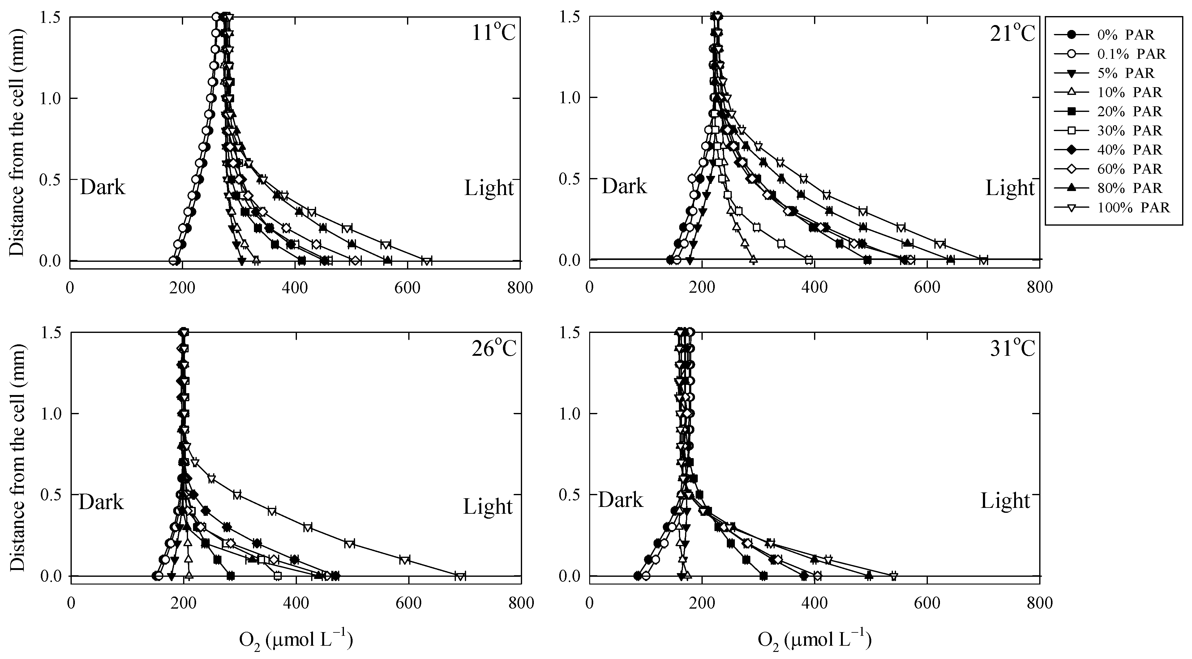
The O2 profiles near the mat of P. fragile were measured at four temperatures (11 °C, 21 °C, 26 °C, and 31 °C) while changing the irradiance (Figure 3). The O2 profiles in the DBL varied according to the photosynthesis and respiration of P. fragile, i.e., the O2 concentration increased (production) or decreased (respiration) close to the P. fragile mat surface (z = 0) under light and dark conditions. The DBL thickness ranged from 200 to 400 μm at 11–31 °C (Figure 3). The O2 concentrations at the mat surface under dark conditions decreased with increasing temperature (189 μmol L−1 at 11 °C, 143 μmol L−1 at 21 °C, 156 μmol L−1 at 26 °C, and 85.6 μmol L−1 at 31 °C). The lowest O2 concentration was recorded at 31 °C and corresponded to an air saturation of 44.2% in the dark. We measured the O2 profiles while increasing the irradiance up to 1250 μmol photon m−2 s−1 from the dark condition. O2 concentrations on the mat became saturated with increasing irradiance, and reached 211% to 344% over the four temperature intervals.
The O2 fluxes in the dark at each temperature ranged from –0.53 ± 0.01 to –1.63 ± 0.07 mmol m−2 h−1, and increased with increasing water temperature. Under light conditions, the O2 fluxes ranged from 0.46 ± 0.04 to 3.8 ± 0.09 mmol m−2 h−1 at 11 °C, 0.92 ± 0.09 to 5.25 ± 0.12 mmol m−2 h−1 at 21 °C, 0.10 ± 0.00 to 8.14 ± 0.02 mmol m−2 h−1 at 26 °C, and 0.62 ± 0.12 to 10.0 ± 0.38 mmol m−2 h−1 at 31 °C, which showed that the O2 flux also varied with increasing temperature. Up to PAR value < 5% (up to 83 μmol photon m−2 s−1), O2 production by photosynthesis was lower than respiration, resulting in a negative slope in the DBL (Figure 3).

3.2. Effects of Irradiance and Temperature on Respiration and Photosynthesis

The relationships between O2 production and irradiance (P–I curve) at each temperature are shown in Figure 4 and Table 1. The P(E) values at 11–21 °C reached asymptotic values at >500 μmol photon m−2 s−1, but gradually increased at higher temperatures (>21 °C). The maximum P(E) ranged from 4.01 ± 0.23 to 14.1 ± 1.16 mmol m−2 d−1, and varied with increasing water temperature.
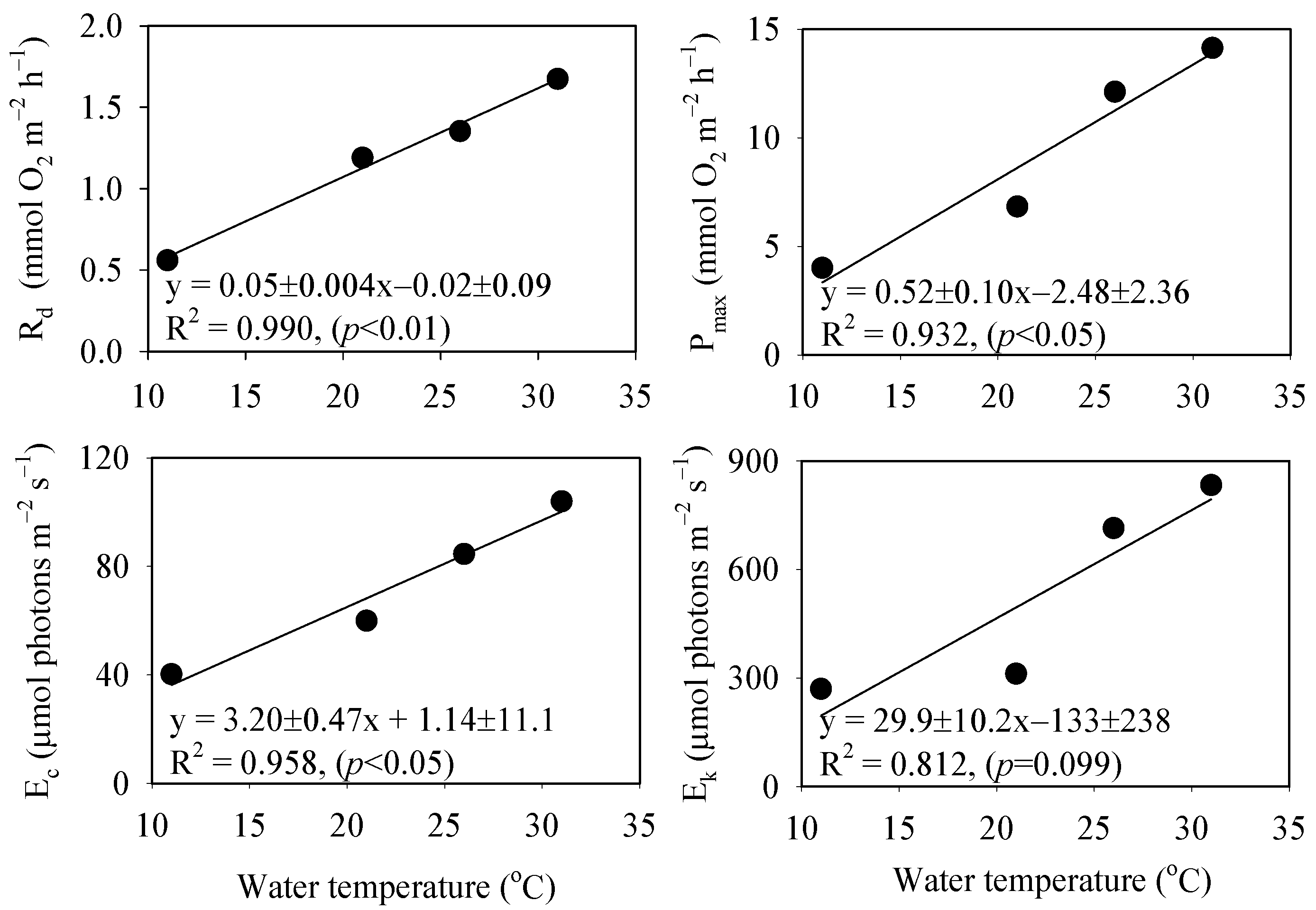
The Rd was in the range of –0.56 ± 0.18 to –1.67 ± 0.26 mmol m−2 d−1, and was highest at 31 °C (3 times that recorded at 11 °C). Similarly, the Pmax in the P–I curve was correlated with water temperature increases and reached a maximum value (14.1 mmol m−2 d−1) at 31 °C (about 3.5 times that recorded at 11 °C). All parameters pertaining to photosynthesis exhibited a good linear correlation with the increase in water temperature (Figure 5).

4. Discussion

4.1. O2 Dynamics in the DBL

The current study examined the O2 dynamics in the DBL on the surface of P. fragile according to changes in water temperature and irradiance. The DBL thickness ranged from 200 to 400 μm, with an O2 concentration of 47–344% air saturation in the MWI. In previous studies, the O2 diffusive flux was affected by DBL thickness, which was altered by the water flow velocity and microtopography [31,32]. In addition, the O2 flux at the MWI was regulated by the photosynthetic activity of algae and DBL thickness. Schubert et al. [33] demonstrated that, despite the increase in DBL thickness (200–600 μm) on the mat of coralline algae resulting from a species-specific increase in protuberance (branch) length observed for L. atlanticum, the O2 fluxes in the DBL were regulated by the photosynthesis rate. In this study, the O2 fluxes in the DBL likely depended on the metabolic rate of P. fragile. The O2 concentration under dark conditions decreased with active respiration because of a temperature increase. In light conditions, the O2 gradient in the DBL increased with the increasing irradiance at four temperatures. In addition, P. fragile was attached to the irregular surface of the rock, but its mat had a flattened thallus, as expected for a smooth surface [26]. Jørgensen and Revsbech [31] suggested that the variation in DBL thickness on a smooth surface with changes in flow velocity (1–20 cm s−1) was not affected by topography. Thus, it can be assumed that there was no interference of the surface roughness on the variation in DBL thickness and that the net O2 flux at the MWI was mainly regulated by the photosynthesis of P. fragile. Moreover, it is considered that the vertical microgradients of O2 at 11–26 °C were influenced by the increase in the net O2 production rate triggered by the increase in irradiance, and a transition layer between the diffusive boundary and the bulk phase being reflected by the O2 divergence was caused by photosynthetic activity [34,35].

4.2. Rates of Respiration and Photosynthesis of P. fragile

Our results suggest that the photosynthesis characteristics of P. fragile were altered by water temperature and irradiance. The correlations between the photosynthesis parameters (Pmax, Ec, Ek, and Rd) and the water temperature variation are shown in Figure 5. Water temperature is an important factor that controls the photosynthesis of CCA [11,15,36]. The increase in water temperature was significantly correlated with parameters such as Pmax, Ec, and Rd (Figure 5). The physiological responses of P. fragile to high light and temperature conditions were similar to those of the coralline algae found in tropical zones, rather than those found in temperate regions. The optimal water temperature for photosynthesis efficiency of coralline algae distributed in temperate regions is <26 °C (Table 2). For example, the Pmax of Phymatolithon lusitanicum in Portugal was highest at 24 °C, and was decreased at higher water temperatures [15]. For Lithophyllum yessoense in Japan, the optimal water temperature for Pmax (5.70 mmol m−2 h−1) was 15 °C [36]. Moreover, the optimal temperature for the maximum photosynthesis capacity of Hildenbrandia rubra and L. yessoense distributed in Korea was 20–25 °C [24]. By contrast, the optimal temperature for the Pmax of Neogoniolithon sp. (rhodolith), Amphiroa tribulus, and Lithothamnion sp. (CCA) distributed in Mexico was 30 °C [14].
In turn, the increase in Ek observed at 11–21 °C was relatively smaller than the changes detected for other parameters, and the correlation between Ek and the increase in water temperature was not statistically significant (Figure 5). However, Ek increased significantly at 26–31 °C and the Ek value of P. fragile was relatively higher than that of other coralline algae in temperate regions and similar to that of coralline algae in tropical zones [15,36,37]. Burdett et al. [38] found that the Ek of Porolithon sp., a species inhabiting Aqaba Bay in Egypt, where the highest water temperature is 29 °C, was ~700 μmol photons m−2 s−1. The Ek parameters based on the P–I curve represent the adaptability to light [22,36,39]. A higher Ek value at a high water temperature indicates a strong dependence on high irradiance; moreover, it suggests that photosynthetic and photoprotective mechanisms minimize photodamage [36,38]. In addition, the physiological responses of P. fragile were similar to the increase in the maximum photosynthetic rate and Ek of Skeletonema costatum observed with increasing temperature [40]. This species-specific response is a key factor associated with the physiological adaption of Skeletonema costatum to temperature variation that enables its distribution in a wide range of oceanic regions [40]. In a previous study, P. fragile was shown to belong to the subfamily Mastophoridae together with Neogoniolithon sp. (rhodolith), and to be representative of coralline algae in southeast Australia [26]. Therefore, it is likely that the increase in Pmax and Ek of P. fragile observed at an elevated water temperature was the result of the excellent adaptability of this species to high temperature and irradiance. Consequently, differences between the photosynthetic characteristics of P. fragile and those of coralline algae in temperate regions may be related to the physiological response associated with species-specific factors, which allow the wide distribution of P. fragile.

4.3. Environmental Implications for the Coastal Ecosystem

Whitening may disturb the benthic ecosystem and result in a reduction of biological diversity, biomass, and habitats in the coastal ecosystem [1,41]. Although whitening seems to be related to artificial effects (eutrophication, climate change, etc.), its mechanisms are still under investigation. Coastal waters are the most reactive place for carbon cycles in the ocean. Therefore, alterations in ecological functions in the benthic layer can change the biogeochemical cycles of organic carbon in coastal waters.
In fact, isoyake (the word for whitening in Japan) has reduced commercial seaweed (Gelidium, Saccharina, Eckonia, and Sargassum) levels over the past couple of decades, and resulted in a decrease in fisheries production [42]. Recently, whitening has spread throughout Korean coastal waters (from Jeju Island to Dokdo Island), and kelp forest restoration projects have been initiated to recover the benthic ecosystem [2,3,43]. However, the results of these efforts remain unclear because the physiological characteristics of CCA and the precise mechanisms associated with whitening outbreaks are unknown. Notably, however, the intrusion of warm currents into coastal waters may be an important factor in the occurrence of whitening [2].
Our results suggest that P. fragile has great adaptability to high temperature and irradiance, in contrast to the CCA found in temperate waters. The increase in the annual mean surface-water temperature around Korea was approximately three times higher than the global trend during the past 50 years, which may be related to climate change [44]. Thus, considering the seasonal variability in irradiance and temperature, P. fragile can likely sustain photosynthesis in Korean coastal waters and be a dominant species associated with whitening. Furthermore, changes in the biogeochemical cycles of carbon may weaken the benthic-coupling effect in coastal waters.

5. Conclusions

To understand the process of algal whitening in coastal waters, we used microprofiling techniques to estimate the photosynthetic parameters (net O2 production, respiration, Ec, and Ek) of P. fragile according to changes in temperature and irradiance. The net O2 production rates were gradually increased with the increase in irradiance, without photoinhibition. All photosynthetic parameters displayed a good linear correlation with water temperature, which indicated that the optimal temperature for each parameter was >31 °C. The coastal ecosystem of Jeju Island is being destabilized. For example, Alveopora japonica, which is a subtropical coral species, is rapidly spreading in the coastal waters of Jeju [45]. The genus Ornithocercus, which encompasses tropical oceanic dinoflagellate species, accounts for 97.9% of the total abundance of dinoflagellate species. Moreover, subtropical and tropical fish species, e.g., Siganus fuscescens, Apogon semilineatus, and Pomacentrus coelestis, account for approximately 88% of the total fish abundance [46,47]. These results may be related to the climate change in the coastal region of Jeju. Our findings suggest that the flourishing of P. fragile in hard-bottom habitats will be sustained at high temperatures and high levels of irradiance, which also implies that algal whitening in coastal waters will spread under the conditions produced by climate change. In the future, the effect of CaCO3 cycles and nutrients on the photosynthesis of CCA should be examined to advance the understanding of ecological functions in coastal waters [16].

Author Contributions

J.-W.B.: conceptualization, writing—original draft preparation, visualization, formal analysis. J.S.L.: conceptualization, writing—review and editing, investigation, funding acquisition. S.-H.K.: conceptualization, investigation, visualization. T.L.: investigation, methodology. S.W.J.: methodology, formal analysis. W.-C.L.: funding acquisition, resources. K.-T.K.: formal analysis, funding acquisition. S.-U.A.: writing—review and editing. All authors have read and agreed to the published version of the manuscript.

Funding

This research was funded by the Korea Institute of Ocean Science and Technology (KIOST; PEA0012 and PG52911) and was supported by the National Institute of Fisheries Science (NIFS; R2022060).

Institutional Review Board Statement

Not applicable.

Informed Consent Statement

Not applicable.

Data Availability Statement

The data used to support the findings of this study are available from the corresponding author upon request.

Acknowledgments

The authors gratefully acknowledge the financial support from the Korea Institute of Ocean Science and Technology and the National Institute of Fisheries Science.

Conflicts of Interest

The authors declare no conflict of interest.

References

  1. NIFS. A Study of Restoration of Barren Ground in Jeju Island; NIFS: Busan, Korea, 2007. [Google Scholar]
  2. Kim, M.-G.; Sin, J.-G.; Cha, J.-H. Variation of species composition of benthic algae and whitening in the coast of Dokdo Island during summer. Algae 2004, 19, 69–78. [Google Scholar] [CrossRef]
  3. Kim, Y.-D.; Park, M.-S.; Yoo, H.-I.; Min, B.-H.; Moon, T.-S.; Choi, H.-G. Seasonal variation in subtidal seaweed community structure at Hajung, on the southeast coast of Korea. Korean J. Fish. Aquat. Sci. 2011, 44, 740–746. [Google Scholar] [CrossRef] [Green Version]
  4. Hwang, S.-I.; Kim, D.-K.; Sung, B.-J.; Jun, S.-K.; Bae, J.-I.; Jeon, B.-H. Effects of climate change on whitening event proliferation the coast of Jeju. Korean J. Environ. Ecol. 2017, 31, 529–536. [Google Scholar] [CrossRef]
  5. Roberts, R.D.; Kühl, M.; Glud, R.N.; Rysgaard, S. Primary production of crustose coralline red algae in a high Arctic fjord. J. Phycol. 2002, 38, 273–283. [Google Scholar] [CrossRef] [Green Version]
  6. Chisholm, J.R. Primary productivity of reef-building crustose coralline algae. Limnol. Oceanogr. 2003, 48, 1376–1387. [Google Scholar]
  7. Martin, S.; Charnoz, A.; Gattuso, J.-P. Photosynthesis, respiration and calcification in the Mediterranean crustose coralline alga Lithophyllum cabiochae (Corallinales, Rhodophyta). Eur. J. Phycol. 2013, 48, 163–172. [Google Scholar] [CrossRef] [Green Version]
  8. Johansen, H.W. Coralline Algae, a First Synthesis; CRC Press: Boca Raton, FL, USA, 2018. [Google Scholar]
  9. Gattuso, J.-P.; Allemand, D.; Frankingnoulle, M. Photosynthesis and calcification at cellular, organismal and community levels in coral reefs: A review on interactions and control by carbonate chemistry. Am. Zool. 1999, 39, 160–183. [Google Scholar] [CrossRef] [Green Version]
  10. King, R.J.; Schramm, W. Calcification in the maerl coralline alga Phymatolithon calcareum: Effects of salinity and temperature. Mar. Biol. 1982, 70, 197–204. [Google Scholar] [CrossRef]
  11. Wilson, S.; Blake, C.; Berges, J.A.; Maggs, C.A. Environmental tolerances of free-living coralline algae (maerl): Implications for European marine conservation. Biol. Conserv. 2004, 120, 279–289. [Google Scholar] [CrossRef]
  12. Martin, S.; Castets, M.-D.; Clavier, J. Primary production, respiration and calcification of the temperate free-living coralline alga Lithothamnion corallioides. Aquat. Bot. 2006, 85, 121–128. [Google Scholar] [CrossRef]
  13. Egilsdottir, H.; Olafsson, J.; Martin, S. Photosynthesis and calcification in the articulated coralline alga Ellisolandia elongata (Corallinales, Rhodophyta) from intertidal rock pools. Eur. J. Phycol. 2015, 51, 59–70. [Google Scholar] [CrossRef] [Green Version]
  14. Vasquez-Elizondo, R.M.; Enriquez, S. Coralline algal physiology is more adversely affected by elevated temperature than reduced pH. Sci. Rep. 2016, 6, 19030. [Google Scholar] [CrossRef] [PubMed] [Green Version]
  15. Sordo, L.; Santos, R.; Barrote, I.; Freitas, C.; Silva, J. Seasonal photosynthesis, respiration, and calcification of a temperate maërl Bed in Southern Portugal. Front. Mar. Sci. 2020, 7, 136. [Google Scholar] [CrossRef]
  16. Yoshioka, S.; Kato, A.; Koike, K.; Murase, N.; Baba, M.; Liao, L.M. Effects of water temperature, light and nitrate on the growth of sporelings of the non-geniculate coralline alga Lithophyllum okamurae (Corallinales, Rhodophyta). J. Appl. Phycol. 2020, 32, 1923–1931. [Google Scholar] [CrossRef]
  17. Martin, S.; Clavier, J.; Chauvaud, L.; Thouzeau, G. Community metabolism in temperate maerl beds. I. Carbon and carbonate fluxes. Mar. Ecol. Prog. Ser. 2007, 335, 19–29. [Google Scholar] [CrossRef]
  18. Kim, J.H.; Kim, N.; Moon, H.; Lee, S.; Jeong, S.Y.; Diaz-Pulido, G.; Edwards, M.S.; Kang, J.H.; Kang, E.J.; Oh, H.J. Global warming offsets the ecophysiological stress of ocean acidification on temperate crustose coralline algae. Mar. Pollut. Bull. 2020, 157, 111324. [Google Scholar] [CrossRef]
  19. Hofmann, L.C.; Koch, M.; de Beer, D. Biotic control of surface pH and evidence of light-induced H+ pumping and Ca2+-H+ exchange in a tropical crustose coralline alga. PLoS ONE 2016, 11, e0159057. [Google Scholar] [CrossRef]
  20. Revsbech, N.P.; JØrgensen, B.B.; Brix, O. Primary production of microalgae in sediments measured by oxygen microprofile, H14CO3-fixation, and oxygen exchange methods. Limnol. Oceanogr. 1981, 26, 717–730. [Google Scholar] [CrossRef]
  21. Revsbech, N.P.; Jorgensen, B.B. Photosynthesis of benthic microflora measured with high spatial resolution by the oxygen microprofile method: Capabilities and limitations of the method. Limnol. Oceanogr. 1983, 28, 749–756. [Google Scholar] [CrossRef]
  22. Kwon, H.K.; Yang, H.S.; Yoon, Y.H.; Choi, O.I.; Choi, I.H.; Oh, S.J. Characteristics of marine environment and primary productivity of phytoplankton in the seaweed bed of northwestern coast of Jeju Island during autumn 2014. Sea 2015, 20, 180–191. [Google Scholar] [CrossRef] [Green Version]
  23. Larkum, A.W.D.; Koch, E.M.W.; Kühl, M. Diffusive boundary layers and photosynthesis of the epilithic algal community of coral reefs. Mar. Biol. 2003, 142, 1073–1082. [Google Scholar] [CrossRef]
  24. Song, J.N.; Park, S.K.; Heo, J.S.; Oh, J.C.; Kim, Y.S.; Choi, H.G.; Nam, K.W. Effects of temperature on the spore release and growth of Lithophyllum yessoense and Hildenbrandia rubra. Korean J. Fish. Aquat. Sci. 2013, 46, 296–302. [Google Scholar] [CrossRef] [Green Version]
  25. Song, J.N.; Park, S.K.; Oh, J.C.; Yoo, H.I.; Kim, Y.S.; Choi, H.G.; Nam, K.W. The effects of environmental factors on the growth of Lithophyllum yessoense and Hildenbrandia rubra sporelings in laboratory culture. Korean J. Fish. Aquat. Sci. 2013, 46, 827–834. [Google Scholar] [CrossRef] [Green Version]
  26. Penrose, D.; Woelkerling, W.J. Pneophyllum fragile in southern Australia: Implications for generic concepts in the Mastophoroideae (Corallinaceae, Rhodophyta). Phycologia 1991, 30, 495–506. [Google Scholar] [CrossRef]
  27. Lorenzen, J.; Glud, R.N.; Revsbech, N.P. Impact of microsensor-caused changes in diffusive boundary layer thickness on O2 profiles and photosynthetic rates in benthic communities of microorganisms. Mar. Ecol. Prog. Ser. 1995, 119, 237–241. [Google Scholar] [CrossRef]
  28. Revsbech, N.P. An oxygen microsensor with a guard cathode. Limnol. Oceanogr. 1989, 34, 474–478. [Google Scholar] [CrossRef]
  29. Kühl, M.; Cohen, Y.; Dalsgaard, T.; Jørgensen, B.B.; Revsbech, N.P. Microenvironment and photosynthesis of zooxanthellae in scleractinian corals studied with microsensors for O2, pH and light. Mar. Ecol. Prog. Ser. 1995, 117, 159–172. [Google Scholar] [CrossRef]
  30. Webb, W.L.; Newton, M.; Duane, S. Carbon dioxide exchange of Alnus rubra. A mathematical model. Oecologia 1974, 17, 281–291. [Google Scholar] [CrossRef]
  31. Jørgensen, B.B.; Revsbech, N.P. Diffusive boundary layers and the oxygen uptake of sediments and detritus. Limnol. Oceanogr. 1985, 30, 111–122. [Google Scholar] [CrossRef]
  32. Jørgensen, B.B.; Des Marais, D.J. The diffusive boundary layer of sediments: Oxygen microgradients over a microbial mat. Limnol. Oceanogr. 1990, 35, 1343–1355. [Google Scholar] [CrossRef] [Green Version]
  33. Schubert, N.; Hofmann, L.C.; Almeida Saa, A.C.; Moreira, A.C.; Arenhart, R.G.; Fernandes, C.P.; de Beer, D.; Horta, P.A.; Silva, J. Calcification in free-living coralline algae is strongly influenced by morphology: Implications for susceptibility to ocean acidification. Sci. Rep. 2021, 11, 11232. [Google Scholar] [CrossRef] [PubMed]
  34. Lichtenberg, M.; Nørregaard, R.D.; Kühl, M. Diffusion or advection? Mass transfer and complex boundary layer landscapes of the brown alga Fucus vesiculosus. J. R. Soc. Interface 2017, 14, 20161015. [Google Scholar] [CrossRef] [PubMed] [Green Version]
  35. Guy-Haim, T.; Silverman, J.; Wahl, M.; Aguirre, J.; Noisette, F.; Rilov, G. Epiphytes provide micro-scale refuge from ocean acidification. Mar. Environ. Res. 2020, 161, 105093. [Google Scholar] [CrossRef] [PubMed]
  36. Ichiki, S.; Mizuta, H.; Yasui, H.; Yamamoto, H. Effects of irradiance and water temperature on the photosynthesis and growth of the crustose coralline alga Lithophyllum yessoense Foslie (Corallinales, Rhodophyceae). Bull. Fish. Sci. 2001, 52, 103–109. [Google Scholar]
  37. Martin, S.; Gattuso, J.-P. Response of mediterranean coralline algae to ocean acidification and elevated temperature. Glob. Chang. Biol. 2009, 15, 2089–2100. [Google Scholar] [CrossRef]
  38. Burdett, H.L.; Keddie, V.; MacArthur, N.; McDowall, L.; McLeish, J.; Spielvogel, E.; Hatton, A.D.; Kamenos, N.A. Dynamic photoinhibition exhibited by red coralline algae in the Red Sea. BMC Plant Biol. 2014, 14, 139. [Google Scholar] [CrossRef] [Green Version]
  39. Nguyen, H.T.T.; Pritchard, D.W.; Desmond, M.J.; Hepburn, C.D. Coralline photosynthetic physiology across a steep light gradient. Photosynth. Res. 2022. online ahead of print. [Google Scholar] [CrossRef]
  40. Kirk, J.T. Light and Photosynthesis in Aquatic Ecosystems; Cambridge University Press: Cambridge, UK, 1994. [Google Scholar]
  41. Kim, W.; Kim, S.H.; Baek, S.-I.; Kim, H.; Oh, J. A Preliminary Study on Benthic Mapping of Uljin Coast Based on Airborne Hyperspectral Imagery Towards the Whitening Detection. J. Coast. Res. 2021, 114, 464–468. [Google Scholar] [CrossRef]
  42. Fujita, D. Current status and problems of isoyake in Japan. Bull. Fish. Res. Agen. 2010, 32, 33–42. [Google Scholar]
  43. NIFS. Study on the Status of Whitening Occurrence in the Waters of Korea; NIFS: Busan, Korea, 2009. [Google Scholar]
  44. KMA. Korea Climate Change Assessment Report 2020: Climate Change Impact and Adaptation; Ministry of Environment: Seoul, Korea, 2020.
  45. Ju, S.-J.; Kim, S. Assessment of the Impact of Climate Change on Marine Ecosystem in the South Sea of Korea II. Ocean. Polar Res. 2013, 35, 123–125. [Google Scholar] [CrossRef]
  46. Kim, H.-S.; Jung, M.-M.; Lee, J.-B. The Korean Peninsula Warming Based on Appearance Trend of Tropical Dinoflagellate Species, Genus Ornithocercus. Sea 2008, 13, 303–307. [Google Scholar]
  47. Myoung, J.G.; Kim, N.-G.; Myoung, S.-H.; Kim, J.-K.; Lee, Y.-U.; Kim, B.-I. Comparison of the species composition of tropical and subtropical fish observed at the coast of Dokdo and Jejudo by scuba diving. Underw. Sci. Technol. 2017, 16–17, 1–11. [Google Scholar]
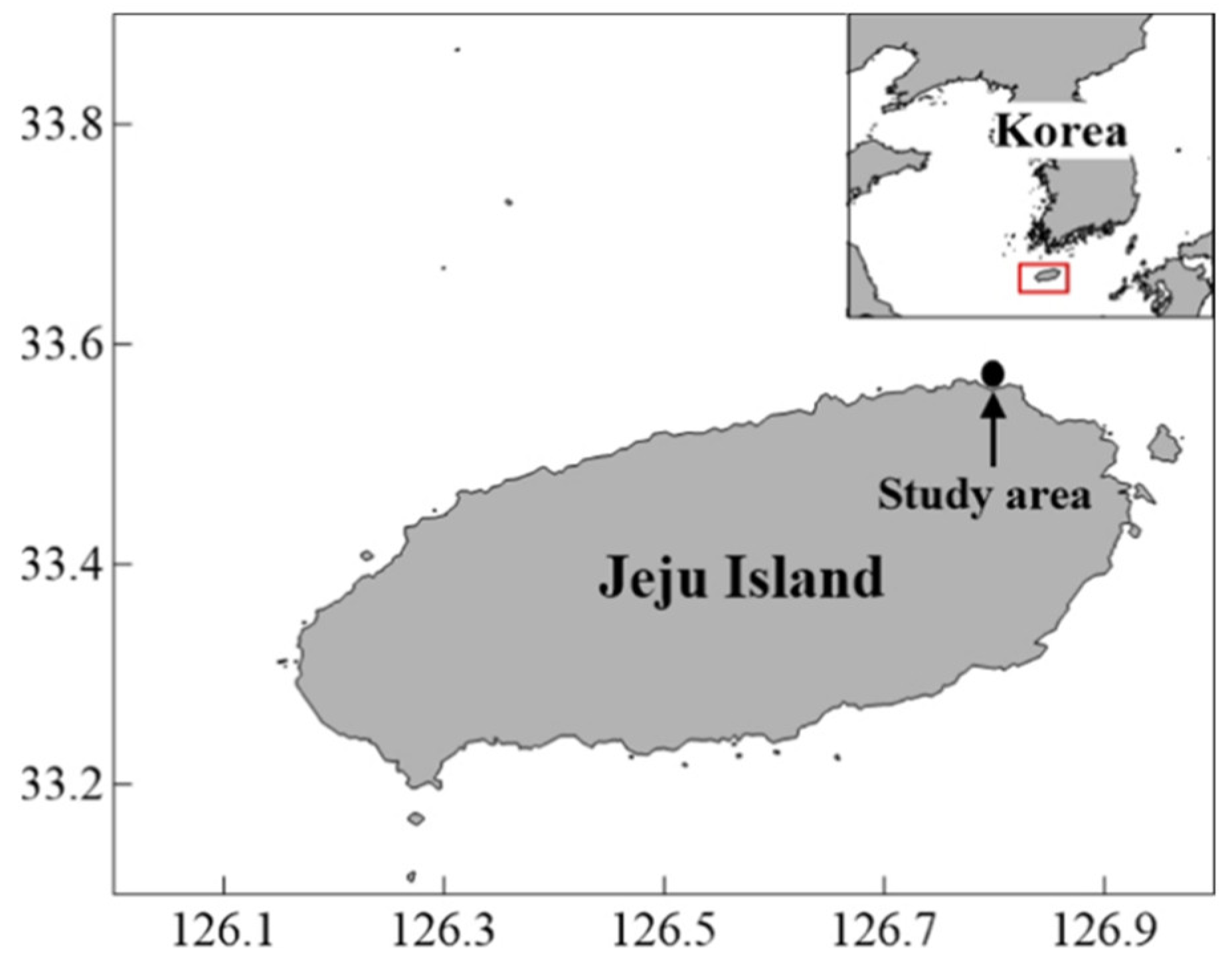
Figure 1. Map showing the study site.
Jmse 10 00851 g001
Figure 2. Images of the experimental setup. The O2 profiles were measured using an oxygen microsensor with control of the water temperature and irradiance in a constant-flow chamber.
Jmse 10 00851 g002
Figure 3. O2 variation with irradiance at each temperature (11 °C, 21 °C, 26 °C, and 31 °C). The error bars represent the standard error in triplicate reading of O2 microsensor.
Jmse 10 00851 g003
Figure 4. Net O2 production–irradiance relationships of Pneophyllum fragile at 11–31 °C. The solid lines represent the curve fit of the equation P(E) = Pmax[1 − exp(–αEd/Pmax)] + Rd, where Rd is the O2 respiration rate. Ek is the saturation irradiance, which was calculated as Ek = Pmax/α. Ec is the compensation irradiance, which is the irradiance at P(E) = 0.
Jmse 10 00851 g004
Figure 5. Photosynthesis characteristics–temperature relationships of Pneophyllum fragile.
Jmse 10 00851 g005
Table 1. Photosynthetic parameters of Pneophyllum fragile at 11–31 °C. Factors: Rd (respiration in the dark), Pmax (maximum photosynthesis capacity), Ec (compensation irradiance), and Ek (saturating irradiance).
Parameter/Temperature11 °C21 °C26 °C31 °C
Rd (mmol m−2 h−1)−0.56 ± 0.18−1.19 ± 0.37−1.35 ± 0.37−1.67 ± 0.26
Pmax (mmol m−2 h−1)4.01 ± 0.236.83 ± 0.5112.1 ± 1.4614.1 ± 1.16
Ec (μmol photon m−2 s−1)40.360.084.6104
Ek (μmol photon m−2 s−1)270312714833
Table 2. Photosynthesis rate, saturation irradiance, and optimal temperature measured for selected coralline algae from different regions. The values were normalized to mmol O2 m−2 h−1 for comparison. Ek is given in μmol photons m−2 s−1.
SpeciesRegionTemperature (°C)Reference
Phymatolithon lusitanicumPortugal24[15]
Lithophyllum yessoenseJapan15[36]
Lithophyllum yessoenseKorea20[24]
Hildenbrandia rubra25
Neogoniolithon sp. (rhodolith)Mexico30[14]
Amphiroa tribulus
Lithothamnion sp. (CCA)
Pneophyllum fragileJeju≥31This study
Publisher’s Note: MDPI stays neutral with regard to jurisdictional claims in published maps and institutional affiliations.