Article

Genome Sequencing of Streptomyces griseus SCSIO PteL053, the Producer of 2,2′-Bipyridine and Actinomycin Analogs, and Associated Biosynthetic Gene Cluster Analysis

1
CAS Key Laboratory of Tropical Marine Bio-Resources and Ecology, Guangdong Key Laboratory of Marine Materia Medica, RNAM Centre for Marine Microbiology, South China Sea Institute of Oceanology, Chinese Academy of Sciences, 164 West Xingang Road, Guangzhou 510301, China
2
Southern Marine Science and Engineering Guangdong Laboratory (Guangzhou), No. 1119, Haibin Rd., Nansha District, Guangzhou 511458, China
*
Author to whom correspondence should be addressed.
J. Mar. Sci. Eng. 2023, 11(2), 396; https://doi.org/10.3390/jmse11020396
Submission received: 27 December 2022 / Revised: 27 January 2023 / Accepted: 6 February 2023 / Published: 10 February 2023
(This article belongs to the Special Issue Advanced Studies in Marine Natural Products)

Abstract

:
Marine symbiotic actinomycetes play a key role in drug development and their ecological niches can influence a variety of natural product biosynthesis, providing potential defensive benefits. In this study, we report the whole-genome sequence analysis of marine gastropod mollusk Planaxis sp.-associated Streptomyces griseus SCSIO PteL053, which harbors 28 putative biosynthetic gene clusters (BGCs). Among them, two BGCs encoded by a hybrid non-ribosomal peptide (NRPS)/polyketide (PKS) synthetase and non-ribosomal peptide synthetase (NRPS) are responsible for the synthesis of the known therapeutic metabolites 2,2′-bipyridine and actinomycin analogs, respectively. Detailed bioinformatics analysis revealed the putative BGCs and the functions of the involved genes in the biosynthesis of the known compounds SF2738D (1), SF2738F (2), actinomycin D (3), and Actinomycin X (4). In the present study, complete-genome sequencing allowed us to rediscover known, clinically useful secondary metabolites in the newly isolated Streptomyces griseus SCSIO PteL053.

1. Introduction

Natural products (NPs) of microbial origin are the main reservoir of agricultural and therapeutic agents [1]. Over the past decade, numerous unique NPs were discovered in marine invertebrate-associated microorganisms, which suggests that actinomycetes, particularly Streptomyces sp., are a main source of novel NPs [2,3]. The understanding of interspecies interactions and their ecological contexts has created new opportunities for the discovery of natural products [4]. Actinomycetes build effective symbiotic relationships with marine invertebrates, which drives the synthesis of new classes of antibiotics and anticancer agents [5]. A substantial number of marine drugs, including trabectedin and eribulin, were isolated from microbial symbionts associated with marine animals. Recently, whole-genome sequencing analyses implied that most actinomycetes possess more than 20 biosynthetic gene clusters (BGCs). Genome-guided strategy is useful for the identification of genetic loci and their encoded gene products with new biosynthetic potential [6]. Recently, numerous NPs, including olimycin, mycemycin, and atratumycin, were yielded by a combination of gene sequencing, components, and a metabolic engineering approach by our group [7,8,9].
Bipyridine (2,2′-BP) is encoded by non-ribosomal peptide (NRPS)/polyketide synthetase (PKS), whereas actinomycin is biosynthesized by non-ribosomal peptide synthetase (NRPS). Bipyridine is a unique molecular scaffold composed of natural compounds from the actinobacteria caerulomycin and collismycin, whose 2,2′-BP core typically contains a di- or tri-alternative A ring and an unaltered B ring. The neuro-protective, anti-inflammatory, immunosuppressive, antibacterial, and antitumor activities of these compounds have been previously reported [10]. Actinomycin D and its analogs are an important family of chromopeptide lactone compounds with the same actinocin moiety but different amino acid residues on the pentapeptide lactone backbone. Many actinomycins exert strong antibacterial and antitumor action [11,12]. Among them, actinomycin D is a well-known drug and an FDA-approved chemotherapeutic agent against rare and difficult-to-treat cancers, namely gestational trophoblastic neoplasia, Wilms tumors in children, and rhabdomyosarcoma [12,13,14]. During our research on marine symbiotic actinomycetes, the Streptomyces griseus SCSIO PteL053 strain was isolated from a marine-derived gastropod mollusk Planaxis sp. The results of the bioinformatics analysis indicated the potential of the strain to produce bipyridine and actinomycin analogs. Here, we report (i) the whole-genome sequence of the marine invertebrate-associated strain SCSIO PteL053; (ii) the production of the known potential compounds bipyridine (2,2′-BP) and actinomycin (14) by 2,2′-Bipyridine and actinomycin BGCs; and (iii) two major known NP-encoded BGCs (termed, herein, cls and acd), which are confirmed by a bioinformatics-based approach, and the biosynthetic pathways for compounds 14.

2. Materials and Methods

2.1. General Methods

A 1260 infinity system (Agilent, Santa Clara, CA, USA) equipped with a Phenomenex Prodigy ODS (2) column (150 × 4.6 mm, 5 μm, Phenomenex Inc., Torrance, CA, USA) was used for HPLC-based analyses. A Primaide 1110 solvent delivery module equipped with a 1430 photodiode array detector (Hitachi, Tokyo, Japan) and a YMC-Pack ODS-A column (250 × 10 mm, 5 µm) was used for semi-preparative HPLC. A MaXis Q-TOF mass spectrometer (Bruker, Billerica, MA, USA) was used to acquire high-resolution mass spectral data. NMR spectra were recorded with an Avance-700 spectrometer (Bruker) at 700 MHz for 1H nuclei and 125 MHz for 13C nuclei. Chemical shifts (δ) are provided with reference to tetramethylsilane [12]. X-ray single-crystal data were obtained with an XtaLAB PRO MM007HF X-ray diffractometer with APEX II CCD by Cu Ka radiation. Silica gel mesh 100–200 was used for column chromatography (CC). All chemicals and solvents used in this study were analytical and chromatographic grade.

2.2. Sample Collection and Strain Identification

The SCSIO PteL053 isolate was obtained from Planaxis sp., an oceanic invertebrate collected in Daya Bay, Guangdong Province, China (33′22° Latitude N; 31′15° Longitude E) using actinomycete isolation agar media, 0.01% L-asparagine, 0.2% D-mannitol, 0.01% FeSO4·7H2O, 0.01% MgSO4·7H2O, 0.05% K2HPO4, 3.0% CaCO3, 3.0% sea salt, 1.8% agar, pH 7.0–7.2. The strain was further pure-cultured using the repeated streak-plate technique on a modified ISP2 agar plate (0.4% yeast extract, 0.4% glucose, 1% malt meal, 3.0% sea salt, 2% agar, pH 7.2–7.4) and kept in 15% (v/v) glycerol at −80 °C for subsequent study.

2.3. Whole-Genome Sequencing and In Silico Analysis

Complete-genome scanning and notation of strain SCSIO PteL053 were obtained using single-molecule real-time (SMRT) sequencing technology (PacBio) at Shanghai Majorbio Bio-Pharm Technology Co., Ltd., Shanghai, China. As a result, 296,471 filtered reads with a total data size of 3639599253 bp were generated. Afterward, they were assembled into the shortest contig using an ordered genome assembly operation (HGAP) [15]. The BGCs were analyzed using AntiSMASH (AntiSMASH 5.0, available at http://antismash.secondarymetabolites.org/ accessed on 22 September 2021) [15]. FramePlot (FramePlot 4.0 beta, available at http://nocardia.nih.go.jp/fp4/ accessed on 22 September 2021) was used to analyze the ORFs, whose functions were predicted using the online BLAST tool (http://blast.ncbi.nlm.nih.gov/ (accessed on 22 September 2021)).

2.4. Metabolic Profile of the Strain by LC-MS and Screening of Desired Compounds

Different production media were screened for the presence of bipyridine (2,2′-BP) and actinomycin analogs from the strain (Table S1). Initially, the strain was grown on an ISP2 agar medium. Subsequently, the 7-day-old mycelium and spore cultures were aseptically transferred into 50 mL of different media and incubated at 28 °C for 8 days with agitation at a rate of 180–200 rpm. Then, the whole cell culture was extracted with double the volume of 2-butanone, and the solvent phase was concentrated using the evaporation method. The concentrated crude extract was dissolved in methanol, and 30 µL of the sample was injected into the LC-HRESIMS analysis (a linear gradient from 10% to 100% MeCN in 30 min with a flow rate of 1.0 mL/min), with UV detection at 254 nm. The MS experiment was conducted in positive ion mode. The analytical conditions of the mass spectrometry were as follows: spray voltage, −3.50 kV; detector voltage, 1.65 kV; pressure of the TOF region, 1.6 × 10⁻⁴ Pa; pressure of the IT, 1.5 × 10⁻² Pa; rotary pump (RP) area vacuum, 70.0 Pa; drying gas pressure, 100.0 kPa; nebulizing gas (N2) flow, 1.5 L/min; curved desolvation line (CDL) collision energy, 50%; and q = 0.251; scan range, m/z 100–2000 for MS.

2.5. Scale Up and Isolation of Active Compounds

Seed culture was prepared by inoculating the mycelium of Streptomyces griseus SCSIO PteL053 into a 250 mL conical flask consisting of 50 mL of suitable AM3 production medium with an agitation rate of 250 rpm at 28 °C for 2 days. Afterward, 10% of the culture was transferred into 200 mL of AM3 medium, and a total of 30 L was kept at 28 °C for 7 days at an agitation rate of 200 rpm. Then, the cell debris and supernatant were separated by centrifugation at 4000× g for 20 min and further solvent extraction was carried out with acetone and 2-butanone. The different organic extracts were concentrated using a rotational vacuum concentrator and the residues were combined. Further, these samples were introduced to silica column chromatography (CC; length 50 cm and diameter 7 cm) and eluted with CHCl3-CH3OH ratios (100:0, 98:2, 96:4, 94:6, 92:8, 90:10, 80:20, 70:30, 60:40, 50:50, (v/v) each fraction 500 mL) to provide ten fractions (Fr. A1–Fr. A10). Fractions A1 and A2 were pooled and subjected to another silica CC with petroleum ether–ethyl acetate ratios (100:0, 90:10, 80:20, 70:30, 60:40, 50:50, 40:60, 30:70, 20:80, 10:90, and 100:0 (v/v) each fraction 250 mL) to obtain the fractions Fr. B1–Fr. B11. The fractions Fr. B4–Fr. B8 were pooled and further purified by HPLC (semi-preparative) and eluted with 10% MeCN, with a flow rate of 2.5 mL/min and UV detection at 254 nm, which yielded compound 1 (4.5 mg) and compound 2 (5 mg). Similarly, fractions A3 and A4 were mixed and, subsequently, subjected to semi-preparative HPLC using the previously described method to yield compound 3 (9 mg) and compound 4 (3 mg) [11].

2.6. X-ray Crystallography of Compounds 1 and 2

White prismatic crystals of compounds 1 and 2 were acquired in the petroleum ether–ethyl acetate (50:50) ratio. High-quality crystal was taken and recorded using an XtaLAB PRO MM007HF X-ray diffractometer with APEXII CCD by Cu Kα radiation. The crystals were mounted at 99.99 (10) K during the data archive. Crystal structures were decoded using the ShelXT program with the dual-solution method and the structure was refined using full-matrix least-squares difference Fourier techniques [16].

3. Results and Discussion

3.1. Isolation and Identification of Strain

The strain SCSIO PteL053 (Figure S1) was obtained from the marine-derived gastropod mollusk Planaxis sp., an oceanic invertebrate in the Daya Bay in the South China Sea, using an ISP2 agar medium augment with 3% sea salt. The 16S rDNA analysis showed that the newly isolated strain has a 99.73% sequence similarity to the strain Streptomyces griseus (Figure S2). The 16S rDNA region was derived from the whole-genome sequence data.

3.2. Genome Sequencing and Elucidation of Strain SCSIO PteL053

The complete-genome sequence data set plays an essential role in the identification of novel compounds and also provides information about secondary metabolites [17,18]. The bacterial genome consists of a linear chromosome with 8,040,759 base pairs and the absence of plasmid, with average GC contents of 71.60% (Figure 1 and Table 1). The genome contains 67 tRNA, 19 rRNA, and 7077 coding genes.
The AntiSMASH [18] analysis revealed that the genome of the Streptomyces griseus SCSIO PteL053 encodes 28 BGCs for the different secondary metabolites (Figure 1 and Table S2). Most of the gene clusters were distributed in the two sub-telomeric regions of the genome [19]. The 28 BGCs spanned 0.75 Mb or 10.71% of the total genome, including two Type III PKSs, seven NRPSs, two PKS-NRPSs, four terpenes, two siderophores, two bacteriocin, three hybrid BGCs, two lanthipeptides, lasso peptides, and other types of BGCs (Table S2). Among these, NRPS cluster 6 is responsible for the synthesis of actinomycin D and PKS-NRPS cluster 19, whose products are collismycins.

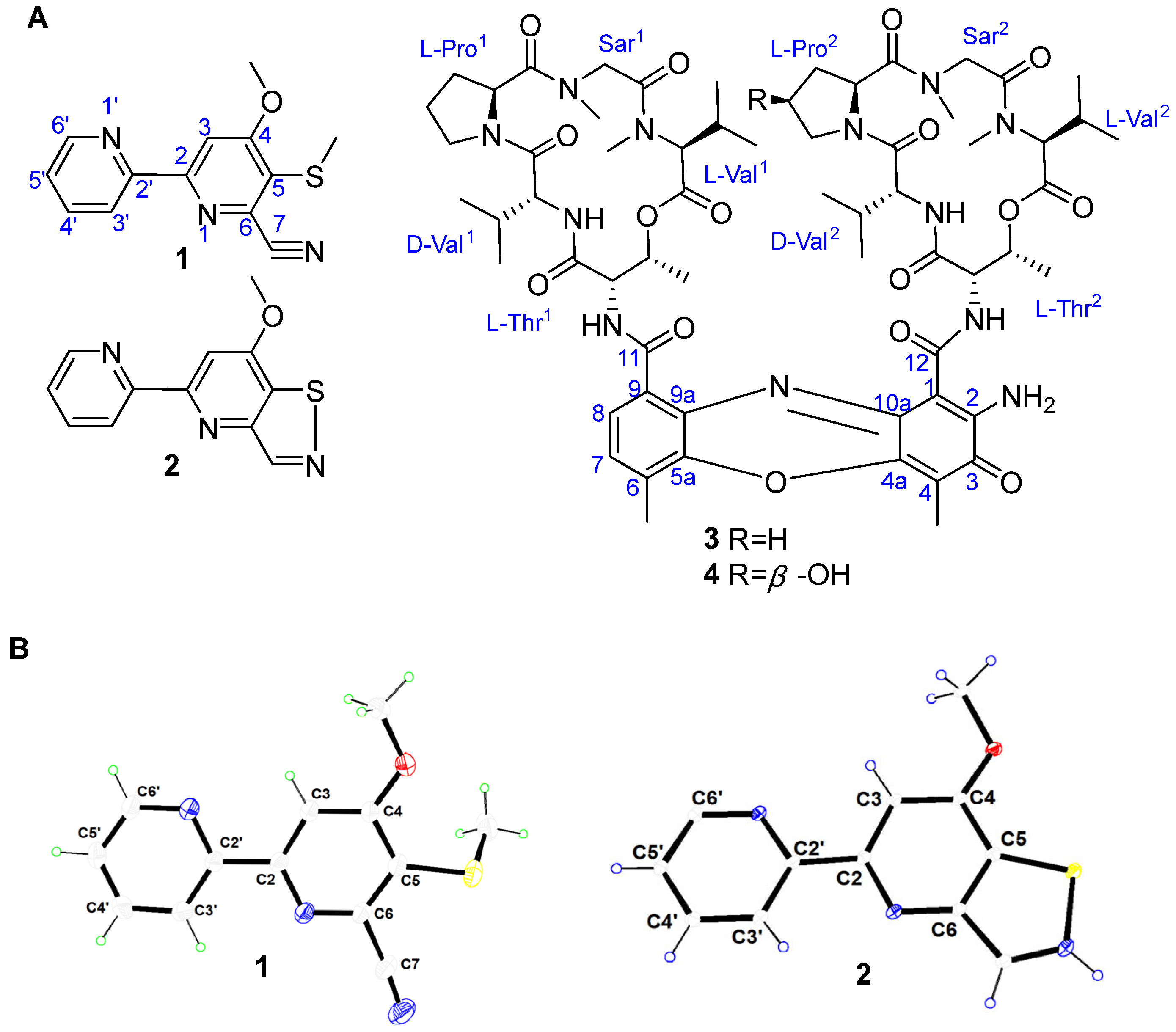
3.3. Production, Isolation, and Elucidation of 2,2′-BP and Actinomycin Analogs

The LC-HRESIMS analysis of the fermentation broth culture of the isolate indicated that the desired compounds were effectively produced under standard culture conditions in an AM3 production medium (Figure S3). In order to isolate and identify the compounds, large-scale fermentation was conducted. Repeating the extractions and concentrations provided a good yield of crude extract (30 g), which was used for purification to yield compounds 14 (Figure 2A).
Compound 1 has the molecular formula C13H11N3OS due to the HRESIMS data at m/z 258.0697 ([M + H]+, cald for 258.0696) and m/z 280.0515 ([M + Na]+, cald for 280.0515). The 1H spectrum showed two distinct downfield-shifted methyl proton signals at δH 2.53 (3H, s, 4-OMe) and δH 4.14 (3H, s, 5-SMe), corresponding to a thiomethyl and methoxy group in the structure, which was in accordance with the 13C shifts at δC 18.6 (4-OMe) and δC 58.2 (5-SMe). The four olefinic protons in the spin system at δH 8.42 (1H, d, J = 8.0 Hz, H-2′), 7.97 (1H, m, H-3′), 7.49 (1H, dd, J = 6.9, 4.7, H-4′), and 8.68 (1H, d, J = 4.7 Hz, H-5′) showed the existence of a 2-substituted pyridine ring. The appearance of a singlet proton at δH 8.22 (1H, s, H-2), together with the chemical shifts of eleven carbons downfield of the 13C spectrum, indicated the possibility of a pyridine. The compound was determined to be SF2738D by comparing its formula and the chemical shifts of the NMR spectroscopic data (Figures S4 and S5, Table S3) with those reported previously [20], which was further verified by the X-ray analysis of the crystals of the compound (Table S4).
The HRESIMS ion peaks at m/z 244.0545 ([M + H]+, calcd for 244.0539) and m/z 266.0360 ([M + Na]+, calcd for 266.0359) supported the molecular formula of C12H9N3OS for compound 2. The 1H and 13C NMR data were similar to compound 1, except for the absence of the thiomethyl group and a non-protoned carbon at C-7, the existence of an additional sp2 CH group at δH 9.16 (1H, s, H-7), and δC 158.2 (C-7). These examinations, together with the comparison of the 1H and 13C NMR spectra (Figures S6 and S7, Table S3) with those reported previously, allowed the identification of the compound as SF2738F [20]. The structure of the compound was also demonstrated using X-ray analysis of the crystals of the compound, which were obtained from the petroleum ether–ethyl acetate (50:50) ratio.
For compounds 3 and 4, the molecular formulas were identified as C62H86N12O16 and C62H86N12O17 due to the HRESIMS peaks at m/z 1255.6355 and 1269.6269. The 62 carbon signals of compounds 3 and 4 in the 13C NMR spectrum, combined with the UV absorptions at 256 nm and 442 nm, indicated the actinomycin skeleton of the compounds. The structures of the compounds were determined to be actinomycin D (3) and actinomycin X (4) from the comparison of the NMR data (Figures S8–S11, Table S4) with the compounds we isolated previously [21].

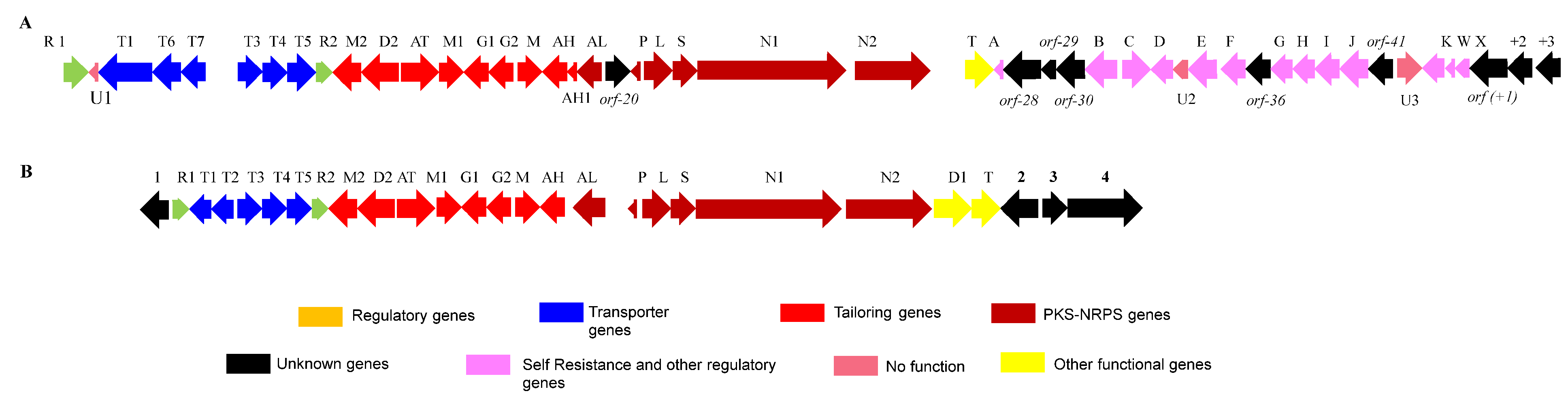
3.4. Bioinformatics Analysis of the Putative 2,2′-BP BGC

For further identification of the putative BGC for the biogenesis of SF2738D (1) and SF2738F (2), all 28 BGCs were screened. Genome cluster 19, named cls, revealed the combined PKS/NRPS gene product collismycin A, which showed an 81% gene sequence similarity to the already reported clm BGC from Streptomyces sp. CS40 [22]. The putative cls BGC contained 49 open reading frames (ORFs), among which, 41 were involved in SF2738D (1) and SF2738F (2) biosynthesis. The genetic organization of the cls BGC is shown in Figure 3 in which the different-colored genes indicate their proposed function shown in Table 2.
The putative cls BGC contained six core synthetic genes, clsAL, clsP, clsL, clsS, clsN1, and clsN2, which were conserved among 2,2′-BP-producing actinomycete species [22]. The biosynthesis of the compounds begins with the production of picolinic acid (PA) from l-lysine with the help of the clsL, clsS, and clsAL genes. Among them, the biosynthetic genes clsL and clsS encode l-lysine 2 aminotransferase and sarcosine oxidase enzymes, respectively.
ClsP was identical to a stand-alone acyl carrier protein (ACP) of collismycin A biosynthesis [22]. Generally, clsp is encoded within the cluster but in the case of actinomycin biosynthesis, it acts as a stand-alone process like AcmD [23]. In this strain, clsP was determined to encode ACP incriminate PA from ClsAL to ClsN1 in the biosynthetic pathway. Gene clsN1 contained seven domains, which were assembled into two modules: a PKS extender module consisting of ketosynthase (KS), acyltransferase (AT), and ACP domains and an NRPS module containing condensation or cyclization (Cy), adenylation (A), a peptidyl carrier protein (PCP), and epimerization (E) domains. Gene clsN2 encoded single-modular NRPS, which consisted of Cy-A-PCP domains. Genes clsN1 and clsN2 were very similar to the previously reported clmN1 and clmN2 in the collismycin A BGC [22]. These gene products may be used as a PA for the formation of ring B, a 2,2′-bipyridyl scaffold, for the biosynthesis of SF2738D and SF2738F. The stand-alone putative type II thioesterase encoded by the clsT gene was identical to the reported clmT of the collismycin A biosynthesis. This gene acted by releasing imperfectly added amino acids from the PCP domain of the NRPS protein or by detaching the acetyl species from the ACP domain to restore the free thiol groups [24]. Recently, a trans-acting flavoprotein-related assembly channel for 2,2′-bipyridine ring formation and differentiation was reported for dethiolated (CAE) and thiolated (COL) compounds [25]. Based on the deduced gene function correlations, we suggest that ClsT may participate in the release of the growing polyketide/non-ribosomal peptide chain from ClsN2.
The upstream region of the core biosynthetic gene begins from clsM2 to clsAH1, which is a total of nine genes. Among these, clsAH-encoded aminohydrolase was identical to the ClmAH gene from Streptomyces sp.CS40; ClmAH is mandatory for detaching the extra leucine residue incorporated during biosynthesis before the formation of collismycin C, which has a carboxylic group at the C6 position. The important role of ClmAH has been confirmed by mutation analysis [22]. ClmAH could be involved in the removal of the leucine residue before the formation of the oxime group.
Another set of upstream-region genes includes clsG1 and clsG2, which are encoded by two dehydrogenase components that are identical to ClmG1 and ClmG2 of collismycin A biosynthesis from Streptomyces sp.CS40. Similarly, ClmG1 and ClmG2 showed a high sequence homology of GriC and GriD involved in carboxylic acid reduction reactions in grixazone biosynthesis [26,27]. Based on this gene function correlation, the homology of ClsG1 and ClsG2 indicated that they were catalyzed by similar reactions during the formation of collismycin analogs. The determined product of the clsAT gene-encoded putative aminotransferase was identical to ClmAT of the reported collismycin biosynthesis from Streptomyces sp.CS40. The ClmAT domain comes under the class III family type of protein, which is associated with the pyridoxal-phosphate-mediated decarboxylation or transamination reaction of cationic amino acid and its derivatives [22,27]. Accordingly, it is clear that ClsAT is mainly involved in oxime group formation in the biosynthesis of collismycins. The clsD2 encodes dehydrogenase homology to ClmD2 in collismycin A BGC. ClsD2 is an essential component in the oxidization of the hydroxymethyl group in position 5 of collismycin analogs to the aldehyde group, which is the complex substrate for the action of aminotransferase by ClsAT [27]. This protein is hopefully involved in the oxime formation in the crucial step of collismycin biosynthesis and its function confers the recovery of the spontaneous generation of shunt products in the main pathway [27]. Gene clsM encodes oxidoreductase with a high-level resemblance to ClmM, a putative-FAD-dependent oxidoreductase, and monooxygenase [22]. ClmM controls the UbiH domain in 2-polyprenyl-6-methoxyphenol hydroxylases and also FAD-binding oxidoreductases, which showed that clsM encodes putative monooxygenase but is not involved in the 2,2′-bipyridyl structure [28]. ClsM is important for the subsequent steps of collismycin construction, perhaps by catalyzing the formation of the oxime group. Gene clsM1 encodes S-methyltransferase and clsM2 encodes O-methyltransferase. Based on the structures of SF2738D and SF2738F, clsM2 is associated with the generation of the methoxy group in position C-4 of collismycin, and clsM1 is involved in the conversion of SF2738D from intermediate/SF2738F. The determined product of clsAT gene-encoded putative aminotransferase showed a 98% similarity with ClmAT of the reported collismycin biosynthesis from Streptomyces sp. CS40. The ClmAT domain comes under the class III family type of protein, which is associated with the pyridoxal-phosphate-mediated decarboxylation or transamination process of cationic amino acid and its derivatives [22,27]. Accordingly, it is clear that ClsAT mainly participates in oxime group formation in the biosynthesis of collismycin compounds 1 and 2. These upstream genes are involved in tailoring the enzymatic reactions for SF2738D and SF2738F biosynthesis. Gene clsAH1 encodes putative amidohydrolase. The amidohydrolase is included in the Metallo-dependent hydrolase family of proteins, which participates in the hydrolysis of the C-N bond (peptic and non-peptic) through the nucleophilic attack of a hydroxyl group [22,29]. According to the proposed pathway, the action of the enzyme is involved in the dehydration of the aldoxime intermediate from collismycin A, resulting in the formation of SF2738D and SF2738F. Nitrile groups, as part of the structure of natural products, have been studied [30,31] and a route for the nitrile group formation has been proposed [22,32].
The sets of genes that reside on the left-hand side of the cls gene cluster include two regulatory genes and six transporter genes. The detailed annotation indicated that two genes, clsR1 and clsR2, which encode the putative TetR and LuxR family transcriptional regulators, were identical to ClmR1 and ClmR2 in collismycin A biosynthesis from Streptomyces sp. CS40 [22,33]. The putative gene products also showed strong similarities to CrmR1 and CrmR2 from Actinoalloteichus cyanogriseus WH1-2216-6 [28,34]. TetR regulators act as a transcriptional repressor, which regulates drug metabolism, cellular metabolic activity, and collismycin biosynthesis through the inhibition of biosynthetic genes by clsR2 repression. Similarly, a homolog of the clmR1 protein encoded a transcriptional regulator of the TetR protein, which has been reported previously [22]. ClsR2 is involved in the positive regulation of collismycin product formation [33]. The putative transporter genes clsT1, clsT3, clsT4, and clsT5 showed strong similarity with consecutive genes from Streptomyces sp. CS40 [22]. Based on the correlation, the gene products are co-transcribed and interact to form whole ABC cassettes for a single collismycin transport system. In addition, the two genes clsT6 and clsT7 both encoded ABC transporter ATP-binding domains; their determined gene products have been previously identified as membrane-associated proteins (MdlB) in Pseudomonas putida [35]. The cls BGC also consists of additional genes that reside throughout the gene cluster and encode unrelated proteins, which are hypothetically supported in collismycin biosynthesis.
Based on the available information and determined functions of genes in the cls BGC, we propose putative biosynthetic pathways for SF2738D (1) and SF2738F (2) (Figure 4). Three different stages are involved in the assembly of SF2738D (1) and SF2738F (2). In the first phase, skeleton assembly is initiated by biosynthesis and the activation of PA from a precursor, l-lysine, with the assistance of three enzymes ClsL, ClsS, and ClsAL. Afterward, the activated picolinic CoA is supplied to the hybrid PKS/NRPS system by a stand-alone ClsP. Therefore, collismycin’s first pyridine ring is formed. In the second phase, the hybrid PKS/NRPS system (ClsN1 and ClsN2) is associated with the incorporation of a malonate unit, cysteine, and extra leucine residue to provide 2,2′-BP-l-leucine. The N-acetyl group of cysteine is ultimately attached to PA via the carbonyl group to make C2-N1 bonding, and its intermediate undergoes a further intramolecular cyclization reaction to provide a seven-membered sulfur-containing heterocycle ring. Later, a six-membered intermediate is formed by the release of the SH group via a subsequent rearrangement process for the biosynthesis of the skeleton structure. A similar type of reaction has been reported for the biosynthesis of caerulomycins and collismycins [28]. In the end phase, the leucine residue may be eliminated by ClsAH, and then the reduction of the carboxylic group to an aldehyde group by ClsG1 and ClsG2. The oxime group formation is catalyzed by ClsAT and the final methylation of the hydroxyl group at the C4 by ClsM2 to yield collismycin A [22,27]. Collismycin A is catalyzed by ClsAH1 and ClsM1 to obtain the final products SF2738D (1) and SF2738F (2).

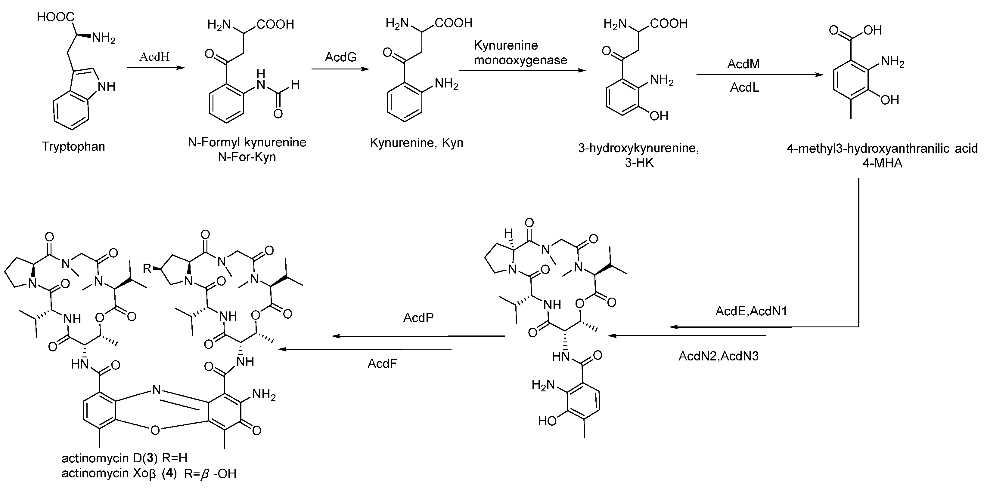
3.5. Bioinformatics Analysis of the Putative Actinomycin BGC

The complete genome of S. griseus SCSIO PteL053 was used to locate the entire BGC coding for potent antitumor actinomycin biosynthesis by computational analysis. Genomic DNA with a size of 50 kb was predicated as a putative actinomycin BGC (termed, herein, acd). Among these data, 20 open reading frames (ORFs) were proposed to be involved in the biosynthesis of actinomycin. The genetic organization of the strain was compared with known actinomycin producers [11,16,36,37] (Figure 5) and the determined gene product functions were noted (Table 3). They showed a strong similarity to homologs of previously reported actinomycin BGC (acd) from Streptomyces chrysomallus [16].
The acd cluster contained five consecutive NRPS genes (acdD, acdE, acdN1, acdN2, and acdN3), which are the homologs of acnR, acnD, acnA, acnB, and acnC in S. chrysomallus responsible for the synthesis of the 4-MHA-pentapeptide halves for actinomycin. This pentapeptide was synthesized from the precursor unit, a 4-methyl-3-hydroxy-anthranilic acid (4-MHA), along with five different amino acids (l-threonine, d-valine, l-proline, sarcosine, and l-methylated valine). Similarly, the action of five consecutive NRPS genes has been reported in the biological synthesis of actinomycin D from mangrove-derived Streptomyces costaricanus SCSIO ZS0073 [11]. The AcdD protein is homologous to the MbtH protein, which is similar to the protein from the Mycobacterium tuberculosis variant bovis [38]. It is found in many NRPS gene clusters and is involved in adenylation reactions [39,40,41]. The acdN1 gene encoded the adenylation protein and the acdE gene encoded the 4-MHA carrier protein. The acdN2 and acdN3 genes encoded the non-ribosomal peptide synthetase enzyme, which was indicated in the peptide chain assembly and release process from the NRPS system. In the downstream of the NRPS, there were four other consecutive genes, acdG, acdH, acdL, and acdM, which had high similarity with enzymes involved in the biosynthesis of 4-MHA. These gene homologs have been shown to play a role in the 4-MHA biosynthesis process in the actinomycin-producing strain S. antibioticus IMRU3720 [16]. The acdF gene was predicted to be involved in the dimerization of actinomycin halves during actinomycin synthesis according to recent reports [11].
A group of genes, including acdO, acdR, acdQ, acdT1, acdT2, and acdT3, located at the end of the right-arm cluster, had regulatory and protective roles. Within the acd BGC, the acdO gene encoded the LmbU regulatory protein. The LmbU gene was previously shown to be involved in lincomycin biosynthesis in S. lincolnensis 78-11 [42]. In addition, the inactivation of the homologous acnO gene in a mutant strain of S. costaricanus SCSIO ZS0073 showed a complete shutdown of actinomycin D production, which demonstrates the importance of the positive regulatory role of the gene [11]. The acdR gene encoded the TetR family of proteins, which play a transcriptional activator and repressor role to regulate the production of secondary metabolites [43,44]. As previously indicated, the trdK gene was found to be highly similar to the TetR protein associated with tirandamycin synthesis in Streptomyces sp. SCSIO 1666 and its inactivation led to an increase in tirandamycin titers [45]. Apart from this, the essential role of acnR in actinomycin D biosynthesis in S. costaricanus SCSIO ZS0073 has been validated by gene inactivation studies [11]. Apart from these genes in the acd cluster, we found three putative transporter genes, namely acdT1, acdT2, and acdT3. Based on the analysis, the acdT1-encoded ATP-binding cassette (ABC) transporter was found to be similar to the drrA gene from daunorubicin-producing Streptomyces peuceticus [46]. Likewise, acnT2, which encoded an ABC-type 2 transporter permease, was found to be similar to the drrB gene from Streptomyces peuceticus [46,47]. acnT3 encoded a 753 amino acid DNA-binding domain that was similar to the UvrA-like protein found in E. coli [48]. The inactivation of three genes, acnT1, acnT2, and acnT3, in the acn BGC resulted in no significant impact on actinomycin D biosynthesis in S. costaricanus SCSIO ZS0073 [11]. Accordingly, we suggest that these genes are mainly involved in the transport of actinomycin analogs to the environment but not in actinomycin biosynthesis. The gene acnQ located upstream of the transporter genes was predicted to encode siderophore-interacting proteins to regulate the intracellular concentrations of metabolites. Hence, the inactivation of acnQ had no impact on actinomycin D production, which has recently been reported in S. costaricanus SCSIO ZS0073 [11]. Based on the available data, we suggest a putative biosynthetic pathway leading to the production of actinomycin D (3) and actinomycin X (4) in S. griseus SCSIO PteL053 (Figure 6). The acd BGC also consisted of a few additional genes throughout the gene cluster that encoded unrelated proteins, which are either directly or hypothetically involved in biosynthesis.

4. Conclusions

In the present study, we attained the whole genome sequence of S. griseus SCSIO PteL053, whose average GC content was 71.6%. The marine invertebrate-associated strain harbors 28 putative BGCs. Here, we characterized two putative BGCs, which were predicted to generate pharmaceutically important 2,2′-BP and actinomycin analogs. The study also provided good evidence to show that the isolated strain has a close association with marine invertebrates and can protect these soft-bodied organisms from bacterial infection through the production of prevalent antibiosis metabolites. These interactions are driven by the biosynthesis of specialized secondary metabolites, which exhibit biological activity and contribute to the chemical diversity in highly diverse environments [5,49]. Understanding these mutualistic associations and the ecology of NPs can provide more insights into the future direction of NP discovery. Furthermore, this work highlights the importance of genome mining techniques in identifying BGCs and potential lead metabolites for drug discovery from wild strains.

Supplementary Materials

The following supporting information can be downloaded at https://www.mdpi.com/article/10.3390/jmse11020396/s1. Figure S1: Aerial view of SCSIO PteL053; Figure S2: Phylogenetic tree view of Streptomyces sp. SCSIO-PteL053 based on 16S rDNA gene sequence by the Maximum Likelihood method; Figure S3: LC-HRESIMS profile of AM3 fermentation broth from S. griseus SCSIOPteL053; Figures S4–S11: 1D NMR spectra of compounds 14; Table S1: List of production media used in this study; Table S2: AntiSMASH-predicted BGCs for Streptomyces sp. SCSIO PteL053; Table S3: 1H and 13C (700 MHz and 175 MHz) NMR data of compounds 1 and 2 in CD3OD; Table S4: 1H and 13C (700 MHz and 175 MHz) NMR data of compounds 34 in DMSO-d6.

Author Contributions

G.G. performed the experiments and wrote the manuscript. Z.Z. and Z.Y. analyzed the bioinformatics data. X.Z., J.M. and P.S.K. helped to analyze the XRD and NMR data. J.J. and C.S. supervised the work, analyzed the data, and refined the manuscript. All authors have read and agreed to the published version of the manuscript.

Funding

This research was funded by the National Natural Science Foundation of China (22177117, 82022067), the Key Science and Technology Project of Hainan Province (ZDKJ202018), and the Key Special Project for the Introduced Talents Team of the Southern Marine Science and Engineering Guangdong Laboratory (Guangzhou) (GML2019ZD0406).

Institutional Review Board Statement

Not applicable.

Informed Consent Statement

Not applicable.

Data Availability Statement

The entire genome sequence of the strain was deposited in Genbank with accession number CP059136. The crystal data of compounds 1 and 2 were deposited in the Cambridge Crystallographic Data center with the deposition numbers CCDC 2035511 and CCDC 2035512, respectively. The original contributions presented in the study are included in the article/Supplementary Materials and further inquiries can be directed to the corresponding author.

Conflicts of Interest

The authors declare no conflict of interest.

References

  1. Berdy, J. Bioactive microbial metabolites. J. Antibiot. 2005, 58, 1–26. [Google Scholar] [CrossRef] [PubMed]
  2. Carroll, A.R.; Copp, B.R.; Davis, R.A.; Keyzers, R.A.; Prinsep, M.R. Marine natural products. Nat. Prod. Rep. 2019, 36, 122–173. [Google Scholar] [CrossRef] [PubMed]
  3. Kumar, P.S.; Ling, C.Y.; Zhou, Z.B.; Dong, Y.L.; Sun, C.L.; Song, X.Y.; Wong, N.K.; Ju, J.H. Chemical Diversity of Metabolites and Antibacterial Potential of Actinomycetes Associated with Marine Invertebrates from Intertidal Regions of Day Bay and Nansha Islands. Microbiology 2020, 89, 483–492. [Google Scholar] [CrossRef]
  4. Vander Meij, A.; Worsley, S.F.; Hutchings, M.I.; Van Wwzel, G.P. Chemical ecology of antibiotic production by actinomycetes. FEMS Microbiol. Rev. 2017, 41, 392–416. [Google Scholar] [CrossRef] [PubMed]
  5. Gulder, T.A.M.; Moore, B.S. Chasing the treasures of the sea-bacterial marine natural products. Curr. Opin. Microbiol. 2009, 12, 252–260. [Google Scholar] [CrossRef]
  6. Zazopoulos, E.; Huang, K.; Staffa, A.; Liu, W.; Bachmann, B.O.; Nonaka, K.; Ahlert, J.; Thorson, J.S.; Shen, B.; Farnet, C.M. A genomics-guided approach for discovering and expressing cryptic metabolic pathways. Nat. Biotechnol. 2003, 21, 187–190. [Google Scholar] [CrossRef]
  7. Sun, C.; Zhang, C.; Qin, X.; Wei, X.; Liu, Q.; Ju, J. Genome mining of Streptomyces olivaceus SCSIO T05: Discovery of olimycins A and B and assignment of absolute configurations. Tetrahedron 2018, 74, 199–203. [Google Scholar] [CrossRef]
  8. Sun, C.; Yang, Z.; Zhang, C.; Liu, Z.; He, J.; Liu, Q.; Zhang, T.; Ju, J.; Ma, J. Genome Mining of Streptomyces atratus SCSIO ZH16: Discovery of Atratumycin and Identification of Its Biosynthetic Gene cluster. Org. Lett. 2019, 21, 1453–1457. [Google Scholar] [CrossRef]
  9. Zhang, C.; Yang, Z.; Qin, X.; Ma, J.; Sun, C.; Huang, H.; Li, Q.; Ju, J. Genome Mining for Mycemycin: Discovery and Elucidation of related Methylation and Chlorination Biosynthetic Chemistries. Org. Lett. 2018, 20, 7633–7636. [Google Scholar] [CrossRef]
  10. Chen, D.; Zhao, Q.; Liu, W. Discovery of caerulomycin/ collismycin-type 2,2′-bipyridine natural products in the genomic era. J. Ind. Microbiol. Biotechnol. 2019, 46, 459–468. [Google Scholar] [CrossRef]
  11. Liu, M.; Jia, Y.; Xie, Y.; Zhang, C.; Ma, J.; Sun, C.; Ju, J. Identification of the actinomycin D Biosynthetic Pathway from Marine-Derived Streptomyces costaricanus SCSIO ZS0073. Mar. Drugs 2019, 17, 240. [Google Scholar] [CrossRef] [PubMed]
  12. Yao, Z.; Sun, C.; Xia, Y.; Wang, F.; Fu, L.; Ma, J.; Li, Q.; Ju, J. Mutasynthesis of Antibacterial Halogenated Actinomycin Analogues. J. Nat. Prod. 2021, 84, 2217–2225. [Google Scholar] [CrossRef] [PubMed]
  13. Farber, S.; D’Angio, G.; Evans, A.; Mitus, A. Clinical studies of actinomycin D with special reference to Wilms tumor in children. J. Urol. 2002, 168, 2560–2562. [Google Scholar] [CrossRef]
  14. Mondick, J.T.; Gibiansky, L.; Gastonguay, M.R.; Skolnik, J.M.; Cole, M.; Veal, G.J.; Boddy, A.V.; Adamson, P.C.; Barrett, J.S. Population pharmacokinetic investigation of actinomycin D in children and young adults. J. Clin. Pharmacol. 2008, 48, 35–42. [Google Scholar] [CrossRef] [PubMed]
  15. Blin, K.; Shaw, S.; Steinke, K.; Villebro, R.; Ziemert, N.; Lee, S.Y.; Medema, M.H.; Weber, T. AntiSMASH 5.0: Updates to the secondary metabolite genome mining pipeline. Nucleic Acids. Res. 2019, 47, W81–W87. [Google Scholar] [CrossRef]
  16. Keller, U.; Lang, M.; Crnovcic, I.; Pfening, F.; Schauwecker, F. The actinomycin biosynthetic gene cluster of Streptomyces chrysomallus: Agenetic hall of mirrors for synthesis of a molecule with mirror symmetry. J. Bacteriol. 2010, 192, 2583–2595. [Google Scholar] [CrossRef] [PubMed]
  17. Chin, C.S.; Alexander, D.H.; Marks, P.; Klammer, A.A.; Drake, J.; Heiner, C.; Clum, A.; Copeland, A.; Huddleston, J.; Eichler, E.E.; et al. Nonhybrid, finished microbial genome assemblies from long-read SMRT sequencing data. Nat. Methods 2013, 10, 563–569. [Google Scholar] [CrossRef] [PubMed]
  18. Li, Y.; Zhang, C.; Ju, J.; Ma, J. Genome sequencing of Streptomycsatratus SCSIO ZH16 and activation, production of nocardamine via metabolic engineering. Front. Microbiol. 2018, 9, 1269. [Google Scholar] [CrossRef]
  19. Low, Z.J.; Pang, L.M.; Ding, Y.; Cheang, Q.W.; Le mai Hoang, K.; Thitran, H.; Li, J.; Liu, X.W.; Kanagasundaram, Y.; Yang, L.; et al. Identification of a biosynthetic gene cluster for the polyene macrolactam sceliphrolactam in a Streptomyces strain isolated from mangrove sediment. Sci. Rep. 2018, 8, 1594. [Google Scholar] [CrossRef]
  20. Lee, J.H.; Kim, E.; Choi, H.; Lee, J. Collismycin C from the Micronesian marine Bacterium Streptomyces sp. MC025 Inhibits Staphylococcus aureus Biofilm Formation. Mar. Drugs 2017, 15, 387. [Google Scholar] [CrossRef] [Green Version]
  21. Song, X.; Jiang, X.; Sun, J.; Zhang, C.; Zhang, Y.; Lu, C.; Ju, J. Antibacterial secondary metabolites produced by mangrove-derived actinomycete Streptomyces costaricanus SCSIO ZS0073. Nat. Prod. Res. Dev. 2017, 29, 410–414. [Google Scholar]
  22. Garcia, I.; Vior, N.M.; Brana, A.F.; Gonzalez-sabin, J.; Rohr, J.; Moris, F.; Mendez, C.; Salas, J.A. Elucidating the biosynthetic pathway for the polyketide-nonribosomal peptide Collismycin A: Mechanism for formation of the 2,2′-bipyridyl ring. Chem. Biol. 2012, 19, 399–413. [Google Scholar] [CrossRef] [PubMed]
  23. Pfennig, F.; Schauwecker, F.; Keller, U. Molecular characterization of the genes of actinomycin synthatase I and of a 4-methyl-3-hydroxyanthranilic acid carrier protein involved in the assembly of the acylpeptide chain of actinomycin in Streptomyces. J. Biol. Chem. 1999, 274, 12508–12516. [Google Scholar] [CrossRef] [PubMed]
  24. Schwarzer, D.; Mootz, H.D.; Linne, U.; Marahiel, M.A. Regeneration of misprimednonribosomal peptide synthetases by type II thioesterases. Proc. Natl. Acad. Sci. USA 2002, 99, 14083–14088. [Google Scholar] [CrossRef] [PubMed]
  25. Pang, B.; Liao, R.; Tang, Z.; Guo, S.; Wu, Z.; Liu, W. Caerulomycin and collismycin antibiotics share a trans-acting flavoprotein-dependent assembly line for 2.2′-bipyridine formation. Nat. Commun. 2021, 12, 3124. [Google Scholar] [CrossRef]
  26. Suzuki, H.; Ohnishi, Y.; Horinouchi, S. GriC and GriD constitute a carboxylic acid reductase involved in grixazone biosynthesis in Streptomyces griseus. J. Antibiot. 2007, 60, 380–387. [Google Scholar] [CrossRef]
  27. Garcia, I.; Vior, N.M.; Gonzalez-sabin, J.; Brana, A.F.; Rohr, J.; Moris, F.; Mendez, C.; Salas, J.A. Engineering the biosynthesis of the polyketide-nonribosomal peptide Collismycin A for generation of analogs with neuroprotective activity. Chem. Biol. 2013, 20, 1022–1032. [Google Scholar] [CrossRef]
  28. Qu, X.; Pang, B.; Zhang, Z.; Chen, M.; Wu, Z.; Zhao, Q.; Zhang, Q.; Wang, Y.; Liu, Y.; Liu, W. Caerulomycins and collismycins share a common paradigm for 2,2′ bipyridine biosynthesis via an unusual hybrid polyketide-peptide assembly logic. J. Am. Chem. Soc. 2012, 134, 9038–9041. [Google Scholar] [CrossRef]
  29. Holm, L.; Sander, C. An evolutionary treasure: Unification of a broad set of amidohydrolases related to urease. Proteins 1997, 28, 72–82. [Google Scholar] [CrossRef]
  30. Fleming, F.F. Nitrile- containing natural products. Nat. Prod. Rep. 1999, 16, 597–606. [Google Scholar] [CrossRef]
  31. Bezerragomes, P.; Nett, M.; Dahse, H.M.; Sattler, I.; Martin, K.; Hertweck, C. Bezerramycins A-C, antiproliferative ohenoxazinones from Streptomyces griseus featuring carboxy, Carboxamide or nitrile substituents. Eur. J. Org. Chem. 2010, 2, 231–235. [Google Scholar]
  32. Olano, C.; Moss, S.J.; Brana, A.F.; Sheridan, R.M.; Math, V.; Weston, A.J.; Mendez, C.; Leadlay, P.F.; Wilkinson, B.; Salas, J.A. Biosynthesis of the angiogenesis inhibitor borrelidin by Streptomyces parvulus Tu 4055: Insights into nitrile formation. Mol. Microbiol. 2004, 52, 1745–1756. [Google Scholar] [CrossRef] [PubMed]
  33. Vior, N.M.; Olano, C.; Garcia, I.; Mendez, C.; Salas, J.A. Collismycin A biosynthesis in Streptomyces sp. CS40 is regulated by iron levels through two pathway- specific regulators. Microbiology 2014, 160, 467–478. [Google Scholar] [CrossRef] [PubMed]
  34. Martinez, A.; Egea, P.U.; Medina, M.; Garcia, E.; Perez, J.; Fernandez, R.I.; Medarde, A.; Canedo, L.M.; Romero, F.; Castro, A.; et al. Use of Collismycin and Derivatives There of as Oxidative Stress Inhibitors. U.S. Patent WO2007017146A3, 26 July 2007. [Google Scholar]
  35. Xu, Y.; Mitra, B. A highly active, soluble mutant of the membrane associated (S)- mandelate degydrogenase from Pseudomonas putida. Biochemistry 1999, 38, 12367–12376. [Google Scholar] [CrossRef] [PubMed]
  36. Crnovcic, I.; Ruckert, C.; Semsary, S.; Lang, M.; Kalinowski, M.; Keller, U. Genetic interrelations in the actinomycin biosynthetic gene clusters of Streptomyces antibioticus IMRU 3720 and Streptomyces chrysomallus ATCC11523, producers of actinomycin X and actinomycin C. Adv. Appl. Bioinformatics Chem. 2017, 10, 29–46. [Google Scholar] [CrossRef] [PubMed]
  37. Peng, Q.; Gao, G.; Lu, J.; Long, Q.; Chen, X.; Zhang, F.; Xu, M.; Liu, K.; Wang, Y.; Deng, Z.; et al. Engineered Streptomyces lividans strains for optimal identification and expression of cryptic biosynthetic gene clusters. Front. Microbiol. 2018, 9, 3042. [Google Scholar] [CrossRef]
  38. Garnier, T.; Eiglmeier, K.; Camus, J.C.; Medina, N.; Mansoor, H.; Pryor, M.; Duthoy, S.; Grondin, S.; Lacroix, C.; Monsempe, C.; et al. The complete genome sequence of Mycobacterium bovis. Proc. Natl. Acad. Sci. USA 2003, 100, 7877–7882. [Google Scholar] [CrossRef]
  39. Baltz, R.H. Function MbtH homologs in nonribosomal peptide biosynthesis and applications in secondary metabolite discovery. J. Ind. Microbiol. Biotechnol. 2011, 38, 1747–1760. [Google Scholar] [CrossRef]
  40. Felnage, E.A.; Barkei, J.J.; Park, H.; Podevels, A.H.; McMahon, M.D.; Drott, D.W.; Thomas, M.G. MbtH-like proteins as integral components of bacterial nonribosomal peptide synthetases. Biochemistry 2010, 49, 8815–8817. [Google Scholar] [CrossRef]
  41. Imker, H.J.; Krahn, D.; Clerc, J.; Kaiser, M.; Walsh, C.T. N-Acylation during glidobactin biosynthesis by the tridomainnonribosomal peptide synthetase module GlbF. Chem. Biol. 2010, 17, 1077–1083. [Google Scholar] [CrossRef]
  42. Peschke, U.; Schmidt, H.; Zhang, H.Z.; Pieperserg, W. Molecular characterization of the lincomycin-production gene cluster of Strptomyceslincolnensis 78-11. Mol. Microbiol. 1995, 16, 1137–1156. [Google Scholar] [CrossRef] [PubMed]
  43. Ramos, J.L.; Martinez-Bueno, M.; Molina-Henares, A.J.; Teran, W.; Watanabe, K.; Zhang, X.; Gallegos, M.T.; Brennan, R.; Tobes, R. The tetR family of transcriptional repressors. Microbiol. Mol. Biol. Rev. 2005, 69, 326–356. [Google Scholar] [CrossRef] [Green Version]
  44. Bassler, B.L.; Losick, R. Bacterially speaking. Cell 2006, 125, 237–246. [Google Scholar] [CrossRef]
  45. Mo, X.; Wang, Z.; Wang, B.; Ma, J.; Huang, H.; Tian, X.; Zhang, C.; Ju, J. Cloning and characterization of the biosynthetic gene cluster of the bacterial RNA polymerase inhibitor titrandamycin from marine-derived Streptomyces sp. SCSIO 1666. Biochem. Biophys. Res. Commun. 2011, 406, 341–347. [Google Scholar] [CrossRef] [PubMed]
  46. Hutchinson, C.R.; Colombo, A.L. Genetic engineering of doxorubicin production in Streptomyces peucetius: A review. J. Ind. Microbiol. Biotechnol. 1999, 23, 647–652. [Google Scholar] [CrossRef] [PubMed]
  47. Kaur, P.; Russell, J. Biochemical coupling between the DrrA and DrrB proteins of the doxorubicin efflux pump of Streptomyces peucetius. J. Biol. Chem. 1998, 273, 17933–17939. [Google Scholar] [CrossRef]
  48. Husain, I.; Van Houten, B.; Thomas, D.C.; Sancar, A. Sequences of Escherichia coli uvrA gene and protein reveal two potential ATP binding sites. J. Biol. Chem. 1986, 261, 4895–4901. [Google Scholar] [CrossRef]
  49. Florez, L.V.; Biedermann, P.H.W.; Engl, T.; Kaltenpoth, M. Defensive symbioses of animals with prokaryotic and eukaryotic microorganisms. Nat. Prod. Rep. 2015, 32, 904–936. [Google Scholar] [CrossRef] [Green Version]
Figure 1. The whole genome of S. griseus SCSIO PteL053. The five circles (inner to outer) correspond to GC skew, GC content, putative BGC distribution, reverse strand CDSs, forward strand CDSs.
Jmse 11 00396 g001
Figure 2. The structures of compounds 14 (A) and compounds 1 and 2 by X-ray crystal (B).
Jmse 11 00396 g002
Figure 3. Genetic organization of the putative 2,2′-BP BGCs from S. griseus SCSIO PteL053 (A) and Streptomyces sp. CS40 (B).
Jmse 11 00396 g003
Figure 4. Proposed biosynthetic pathway of SF2738D and SF2738F in S. griseus SCSIO PteL053.
Jmse 11 00396 g004
Figure 5. Genetic organization of actinomycin D biosynthesis gene clusters (BCGs) of S. griseus SCSIO PteL053 (A), Streptomyces costaricanus SCSIO ZS0073 (B), Streptomyces chrysomallus ATCC11523 (C), Streptomyces antibioticus IMRU 3720 (D), and Streptomyces parvulus (E).
Jmse 11 00396 g005
Figure 6. Proposed biosynthetic pathway of actinomycin in S. griseus SCSIO PteL053.
Jmse 11 00396 g006
Table 1. Genome features of S. griseus SCSIO PteL053.
FeaturesValue
Genome size (bp)8,040,759
GC content (%) 71.60
Protein-coding genes7077
Size of protein-coding gene (bp)7,075,212
rRNAs19
tRNAs67
Table 2. Determined functions of ORFs in the cls BGC.
ORFSizeProposed FunctionID/SIProtein Homolog and Origin
clsR1185Putative TetR transcriptional regulator98/98ClmR1, (CCC55902.1); Streptomyces sp. CS40
clsU1118Uncharacterized protein50/64CUP04914.1; Flavonifractorplautii
clsT1581Putative ABC transporter, ATPase, and permease99/100ClmT1, (CCC55903.1; Steptomyces sp. CS40
clsT6586ABC transporter, ATP-binding protein99/99MdlB, WP_032768595.1; Streptomyces sp. CNS654
clsT7569ABC transporter, ATP-binding protein100/100MdlB, WP_032768595.1; Streptomyces sp. CNS654
clsT3322Putative ABC transporter, solute-binding protein96/98ClmT3, CCC55905.1; Steptomyces sp. CS40
clsT4327Putative ABC transporter, permease component99/99ClmT4, CCC55906.1; Steptomyces sp. CS40
clsT5261Putative ABC transporter, ATP-binding protein98/99ClmT5, CCC55907.1; Steptomyces sp. CS40
clsR2168LuxR family transcriptional regulator98/98ClmR2, CCC55908.1; Streptomyces sp. CS40
clsM2342O-methyltransferase96/98ClmM2, CCC55909.1; Streptomyces sp. CS40
clsD2532Putative dehydrogenase97/98ClmD2, CCC55910.1; Streptomyces sp. CS40
clsAT544Putative aminotransferase98/98ClmAT, CCC55911.1; Streptomyces sp. CS40
clsM1328S-Methyltransferas97/98ClmM1, CCC55912.1; Streptomyces sp. CS40
clsG1462GriD-like dehydrogenase component96/98ClmG1, CCC55913.1; Streptomyces sp. CS40
clsG2343GriC-like dehydrogenase component98/99ClmG2, CCC55914.1; Streptomyces sp. CS40
clsM397Putative FAD-dependent oxidoreductase97/98ClmM, CCC55915.1; Streptomyces sp. CS40
clsAH413Amidohydrolase family protein96/98ClmAH, CCC55916.1; Streptomyces sp. CS40
clsAH189Amidohydrolase family protein99/99WP_032768579.1; Streptomyces sp. CNS654
clsAL562Putative acyl CoA ligase98/98ClmAL, CCC55917.1; Streptomyces sp. CS40
orf-2063Hypothetical protein45/54PAZ10747.1; Streptomyces sp. SA15
clsP86Putative free-standing acyl carrier protein96/97ClmP, CCC55918.1; Streptomyces sp. CS40
clsL458L-Lysine 2-aminotransferase97/98ClmL, CCC55919.1; Streptomyces sp. CS40
clsS393Monomeric sarcosine oxidase97/97ClmS, CCC55920.1; Streptomyces sp. CS40
clsN12552Non-ribosomal peptide synthetase/polyketide synthase hybrid protein97/98ClmN1, CCC55921.1; Streptomyces sp. CS40
clsN21331Putative non-ribosomal peptide synthetase82/84ClmN2, CCC55922.1; Streptomyces sp. CS40
clsT242Putative type II thioesterase98/99ClmT, CCC55924.1; Streptomyces sp. CS40
clsA375PD-(D/E)XK nuclease family protein50/62NEB63020.1; Streptomyces diastaticus
orf-28257Hypothetical protein97/98WP_032768566.1; Streptomyces sp. CNS654
orf-29302Hypothetical protein98/98WP_032768565.1; Streptomyces sp. CNS654
orf-30302Hypothetical protein95/96WP_032768565.1; Streptomyces sp. CNS654
clsB224GNAT family N-acetyltransferase100/100RimI, WP_050487033.1; Streptomyces sp. CNS654
clsC354Alpha/beta hydrolase100/100AXE1, WP_100562610.1; Streptomyces sp. CB02613
clsD167Helix-turn-helix transcriptional winged regulator99/99MarR, WP_093443708.1; Streptomyces sp.Cmuel-A718b
clsU2-Unknown function--
clsE4363-phytase99/99SCF80346.1; Streptomyces sp. Cmuel-A718b
clsF275Endo alpha-1,4 polygalactosaminidase99/100WP_032768511.1; Streptomyces sp. CNS654
orf-37220hypothetical protein98/99WP_032768508.1; Streptomyces sp. CNS654
clsG308NAD(P)-dependent oxidoreductase98/99WcaG, WP_032768507.1; Streptomyces sp. CNS654
clsH308NAD(P)-dependent oxidoreductase81/82WcaG, WP_032768507.1; Streptomyces sp. CNS654
clsI237Nucleotidyltransferase family protein100/100GCD1, WP_032768506.1; Streptomyces sp. CNS654
orf-41333SDR family NAD(P)-dependent oxidoreductase99/99WP_032768503.1; Streptomyces sp. CNS654
clsJ468Hypothetical protein99/99WP_032768501.1; Streptomyces sp. CNS654
clsU3-Unknown function--
clsK129DUF3492 domain-containing protein100/100RfaB, WP_032768499.1; Streptomyces sp. CNS654
clsW60DUF3492 domain-containing protein93/93RfaB, WP_043252613.1; Streptomyces vinaceus
clsX41DUF3492 domain-containing protein100/100RfaB, WP_032768499.1; Streptomyces sp. CNS654
orf (+1)524Hypothetical protein84/85HSNSD, WP_032768497.1; Streptomyces sp. CNS654
orf (+2)
orf(+3)
176
175
Hypothetical protein
93/94
100/100
HSNSD, WP_096629522.1; Streptomyces sp. WZ.A104, WP_032768496.1; Streptomyces sp. CNS654
Table 3. Determined functions of ORFs in the acd BGC.
ORFSizeProposed FunctionID/SIProtein Homologue and Origin
acdU3210Hypothetical protein30/49GLYR1; Drosophila pseudoobscura (Q29NG1.2)
acdU4187Hypothetical protein27/36SWS; Drosophila mojavensis (B4L535.1)
acdD66MbtH protein63/77MbtH; Mycobacterium tuberculosis variant bovisAF2122/97(P59965.1)
acdE784-MHA carrier protein25/54AflC; Aspergillus parasiticus SU-1 (Q12053.1)
acdN1410Acyl–CoA ligase44/60Schizosaccharomyces pombe 972h- (O74976.1)
acdN22537Non-ribosomal peptide synthetase44/60DhbF; Bacillus subtilis subsp. subtilis str. 168 (P45745.4)
acdN34250Non-ribosomal peptide synthetase29/45Metarhiziumrobertsii ARSEF 23 (E9FCP4.2)
acdF210Hypothetical protein(DA)29/45Schizosaccharomyces pombe 972h (O13799.1)
acdG299Arylformamidase34/50Afmid; Danio rerio (Q566U4.2)
acdH285Tryptophan 2,3-dioxygenase43/58KynA; Polaromonas sp. JS666 (Q126P7.1)
acdL420Kynureninase44/62KynU; Pseudomonas fluorescens (P83788.1)
acdM347Methyltransferase54/70AcmL; Streptomyces lavendulae NRRL 11,002 (ABI22137.1)
acdP435Cytochrome P45049/67CypA; Saccharopolyspora erythraea NRRL 2338 (P33271.1)
adnU63Ferredoxin45/60SoyB; Streptomyces griseus (P26910.1)
acdO216LmbU-like protein99/99LmbU: Streptomyces parvus (WP_167533479)
acdR281TetR family transcriptional regulator35/53TetR; Vibrio anguillarum (P51560.1)
acdQ289Siderophore-interacting protein33/48Mb2919c; Mycobacterium tuberculosis variant bovis AF2122/97 (P65050.1)
acdT1327ABC transporter, ATP-binding protein46/60DrrA; Streptomyces peucetius(P32010.1)
acdT2255ABC-2-type transporter27/44DrrB; Streptomyces peucetius (P32011.1)
acdT3753ATP-binding protein53/70UvrA; Methanothermobacterthermautotrophicus str. Delta H (O26543.1)
Disclaimer/Publisher’s Note: The statements, opinions and data contained in all publications are solely those of the individual author(s) and contributor(s) and not of MDPI and/or the editor(s). MDPI and/or the editor(s) disclaim responsibility for any injury to people or property resulting from any ideas, methods, instructions or products referred to in the content.

