Article

Paleontological Evidence for Dinoflagellates and Ciliates as Early Eukaryotes

Geosciences Department, University of Oslo, 0316 Oslo, Norway
J. Mar. Sci. Eng. 2023, 11(3), 533; https://doi.org/10.3390/jmse11030533
Submission received: 4 January 2023 / Revised: 21 February 2023 / Accepted: 24 February 2023 / Published: 28 February 2023
(This article belongs to the Special Issue Marine Phytoplankton and Their Evolution)

Abstract

:
Molecular trees and geochemical markers suggest the divergence of dinoflagellates as early eukaryotes (~650 million years ago), but the traditional fossil record of cysts (dinocysts) starts during the Triassic (~230 million years ago). A re-evaluation of the pre-Triassic record shows that many acritarchs (microfossils of uncertain affinities) are dinocysts representing “missing” fossil evidence. Traditional diagnostic criteria for dinocysts, based on morphologic comparisons with motile stages, are biased towards thecate species. The approach proposed here, based on the more natural comparison with living cysts, includes athecate species. Many living cysts of athecate species would be “acritarchs” if found as fossils, and many earlier acritarchs would be accepted as dinoflagellate cysts if found living. The earliest acritarchs represent an innovation with profound implications for evolution: a cell wall of sporopollenin-like material enabling survival from microbial attack, in a then microbial-dominated world. Related cell wall material most likely evolved as protection for crucial stages in sexual reproduction (e.g., cysts in ciliates and dinoflagellates, and spores and pollen in algae and plants). Ciliates and dinoflagellates may have evolved in response to extreme climatic conditions in the Cryogenian, where a robust resting cyst would be advantageous. Thecate dinoflagellates most likely evolved from athecate forms, possibly in response to predatory pressure.

1. Introduction

Advances in molecular analysis are challenging paleontologists to provide the calibration points for major divergent points in evolution. This is particularly the case for the prokaryote/eukaryote transition (PET) around 2 billion years ago and early eukaryote development [1,2,3]. Phylogenetic trees produced from molecular analyses (molecular trees) identify groups of early eukaryotes and estimate their time of divergence [1,4]. However, most of these groups have no known fossil record. Furthermore, the affinities of the sometimes numerous fossils documented from this period are largely unknown. Dinoflagellates are the notable exception. They are identified in molecular trees diverging after ciliates around 650 Ma, within the early eukaryote group of alveolates [1,2,3], and they have an extensive fossil record (hereafter called the traditional record) [4]. Living dinoflagellates have distinctive features, suggesting possible early eukaryote affinity: the “apparent primitiveness” referred to by Taylor [5], and geochemical markers detected in ancient rocks suggest further support for this [6,7]. However, the traditional dinoflagellate fossil record begins in the Triassic (around 230 Ma), leaving around a 400 million year period (hereafter called the “missing paleontological evidence”) between the emergence of dinoflagellates suggested by molecular trees and the traditional fossil record.
If dinoflagellates evolved for more than 400 million years without leaving a fossil record, this would raise interesting questions. However, the fossil record between the PET and the first traditional fossil dinoflagellates (dinocysts) includes a prominent group of microfossils: the acritarchs, with uncertain affinities. Many investigators have speculated that the acritarchs may include early fossil dinoflagellates lacking the definitive criteria for dinocysts [4,8,9]. Here, I critically review the criteria currently separating acritarchs from dinocysts, introduced in the early 1960s, and suggest modifications based on the wealth of biological and paleontological information published since. The application of modified criteria reveals some of the long-suspected “missing” evidence for dinoflagellates and probably ciliates, helping to identify dinoflagellates as one of the major extant groups associated with early eukaryote evolution.

2. Reassessing the Relationship between Fossil Dinoflagellates and Acritarchs

2.1. The Need for Reassessment

As will be shown in Section 2.2, the traditional record of dinoflagellates is made up of fossil cysts (dinocysts) showing affinity with dinoflagellate motile stages (Figure 1 shows some examples). Acritarchs were defined as lacking indications of such affinity (Section 2.6). I exposed the need for the reassessment of acritarch affinities in 1976 when I incubated a living acritarch from modern sediments in laboratory cultures which was identical with one first discovered as a fossil in Eocene sediments, and it excysted to form a dinoflagellate [10,11] Biological and paleontological information developed since then provides a sound basis for re-examining the criteria traditionally separating dinocysts and acritarchs.
Evitt introduced the criteria still used to define fossil dinoflagellates and acritarchs as a pivotal contribution to aquatic palynology [12,13,14]. Palynology, the study of acid-resistant microfossils [15], previously showed a long record of fossil microplankton including a large group of spherical spiny forms of unknown affinities assigned the name “hystrichospheres” (spiny sphere). From within these, Evitt provided criteria for recognizing a new group: fossil dinoflagellate cysts. However, many hystrichospheres did not meet Evitt’s criteria, and he created an informal group, the acritarchs, to accommodate these [8,13] (and references therein). He noted that more acritarchs might prove to be dinocysts as the subject developed [13].

2.2. Limitations to the Criteria Used to Define Dinocysts

We now recognize that many dinoflagellate life cycles include non-motile cysts/zygotes (Figure 1) produced in sexual reproduction and protected by highly resistant walls that produce the fossil record [16]. However, work here draws attention to the critical fact that Evitt [12] unavoidably relied on comparison with living motile dinoflagellate morphology to establish the affinities of his fossil cysts. Information from several hundred living cysts since revealed how this comparison with motiles imposed limits on Evitt’s criteria for dinocysts (discussed later). Only three living dinoflagellate cysts were described in the literature when Evitt discovered fossil cysts. Dinoflagellates at that time were described from over one hundred years of observations of motile stages from plankton, but plankton workers were practically unaware of the non-motile cysts [14].
Evitt observed openings in the walls of some hystrichospheres that he interpreted as excystment openings, suggesting they were fossil cysts. Furthermore, he observed features relating some forms to motile dinoflagellates reported in the literature. Hence, he introduced dinoflagellate cysts into micropaleontology. Evitt cited the only two articles on living cysts available at that time [14]. However, descriptions and illustrations of the three published cysts were from cysts with cell contents that obscured features that Evitt subsequently introduced as diagnostic for identifying fossil cysts (discussed later). The previous articles showed that dinoflagellates could produce cysts, but did not otherwise influence the criteria Evitt introduced for recognizing dinocysts. It is a major point to this reassessment that Evitt relied almost entirely on the morphology of motile stages, not cysts, to establish dinoflagellate affinities for the fossil cysts, and that the diagnostic criteria he proposed reflect this.
The traditional fossil record thus consists of cysts heavily biased towards reflected motile-stage morphology rather than living cyst morphology that now offers the more natural basis for recognizing dinocysts, presented here.

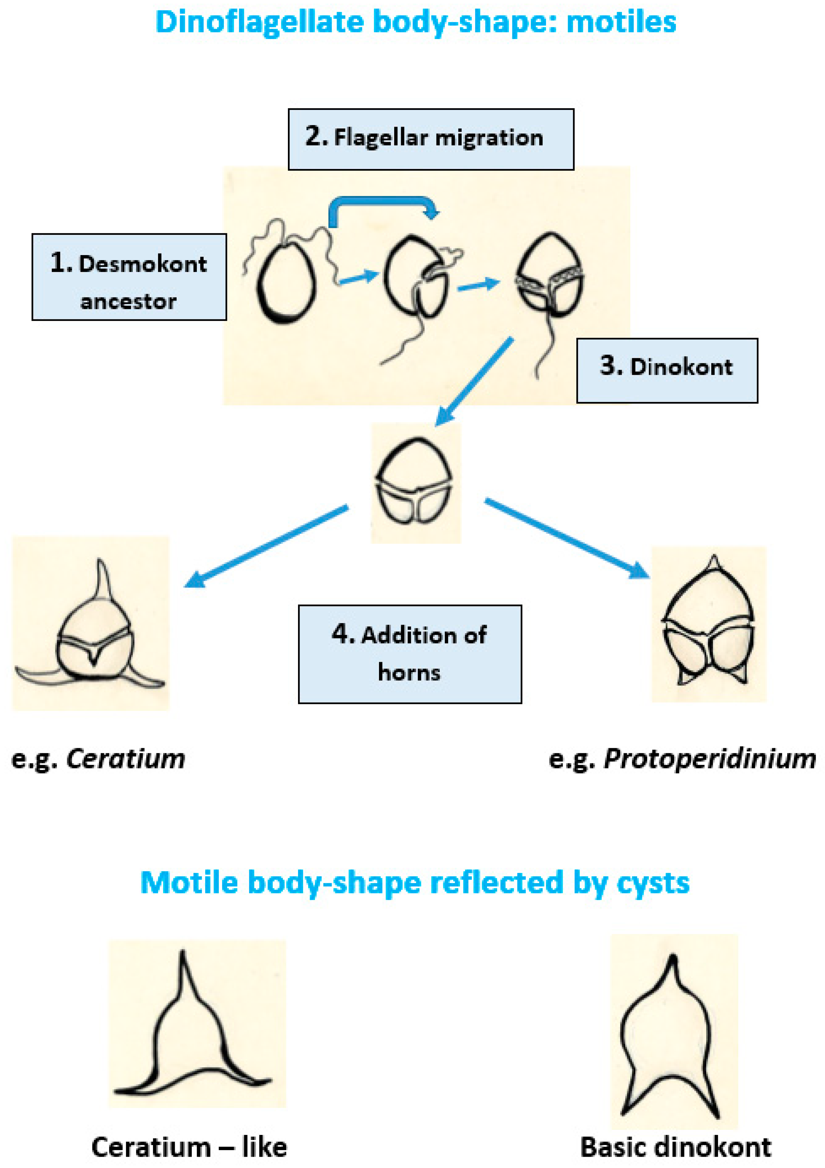
2.3. Morphology of Dinoflagellate Motile Stages Reflected by Fossil Cysts

Dinoflagellates are characterized by diagnostic genomic, physiological, and biochemical features in addition to morphology, but paleontology relies solely on the morphology of the fossils (Figure 1 shows examples). The detailed morphology of living motiles and fossil dinocysts is widely reported in the literature [15,16,17,18,19]. Here, I focus on the basic features used to identify dinocysts: body shape and wall structure including spines (processes) and ornamentation. The basic body shape of most living motile dinoflagellates is ovoid to roundly pyriform (pear-shaped), usually more pointed toward the anterior end and often indented or lobed at the posterior end. The basic shape may be modified by horns or spines projecting from the apex (usually one single) or antapex (1–2 horns and up to several spines) (Figure 2 shows how motile stage body shape may have developed from ancestral flagella migration).
Taylor described an impressively wide range of morphologic variance around this basic form [19]. The flagella impose distinctive features on the body shape. Dinoflagellate life cycles generally include a motile stage with two structurally different flagella: one beating sideways around the cell, the other beating backwards. Grooves in the wall (furrows) house the flagella: the transverse flagellum in the cingulum around the cell and the longitudinal flagellum in the sulcus leading behind. The furrows often produce a distinctive “T” or “spiral shape” where they meet at the position on the wall where the flagella arise in near proximity to each other. This arrangement in dinoflagellates differs from other bi-flagellated groups where both flagella work in tandem, either drawing the cell forward or driving it from behind, requiring no such grooves. A few dinoflagellates are more or less bilaterally symmetrical, but most show some degree of asymmetry with a marked tendency towards greater development on the left side. Some species show posterior lobes or spines that are larger on the left side than the right (e.g., in many species of Ceratium). The distinctive body forms of several living dinoflagellate motiles are diagnostic when reflected by fossil dinocyst genera (e.g., Pseudoceratium reflecting Ceratium (Figure 2 and Figure 3t–u) and Dinogymnium reflecting Gymnodinium) [18].
Wall structures, including processes and ornament, reflecting features seen in living motiles are the main criteria for identifying most dinocysts. Differences in wall structure define two distinct groups of motiles: thecate (armored) and athecate (unarmored) dinoflagellates. The outer wall of thecate dinoflagellates is composed of a series of plates, seen in light microscopy (Figure 1a). The arrangement of these plates (plate tabulation) is important for the classification of thecate dinoflagellates. Athecate dinoflagellates, by comparison, show no such plate covering in light microscopy (Figure 1b), though sections through the wall show interesting structural comparisons with thecae under the outer wall. Classification of athecate forms therefore relies on other features such as body shape. Evitt [12] recognized several dinocysts from body shape, but most were based on “reflected” tabulation, i.e., features such as ridges on the wall or other ornamentation that demonstrated affinities with plate patterns in thecate dinoflagellates (Figure 1j). He also showed how the shape of the opening in the cell wall that had allowed the motile-stage to exit (excyst) also reflected tabulation (often the only feature reflecting tabulation) (Figure 1q). He termed this the “archeopyle” (ancient opening) and its shape has proven to be a consistently useful diagnostic feature for the identification and classification of dinocysts [12,18]. As a result, Evitt’s criteria for dinocysts are heavily biased toward cysts of thecate dinoflagellates, whereas the identification of the few cysts of athecate species relies on body shape alone.
Thus, the traditional record of the group is almost entirely composed of cysts which reflect some plate tabulation representing thecate dinoflagellates. A few, by their body shapes, represent athecate dinoflagellates. Other forms in the palynological record of microplankton not showing these features are classified as acritarchs.

2.4. Living Dinoflagellate Cyst Morphology

Evitt’s publications in the 1960s caused a surge in studies of living cysts [14]. Wall and Dale [20] carried out the first extensive studies of living cysts, using incubation experiments, and many researchers have since added a wealth of information [21,22]. The results of early incubation experiments supported Evitt’s observations and the criteria he suggested. Wall and Dale linked many living cysts with their motile stages and, using Evitt’s criteria, were able to link the biological classification of dinoflagellates based on motile stages with the dinocyst record to describe some basic lineages for the group [20]. Focusing on living equivalents for Evitt’s dinocysts in these early studies inadvertently resulted in a strong bias towards cysts of thecate species. Nevertheless, dinocyst morphology was not well known at that time, and many living forms were selected for incubation based on their “hystrichosphere” morphology. Many spiny forms, showing their dinoflagellate affinity only after incubation, revealed an archeopyle reflecting plate tabulation, and a few regarded as acritarchs incubated to give athecate dinoflagellates [20]. This, together with the wealth of information from subsequent experiments, shows limits to the traditional criteria used to recognize dinocysts. A few living cysts closely reflect motile body shape (Figure 1g,r) [20] (pl. 4, 11–14), but many do not (e.g., the many simple unornamented spherical brown Protoperidinium cysts described in the literature (e.g., Figure 1q), and other spherical cysts with many processes (e.g., Figure 1s) [20] (pl. 2, 27, 29; pl. 4, 2, 19, 20). Many living cysts reflect plate tabulation, but this is restricted to very few genera, mainly Gonyaulax (Figure 1i) and Protoperidinium (Figure 1r) [4,20].
Early studies of living cysts revealed other important limitations for interpreting the fossil record. For example, not all living cysts are fossilizable, and even some of those showing well-established morphological links to fossil cysts may be incapable of contributing directly to the fossil record [20]. Some species of Alexandrium, for example, have less resistant walls and degrade before incorporation into the sedimentary record. Other species are less resistant to the rigors of diagenesis after burial in sediment [23,24]. This likely explains some gaps in the fossil record. It also suggests that the ability to fossilize may have varied through time, and, therefore, the morphology of all living cysts is relevant when comparing living and fossil cysts.
The cyst and motile stages have very different functions: non-motile resting stages to settle eventually in bottom sediments, versus motility in the water column for the motile-stage, and their morphologies reflect this. Given their different niches, it would not be surprising that cyst and motile stages would be subjected to different evolutionary pressures over time. The reflected plate tabulation of fossil and living cysts is presumed to be non-functional and ornamentation rather than actual plates, apart from some archeopyle structures showing “plate sutures and separate plates” (Figure 1i) [25]. However, many living cysts have “spines” (processes) projecting from the wall which are not found in motiles (Figure 1f,s), raising questions of functionality. These processes may substantially increase their overall size, possibly dissuading predators seeking smaller or non-spiny prey. If processes are adapted for increasing resistance, in accordance with Stoke’s Law, they may increase possible lateral transport, extending the population range. Particles attaching to the processes during transport through the water column may increase size and mass, thereby accelerating sinking and potentially helping to maintain local seed- beds in bottom sediments. Cysts with a dense covering of long, rigid, thickly-branched processes such as some Spiniferites species (Figure 1f) [26] elicit speculation that the processes may provide space around partially buried cysts in sediments that helps the emerging amoeboid motile (Figure 1d) to reach the water column through an opening in the network of processes associated with the archeopyle. Maintaining a more porous local microenvironment around the buried cyst may help to avoid anaerobiosis that would be detrimental to excystment [27,28].
Though Evitt’s criteria certainly established dinocysts as unequivocal fossil dinoflagellates, this discussion shows that they proved inadequate for recognizing many living cysts. Evitt’s main criteria were body shape and reflected plate tabulation, including archeopyles. Living cysts often have simple body-shapes (spherical to oblong), with or without processes, but lack wall ornamentation reflecting plate tabulation. The only feature distinguishing such cysts from acritarchs is the archeopyle (Figure 1l–q,u).

2.5. Reassessing The Archeopyle in Fossil Cysts

All cysts are presumed to possess an opening to allow the germinating motile-stage to emerge (excyst). Evitt [12] (p. 389) defined the archeopyle as “the opening in a fossil dinoflagellate …formed by the release of a single plate or group of plates”. He later revised the definition to “an excystment aperture in the wall of a dinoflagellate cyst” and noted that some archeopyles do not reflect plate tabulation, and some cysts do not even show archeopyles [25] (p. 14). Living cysts show examples conforming to all these categories. In many living cysts, the sutures do not surround a portion of the wall but simply follow a line of dehiscence expressed as a single straight or zig-zag split (Figure 1l,o,p,u) [20,29,30,31]. Some fossils also show the same (Figure 1m) [4,18]. Such archeopyles resemble “splits” rather than clearly defined openings. Matsuoka introduced the terms “chasmic” and “tremic” for some of these, and they are included as archeopyles in dinocysts from a broader understanding of the term than originally defined [18,29]. Unsurprisingly, split-like archeopyles without reflected plate tabulation are the only type found in living athecate species so far (Figure 1k,p,u) [20,29,31], but they also occur in some thecate species (Figure 1o) (e.g., Diplopsalopsis latipeltata) (Figure 1l) [30].
The most surprising observation from studying archeopyles in living cysts is how difficult it is to find archeopyles in some species. I did not observe any opening or split in the living acritarch that proved to be a cyst [10]. This was despite the close examination of more than seventy cysts shortly after the emergence of the motile, including some using scanning electron microscopy. I presumed that excystment must have occurred through a simple split that remained undetected. Though I did not draw attention to this in the original study, I now consider it highly relevant here. Since my first observations, this species (re-named Pentapharsodinium dalei) is recognized as a common cold-water dinoflagellate indicator species [32], and I and many other workers have observed many thousands of empty cysts from cultures and recent and Quaternary assemblages. Despite these numerous observations, I am not aware of any recorded archeopyle. In other species, only one in the thousands examined reveals an archeopyle ([33], and Rochon, A., personal comment 2022). Archeopyles also proved to be difficult to find in some fossil cysts [4,17,25]. Infrequent or absent archeopyles in dinocysts were often presumed to be caused by the poor preservation and/or orientation of specimens on slides, but this explanation is harder to invoke in living cultures or well-preserved recent cysts, where specimens may be manually rotated in wet preparations. In some cases, in recent sediments, cysts may not excyst due to unfavorable conditions, but it is doubtful that this explains all the observations of rare or absent archeopyles. In living cysts, I consider this observation a reflection of poorly understood fundamental aspects of archeopyle formation and excystment and this is significant, given that archeopyles are often used as criteria for differentiating cysts from acritarchs.
The living cysts considered here are diploid zygotes in the sexual reproduction of haploid vegetative species (Figure 1c) [16]. They sink through the water column and accumulate as benthic resting stages before excysting (Figure 1d–e) to re-establish motile populations in plankton. Protected by a highly resistant wall and packed with food storage products such as starch grains and lipid globules (Figure 1c), some cysts are believed to live in sediments for at least a hundred years [34]. The processes that transform this robust resting stage into a free-swimming motile and an empty cyst involve overcoming demanding challenges. Energetic “Brownian movement” is observed prior to excystment, as cell contents form the complex structures of the motile amoeboid stage [16,20]. Temperature is one factor that triggers excystment [16], but the processes that trigger and affect excystment remain largely unknown. Are environmental signals transmitted through such a robust wall? How is the highly resistant cell wall “disrupted” along genomically predetermined lines of dehiscence, and can the process possibly reverse in some cases, “closing the door behind”?
Archeopyles in living cysts now include a broad spectrum from an angular opening with a “cap” (operculum), and various forms of sutures reflecting plate tabulation to simple splits, or, importantly to this discussion, no visible trace of any breach in the wall after excystment. This confirms Evitt’s revised definition [25], and that archeopyles reflecting tabulation are only diagnostic for differentiating some cysts from acritarchs.

2.6. The Acritarchs

The division of organic-walled fossil microplankton into dinocysts and acritarchs [13] resulted in two separated fields of research, each with its own specialists. This separation of scientific effort likely helped to delay the consideration of some of the issues discussed here [35] (pp. 27, 29). Nevertheless, to date, acritarch specialists have produced a comprehensive literature that allows the comparison of acritarchs and dinoflagellate cysts. Strother, in an introduction to the acritarchs, shows how acritarch studies have progressed along very similar lines to those of dinocysts: the documentation and classification of morphotypes, and investigation of stratigraphic and paleoenvironmental distributions [8]. The acritarchs have proved useful as biostratigraphic indicators, particularly from the Precambrian through the Paleozoic [8]. Many studies collectively show a broad stratigraphic coverage from the Proterozoic to the present, but with progressively fewer species after the traditional record of dinocysts starts in the Middle Triassic [8] (refs. therein). Many acritarchs show palaeoecological and paleogeographic distributions supporting their general interpretation as microplankton, and Strother accepts that most acritarchs represent the cysts of phytoplankton [8] (p. 82).
The criteria used to characterize acritarchs are the same as those used for dinocysts: body shape, wall structure including ornamentation, and openings in the wall (in acritarchs called excystment apertures) (Figure 3 shows some examples). Seven types of excystment apertures are recognized [8] (pp. 83–88). Therefore, the only remaining difference in criteria separating dinocysts and some acritarchs is the presence of an archeopyle as defined for dinocysts versus an “excystment aperture” defined for acritarchs. Traditional archeopyles reflecting the plate tabulation of motile dinoflagellates were easily distinguished from any structure in acritarchs that by definition lacked affinity with any known group. However, the excystment apertures illustrated by Strother [8] fit within the broader definition of archeopyles discussed above here, and may be considered as possible evidence of dinoflagellate affinity (Figure 1n and Figure 3e,r).
A broader definition of the archeopyle provides a more realistic criterion for recognizing dinocysts. Virtually any consistent split or opening in the wall is now eligible for consideration as an archeopyle in dinocysts, consistent with evidence from living cysts. Dinocyst workers accepted the slit-like chasmic and trematic archeopyles introduced by Matsuoka for athecate species [29] as diagnostic for dinocysts [18]. However, they noted that, lacking reflected tabulation, their dinoflagellate affinities would not be recognized unless linked to a dinoflagellate life cycle [18]. Acritarch workers included most split-like “excystment openings” as “simple lateral ruptures”, and some as “medium splits” or “epitychs” [8]. The morphologic overlap between the split-like excystment apertures in acritarchs and dinocysts suggested here was not specifically addressed by Strother, but he suggests that acritarchs with more or less regular excystment openings may eventually be matched with dinoflagellates [8] (p. 99). My impression of the current status is that both groups of palynologists generally agree that acritarchs probably include some dinocysts. Nevertheless, the archeopyle has persisted as an important criterion often separating dinocysts and acritarchs in the traditional record, in effect acting as an artificial barrier to recognizing fossil dinoflagellate cysts within the acritarchs. Removing this barrier allows us to address the unavoidable fact that some living cysts would be classified as “acritarchs” if recovered from the geological record.
Regarding the application of his revised archeopyle definition, Evitt predicted that some dinoflagellate cysts not showing archeopyles reflecting tabulation or other recognizable dinoflagellate-like features would be referred to the acritarchs [25]. Acritarchs were a group with “uncertain biological affinities”, “a receptacle for unknowns until their affinities can be established” [25]. The dinocysts were all “acritarchs” prior to Evitt’s contribution emphasizing reflected tabulation as certain evidence of dinoflagellate affinities. Given the “missing paleontological record” considered here, it is pertinent to further explore the correlative implied by Evitt’s prediction: that some of the acritarchs in the long record of life indeed may be dinoflagellate cysts.

3. The Fossil Record of Early Life

3.1. Bacteria and Cyanobacteria

The fossil record of life begins in the early Archean (3500–3000 Ma) with an extremely long period of single-celled prokaryote microorganisms through to the Mesoproterozoic (1600–1000 Ma). The earliest microfossils are accepted to have been bacteria, followed by representatives of the cyanobacteria [36,37], based on comparative morphology and habitats with living organisms. Living bacteria and cyanobacteria are composed of biodegradable material not expected to fossilize, but many species of cyanobacteria are covered by a polysaccharide-rich sheath that sometimes may be preserved in Neoproterozoic rocks [38]. The original direct evidence for their early occurrence comes from transparent thin sections of siliceous rocks (e.g., cherts) that formed under exceptional geochemical conditions [37]. Further evidence was added recently by discoveries of mineralized tubes in ancient fossil hydrothermal vents comparable to tubes produced by bacteria living in hydrothermal vents today [39]. More widespread fossil “mounds” comparable to those produced by living mat-forming cyanobacteria (e.g., living stromatolites in Australia) support the identity of cyanobacteria as being among the earliest known living organisms [40].

3.2. The First Appearance of the Acritarchs—A New Group and a New Strategy

The fossil record of early life changed abruptly around 2.1 billion years ago with the appearance of the earliest palynomorphs, the acritarchs [37,40]. This occurred within the period of early eukaryote evolution: one of the foremost advances in the history of life. Palynomorphs are the highly resistant microscopic remains of organisms extracted from sedimentary rocks using harsh acid treatments to remove the minerals. Palynology, the study of acid-resistant microfossils, is one of the prominent fields in micropaleontology. The main groups of palynomorphs studied include plant spores and pollen, the resting stages of some aquatic protists, notably dinoflagellates, and the acritarchs. The walls of spores and pollen are composed of complex organic material referred to as sporopollenin, which is highly resistant to fungal and bacterial attacks. The walls of many dinoflagellate cysts are protected by sporopollenin-like material referred to as dinosporin [18]; other dinoflagellate cysts have calcareous walls destroyed by palynological treatments, and therefore, are not included in this discussion [16]. The walls of acritarchs are considered to include sporopollenin-like material [8]. Thus, the first appearance of acritarchs in the geological record marks the development of an innovation in life strategies with profound implications for evolution: a cell wall that enabled survival from microbial attack in the then microbial world. The persistence of an evolving record of ever more complex palynomorph morphology from acritarchs in the Proterozoic through to the dinocysts, plant spores, pollen and dinoflagellate cysts in the microplankton of today showing little or no evidence of microbial attack, attests to the success of this strategy.
The earliest acritarchs are much larger than earlier bacteria and cyanobacteria (most >10 µm), and possess a uniquely different cell wall chemistry, suggesting that their appearance marks the first evidence of a possible new group of organisms after almost two billion years of dominance by bacteria and cyanobacteria. Identifying the natural affinities of acritarchs would, therefore, add valuable information to our understanding of the paleontological record of early evolution. However, the loose definition of the acritarchs as “palynomorphs of uncertain affinities” opens for the inclusion of diverse groups of different affinities. This adds complications but does not preclude applying the standard methods for investigating natural affinities of fossils: comparing the morphologies and paleoenvironments of fossils with living organisms and their habitats.
We may now investigate the natural affinities of the acritarchs based on a modern day understanding of by far the most prevalent living aquatic group producing sporopollenin-like palynomorphs: the dinoflagellates.

3.3. The Role of Sporopollenin-Like Walls in Dinoflagellate Cysts and Acritarchs

As noted previously, acid-resistant walls are a common feature of dinocysts, acritarchs and other palynomorphs. The composition of this wall material has received more attention as geochemical techniques have improved. Studies so far suggest that sporopollenin in plant spores and pollen, and dinosporin in dinocysts, are general terms applied to what is most likely a unique and evolving group of macromolecules (biopolymers) [41] first produced by early acritarchs. If a detailed record of wall composition eventually reveals systematic changes through time, it would prove useful for biostratigraphy. It would also add a further line of evidence for the evolutionary relationships between fossil dinocysts and acritarchs.
I suggest that the innovative development of a sporopollenin-like wall by acritarchs that initiated a long fossil record of palynomorphs, including dinoflagellate cysts, supports other observations of shared ancestry between the two groups. Furthermore, I suggest that this unique group of compounds that provides cell protection for vital stages in sexual reproduction for living dinoflagellates also served a similar function in early dinocysts, and spores and pollen throughout the long history of plants. Sporopollenin-like cell walls most likely evolved as an innovative protection for stages of sexual reproduction in some eukaryotes represented by the early acritarchs. Each of several early groups of eukaryotes eventually then developed its own unique form of sporopollenin-like material as it evolved. If true, this is evidence of early sexuality, one of the monumental developments in eukaryotes enabling life as we know it today.
The emergence of sporopollenin-like material in early acritarchs thus marks a major evolutionary event. The simple morphology of these earliest acritarchs, spheromorphs lacking ornamentation and excystment openings, provide no evidence for their affinity. However, their ability to produce sporopollenin-like walls strongly suggests an evolutionary link to dinoflagellates, later diverging within the alveolates.

4. Discussion

4.1. The Belief That Some Acritarchs May Be Dinoflagellate Cysts Is Not New

Evitt [12,25] predicted that some acritarchs might prove to be dinoflagellate cysts. Wall and Dale [20] (p. 284) observed that cysts of some living athecate dinoflagellates lack distinctive archeopyles and reflected tabulation, such that they resembled acritarchs. Lister [35] presented an early comprehensive challenge to Evitt’s separation of dinocysts and acritarchs, and he and several others around that time introduced the basic arguments used by many since to speculate on overlap between the two groups. Lister particularly focused on the similarities between excystment openings in acritarchs and archeopyles in some dinocysts. The acanthomorph acritarchs are often cited as likely to include dinocysts [8,35], and there are several suggestions of older acritarchs identified as probable dinoflagellate cysts [42]. A comprehensive review of dinoflagellate phylogeny in 1999 accepted that speculations regarding dinoflagellate affinities within early acritarchs might prove correct, but concluded that they lacked convincing paleontological evidence [9].

4.2. A New Approach

This reassessment proposes a new approach prompted by new evidence from living dinoflagellate cysts, in the context of mounting biological and geochemical evidence supporting a much earlier emergence for the group than shown by the traditional record. The “new information” referred to here is the information developed since the introduction of criteria defining fossil cysts and acritarchs, some sixty years ago. The objective here is to highlight possible missing paleontological evidence for dinoflagellates prior to the traditional record. The results show that whereas the traditional interpretation [4] presents an important fossil record for thecate forms, it shows little evidence for athecate forms, which account for around half of living species, and no record for the proposed first two thirds of evolution for the group. Seen from a perspective of living cysts, the traditional approach seems disproportionally influenced by thecate dinoflagellates. Many living cysts, including some from thecate species, do not show reflected tabulation and they are generally simpler in form than cysts of thecate species (examples shown in Figure 1). Most are more or less spherical, with ornamentation that when present is often composed of unbranched processes, and have simple split archeopyles that may only rarely be seen. This simpler cyst type is characteristic for all known cysts of living athecate species, and corresponds to the morphology of many acritarchs from before the traditional record. Broader criteria suggested here recognize split archeopyles in both dinocysts and acritarchs and, together with other features discussed below, they identify dinoflagellate affinities in acritarchs.
A comprehensive application of these criteria to reassess the many hundreds of described acritarchs is beyond the scope of this article. The main aim is to present a new approach, illustrated by a few examples that should help others provide more details of the missing early dinoflagellate record. This new approach begins with the living dinoflagellate cysts, based on the principle that not only are all life cycle stages potential indicators of affinities, but the cysts are the equivalent stage comparable with the fossils. The traditional record provides an unequivocal fossil record, based largely on complex details of plate tabulation only found in dinoflagellates. This discussion shows that whereas cyst morphology often is less complex, it, too, is distinctive for the group. Distinctive body shapes are acceptable criteria that show dinoflagellate affinities in some species, for example in the thecate genus Ceratium and the athecate genus Gymnodinium. They are equally acceptable when reflected in their cysts.
The approach here accepts distinctive body shapes and other non-thecal reflections seen in living cysts as indicators of dinoflagellate affinities. Searching the dinocyst and acritarch literature for comparable morphologies reveals evidence for a plausible fossil record of dinoflagellates long before the traditional record begins (Figure 1 and Figure 3).

4.3. Examples of Early Dinoflagellate Affinities in Acritarchs

4.3.1. Micrhystridium: A Basic Cyst Morphotype Persisting from Early Dinoflagellate Evolution

The living “acritarch” of Pentapharsodinium dalei [10] is an example of an ancient morphotype persisting to the present. The “acritarch”, from Eocene sediments, showed morphology allowing informal classification as a species of Micrhystridium within the acanthomorph acritarchs (Figure 3a) [10]. Its morphology comprises a small spherical body (vesicle) with numerous processes, some of which may bifurcate at any position along their length. On incubation, the living cyst produced a thecate dinoflagellate, P. dalei. Acanthomorph acritarchs are a long-ranging, large, and diverse group including many of the spiny vesicles regularly recorded from Neoproterozoic [8,40,42,43] to recent sediments. Micrhystridium is a recognized morpho-group (acritarch genus) that includes many small spiny forms comparable to the cyst of P. dalei, which in my opinion, are dinocysts. They occur regularly from the Early Cambrian [40] through the Early to Mid-Paleozoic [44], and from the Mesozoic to recent, with a direct link to living dinoflagellates [10]. Their persistence through to the living cysts strengthens the case for acceptance of similar Micrhystridium-like forms as dinoflagellate cysts.
P. dalei belongs to an extant group of small spherical spiny cysts that very rarely show archeopyles (Figure 3b shows an example) [33]. Micrhystridium in the acritarch record also rarely if ever shows its split-like archeopyle. The examination of freshly excysted cysts in future incubation experiments should show whether the archeopyle sometimes remains invisible after excystment. The absence of an archeopyle is one of the principal diagnostic criteria traditionally separating acritarchs from cysts but in accordance with this discussion, this no longer applies. The difficulty experienced in documenting archeopyles in some living cysts suggests it may be at least as difficult to document for acritarchs in older sediments, and infrequent archeopyles may prove to be a common feature for some dinocysts and acritarchs. Rather than serving as an artificial barrier previously upholding separation of the two groups, this would show further evidence of their shared affinity.
Some early species of Micrhystridium include simple split-like archeopyles (Figure 3e), [45], as do some living cysts that would be “micrhystridia” if found in the fossil record (e.g., the athecate Polykrikos hartmannii (Figure 3c) [26]). Where more recent Micrhystridium-like cysts show archeopyles reflecting tabulation, as in the small, spherical, spiny cysts [33], this, in turn, reflects dinoflagellate evolution eventually producing thecate motiles. I consider the micrhystridia as a simple morphotype that has persisted successfully as the cyst of dinoflagellates from early in their divergence within the alveolates through to the present. From this perspective, the details of process numbers and types, and archeopyle forms that have allowed specialists to “speciate” micrhystridia should show possible evolutionary traits. I consider this an example of basic morphology retained in the cyst stage, possibly with its own morphologic evolution in process types or archeopyles, whereas the motile stage evolved into different lineages. Evolutionary pressures may act differently on the very different life cycle stages in cyst-forming dinoflagellates, and the simple micrhystridium-like cyst seems to have proved adequate with only minor morphological change. Alternatively, this may be cyst morphology repeated in progressive intervals of evolution. P. dalei shows a combination of an “ancient” acritarchous cyst morphology with a far more recent thecal morphology, considered here to represent significant evidence for the basal position of Pentapharsodinium in phylogenetic trees [4] (figure 5). This suggests that cyst morphology may have evolved somewhat independently from motile-stage morphology. The genus Pentapharsodinium is an example where even though living species place closely together in molecular analyses [4], their cysts are morphologically very different. P. dalei has an organic-walled “acritarchous” cyst [10] and Pentapharsodinium tyrrhanicum has a calcareous cyst-wall and a simple spherical inner acid-resistant organic membrane with no processes, ornament, or archeopyle [46]. Other groups of living dinoflagellates show similar examples where cysts show markedly more morphological variance than their motile cells [11]. For example, the Gonyaulax spinifera complex [16] (figure 45) [47], in which the motile cells morphologically classify as one species and cluster closely in molecular trees, whereas the cysts were classified in different genera by palynologists [16].
Micrhystridium is one of the few forms to persist through the Late Permian extinction interval preceding the evidence of thecate dinoflagellates at the start of the traditional record, indicating its resilience as a cyst form [45]. P. dalei cysts proved to be the most resilient dinoflagellate cysts in incubation experiments with older sediments, maintaining viability for up to 100 years [48]; probably enough to survive a catastrophic event such as the bolide impact at the K/T boundary [48].

4.3.2. Processes on Cysts and Acritarchs Reflecting Horns and Cingulum from Motile Dinoflagellates

Horns are a distinctive feature of dinoflagellate body shape also shared by some living cysts, dinocysts, and early acritarchs (Figure 4). Motile stages in several major genera such as Protoperidinium and Ceratium develop horns, usually one at the apex and two or more at the antapex (Figure 3f and Figure 4a). Other motiles are more spindle-shaped with one horn at each pole (e.g., Oxytoxum). In some living cysts distinctive processes reflect horns. Ceratium cysts reflect the apical and antapical horns of the motile stage (Figure 2 and Figure 3t), and the same characteristic body shape is reflected in fossil cysts of Pseudoceratium (Figure 3u) [49] and acritarchs assigned to Veryhachium (Figure 3r—a living cyst, and Figure 3v—an acritarch). These acritarchs are convincing examples of pre-Mesozoic dinoflagellate cysts. In some living cysts, additional processes are developed at the position of shoulders/cingulum in the motile stage (Figure 4b,c). Figure 3f shows a living Protoperidinium cyst with two processes reflecting the shoulder/cingulum positions of the motile stage, and Figure 3p shows an acritarch with the same characteristic body shape identifying it as a dinoflagellate cyst. Other living cysts show more processes in the shoulder/cingulum position. Figure 3g,h show living cysts with a row of processes in the shoulder/cingulum position, and K. Zonneveld provides further illustrations for comparison with the fossils [26]. The row of processes likely reflects the cingulum in the motile stage. The living cyst of Protoperidinium conicum has two rows of processes clearly reflecting the cingulum (Figure 3i and Figure 4d). These are living cysts with complex features reflecting unique dinoflagellate morphology. The Ordovician acritarch genus Barakella Cramer & Diéz 1977 (Figure 3l–n) shows similarities with the living Stelladinium, including some distinctly robust processes similar to those reflecting horns on living cysts and a bipolar distribution of processes with a process-free space between comparable to the reflected cingulum in Figure 3i, although processes are not in clearly distinguished rows. The longitudinal striations in the process-free area are interesting, since at least one living cyst-forming dinoflagellate species, Gonyaulax scrippsae, has similar striations in the motile stage cingulum [20] (p. 271). Yan et al. provide excellent photomicrographs showing how B. fortunata [50] (pp. 1, 1–4) with only four processes suggests a possible common ancestor with earlier trapezoidal acritarchs. Processes are comparable in the living cysts and the acritarchs: long, with broad bases open to the vesicle, and thinning to pointed solid tips. The Cretaceous dinocyst Nyktericysta davisii shows a directly comparable morphology (Figure 3o) [18] (pp. 3, 20). Some Early Jurassic fossil cysts reflect horns and shoulders ornamented with branched processes (Figure 3q). Although these examples of processes possibly reflecting a cingulum in acritarchs are not as clearly indicative of dinoflagellate affinity as are the examples of horns, they are included here to encourage further work to explore the possibility.

4.3.3. Paleontological Evidence for Early Ciliates

The acritarch record shows that some of the earliest excystment openings were distinctive circular “holes”, pylomes (Figure 3w), [8]. Early palynologists recognized the distinction between pylomes and angular or split archeopyles [12,25]. In our early incubation experiments seeking living dinocysts [20] I incubated one cyst-like organism, Hexasterias problematica, from Woods Hole, USA sediments, and it produced an unidentified ciliate. This was never followed further in projects concentrated on dinocysts, but the freshly excysted Hexasterias showed a very distinctive pylome, unlike any dinoflagellate cyst recognized at the time. Recent work using molecular methods and wall chemistry has confirmed that this species and Halodinium verrucatum, recorded over the past decades as acritarchs in palynology and plankton studies, are ciliate cysts [51]. H. problematica (Figure 3x) and five species of Halodinium also show pylomes, with or without an operculum [51]. I propose that pylomes are a diagnostic feature of ciliates identifying the group within the early acritarchs. Their recorded occurrence prior to dinocysts identified here suggests possible support for their earlier divergence within the alveolates proposed in molecular trees [1,2,4], but more observations are needed to confirm this. Galeate acritarchs are examples of these suggested ciliate cysts [42] (figure 4B) [52]. Palaeostomocystis subtilitheca from the Holocene of Greenland probably represents the cyst of an extant heterotrophic ciliate [53].

4.4. Suggestions for Future Work

  • Freshly excysted cysts of living dinoflagellates in incubation experiments should be carefully examined for archeopyles. Pentapharsodinium dalei is a particular example where questions raised here of rare or absent archeopyles in some species could be investigated.
  • The acritarch record of the late Paleozoic/Early Triassic prior to the first traditional dinocysts in the Middle Triassic is particularly interesting. Acritarchs should be examined for evidence of transition to thecate dinoflagellates, from the perspective suggested here that dinoflagellates were present before the traditional record starts.
  • Many previous palynological studies of Mesozoic and Cenozoic samples were concentrated on traditional dinocysts useful for biostratigraphy. Acritarchs were often seen and sometimes recorded, but seldom studied in detail. The research here suggests the value of a new approach that investigates the whole acritarch record for evidence complementing the traditional dinocyst record. The Late Cretaceous to Late Eocene is an interesting interval where acritarchs identified as Micrhystridium occur together with Micrhystridium-like dinoflagellate cysts identified as Impletosphaeridium [54]. More research is needed to clarify the morphologic differences within these groups.
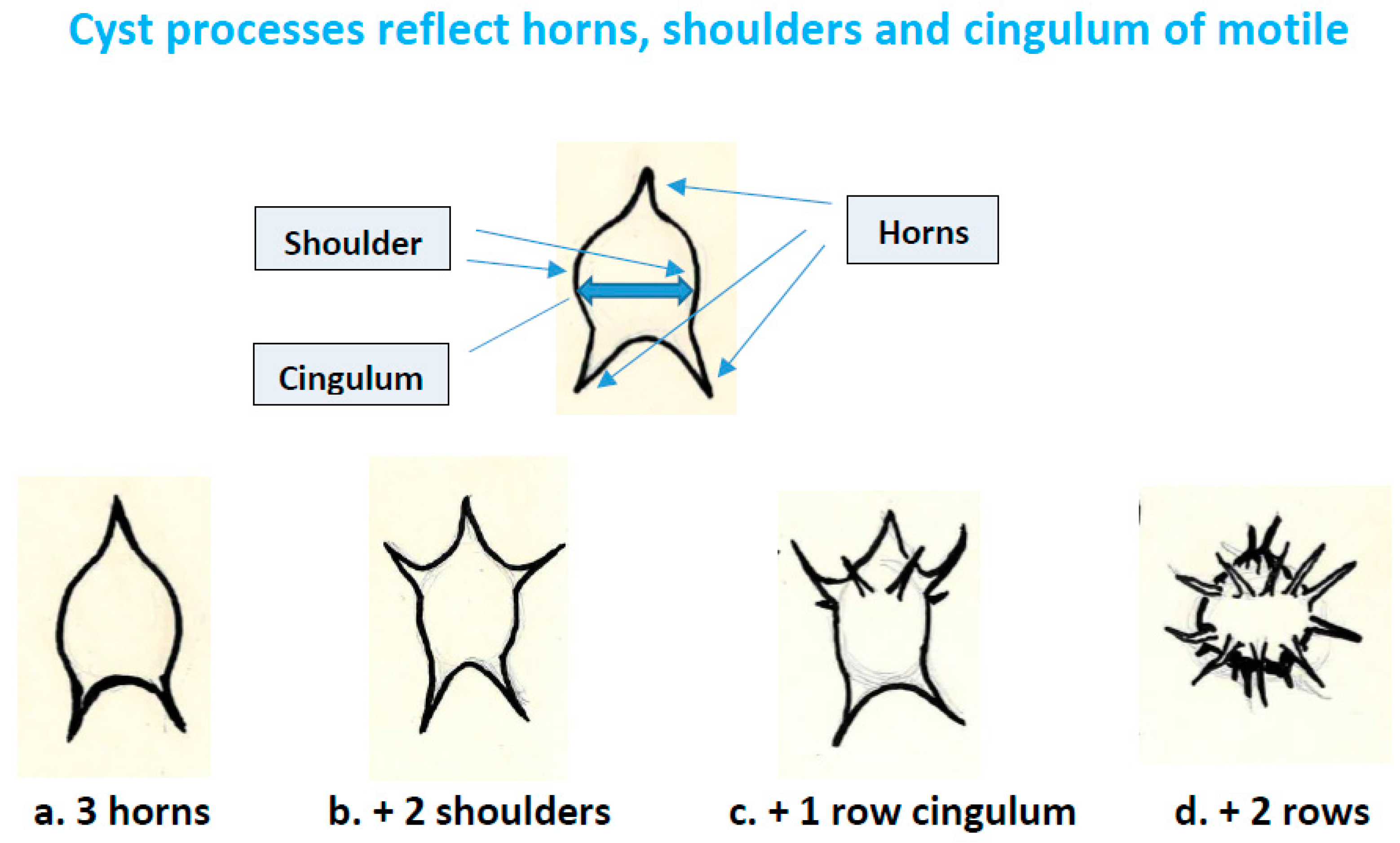
  • Spinose acritarchs (acanthomorphs) should be examined for evidence of processes reflecting horns and cingulum. Horns may be represented by more prominent processes, and by their critical positions (1 for apex, usually 2 but possibly more for antapex, and shoulders); the cingulum by rows of processes separated towards the poles (Figure 4). The acritarch genus Dorsennidium Wicander 1974 may prove to be an example [55].
  • The research presented here has obvious implications for the application of the traditional dinoflagellate record in other studies. For example, previous interpretations of pre-Mesozoic dinosteranes were considered unlikely to originate from dinoflagellates partly because they predate “unambiguous fossils” [56], but the research here suggests that could be circular reasoning. The research here also challenges studies of phytoplankton evolution that treat acritarchs and dinoflagellate cysts in the traditional record as separate groups. For example, one line of research recognizes two basic groups of plankton (referred to as “red”, including dinoflagellates, or “green”, including acritarchs) and links a switch between relative amounts of these groups to paleo nutrients [57] (and references therein). There is still much that we do not know about how morphology persists or is repeated within dinoflagellate cysts, but the research here proposes significant overlap between the two traditionally separated groups as an alternative perspective to be considered in future studies such as these.
  • Gilan Attaran-Fariman et al. [58] described the living athecate Gymnodinium trapeziforme with a trapezoidal-shaped cyst with a micro reticulate wall reflecting alveolae, cingulum and sulcus, and with a split archeopyle in the sulcal region. This most likely represents a living cyst form with morphological linkage to trapezoidal acritarchs. The closely related group of micro reticulate Gymnodinium species should be closely examined for similar morphologies comparable with dinocysts and acritarchs.

5. Concluding Remarks

The traditional dinoflagellate fossil record of dinocysts from the Mid Triassic to the present covers only approximately the past one third of the evolutionary timespan suggested for the group by molecular analysis and bio-geochemical evidence. The acritarchs are a prominent group of microfossils of uncertain affinities covering the whole timespan, long suspected of possibly including early dinoflagellates not conforming to criteria defining dinocysts. Other researchers have identified a few other groups of organisms from within the large diverse group of the acritarchs [8,59]. The evidence now available opens a new approach, applying broader criteria based on the more natural comparison with living dinoflagellate cysts. This shows definitively overlapping morphologies between living cysts, some dinocysts in the traditional record, and some earlier acritarchs regarded here as dinoflagellate cysts. The present discussion identifies large groups of acritarchs as dinocysts, representing “missing evidence” of the early evolution of the group. More research should reveal details of early dinoflagellate evolution, and the limited outline presented here suggests some possibilities.
The proposed timing of dinoflagellate divergence, around 650 Ma, corresponds approximately with the Cryogenian–Ediacaran boundary. This marked a transition from a period including the most extensive glaciations ever recorded on Earth to warmer oceans and greatly accelerated evolution of early life [60]. The earliest acritarchs are large unornamented leiospheres and acanthomorphs that preceded the Cryogenian glaciations. These forms show no evidence of dinoflagellate affinities, and they are much larger than later acritarchs here regarded as dinocysts. They most likely include early eukaryotes preceding dinoflagellates. They are followed by ornamented leiospheres (some with pylomes, interpreted here as ciliate cysts) and smaller acanthomorphs closer to dinocyst morphologies.
The research here reveals a comprehensive record of evidence for dinoflagellates long before the traditional fossil record, broadly supporting the molecular and geochemical evidence. Many earlier acritarchs, particularly acanthomorphs, show their dinophycean affinities by split-like archeopyles and/or basic body forms otherwise found in living dinoflagellate cysts today. This early record suggests that dinoflagellates and ciliates may have diverged within the alveolates during the Cryogenian, when a robust resting cyst would increase likelihood for survival from harsh climatic conditions. Early dinoflagellates seem to have diversified in the following period of accelerated evolution of life in the Ediacaran and the Cambrian Explosion of Life. Following a peak of species in the Silurian, diversity decreased later in the Paleozoic prior to evidence of the appearance of thecate dinocysts and the start of the traditional record for the group in the Mid Triassic.
The early record suggests possible answers to one of the most discussed questions regarding dinoflagellate evolution: Did thecate dinoflagellates evolve from athecate forms? [56]. The early dinocysts identified here have split archeopyles characteristic of athecate living species, suggesting the likely dominance of athecate ancestors long before thecates emerged by the Mid Triassic. The presence of basic dinokaryont body forms in the early record suggests that motile stages likely resembled similar morphologies to living dinoflagellates, with cingulum and sulcus already accommodating two differently operating flagella. The timing of thecate development implied by the radiation of dinocysts in the traditional record is interesting because it corresponds to the Mesozoic radiations in two other major phytoplankton groups in modern oceans, the coccolithophorids and the diatoms [57]. Thecae are generally believed to protect the motile stage of dinoflagellates (4), and the mineralized “hard parts” of coccolithophorids and diatoms also may have developed for protection [61], suggesting that pressure from predators may have influenced the community structure of modern-day phytoplankton. Therefore, the traditional fossil record may represent development of the evolutionary advantage to the group of increased protection for the motile stage by a theca in addition to a heavily protected cyst. Since many acritarchs were athecate dinoflagellates, this would account for the observed large reduction in acritarch species, as dinocysts of thecate species increased [4]; the new thecate forms would have had evolutionary advantages for surviving predation. However, the possibility of the loss of fossilization potential in athecate species cannot be ruled out, either. Living thecate and athecate species both include split archeopyles, but many athecate species do not seem capable of fossilization, suggesting that, like Ceratium, they may have lost this capability.
One further point should be recognized when evaluating the dinoflagellate fossil record: cyst formation is only one possible survival strategy within the group. Around ten percent of the probably more than two thousand living species of dinoflagellates are known to produce resting cysts, and less than one percent produce acid resistant cysts capable of contributing to the fossil record. Many new species are discovered regularly and the total species numbers often cited from literature [21] are outdated [22]. Furthermore, many of the acid-resistant cysts belong to just the two genera Gonyaulax and Protoperidinium. Thus, evolutionary processes affecting most species would presumably go undetected in the fossil record, and there is no reason to presume that the earlier record is complete. This has potentially interesting implications for interpreting the fossil record. We may expect large gaps in the evolutionary record, limiting possibilities to reconstruct long-term phylogenies for most lineages. However, the powerful potential for the resistant cysts to survive major extinction events most likely resulted in them exerting a disproportionately large effect on the overall evolution of the group. A plausible scenario for dinoflagellate evolution could involve at least five major steps resulting from the five prominent extinction events of the Phanerozoic [62]. Each step would include: reduced diversity following the event, heavily influenced by surviving resistant cyst-formers; followed by increased diversity including development of less resistant cysts, or no cysts, in species using less energy for cyst-formation; before another event again reduced diversity back towards resistant cyst formers. This may explain the observed persistence of some cyst types through extinction events, with long periods of evolution before the next event. It may also explain the present situation with dominance by the few resistant thecate cyst types and the reduced amount of less resistant athecate cyst types.
The chapter by Riding et al. in this volume [4] demonstrates the scientific value of integrating molecular information with the fossil record of dinoflagellates. However, the traditional fossil record applied in that chapter starts in the Middle Triassic, around 400 million years after the point of divergence proposed for dinoflagellates by molecular evidence [4]. A quote from [4] helps explain this discrepancy: “A polygonal network of ridges, angular openings, processes, and long slit-like openings do not a dinoflagellate make”. As shown here, many living cysts are characterized by just such features (e.g., Figure 1k,l,o,p,t,u and Figure 3c,t). Many of these are produced by athecate dinoflagellates, and comparable features characterize many pre-Mesozoic acritarchs (e.g., examples in Figure 1 and Figure 3). The authors in [4] are only “convinced” of dinoflagellate affinities by fossils reflecting motile stage morphology (almost exclusively thecate), and, therefore, overlook the comprehensive acritarch record reflecting athecate cyst morphology.
The chapter here reveals a fossil record back to the Precambrian. This reassessment provides the missing paleontological evidence, suggesting an approximate calibration point for dinoflagellate divergence around the time before the Cryogenian–Ediacaran boundary at about 650 Ma. The fossil record of “acritarchs” complements molecular evidence showing that dinoflagellates are one of the earliest groups of extant eukaryotes, presenting unique opportunities for biologists and paleontologists investigating evolutionary processes. The acritarchs are defined as “of uncertain affinity”. The traditional record correctly asserts that dinocysts reflecting plate tabulation on motile stages are accepted as dinoflagellates, as are those few reflecting athecate body shape. Here, I propose the acceptance of the many acritarchs reflecting dinoflagellate cyst morphology as the missing fossil record for the early development of dinoflagellates, probably before thecate forms emerged. If other groups not discussed here showed overlapping morphologies with these acritarchs, it could cause possible uncertainty, but no such other groups are known. The early dinoflagellates identified here are many of the original hystrichospheres, now called acanthomorph acritarchs. They were not identified as dinocysts by Evitt’s early criteria that were based on motile stage morphology. These acanthomorphs and Ceratium-like acritarchs do not meet criteria to identify them with other groups. They would be identified as dinoflagellate cysts if found living today and, therefore, meet the morphological criteria for acceptance as fossil dinoflagellates. The proposed further research suggested here should lead to these and additional acritarchs being reclassified as dinoflagellate cysts.

Funding

This research received no external funding.

Institutional Review Board Statement

Not applicable.

Informed Consent Statement

Not applicable.

Data Availability Statement

No data were created.

Acknowledgments

Some of the ideas, here, were first outlined briefly in a publication [63], and developed further in a keynote address to the DINO 12 Meeting in Gran Canaria, Spain, July 2022. I gratefully acknowledge Amy L. Dale for many useful comments that greatly improved all versions of this work, and I thank the following colleagues who provided photo images for figures: Gilan Attaran, Dept. of Marine Biology, Chabahar Maritime University, Iran (1b, 1k, 1p); Pieter Gurdebeke, Department of Geology, Ghent University, Belgium (3x); Yan Kui, Nanjing Institute of Geology and Palaeontology, Chinese Academy of Sciences, China (3l, 3m, 3n); Wolfram M. Kürschner, University of Oslo, Norway (3d, 3e, 3k); Francine MacCarthy and Olena Volik, Brock University, Canada (3t); Palsys.org, Utrecht, The Netherlands (3o); Bas van de Schootbrugge, Utrecht University, The Netherlands, (3q); Paul K. Strother, Boston College Weston Observatory, USA (1n, 3p, 3s, 3w); Robert Williams, Norwegian Petroleum Directorate, Norway (1h, 1j, 1m, 3u, 3v, 3r). I thank five reviewers for suggestions that greatly improved the manuscript.

Conflicts of Interest

The author declares no conflict of interest.

References

  1. Bhattacharya, D.; Yoon, H.S.; Hedges, S.B.; Hackett, J.D. Eukaryotes (Eukaryota). In The Timetree of Life; Hedges, S.B., Kumar, S., Eds.; Oxford University Press: Oxford, UK, 2009; pp. 116–120. [Google Scholar]
  2. Butterfield, N.J. Early evolution of the eukaryota. Palaeontology 2015, 58, 5–17. [Google Scholar] [CrossRef]
  3. Porter, S.M. Insights into eukaryogenesis from the fossil record. Interface Focus 2020, 10, 20190105. [Google Scholar] [CrossRef] [PubMed]
  4. Riding, J.B.; Fensome, R.A.; Soyer-Gobillard, M.-O.; Medlin, L.K. A review of the dinoflagellates and their evolution from fossils to modern. J. Mar. Sci. Eng. 2022, 11, 1. [Google Scholar] [CrossRef]
  5. Taylor, F.J.R. General group characteristics; special features of interest; short history of dinoflagellates study. In The Biology of Dinoflagellates; Taylor, F.J.R., Ed.; Botanical Monographs; Wiley-Blackwell: Hoboken, NJ, USA, 1987; Volume 21, pp. 1–23. [Google Scholar]
  6. Talyzina, N.M.; Moldowan, J.M.; Johannisson, A.; Fago, F.J. Affinities of Early Cambrian acritarchs studied by using microscopy, fluorescence flow cytometry and biomarkers. Rev. Palaeobot. Palynol. 2000, 108, 37–53. [Google Scholar] [CrossRef]
  7. Moldowan, J.M.; Dahl, J.; Jacobson, S.R.; Huizinga, B.J.; Fago, F.J.; Shetty, R.; Watt, D.S.; Peters, K.E. Chemostratographic reconstruction of biofacies: Molecular evidence linking cyst-forming dinoflagellates with pre-Triassic ancestors. Geology 1996, 24, 159–162. [Google Scholar] [CrossRef]
  8. Strother, P.K. Acritarchs. In Palynology: Principles and Applications; Jansonius, J., McGregor, D.C., Eds.; American Association of Stratigraphic Palynologists Foundation: Dallas, TX, USA, 1996; Volume 1, pp. 81–106. [Google Scholar]
  9. Fensome, R.A.; Saldarriaga, J.F.; Taylor, F.J.R. Dinoflagellate phylogeny revisited: Reconciling morphological and molecular based phylogenies. Grana 1999, 38, 66–80. [Google Scholar] [CrossRef]
  10. Dale, B. New observations on Peridinium faeroense Paulsen (1905), and classification of small orthoperidinioid dinoflagellates. Br. Phycol. J. 1977, 12, 241–253. [Google Scholar] [CrossRef]
  11. Dale, B. Acritarchous cysts of Peridinium faeroense Paulsen: Implications for dinoflagellate systematics. Palynology 1978, 2, 187–193. [Google Scholar] [CrossRef]
  12. Evitt, W.R. Observations on the morphology of fossil dinoflagellates. Micropaleontology 1961, 7, 385–420. [Google Scholar] [CrossRef]
  13. Evitt, W.R. A discussion and proposals concerning fossil dinoflagellates, hystrichospheres, and acritarchs. Nat. Acad. Sci. Proc. USA 1963, 49, 158–164 and 298–302. [Google Scholar] [CrossRef] [Green Version]
  14. Dale, B. From hystrichospheres to dinoflagellate cysts: Scandinavian contributions to E’vitt’s pivotal recognition of fossil dinoflagellate cysts. Palynology 2021, 45, 165–170. [Google Scholar] [CrossRef]
  15. Jansonius, J.; McGregor, D.C. (Eds.) Palynology: Principles and Applications; American Association of Stratigraphic Palynologists Foundation: Dallas, TX, USA, 1996; Volume 1, pp. 1–443. [Google Scholar]
  16. Dale, B. Dinoflagellate resting cysts: «benthic plankton». In Survival Strategies of the Algae; Fryxel, G.A., Ed.; Cambridge University Press: Cambridge, UK, 1983; pp. 69–136. [Google Scholar]
  17. Evitt, W.R. Sporopollenin Dinoflagellate Cysts–Their Morphology and Interpretation; American Association of Stratigraphic Palynologists Foundation: Dallas, TX, USA, 1985; p. 333. [Google Scholar]
  18. Fensome, R.A.; Riding, J.B.; Taylor, F.J.R. Dinoflagellates. In Palynology: Principles and Applications; Jansonius, J., McGregor, D.C., Eds.; American Association of Stratigraphic Palynologists Foundation: Dallas, TX, USA, 1996; Volume 1, pp. 107–169. [Google Scholar]
  19. Taylor, F.J.R. Dinoflagellate morphology. In The Biology of Dinoflagellates; Taylor, F.J.R., Ed.; Botanical Monographs; Blakwell Scientific Publications: Hoboken, NJ, USA, 1987; Volume 2, pp. 24–91. [Google Scholar]
  20. Wall, D.; Dale, B. Modern dinoflagellate cysts and evolution of the Peridiniales. Micropaleontology 1968, 14, 265–304. [Google Scholar] [CrossRef]
  21. Head, M.J. Modern dinoflagellate cysts and their biological affinities. In Palynology: Principles and Applications; Jansonius, J., McGregor, D.C., Eds.; American Association of Stratigraphic Palynologists Foundation: Dallas, TX, USA, 1996; Volume 3, pp. 1197–1248. [Google Scholar]
  22. Belmonte, G.; Rubino, F. Resting cysts from coastal marine plankton. Oceanogr. Mar. Biol. Annu. Rev. 2019, 57, 1–88. [Google Scholar]
  23. Persson, A.; Smith, B.C. Preservation of Dinoflagellate Cysts in Different Oxygen Regimes: Differences in Cyst Survival between Oxic and Anoxic Natural Environments. Phycology 2022, 2, 384–418. [Google Scholar] [CrossRef]
  24. Zonneveld, K.A.F.; Versteegh, G.J.M.; de Lange, G.J. Preservation of organic-walled dinoflagellate cysts in different oxygen regimes: A 10,000 year natural experiment. Mar. Micropaleontol. 1997, 29, 393–405. [Google Scholar] [CrossRef]
  25. Evitt, W.R. Dinoflagellate Studies II. In The Archeopyle; Stanford University Publications: Stanford, CA, USA, 1967; Volume 10, p. 88. [Google Scholar]
  26. Zonneveld, K.A.F.; Pospelova, V. A determination key for modern dinoflagellate cysts. Palynology 2015, 39, 387–409. [Google Scholar] [CrossRef]
  27. Anderson, D.M.; Taylor, C.D.; Armbrust, E.V. The effects of darkness and anaerobiosis on dinoflagellate cyst germination. Limnol. Oceanogr. 1987, 32, 340–351. [Google Scholar] [CrossRef]
  28. Genovesi-Giunti, B.; Laabir, A.; Vaquer, A. The benthic resting cyst: A key actor in harmful dinoflagellate blooms—A review. Vie. Milieu. 2006, 56, 327–337. [Google Scholar]
  29. Matsuoka, K. Archeopyle structures in modern gymnodinialean dinoflagellate cysts. Rev. Palaeobot. Palynol. 1985, 44, 217–231. [Google Scholar] [CrossRef]
  30. Dale, B.; Montresore, M.; Zingone, A.; Zonneveld, K. The cyst-motile stage relationships of the dinoflagellates Diplopelta simmetrica and Diplopsalopsis latipeltata. Eur. J. Phycol. 1993, 28, 129–137. [Google Scholar] [CrossRef]
  31. Ellegaard, M.; Dale, B.; Amorim, A. The acritarchous cyst of the athecate dinoflagellate Warnowia cf. rosea (Dinophyceae). Phycologia 2001, 40, 542–546. [Google Scholar] [CrossRef]
  32. Dale, B. Dinoflagellate cyst ecology: Modeling and geological application. In Palynology: Principles and Applications; Jansonius, J., McGregor, D.C., Eds.; American Association of Stratigraphic Palynologists Foundation: Dallas, TX, USA, 1996; Volume 3, pp. 1249–1275. [Google Scholar]
  33. Radi, T.; Bonnet, S.; Cormier, M.A.; de Vernal, A.; Durantou, L.; Faubert, É.; Head, M.J.; Henry, M.; Pospelova, V.; Rochon, A.; et al. Operational taxonomy and (paleo-)autecology of round, brown, spiny dinoflagellate cysts from the Quaternary of high northern latitudes. Mar. Micropaleontol. 2013, 98, 41–57. [Google Scholar] [CrossRef]
  34. Ribeiro, S.; Berge, T.; Lundholm, N.; Andersen, T.J.; Abrantes, F.; Ellegaard, M. Phytoplankton growth after a century of dormancy illuminates past resilience to catastrophic darkness. Nat. Commun. 2011, 2, 311. [Google Scholar] [CrossRef] [PubMed] [Green Version]
  35. Lister, T.R. The acritarchs and chitinozoa from the Wenlock and Ludlow Series of the Ludlow and Millichope areas, Shropshire. Monogr. Palaeontogr. Soc. 1970, 124, 1–100. [Google Scholar] [CrossRef]
  36. Demoulin, C.F.; Lara, Y.J.; Cornet, L.; François, C.; Baurain, D.; Wilmotte, A.; Javaux, E.J. Cyanobacteria evolution: Insight from the fossil record. Free Radic Biol Med. 2019, 140, 206–223. [Google Scholar] [CrossRef] [PubMed]
  37. Schopf, J.W. Precambrian paleobiology: Precedents, progress, and prospects. Front. Ecol. Evol. 2021, 9, 707072. [Google Scholar] [CrossRef]
  38. Tiwari, M. Organic-walled microfossils from the Chert-phosphate Member, Tal Formation, Precambrian-Cambrian Boundary, India. Precambrian Res. 1999, 97, 99–113. [Google Scholar] [CrossRef]
  39. Dodd, M.S.; Papineau, D.; Grenne, T.; Slack, J.F.; Rittner, M.; Pirajno, F.; O’Neil, J.; Little, C.T.S. Evidence for early life in Earth’s oldest hydrothermal vent precipitates. Nature 2017, 543, 60–64. [Google Scholar] [CrossRef]
  40. Knoll, A. Archean and Proterozoic paleontology. In Palynology: Principles and Applications; Jansonius, J., McGregor, D.C., Eds.; American Association of Stratigraphic Palynologists Foundation: Dallas, TX, USA, 1996; Volume 1, pp. 51–81. [Google Scholar]
  41. Suh, D.Y.; Ashton, N.W. A sporopollenin definition for the genomic age. New Phytol. 2022, 236, 2009–2013. [Google Scholar] [CrossRef]
  42. Butterfield, N.J.; Rainbird, R.H. Diverse organic-walled fossils, including “possible dinoflagellates” from the early Neoproterozoic of Arctic Canada. Geology 1998, 26, 963–966. [Google Scholar] [CrossRef]
  43. Meng, F.; Zhou, C.; Yin, L.; Chen, Z.; Yuan, X. The oldest known dinoflagellates: Morphological and molecular evidence from Mesoproterozoic rocks at Yongji, Shanxi Province. Chin. Sci. Bull. 2005, 50, 1231–1234. [Google Scholar] [CrossRef]
  44. Sarjeant, W.A.S.; Stancliffe, R.P.W. The Michystridium and Veryhachium complexes (Acritarcha: Acanthomorphitae and Polygonomorphitae): A taxonomic reconsideration. Micropaleontology 1994, 40, 1–77. [Google Scholar] [CrossRef]
  45. Van Soelen, E.E.; Kürschner, W.M. Late Permian to Early Triassic changes in acritarch assemblages and morphology in the Boreal Arctic: New data from the Finnmark Platform. Palaeogeogr. Palaeoclimatol. Palaeoecol. 2018, 505, 120–127. [Google Scholar] [CrossRef]
  46. Montresor, M.; Zingone, A.; Marino, D. The calcareous resting cyst of Pentapharsodinium Tyrrhenicum comb.nov. (Dinophyceae). J. Phycol. 1993, 29, 223–230. [Google Scholar] [CrossRef]
  47. Mertens, K.N.; Carbonell-Moore, C. Introduction to Spiniferites Mantell 1850. Palynology 2018, 42, 1–9. [Google Scholar] [CrossRef] [Green Version]
  48. Lundholm, N.; dos Santos Ribeiro, S.I.; Andersen, T.J.; Koch, T.A.; Godhe, A.; Ekelund, F.; Ellegaard, M. Buried alive–germination of up to a century-old marine protist resting stages. Phycologia 2011, 50, 629–640. [Google Scholar] [CrossRef]
  49. Wall, D.; Evitt, W.R. A Comparison of the Modern Genus Ceratium Schrank, 1733, with Certain Cretaceous Marine Dinoflagellates. Micropaleontology 1975, 21, 14–44. [Google Scholar] [CrossRef]
  50. Yan, K.; Li, J.; Molyneux, S.G.; Raevskaya, E.G.; Servais, T. A review of the Ordovician acritarch genus Barakella Cramer & Díez 1977. Palynology 2017, 41, 80–94. [Google Scholar]
  51. Gurdebeke, P.R.; Mertens, K.N.; Takano, Y.; Yamaguchi, A.; Bogus, K.; Dunthorn, M.; Matsuoka, K.; Vrielinck, H.; Louwye, S. The affiliation of Hexasterias problematica and Halodinium verrucatum sp. Nov. to ciliat cysts based on molecular phylogeny and cyst wall composition. Eur. J. Protistol. 2018, 66, 115–135. [Google Scholar] [CrossRef] [Green Version]
  52. Servais, T.; Eiserhardt, K.H. A discussion and proposals concerning the Lower Paleozoic « galeate» acritarch plexus. Palynology 1995, 19, 191–210. [Google Scholar] [CrossRef]
  53. Roncaglia, L. New acritarch species from Holocene sediments in central West Greenland. Grana 2004, 43, 81–88. [Google Scholar] [CrossRef] [Green Version]
  54. Bowman, V.C.; Riding, J.B.; Francis, J.E.; Crame, J.A.; Hannah, M.J. The taxonomy and palaeobiogeography of small chorate dinoflagellate cysts from the Late Cretaceous to Quaternary of Antarctica. Palynology 2013, 37, 151–169. [Google Scholar] [CrossRef] [Green Version]
  55. Stancliffe, R.P.W.; Sarjeant, W.A.S. The acritarch genus Dorsennidium Wicander 1974, emend. Sarjeant, W.A.S.; Stancliffe 1994: A reassessment of its constituent species. Micropaleontology 1996, 42, 151–166. [Google Scholar] [CrossRef]
  56. Janouškovec, J.; Gavellis, G.S.; Burki, F.; Donna, D.; Bachvaroff, S.R.; Gomik, S.G.; Bright, K.J.; Imanian, B.; Strom, S.L.; Delwiche, C.F.; et al. Major transitions in dinoflagellate evolution unveiled by phylotranscriptomics. Proc. Natl. Acad. Sci. USA 2016, 114, E171–E180.56. [Google Scholar] [CrossRef] [Green Version]
  57. Martin, R.E.; Servais, T. Did the evolution of the phytoplankton fuel the diversification of the marine biosphere. Lethaia 2020, 53, 5–31. [Google Scholar] [CrossRef]
  58. Attaran-Fariman, G.; de Salas, M.F.; Negri, A.P.; Bolch, C.J.S. Morphology and phylogeny of Gymnodinium trapeziforme sp. nov. (Dinophyceae): A new dinoflagellate from the southeast coast of Iran that forms microreticulate resting cysts. Phycologia 2007, 46, 644–656. [Google Scholar] [CrossRef]
  59. Colbath, G.K.; Grenfell, H.R. Review of biological affinities of Paleozoic acid-resistant, organic-walled eukaryotic algal microfossils (including “acritarchs”). Rev. Palaeobot. Palynol. 1995, 86, 287–314. [Google Scholar] [CrossRef]
  60. Long, J.; Zhang, S.; Luo, K. Cryogenian magmatic activity and early life evolution. Sci. Rep. 2019, 9, 6586. [Google Scholar] [CrossRef] [Green Version]
  61. Hamm, C.; Smetacek, V. Armor: Why, When, and How. In Evolution of Primary Producers in the Sea; Falkowski, P.G., Knoll, A.H., Eds.; Academic Press: Cambridge, MA, USA, 2007; pp. 311–332. [Google Scholar]
  62. Hallam, A. Mass extinctions in Phanerozoic Time. Geol. Soc. Lond. Spec. Publ. 2007, 140, 259. [Google Scholar] [CrossRef]
  63. Dale, B. The sedimentary record of dinoflagellate cysts: Looking back into the future of phytoplankton blooms. Sci. Mar. 2001, 65 (Suppl. S2), 257–272. [Google Scholar] [CrossRef] [Green Version]
Figure 1. Living and fossil dinoflagellates. (a,b) living motiles; (a) thecate (Gonyaulax), (b) athecate (Gymnodinium trapeziforme); (c) cyst-formation in planozygote (Protoperidinium); (d,e) excystment, note amoeboid stage exit (Gonyaulax); (f) living cyst with robust processes (Gonyaulax); (gu) cysts; (g,h) reflecting motile stage morphology, (g) living cyst with protoperidiniod body shape, (h) fossil cyst with same body-shape (Subtilisphaera L. Cretaceous), (i) living cyst with reflected tabulation (Gonyaulax), (j) fossil cyst with reflected tabulation (Palaeoperidinium, U. Cretacous); (k) living cyst of athecate Gymnodinium trapeziforme showing trapezoidal body-shape, reticulate ornamentation reflecting alveolae and cingulum and sulcus, (l) living cyst of thecate Diplopsalopsis latipeltata showing split archeopyle, (m) fossil cyst with split archeopyle (U. Cretaceous), (n) acritarch with split archeopyle (Lowermost Silurian), (o) living cyst of thecate Diplopelta symmetrica with split-like archeopyle, (p) living cyst of athecate G. trapeziforme with split archeopyle; (qs) features on living cysts showing dinoflagellate affinities; (q) only archeopyle shows dinoflagellate affinity (Protoperidinium), (r) body-shape and archeopyle (Protoperidinium), (s) typical spherical spinose cyst that would be “acritarch” if archeopyle not evident (Gonyaulax); (t,u) athecate cyst with split archeopyle that would be “acritarch” if archeopyle not evident (arrows show archeopyle margin). Scale bar = 10 µm: (b,f,h,i,k,m,p,ru); 20 µm: (a,j,l,n,o,q). See text for more detailed explanations of features shown. Images (a,cg,i,l,o,qu) by the author; see acknowledgements for other credits.
Jmse 11 00533 g001
Figure 2. Examples of dinoflagellate motile body-shapes reflected be cysts.
Jmse 11 00533 g002
Figure 3. Comparable morphologies in living cysts, fossil cysts, and acritarchs. (ae,j) living and fossil Micrhystridium and Micrhystridium-like dinoflagellate cysts; (a) living cyst of Pentapharsodinium dalei, (b) living small spiny cyst of Echinidinium aculeatum, (c) living cyst of athecate Polykrikos hartmanii, (d,e) acritarchs, (e) with split archeopyle (L. Permian/E. Triassic); (fi) living cysts (Protoperidinium) with processes reflecting horns and cingulum of motile stage; (f) Stelladinium stellata with processes reflecting horns and shoulders, (g,h) Stelladinium robusta,with processes also reflecting the cingulum, (i) P. conicum with processes reflecting cingulum, (j) living cyst with split archeopyle comparable with acritarch in e above, (kq) acritarchs and fossil cysts with processes reflecting horns, shoulders and cingulum; (k) acritarch (L. Permian/E. Triassic), (ln) acritarchs (Ordovician), (o) fossil cyst (mid Cretaceous), (p) acritarch (L. Ordovician), (q) fossil cyst (L. Jurassic); (r,tv) living and fossil cysts and acritarchs with affinities to Ceratium; (r,v) acritarch with split archeopyle (Veryachium, E. Silurian), (t) living Ceratium cyst, (u) fossil cyst (Pseudoceratium, L. Cretaceous); (s) acritarch with long branched processes (M. Silurian); (wx) living and fossil ciliate cysts; (w) acritarch (lowermost Silurian), (x) living cyst (Hexasterias). Scale bar = 10 µm: (af,r,v); 20 µm: (ln,p,q,su,w,x); 40 µm: 0. See text for more detailed explanations of features shown. Images (ac,fj), by the author; see acknowledgements for other image credits.
Jmse 11 00533 g003
Figure 4. Reflection of horns, shoulders and cingulum from motile dinoflagellates on cysts.
Jmse 11 00533 g004
Disclaimer/Publisher’s Note: The statements, opinions and data contained in all publications are solely those of the individual author(s) and contributor(s) and not of MDPI and/or the editor(s). MDPI and/or the editor(s) disclaim responsibility for any injury to people or property resulting from any ideas, methods, instructions or products referred to in the content.

