Article

Elemental Distribution in Tissues of Shorthorn Sculpins (Myoxocephalus scorpius) from Kongsfjorden in Svalbard

1
Department of Chemistry and Industrial Chemistry, University of Genoa, Via Dodecaneso 31, 16146 Genoa, Italy
2
National Research Council (CNR) of Italy, Institute of Marine Engineering (INM), Via De Marini 6, 16149 Genoa, Italy
3
National Research Council (CNR) of Italy, Institute for the Study of the Anthropic Impacts and the Sustainability of the Marine Environment (IAS), Via De Marini 6, 16149 Genoa, Italy
*
Author to whom correspondence should be addressed.
J. Mar. Sci. Eng. 2024, 12(12), 2245; https://doi.org/10.3390/jmse12122245
Submission received: 29 October 2024 / Revised: 26 November 2024 / Accepted: 3 December 2024 / Published: 6 December 2024
(This article belongs to the Special Issue Chemical Contamination on Coastal Ecosystems—Edition II)

Abstract

:
The shorthorn sculpin (Myoxocephalus scorpius) is considered a suitable sentinel species for marine pollution in the Arctic due to its ecology and stationary habits. To evaluate its role as a bioindicator for potential natural and anthropic impacts on the marine ecosystem of the Kongsfjorden (Svalbard, Norwegian Arctic), 33 female and male specimens of shorthorn sculpins were collected in July 2018 in proximity of the Ny-Ålesund international research facility and analyzed for the content of 25 major and trace elements and methylmercury (MeHg) in the muscle, liver, gonads, and gills by using spectroscopic techniques. Most elements had their maximum average concentrations in the gills (Al, Cr, Fe, Mn, Na, Ni, Pb, Se, Si, Sr, and V), while the livers featured higher contents of some toxic and heavy metals (As, Cd, Cu, Mo, and Zn). The muscle was characterized by high contents of Ca, K, and Mg, while Ba, Co, and P were mostly concentrated in the gonads. The gonads presented higher concentrations of Cr, K, Mg, Ni, P, and V for the males and Co, Cu, Fe, Mn, and Se for the females. Both the total Hg and MeHg concentrations in the muscle correlated with the fish size, indicating bioaccumulation, although high Se/Hg molar ratios (11.0 ± 2.2) suggested a low toxic potential of mercury.

1. Introduction

Ny-Ålesund is the northernmost human settlement in the world. The first buildings date back to 1901. Coal mining in the area commenced soon after in 1916 and continued for over four decades. In 1962, a large accident resulted in the cessation of operations, and mines were officially closed the following year. After that dramatic event, Ny-Ålesund was completely redesigned from a coal mining community to an international facility devoted to science, receiving official recognition as a base for international research in 1991. Such activity increased in the last 30 years, moving from 1720 research days registered in 1990 to over 13,000 overnight stays by scientists in 2016, with an equal number by support staff and other visitors [1]. In recent decades, Ny-Ålesund also became a major touristic destination for polar sightseeing cruises [2].
Given its history of local anthropic pressure and considering the long-range transport of pollutants from the mid-latitudes to the Arctic, Ny-Ålesund has been at the center of studies on contamination. Multiple environmental matrices and biota have been analyzed, both on land and in the adjacent Kongsfjorden area. Heavy metals have been measured in the atmosphere, soil, lake sediments [3], and surface and core sediments from the Kongsfjorden [4,5,6], seawater [6], suspended particulate matter [7,8], and organisms such as pyhto- and zooplankton [6], macroalgae [6], seabirds [9,10], fish, and benthic invertebrates [11].
The contents of potentially toxic elements (As, Cd, Cu, Hg, Pb, and Se) in a marine sediment profile from Ny-Ålesund showed a trend of rapid increases over the past 100 years, with more serious pollution of Cd, Pb, and Hg, supposedly related to the increased intensity of local human activities [12]. Climate change is also having an effect on local pollution. In the Ny-Ålesund area, accelerated retreat and frontal ablation of the Kongsbreen glacier due to global warming have affected the sediment accumulation rate, especially in the inner part of the Kongsfjorden and increased the release of metals in the fjord. Those chemicals arrived in the Arctic by long-range transport and had been trapped in the ice for decades, but glacial melting over 30 years led to an increase in metals in the marine sediments, with potential repercussions on the marine ecosystems [13].
Metal uptake by biota and their transfer through the food chain strongly depend on metal bioavailability [4], and its accumulation in each organ and tissue differs depending on the environmental concentration and exposure time but also on the metal absorption, regulation, storage, and excretion mechanisms [14,15,16]. Therefore, metal toxicity and potential bioaccumulation call for further studies on metal accumulation and distribution in key species of the local food chain to improve our understanding of the different pathways across marine trophic webs. A group of special interest includes species which are well represented in the area and do not migrate over large distances, such as the shorthorn sculpin (Myoxocephalus scorpius).
M. scorpius, the shorthorn sculpin, is a swim bladderless demersal cottid fish widely distributed in temperate-to-high Arctic waters [17]. Commonly distributed in shallow waters, it is the most abundant species collected along the coastal waters of the Kongsfjorden [18]. A generalist feeder, the shorthorn sculpin mainly preys on amphipods, mollusks, decapods, and polychaetes. However, piscivory and cannibalism have also been recorded for the species, especially in large individuals (total body length > 25 cm). The trophic position of the shorthorn sculpin increases with its body size [19,20,21]. While the shorthorn sculpin mainly preys benthic organisms, such as amphipods and polychaetes, pelagic sources of energy are also accessed; small individuals feed on pteropods, and large ones feed on fish, allowing them to derive up to 50% of their energy intake from pelagic sources. Therefore, the shorthorn sculpin is considered an important secondary to tertiary consumer able to couple multiple trophic pathways in coastal Arctic habitats [21].
Although limited biological information on the species is available from high latitudes, a sexual dimorphism epitomized by distinct colorations and different growth patterns between females and males has been reported for shorthorn sculpin populations across the Arctic [22,23,24,25], which is associated with the male’s early sexual maturation and shorter life span. In general, sex-related differences in nutrition, habitat occupancy, and bioenergetics are acknowledged to play crucial roles in the differential uptake, bioaccumulation, and excretion of pollutants, accounting for differences in the elemental concentrations between males and females [26].
The shorthorn sculpin plays a key ecological role in the local ecosystem, coupling the benthic and pelagic food webs and being included in the diet of local predators such as seabirds (e.g., guillemot) and marine mammals (e.g., ringed seals), to which contaminants may be transferred and accumulated [21,27]. Therefore, since 1995, this species has been included among the target species in the long-term monitoring of Hg under the Greenland CORE program, contributing to the Arctic Monitoring and Assessment Programme (AMAP) of the Arctic Council [28]. The suitability of the shorthorn sculpin as a sentinel species for marine pollution has also been investigated along the coasts of Iceland [29,30], Greenland [24,31,32,33,34,35,36,37,38,39], Canada [40], and Alaska [41], as well as under laboratory conditions [42]. Most of the studies related to metal pollution in the shorthorn sculpin have focused on liver and muscle. Occasionally, the research extended to gills, kidneys, and blood. Also, the analysis of otoliths has recently been explored as a means to assess time-resolved metal loading [29,43]. However, the elements analyzed were not consistent across studies, with As, Cd, Cr, Cu, Hg, Pb, Se, and Zn being the most frequently considered elements. Moreover, concentrations of Ba, Sr, and V have only been reported in the Svalbard area, and this information, restricted to the muscle, is based on the analysis of only three specimens [11].
In the present work, we investigate the distribution of five major elements (Ca, K, Mg, Na, and P), 20 trace elements (Al, As, Ba, Cd, Co, Cr, Cu, Fe, Hg, Mn, Mo, Ni, Pb, Sb, Se, Si, Sn, Sr, V, and Zn) and methylmercury (MeHg) in the muscles, gonads, livers, and gills of shorthorn sculpins collected in the Kongsfjorden (Svalbard) in proximity of the Ny-Ålesund international research facility. This represents the first detailed study on metal accumulation patterns in shorthorn sculpins from the Svalbard. Considering the whole Arctic area, it is one of the most extensive in terms of determined elements, analyzed tissues (no data on the gonads have ever been reported), and evaluation of sex-related differences. Due to the key role of this species in the environment, a detailed knowledge of the elemental distribution and potential contamination in different tissues, depending on size and sex, can represent a fundamental basis to study the health of the local ecosystem.

2. Materials and Methods

2.1. Sample Collection, Pretreatment, and Storage

The sampling was carried out in the Kongsfjorden (Spitsbergen, Svalbard). This is a glacial fjord which lies adjacent to both Arctic and Atlantic water masses and can be subjected to influences from both the outer ends, opening out into the ocean, and the inner ends, abutting the glaciers [44]. The melting and retreat of the glaciers surrounding the fjord (especially Kongsbreen) due to global warming have increased the input of metals in the marine environment [13]. Moreover, the occurrence of activity cruises carried out in the fjord and the presence of the human settlement Ny-Ålesund, which hosts several research stations established by different countries, could affect the local environment. Finally, the fjord features a mixture of boreal and Arctic flora and fauna [27], which are particularly vulnerable to human impacts. For these reasons, the Kongsfjorden represents a highly suitable site for the study of the effects of global anthropogenic activity on the Arctic ecosystem.
Field work was performed in July 2018, after the spring bloom period (starting in April and extending into May) and in a timeframe similar to the one considered in previous works on the local ichthyofauna (e.g., [18]). Shorthorn sculpins were collected by fishing rod from the Ny-Ålesund shoreland in proximity of an old pier (Figure 1). Fish were kept in tanks with running seawater until euthanization by a tricaine methanesulfonate (MS222; Sigma-Aldrich, St. Louis, MO, USA) overdose (C = 250 mg/L). The total body length (TL), wet weight (TWW), and sex were recorded for each fish for length-weight relationship and Fulton’s condition index (Fulton’s CI) calculations, following the work of Blackwell et al. [45]. Individuals were wrapped in aluminum foil, frozen, and stored at −20 °C for later analyses.

2.2. Chemicals and Reagents

Ultrapure water was supplied by a Milli-Q four-column ion exchange system fed by an Elix 3 reverse osmosis system, both of which were from Merck Millipore (Burlington, MA, USA). Suprapur-grade nitric acid (65%) was provided by VWR International (Leuven, Belgium). Suprapur hydrochloric acid (37%), sulfuric acid (96%), hydrobromic acid, and anhydrous sodium sulphate were purchased from Merck (Darmstadt, Germany). The toluene (Sigma-Aldrich) and L-cysteine chlorohydrate (Merck) were of analytical reagent grade. Stannous chloride (15%) was prepared by dissolving the appropriate amount of SnCl2 (Merck) in 2 M HCl. Single-element standard solutions (1000 or 10,000 mg/L) of Al, As, Ba, Ca, Cd, Co, Cr, Cu, Fe, K, In, Lu, Mg, Mn, Mo, Na, Ni, P, Pb, Sb, Se, Si, Sn, Sr, V, and Zn were obtained from Sigma-Aldrich and Merck. The working standard solutions were prepared in 1% (v/v) nitric acid by proper dilution with ultrapure water. For Hg determination, the diluted solutions also contained 1% K2Cr2O7 (VWR International) as a stabilizing agent. A solution of 1000 ppm of CH3HgCl was provided by Alfa Aesar GmbH & Co KG (Karlsruhe, Germany). The solution for daily performance checks and tuning of the inductively coupled plasma mass spectrometer (ICP-MS) was purchased from PerkinElmer (Waltham, MA, USA). The ammonia (99.9995%) for the reaction cell was provided by SIAD (Bergamo, Italy).

2.3. Instrumentation

The inductively coupled plasma atomic emission spectrometer (ICP-AES) used was an axially viewed Varian (Springvale, Australia) Vista PRO. The sample introduction system consisted of a glass concentric K-style pneumatic nebulizer jointed to a glass cyclonic spray chamber (both from Varian). The main operating conditions are listed in Table S1.
The ICP-MS system used was a PerkinElmer Elan DRC II. The sample introduction system consisted of a PFA-ST microconcentric pneumatic nebulizer (Elemental Scientific, Omaha, NE, USA) jointed to a 20 mL inner volume Cinnabar spray chamber (Glass Expansion, Melbourne, Australia). The main operating conditions are listed in Table S2.
The cold vapor atomic absorption spectrometer (CV-AAS) used for Hg determination was a Varian Spectra AA-400 spectrometer equipped with a cold vapor generation system (Varian VGA 76) and a deuterium lamp background correction. The main operating conditions are listed in Table S3.

2.4. Sample Treatment and Analysis

Fish dissection was performed in the laboratory. The specimens were thawed, and the following tissues were collected using acid-cleaned stainless steel dissecting tools: muscle (an aliquot of about 1.5 cm3), liver (whole), gill (lamellae from both main arches), and gonad (whole). All samples were freeze-dried and grinded. Depending on the obtained amounts, individual or pooled samples were considered, as summarized in Table S4. In conclusion, 32 muscle, 20 liver, 15 gonad, and 5 gill samples were obtained and analyzed.
The portions (~100 mg) were weighted with a precision of ±0.1 mg and placed directly into TFM® fluoropolymer digestion vessels, and 5 mL of 65% HNO3 were added. The samples were digested using a CEM (Matthews, NC, USA) MARS 5 microwave digestion system by heating at 200 °C for 15 min (power: 800 W; maximum pressure: 800 psi). The digested samples were then quantitatively transferred into 15 mL PP graduated conical test tubes (VWR International) up to the final volume of 10 mL and stored at +4 °C until analysis. Procedural blanks and certified reference materials were concomitantly prepared. Finally, the solutions were analyzed via ICP-AES (Al, As, Ba, Ca, Cd, Cu, Fe, K, Mg, Mn, Na, Ni, P, Si, Sr, V, and Zn) and ICP-MS (Co, Cr, Mo, Pb, Sb, Se, and Sn), using the internal standard method to compensate for the physical interferences and instrumental drift.
The method used for organic Hg extraction from the tissue was previously reported [46]. Briefly, a total of 150–400 mg of each sample was treated with 2 mL of ultrapure water and 1 mL of HBr and extracted into 7 mL of toluene. Then, 5 mL of the organic phase were back extracted into 5 mL of aqueous cysteine solution (1% L-cysteine chlorohydrate in 12.5% Na2SO4). After the extraction, 3 mL of cysteine extractant solution were added with 3 mL of a 1:1 HNO3:H2SO4 mixture into the fluoropolymer digestion vessels, and MeHg was transformed into Hg2+ by microwave assisted mineralization, which was carried out using the same conditions reported above. The digests were diluted to 10 mL with ultrapure water and stored at +4 °C until analysis. Hg and MeHg were quantified by CV-AAS, using the standard addition method for calibration.

2.5. Quality Control

The accuracy of the analytical procedures was verified by analysis of the certified reference material IAEA-407 (fish homogenate) provided by the International Atomic Energy Agency (Vienna, Austria). The obtained values (Table S5) were in good agreement with the certified ones. Precision ranged from 2 to 35% with an average of (12 ± 10)% (RSD, n = 8). The limits of detection, computed as three times the standard deviation of nine procedural blanks and reported in Table S5, were globally adequate for the actual concentrations in the samples.

2.6. Data Processing

Data were processed using the open-source software R, with the additional package CAT [47]. Principal component analysis (PCA) was performed using the same software tools after log-transformation and autoscaling of the data. The total body length, total wet weight, and element concentrations were regarded as variables, while the samples were regarded as objects. The average data were expressed as the mean ± a 95% confidence interval. To compare the global elemental concentrations in the tissues, the metal pollution index (MPI) was calculated as described by Usero et al. [48]:
M P I = C 1 × C 2 × × C n n
where Cn is the concentration of the element n in the sample (in mg/kg of wet weight, excluding the major elements Ca, K, Mg, Na, and P). All comparisons were performed by means of independent t-tests, and correlations were calculated as Spearman’s ρ. In both cases, p < 0.05 was used as a threshold to assess statistical significance, unless stated otherwise.

3. Results

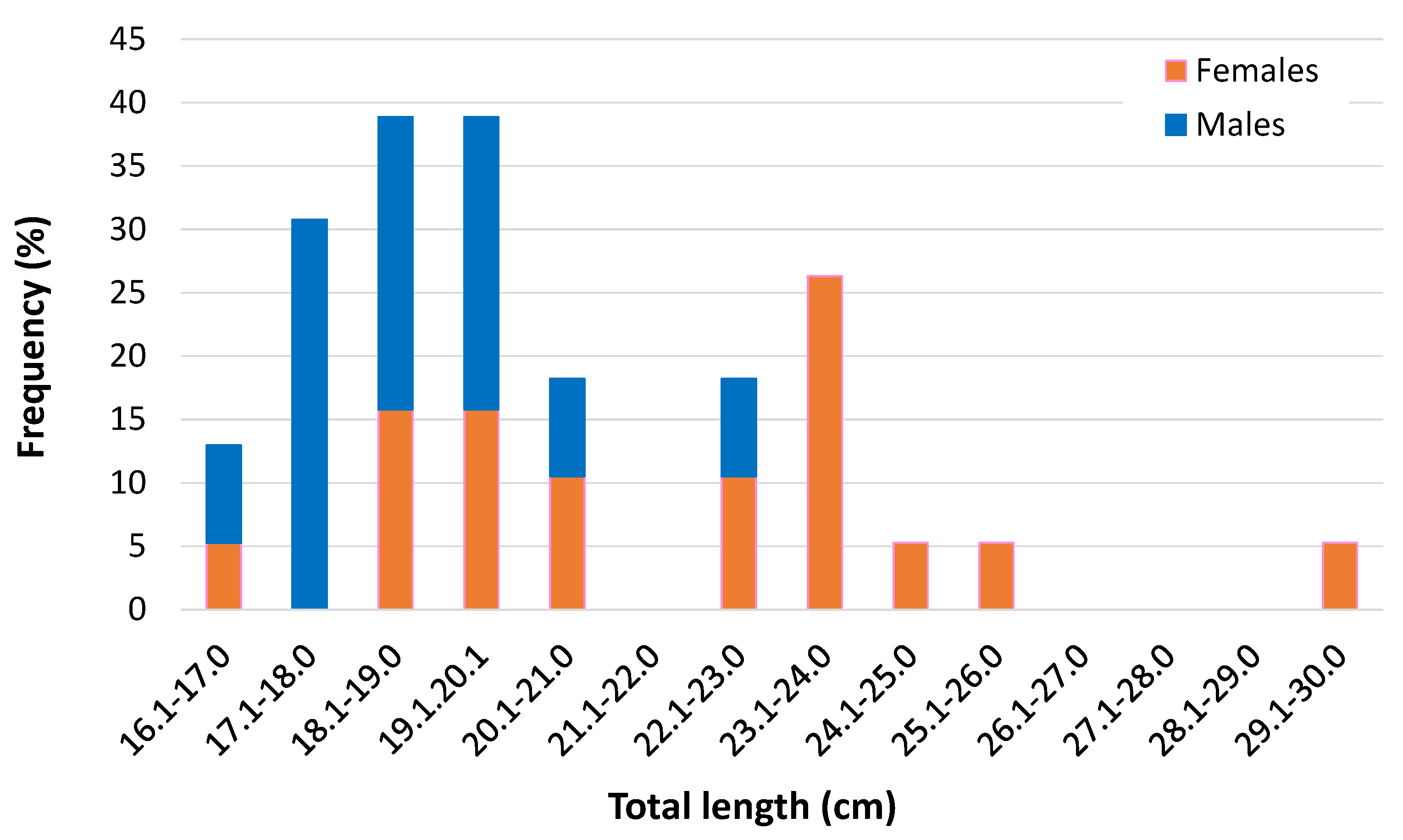
The analyses were conducted on 33 shorthorn sculpin individuals (19 females, 13 males, and 1 immature specimen of an undetermined sex). The biometrics are summarized in Table 1 and reported in full in Table S4, while the length frequency distributions of the females and males are shown in Figure 2. On average, the females were larger than the males, since the former had modal group at 23.1–24.0 cm TL, whereas the latter had a definite modal group at 17.1–18.0 cm TL. This divergence was also reported by Sonne et al. [39] and Nørregaard et al. [24] for sculpins from West Greenland, while the same research groups did not observe sex-specific differences in the biometric parameters of specimens from East Greenland [36,37]. Although no information is currently available on the spawning period of the shorthorn sculpin in Svalbard, it cannot be ruled out that the observed sex-related differences may be due to the females’ increased feeding and gonadal development in preparation for the reproductive season. In both sexes, feeding activity increases during the summer. Consistently, all individuals examined were found in normal to quite good condition, and the Fulton’s CI values were rather homogeneous, with the same mean in females and males (Table 1).

3.1. Measured Concentrations in Tissues

The results obtained from the multi-element analysis of the samples are reported in Table S6 and summarized in Table 2. The speciation analysis of mercury could not be performed for the livers and gonads due to matrix effects. A comparison with data from the literature for other Arctic sites is reported in Table S7.
The maximum average concentrations of the trace elements As, Cd, Cu, Mo, and Zn were detected in the livers, while the muscle had the highest concentrations of the macroelements Ca, K, and Mg and the maximum concentration of Sn. P, the essential trace element Co, and the non-essential trace element Ba had their maximum average concentrations in the gonads. Most of the elements considered in the present work had their maximum average concentrations in the gills (Al, Cr, Fe, Mn, Na, Ni, Pb, Se, Si, Sr, and V). The mean MPIs showed statistically significant differences (p < 0.05) among the tissues, being higher in the gills (4.55 ± 2.82 mg/kg), followed by the liver (2.44 ± 0.57 mg/kg), gonads (1.94 ± 0.49 mg/kg), and muscle (1.26 ± 0.57 mg/kg).
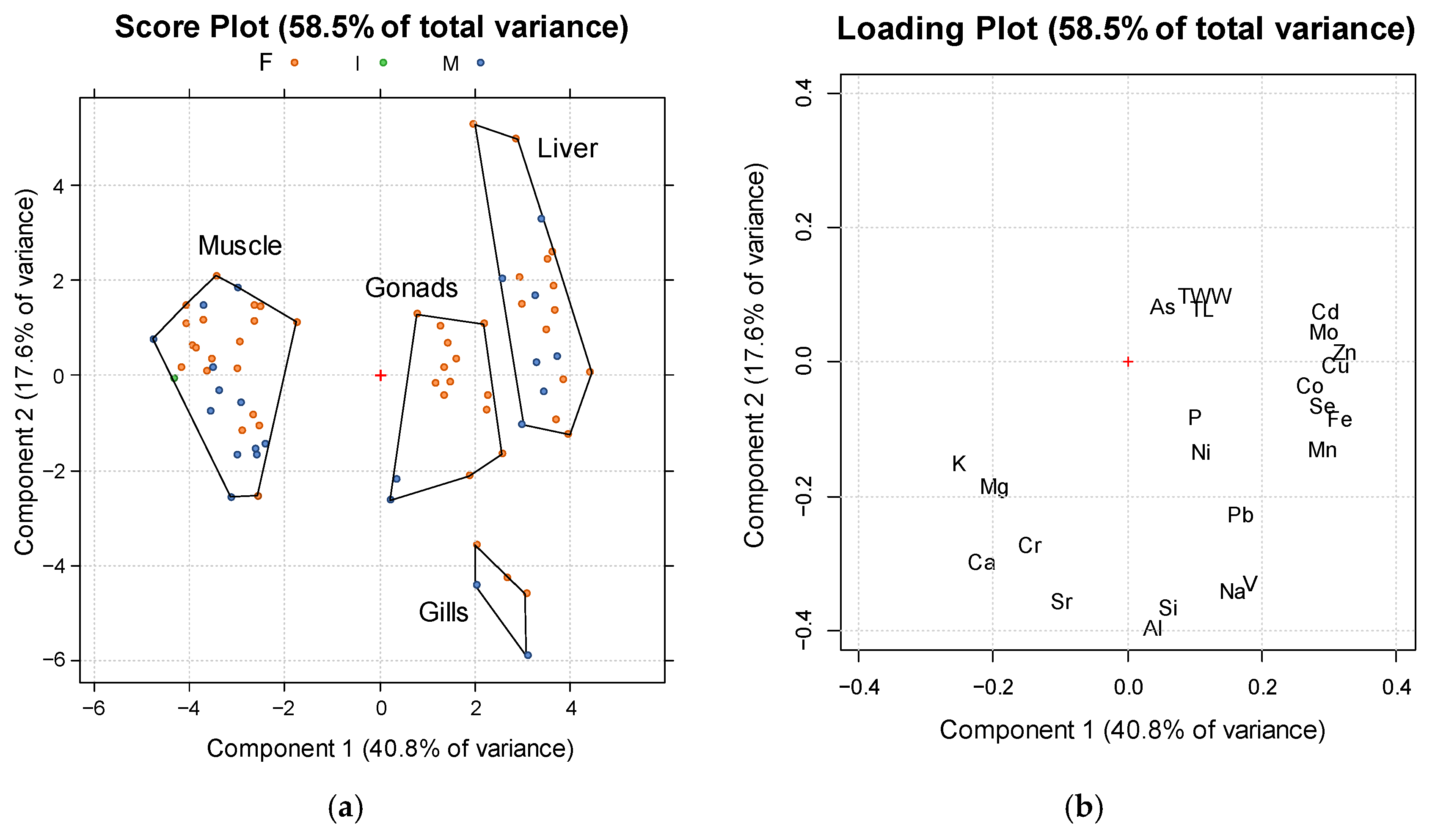
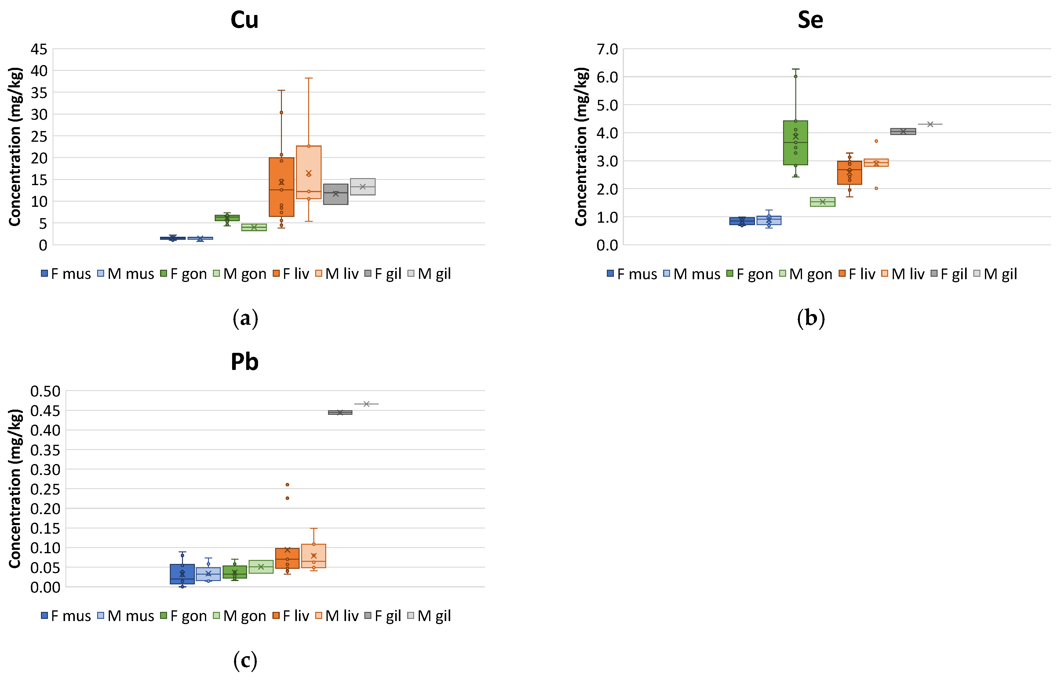
In order to explore the elemental distribution among the tissues more in detail, statistical evaluations were performed. Principal component analysis (PCA) was applied to the dataset, and two principal components were identified, explaining 40.8% and 17.6% of the total variance, respectively. The score and loading plots for PC1 and PC2 are shown in Figure 3a,b, respectively. The score plot (Figure 3a) revealed a good differentiation of the analyzed tissues. In particular, the muscle, gonad, and liver samples were distinguished along PC1, whereas the gill samples were differentiated from the other ones on PC2. By looking at the loading plot (Figure 3b), it can be seen that the main discrimination of the tissues was due to most of the trace elements (Cd, Co, Cu, Fe, Mn, Mo, Se, and Zn) being positively loaded on PC1, resulting in generally increasing concentrations moving from the muscles to the gonads and liver. The Cu concentration in the studied tissues is shown in Figure 4a as a representative example of this trend. The same trend was observed for Cd, Fe, Mo, and Zn. It is worth noting that, while showing the same general pattern in the muscle, gonads, and liver, Co, Mn, and Se presented significantly higher concentrations in the female gonads (Figure 4b for Se).
Considering the other trace elements, Ni, Pb, and V were detected at higher concentrations in the liver than in the muscle, while As had comparable concentrations in these tissues and significantly lower values in the gonads. The opposite trend was found for Cr, Sn, and Sr, which presented higher concentrations in the muscle than in the liver, as well as for Sn and Sr, which presented higher concentrations in the muscle than in the gonads.
The gills were discriminated from the other tissues by higher contents for the elements (Figure 3b) which loaded positively on PC1 and negatively on PC2, such as Al, Na, Pb, Si, V, and to a lesser extent Fe, Mn, and Ni. A representative example of this trend is shown in Figure 4c for Pb. In particular, Al and Si did not show significant differences among the other tissues, except for the gills. Moreover, Ba, Cr, and Sr were also particularly concentrated in the gills.
Finally, the major elements were clearly distinguished and had different trends; K and Mg presented the highest concentrations in the muscle and gonads, Ca’s were highest in the muscle, P’s were highest in the gonads, and Na’s were highest in the gonads and gills.

3.2. Size-Related Elemental Pattern

The loading plot of the PCA (Figure 3b) shows that the body length and body weight of the fish were positively correlated, as confirmed by the Pearson’s coefficient (r = 0.96, p < 0.001, n = 31).
The biometric parameters also correlated with As, while a negative correlation was detected with major elements like Ca, K, and Mg. The relationship between element accumulation and size was investigated by considering the various tissues separately (Figure 5 for representative examples). Arsenic showed a pronounced weight-dependent accumulation in the muscle (ρ = 0.561, p = 0.001, n = 32) and liver (ρ = 0.687, p = 0.001, n = 20). In the muscle, a positive correlation was also found for Hg (ρ = 0.404, n = 32), whereas a negative trend was observed for Ca, K, and Mn (ρ = −0.412, −0.356, and −0.431, respectively, n = 32). Interestingly, a positive correlation was observed for MeHg in the muscle only for female sculpins (ρ = 0.653, p = 0.003, n = 18). In addition, the percentages of MeHg with respect to the total mercury were highly variable in both the male and female groups (11–100%) and showed a slight decrease with the body weight (Figure S1).
Different trends were also observed in the gonads; the Na and Se concentrations increased with the body weight (ρ = 0.532 and 0.539, respectively, n = 15), whereas those of Mg, P, and Zn decreased (ρ = −0.557, −0.750, and −0.596, respectively; p = 0.001 for P and <0.05 for Mg and Zn, n = 15). It is noteworthy that no element accumulated in the liver as a function of the body weight, aside from the already mentioned As. Finally, significant correlations with the biometric parameters in the gills were observed for Hg and Ni (ρ = 0.9 and −0.9, respectively). Although the same trend was observed for Hg in previous studies [36,42], the number of gill-pooled samples analyzed in this work was rather low (n = 5), and the considerations for this tissue should hence be considered purely indicative.

3.3. Elemental Distribution in Male and Female Specimens

From the first two principal components of the PCA, it was possible to observe a discrimination between the male and female gonads along PC2 and, to a lesser extent, PC1 (Figure 3a). By looking at the elements influencing this pattern in the loading plot and observing the corresponding box plots, it was concluded that the elements presenting higher concentrations in the male gonads were Cr, K, Mg (Figure 6a), Ni, P, and V, whereas those having higher levels in the female gonads were Co, Cu, Fe, Mn, and Se (Figure 4b). Regarding the muscle and liver, no sex-based discrimination could be observed in the score plot. However, a univariate analysis showed that Ca, Mn, Sr, and Zn presented higher concentrations in the muscle of the male specimens than in the corresponding tissue of the females, while the element contents in the liver were different for As and Zn, being higher in the female samples (Figure 6b). Finally, the elements presenting higher concentrations in the male gills were Al, Ba, Si, and to a lesser extent Co, Cr, Ni, and Sb. However, the limited available data for this tissue did not allow any robust conclusions to be made.

4. Discussion

4.1. Distribution in Tissues

The elevated MPI and concentrations of several elements (Al, Ba, Ca, Cr, Fe, Mn, Na, Ni, Pb, Se, Si, Sn, Sr, and V) found in the analyzed gills are consistent with the known role of this tissue in the assimilation of elements. In fact, with their large surface area of active exchange, the gills are the dominant route of uptake for waterborne elements [32,49], and they have been used as an indicator for their concentrations in the water (e.g., [50]) and for heavy metal pollution [51]. In the shorthorn sculpin, they have been demonstrated to be the ideal tissue for assessing organismal exposure to Pb [42]. The relatively high elemental concentrations in the gills of the specimens from the Kongsfjorden may be ascribed to the contribution of the fjord environment or to potential local pollution. This is consistent with the findings of high concentrations of anthropogenic (90Sr) and natural (210Pb) radionuclides and heavy metals (Cd, Cu, Fe, Mn, Pb, and Zn) in glacial cryoconites of Spitsbergen [52], which may be transferred into the fjords via meltwater. It is also worth noting that Al accumulation in the gills was detected in the present work. Since Al deposition may cause damage to the gill epithelium and the apoptosis of ion-transporting cells, ultimately leading to ion regulatory and osmoregulatory dysfunction [53], future investigations on the status of the gill epithelium in shorthorn sculpins from the Kongsfjorden may be appropriate to assess the potential damages associated with the accumulation of this element in this tissue.
The observed prevalent hepatic distribution of several toxic elements and heavy metals (As, Cd, Cu, Mo, Pb, and Zn), supported by the relatively high MPI found for this organ, is in agreement with studies on metal accumulation in shorthorn sculpins from other Arctic areas which reported higher elemental concentrations for As, Cd, Co, Cu, Fe, Mn, Ni, Pb, Se, and Zn in the liver than in the muscle [24,29,35,37,40,42]. In fish, metals and trace elements with high affinity for metal-binding proteins (e.g., As, Cu, Pb, and Zn) tend to accumulate with higher short-term rates in metabolically active tissues such as that in the liver [39]. Therefore, this is considered tissue with a high accumulation ability and detoxification capability [54], making it a good indicator of exposure to heavy metals [39] and able to provide information on the mid-term effects of chronic exposure. Our results are also consistent with this indication for shorthorn sculpins [29,32,36,37,39].
Muscles are rather isolated compartments with weak accumulation potential [55], and for this reason, trace elements generally do not accumulate in this tissue, and elemental accumulation is considered indicative of chronic exposure [56]. For example, low levels of Pb accumulation in muscle tissue have been reported for shorthorn sculpins [42,49] and other fish species [15]. Our data support previous findings as, among trace metals, we found only Sn to be mainly concentrated in muscle. In the present work, the concentrations of some elements were higher than those found in specimens from East Greenland [35,37] (As, Co, Cr, Cu, Fe, Hg, Ni, Se, and Zn) and West Greenland [24,29,35] (Cr, Hg, and Se). The maximum Pb levels were generally lower than those from the literature, whereas the Hg content was lower compared with the West Greenland specimens, being comparable with those from Alaska, and higher than those from East Greenland. The Cd and Pb concentrations were lower than the European Union maximum values for the muscle meat of fish (0.050 mg/kg and 0.30 mg/kg for Cd and Pb, respectively) [57]. However, although the mean value of Hg in muscle from the present study was lower than the threshold established by the EU for fish meat (0.50 mg/kg), its concentration in the muscle of four specimens exceeded this limit.
The accumulation of some of the major elements (K, Mg, and P) detected in the gonads might have resulted from the energy storage process related to their maturation. In fish, from 10% to 90% of the body energy is transferred to gonads as a reproduction cost [58]. Although a considerable variation has been described for the length at maturity between populations [22,59], the lengths of most of specimens analyzed in present work (with only one exception) fell in the range of mature adults for European Arctic populations, and therefore gonadal development effort was not unlikely. Differences have been reported among populations for the spawning period. In Kiel Fjord, spawning starts in late November and continues until February. In Newfoundland and in the White Sea, the onset of spawning is in late November and continues until January, and in northern Norway, it commences in the second half of January and continues until early March [60]. While no information is available on the timing of spawning in Svalbard waters, macroscopic observation of the gonads of specimens collected in early July and analyzed in the present work resulted in most of the gonads being at the developing stage. At the beginning of development, feeding activities support the production of new gonadal tissue, allowing their condition to be maintained. Conversely, this condition decreases when the fish get closer to spawning [59]. Finding a high and homogeneous coefficient of condition (Fulton’s CI) in the specimens analyzed (Table 1) supports the hypothesis that initial gonadal development was ongoing during the sampling period. The accumulation of Co in the gonads seems to confirm such a trend, given that Co in the form of vitamin B12 is used by fish for gonadal maturation [61].

4.2. Relationship with Size

A general relationship between element concentrations in the tissues and fish size is not definite or established. Tissue growing more rapidly than the trace metal intake and high metabolic rate in young specimens is usually accounted for to explain the predominance of negative slopes in size–concentration relationships. However, seasonal fluctuations in body condition and fish physiology in general may support opposite or varying trends [62]. In the shorthorn sculpin, the relationship between element concentrations and size is controversial. In muscle, a linear correlation between body size and arsenic concentration was reported [40]. Conversely, Cd and Cu concentrations were found to decrease with body size in fish smaller than 300 g, and no correlation emerged in larger specimens.
The positive trends observed in this work for the muscles confirm what was previously reported for both As [31,40,42] and Hg [29,42]. The same researchers did not observe any trend for Cd [29], Pb [29], or Zn [29,40], which is in agreement with our findings. However, our data did not confirm either the increasing concentration with body weight found for Fe in Danish sculpins under laboratory conditions [42] or the decreasing accumulation of Cu in Canadian specimens [40].
In the liver, the increasing trend of As with fish weight is a typical behavior of this element [63], and it is in agreement with previous findings for shorthorn sculpins from West Greenland and Canada [31,40]. However, contrasting trends have been reported for other trace elements in this tissue, while we did not observe any specific pattern. For example, data for Pb showed an increase [33], a decrease [29,40], or no correlation [42] with the size. Also, for Cd, Se, and Zn, there are conflicting reports of increasing trends [33,42] or unrelated relationships [29,36]. Finally, in contrast with our findings, increasing metal concentrations with weight were observed for Co [33,42], Mn [33], and Ni [33], while decreasing trends were reported for Cu [36,40]. These discrepancies can be attributed to natural variability among organisms, differences in the biometric characteristics of the analyzed populations (age and sex), and their overall physiological condition and food habits [40,49].
The accumulation of As has also been observed in other fish species from the Kongsfjorden [11], and this can be ascribed to local contributions from the Svalbard terrestrial habitats. In fact, both sediments [64] and glacier cryoconites [65] from Kongsfjorden have been reported to display high concentrations of As. Moreover, a high bioavailability of As in the finer fraction of the fjord sediments (<63 μm) was observed both with a sequential selective extraction procedure and using a biomimetic approach [4]. This behavior in sediments was also observed for Cd, Cr, Cu, and Ni [4], and a potential contribution of the habitat to the accumulation of Zn has also been reported [11]. It is noteworthy that the maximum levels of these elements in the analyzed tissues were higher than most of the data in the literature from other Arctic areas (Table S7). This result could indicate a local contribution, due to the geochemical features of the fjord or the anthropic impact from the nearby settlement of Ny-Ålesund and cruise activities carried on in the Kongsfjorden during the summer months. Regarding arsenic, speciation studies are needed to clarify the percentage of toxic inorganic arsenic compared with non-toxic species such as arsenobetaine, usually accounting for more than 80% of the total As content in marine organisms [66].

4.3. Sex-Related Differences

The effect of sex on heavy metal accumulation in various tissues of the shorthorn sculpin has been considered in previous studies, and heterogeneous results have been reported [36,37]. For instance, sex was found to significantly affect the accumulation of hepatic Cu in shorthorn sculpins from West Greenland [39] but not in specimens from East Greenland [36]. The concentration of Se in the liver was higher in males than females in the specimens East Greenland [36], lower in males than females in the individuals from West Greenland [39], and similar in both sexes among specimens from Northeast Greenland [37]. The Pb concentration in the liver seems to not be affected by sex [36,37], as also validated by controlled laboratory exposure experiments [42]. In the muscle, no significant differences were detected in the concentrations of Ag, As, Cd, Co, Cr, Cu, Fe, Hg, Mn, Ni, Pb, Se, or Zn in the Northeast Greenland sculpin population [37]. However, higher concentrations of Cu in the muscles of females than males were found in fish smaller than 300 g from the Canadian Arctic [40].
The higher hepatic concentration of Zn found in females compared with males is in accordance with what was reported for the species in the Canadian Arctic, where the Zn concentrations in the liver were lower for males than females [40]. Conversely, other divergences among specimens of different sexes (higher Ca, Mn, Sr, and Zn concentrations in the muscles of males and a higher hepatic concentration of As in females) were neither studied nor reported in other works. Differences in sex-related element concentrations have been ascribed to differences in growth, age, metabolism, and diet [32,39]. In the case of testis- or ovary-specific accumulation observed in this work (higher concentrations of Cr, K, Mg, Ni, P, and V for males and Co, Cu, Fe, Mn, and Se for females), the differential mechanisms and pathways of male and female gonad maturation may have played a role.
In general, the discrepancies between the results presented in this study and other works could be related to differences in the habitats of the collected fish as well as different characteristics of the considered populations in terms of age and size. For example, the fish analyzed by Nørregaard et al. [37] were comparable to the specimens in this work (19.4–25.6 cm), while those studied by Sonne et al. [39] were longer on average (over 30 cm).

4.4. Mercury Speciation

An increase in the total Hg concentration in the muscle tissue with age was observed by Harley et al. [41] and Hansson et al. [29] in sculpins collected in the Bering Sea and in West Greenland, respectively. These trends reflect the bioaccumulation of this metal within the organisms, both in inorganic and organic form [63]. Regarding organic mercury, the only study reporting MeHg concentrations in the shorthorn sculpin was that of Harley et al. [41], who found a positive correlation between the MeHg concentration and a fish’s age. Since the specimens analyzed in that study were mostly females (11 out of 13) and bigger than those considered in the present work (28–65 cm versus 14–30 cm), our data confirm an accumulation of MeHg in muscle tissue related to size. However, it is still not clear if this trend is sex-dependent, due to the limited number of large male specimens. The average MeHg percentages in the muscle of the sculpins analyzed by Harley et al. [41] were 81–94%, consistent with the indication of a percentage of organic mercury in fish muscle ranging from 70 to 100% [67]. The lower percentages found in this work could be ascribed to differences in the biometric parameters of the analyzed sculpins (younger specimens), in their diet or habitat. Since these are the first data on MeHg in the shorthorn sculpin collected in Svalbard, future studies are needed to provide further insights and ultimately confirm these results. However, the decrease in the percent of MeHg with age was also observed by Harley et al. [41] in both the muscle and internal organs, possibly due to the processes of demethylation of MeHg and storage as inorganic Hg in the tissues, as was already found in other organisms [68,69]. Similarly, a relatively large variation in the percent of MeHg was also found by the same authors in young fish [41], explaining that demethylation of MeHg could occur only after a critical threshold of the total Hg concentration had been exceeded.
It is well known that the bioaccumulation of MeHg is partially compensated by a protective role of Se in the form of selenide, which forms HgSe due to its high affinity with Hg2+ [70]. Hence, the molar Se/Hg ratio in the samples must be computed for a better evaluation of the potential toxicity of the concentration of Hg in fish [71,72]. In particular, when the molar Se/Hg ratio is higher than one, Se is able to diminish Hg accumulation and toxicity, while Hg’s toxic potential is higher with ratios lower than one [71,73]. In the present work, the Se/Hg molar ratios were always higher than one in both the muscle (11.0 ± 2.2) and gills (75.6 ± 6.7). Similar values were obtained by Sonne et al. [39], who found a molar ratio of 10 in the liver of specimens collected in West Greenland, indicating a molar surplus of Se as a detoxification element or mechanism of Hg. Furthermore, the ratios in our samples significantly decreased with an increase in body weight for females (ρ= −0.757, p < 0.001, n = 17, Figure S1d), suggesting the bioaccumulation of Hg with increasing fish sizes, since Se did not exhibit the same behavior.

5. Conclusions

The analysis of shorthorn sculpins collected in the proximity of Ny-Ålesund provided the first detailed information on the metal accumulation patterns in the tissues of this species in the Svalbard. Both the high elemental concentrations and MPIs supported gills as the major uptake route of most metals, while the livers showed tissue where toxic elements were prevalently distributed. Differences in sex-related element concentrations were mostly found in the gonads, and accumulations of As and Hg with growth were detected in the muscle and liver. It is noteworthy that the maximum levels of some elements (e.g., Cd, Cr, Cu, Ni, and Zn) exceeded those reported in other Arctic areas.
The present work provides a snapshot of the elemental distribution in shorthorn sculpins from the Kongsfjorden. While further studies with periodic sampling are needed to investigate the metal contamination pattern year-round and disentangle the contributions from the multiple local sources (sediments, glacial meltwater, and impacts from settlement and cruise activities in the fjord), our data lay the basis for using this species as sentinel species for monitoring the effects of accelerated glacial melting on the local ecosystem.

Supplementary Materials

The following supporting information can be downloaded at https://www.mdpi.com/article/10.3390/jmse12122245/s1. Figure S1: Relationship between Hg accumulation in the muscle and body weight of the shorthorn sculpins. Table S1: ICP-AES operating parameters. Table S2: ICP-MS operating parameters. Table S3: CV-AAS operating parameters. Table S4: Individual and pooled samples (dry weights). Table S5: Analysis of the certified reference materials IAEA-407. Table S6: Elemental concentrations in the fish samples. Table S7: Total body length and elemental concentration in tissues of shorthorn sculpins from Kongsfjorden and other Arctic sites.

Author Contributions

Conceptualization, F.A., P.R., M.G. and L.G.; methodology, F.A., D.D.B., P.R. and L.G.; validation, F.A., P.R. and M.G.; formal analysis and investigation, F.A., F.M. and D.D.B.; resources, F.A., P.R., M.G. and L.G.; data curation, F.A.; writing—original draft preparation, F.A.; writing—review and editing, F.A., D.D.B., P.R., M.G. and L.G.; visualization, F.A. and L.G.; supervision, F.A., P.R. and L.G.; project administration, F.A., P.R. and L.G.; funding acquisition, M.G. and L.G. All authors have read and agreed to the published version of the manuscript.

Funding

This work was carried out in the frame of the PNRA 16_00281 (National Antarctic Research Program) and Research in Svalbard (RiS 10987) projects.

Institutional Review Board Statement

No ethics approval was needed for this study. The fieldwork activity was approved by the governor of Svalbard, sampling of shorthorn sculpins was conducted in compliance with the local regulations relating to harvesting of the fauna of Svalbard, and the capture and handling of animals was in accordance with the act on animal welfare.

Informed Consent Statement

Not applicable.

Data Availability Statement

The original contributions presented in this study are included in the article or Supplementary Materials. Further inquiries can be directed to the corresponding author.

Acknowledgments

The Department of Earth Systems Science and Environmental Technologies of the Italian National Research Council and Kings Bay AS are gratefully acknowledged for their logistic support in Ny-Ålesund. The authors thank Francesco Soggia and Federica Anahi Giussani for their assistance with the CV-AAS and ICP-MS measurements.

Conflicts of Interest

The authors declare no conflicts of interest.

References

  1. Paglia, E. A Higher Level of Civilisation? The Transformation of Ny-Ålesund from Arctic Coalmining Settlement in Svalbard to Global Environmental Knowledge Center at 79° North. Polar Rec. 2020, 56, e15. [Google Scholar] [CrossRef]
  2. Eckhardt, S.; Hermansen, O.; Grythe, H.; Fiebig, M.; Stebel, K.; Cassiani, M.; Baecklund, A.; Stohl, A. The Influence of Cruise Ship Emissions on Air Pollution in Svalbard—A Harbinger of a More Polluted Arctic? Atmos. Chem. Phys. 2013, 13, 8401–8409. [Google Scholar] [CrossRef]
  3. Pedersen, Å.Ø.; Convey, P.; Newsham, K.K.; Mosbacher, J.B.; Fuglei, E.; Ravolainen, V.; Hansen, B.B.; Jensen, T.C.; Augusti, A.; Biersma, E.M.; et al. Five Decades of Terrestrial and Freshwater Research at Ny-Ålesund, Svalbard. Polar Res. 2022, 41, 6310. [Google Scholar] [CrossRef]
  4. Grotti, M.; Soggia, F.; Ianni, C.; Magi, E.; Udisti, R. Bioavailability of Trace Elements in Surface Sediments from Kongsfjorden, Svalbard. Mar. Pollut. Bull. 2013, 77, 367–374. [Google Scholar] [CrossRef]
  5. Grotti, M.; Soggia, F.; Ardini, F.; Bazzano, A.; Moroni, B.; Vivani, R.; Cappelletti, D.; Misic, C. Trace Elements in Surface Sediments from Kongsfjorden, Svalbard: Occurrence, Sources and Bioavailability. Int. J. Environ. Anal. Chem. 2017, 97, 401–418. [Google Scholar] [CrossRef]
  6. Rajaram, R.; Ganeshkumar, A.; Emmanuel Charles, P. Ecological Risk Assessment of Metals in the Arctic Environment with Emphasis on Kongsfjorden Fjord and Freshwater Lakes of Ny-Ålesund, Svalbard. Chemosphere 2023, 310, 136737. [Google Scholar] [CrossRef]
  7. Bazzano, A.; Rivaro, P.; Soggia, F.; Ardini, F.; Grotti, M. Anthropogenic and Natural Sources of Particulate Trace Elements in the Coastal Marine Environment of Kongsfjorden, Svalbard. Mar. Chem. 2014, 163, 28–35. [Google Scholar] [CrossRef]
  8. Bazzano, A.; Ardini, F.; Terol, A.; Rivaro, P.; Soggia, F.; Grotti, M. Effects of the Atlantic Water and Glacial Run-off on the Spatial Distribution of Particulate Trace Elements in the Kongsfjorden. Mar. Chem. 2017, 191, 16–23. [Google Scholar] [CrossRef]
  9. Øverjordet, I.B.; Kongsrud, M.B.; Gabrielsen, G.W.; Berg, T.; Ruus, A.; Evenset, A.; Borgå, K.; Christensen, G.; Jenssen, B.M. Toxic and Essential Elements Changed in Black-Legged Kittiwakes (Rissa Tridactyla) during Their Stay in an Arctic Breeding Area. Sci. Total Environ. 2015, 502, 548–556. [Google Scholar] [CrossRef]
  10. Øverjordet, I.B.; Gabrielsen, G.W.; Berg, T.; Ruus, A.; Evenset, A.; Borgå, K.; Christensen, G.; Lierhagen, S.; Jenssen, B.M. Effect of Diet, Location and Sampling Year on Bioaccumulation of Mercury, Selenium and Cadmium in Pelagic Feeding Seabirds in Svalbard. Chemosphere 2015, 122, 14–22. [Google Scholar] [CrossRef]
  11. Singh, S.M.; Tsuji, M.; Singh, P.; Mulik, R.U. Elemental Composition and Freezing Tolerance in High Arctic Fishes and Invertebrates. Sustainability 2022, 14, 11727. [Google Scholar] [CrossRef]
  12. Yang, Z.; Yuan, L.; Xie, Z.; Wang, J.; Li, Z.; Tu, L.; Sun, L. Historical Records and Contamination Assessment of Potential Toxic Elements (PTEs) over the Past 100 Years in Ny-Ålesund, Svalbard. Environ. Pollut. 2020, 266, 115205. [Google Scholar] [CrossRef] [PubMed]
  13. Mohan, M.; Sreelakshmi, U.; Vishnu Sagar, M.K.; Gopikrishna, V.G.; Pandit, G.G.; Sahu, S.K.; Tiwari, M.; Ajmal, P.Y.; Kannan, V.M.; Abdul Shukkur, M.; et al. Rate of Sediment Accumulation and Historic Metal Contamination in a Tidewater Glacier Fjord, Svalbard. Mar. Pollut. Bull. 2018, 131, 453–459. [Google Scholar] [CrossRef] [PubMed]
  14. Squadrone, S.; Prearo, M.; Brizio, P.; Gavinelli, S.; Pellegrino, M.; Scanzio, T.; Guarise, S.; Benedetto, A.; Abete, M.C. Heavy Metals Distribution in Muscle, Liver, Kidney and Gill of European Catfish (Silurus Glanis) from Italian Rivers. Chemosphere 2013, 90, 358–365. [Google Scholar] [CrossRef]
  15. Wood, C.M.; Farrell, A.P.; Brauner, C.J. (Eds.) Homeostasis and Toxicology of Essential Metals; Fish Physiology; Elsevier: Amsterdam, The Netherlands, 2011; Volume 31A, ISBN 978-0-12-378636-4. [Google Scholar]
  16. Wood, C.M.; Farrell, A.P.; Brauner, C.J. (Eds.) Homeostasis and Toxicology of Non-Essential Metals; Fish Physiology; Elsevier: Amsterdam, The Netherlands, 2011; Volume 31B, ISBN 978-0-12-378634-0. [Google Scholar]
  17. Mecklenburg, C.W.; Lynghammar, A.; Johannesen, E.; Byrkjedal, I.; Christiansen, J.S.; Dolgov, A.V.; Karamushko, O.V.; Mecklenburg, T.A.; Møller, P.R.; Steinke, D.; et al. Marine Fishes of the Arctic Region Volume 1; CAFF Monitoring Series Report 28; Conservation of Arctic Flora and Fauna: Akureyri, Iceland, 2018. [Google Scholar]
  18. Brand, M.; Fischer, P. Species Composition and Abundance of the Shallow Water Fish Community of Kongsfjorden, Svalbard. Polar Biol. 2016, 39, 2155–2167. [Google Scholar] [CrossRef]
  19. Cardinale, M. Ontogenetic Diet Shifts of Bull-Rout, Myoxocephalus Scorpius (L.), in the South-Western Baltic Sea. J. Appl. Ichthyol. 2000, 16, 231–239. [Google Scholar] [CrossRef]
  20. Gray, B.P.; Norcross, B.L.; Beaudreau, A.H.; Blanchard, A.L.; Seitz, A.C. Food Habits of Arctic Staghorn Sculpin (Gymnocanthus Tricuspis) and Shorthorn Sculpin (Myoxocephalus Scorpius) in the Northeastern Chukchi and Western Beaufort Seas. Deep-Sea Res. Pt. II 2017, 135, 111–123. [Google Scholar] [CrossRef]
  21. Landry, J.J.; Fisk, A.T.; Yurkowski, D.J.; Hussey, N.E.; Dick, T.; Crawford, R.E.; Kessel, S.T. Feeding Ecology of a Common Benthic Fish, Shorthorn Sculpin (Myoxocephalus Scorpius) in the High Arctic. Polar Biol. 2018, 41, 2091–2102. [Google Scholar] [CrossRef]
  22. Luksenburg, J.A.; Pedersen, T. Sexual and Geographical Variation in Life History Parameters of the Shorthorn Sculpin. J. Fish. Biol. 2002, 61, 1453–1464. [Google Scholar] [CrossRef]
  23. Datsky, A.V. Biological Features of the Common Fish Species in Olyutorsky-Navarin Region and the Adjacent Waters of the Bering Sea: 4. Family Sculpins (Cottidae). J. Ichthyol. 2017, 57, 341–353. [Google Scholar] [CrossRef]
  24. Nørregaard, R.D.; Bach, L.; Geertz-Hansen, O.; Nabe-Nielsen, J.; Nowak, B.; Jantawongsri, K.; Dang, M.; Søndergaard, J.; Leifsson, P.S.; Jenssen, B.M.; et al. Element Concentrations, Histology and Serum Biochemistry of Arctic Char (Salvelinus Alpinus) and Shorthorn Sculpins (Myoxocephalus Scorpius) in Northwest Greenland. Environ. Res. 2022, 208, 112742. [Google Scholar] [CrossRef] [PubMed]
  25. Datsky, A.V.; Vedishcheva, E.V.; Trofimova, A.O. Features of the Biology of Mass Fish Species in Russian Waters of the Chukchi Sea. 2. Families Pleuronectidae and Cottidae. J. Ichthyol. 2022, 62, 863–884. [Google Scholar] [CrossRef]
  26. Madenjian, C.P.; Rediske, R.R.; Krabbenhoft, D.P.; Stapanian, M.A.; Chernyak, S.M.; O’Keefe, J.P. Sex Differences in Contaminant Concentrations of Fish: A Synthesis. Biol. Sex. Differ. 2016, 7, 42. [Google Scholar] [CrossRef] [PubMed]
  27. Hop, H.; Pearson, T.; Hegseth, E.N.; Kovacs, K.M.; Wiencke, C.; Kwasniewski, S.; Eiane, K.; Mehlum, F.; Gulliksen, B.; Wlodarska-Kowalczuk, M.; et al. The Marine Ecosystem of Kongsfjorden, Svalbard. Polar Res. 2002, 21, 167–208. [Google Scholar] [CrossRef]
  28. AMAP AMAP Assessment 2021: Mercury in the Arctic; Arctic Monitoring and Assessment Programme (AMAP): Tromsø, Norway, 2021; ISBN 978-82-7971-106-3.
  29. Hansson, S.V.; Desforges, J.-P.; van Beest, F.M.; Bach, L.; Halden, N.M.; Sonne, C.; Mosbech, A.; Søndergaard, J. Bioaccumulation of Mining Derived Metals in Blood, Liver, Muscle and Otoliths of Two Arctic Predatory Fish Species (Gadus Ogac and Myoxocephalus Scorpius). Environ. Res. 2020, 183, 109194. [Google Scholar] [CrossRef] [PubMed]
  30. Stephensen, E.; Svavarsson, J.; Sturve, J.; Ericson, G.; Adolfsson-Erici, M.; Förlin, L. Biochemical Indicators of Pollution Exposure in Shorthorn Sculpin (Myoxocephalus Scorpius), Caught in Four Harbours on the Southwest Coast of Iceland. Aquat. Toxicol. 2000, 48, 431–442. [Google Scholar] [CrossRef]
  31. Bohn, A. Arsenic in Marine Organisms from West Greenland. Mar. Pollut. Bull. 1975, 6, 87–89. [Google Scholar] [CrossRef]
  32. Dang, M.; Nørregaard, R.; Bach, L.; Sonne, C.; Søndergaard, J.; Gustavson, K.; Aastrup, P.; Nowak, B. Metal Residues, Histopathology and Presence of Parasites in the Liver and Gills of Fourhorn Sculpin (Myoxocephalus Quadricornis) and Shorthorn Sculpin (Myoxocephalus Scorpius) near a Former Lead-Zinc Mine in East Greenland. Environ. Res. 2017, 153, 171–180. [Google Scholar] [CrossRef]
  33. Dang, M.; Pittman, K.; Bach, L.; Sonne, C.; Hansson, S.V.; Søndergaard, J.; Stride, M.; Nowak, B. Mucous Cell Responses to Contaminants and Parasites in Shorthorn Sculpins (Myoxocephalus Scorpius) from a Former Lead-zinc Mine in West Greenland. Sci. Total Environ. 2019, 678, 207–216. [Google Scholar] [CrossRef]
  34. Dang, M.; Pittman, K.; Sonne, C.; Hansson, S.; Bach, L.; Søndergaard, J.; Stride, M.; Nowak, B. Histological Mucous Cell Quantification and Mucosal Mapping Reveal Different Aspects of Mucous Cell Responses in Gills and Skin of Shorthorn Sculpins (Myoxocephalus Scorpius). Fish. Shellfish. Immun. 2020, 100, 334–344. [Google Scholar] [CrossRef]
  35. Dietz, R.; Riget, F.; Johansen, P. Lead, Cadmium, Mercury and Selenium in Greenland Marine Animals. Sci. Total Environ. 1996, 186, 67–93. [Google Scholar] [CrossRef] [PubMed]
  36. Kaarsholm, H.M.; Verland, N.; Nørregaard, R.D.; Bach, L.; Søndergaard, J.; Rigét, F.F.; Dietz, R.; Hansen, M.; Eulaers, I.; Desforges, J.-P.; et al. Histology of Sculpin Spp. in East Greenland. II. Histopathology and Trace Element Concentrations. Toxicol. Environ. Chem. 2018, 100, 769–784. [Google Scholar] [CrossRef]
  37. Nørregaard, R.D.; Dang, M.; Bach, L.; Geertz-Hansen, O.; Gustavson, K.; Aastrup, P.; Leifsson, P.S.; Søndergaard, J.; Nowak, B.; Sonne, C. Comparison of Heavy Metals, Parasites and Histopathology in Sculpins (Myoxocephalus Spp.) from Two Sites at a Lead-Zinc Mine in North East Greenland. Environ. Res. 2018, 165, 306–316. [Google Scholar] [CrossRef] [PubMed]
  38. Søndergaard, J. Dispersion and Bioaccumulation of Elements from an Open-Pit Olivine Mine in Southwest Greenland Assessed Using Lichens, Seaweeds, Mussels and Fish. Environ. Monit. Assess. 2013, 185, 7025–7035. [Google Scholar] [CrossRef]
  39. Sonne, C.; Bach, L.; Søndergaard, J.; Rigét, F.F.; Dietz, R.; Mosbech, A.; Leifsson, P.S.; Gustavson, K. Evaluation of the Use of Common Sculpin (Myoxocephalus Scorpius) Organ Histology as Bioindicator for Element Exposure in the Fjord of the Mining Area Maarmorilik, West Greenland. Environ. Res. 2014, 133, 304–311. [Google Scholar] [CrossRef]
  40. Bohn, A.; Fallis, B.W. Metal Concentrations (As, Cd, Cu, Pb and Zn) in Shorthorn Sculpins, Myoxocephalus Scorpius (Linnaeus), and Arctic Char, Salvelinus Alpinus (Linnaeus), from the Vicinity of Strathcona Sound, Northwest Territories. Water Res. 1978, 12, 659–663. [Google Scholar] [CrossRef]
  41. Harley, J.; Lieske, C.; Bhojwani, S.; Castellini, J.M.; López, J.A.; O’Hara, T.M. Mercury and Methylmercury Distribution in Tissues of Sculpins from the Bering Sea. Polar Biol. 2015, 38, 1535–1543. [Google Scholar] [CrossRef]
  42. Jantawongsri, K.; Nørregaard, R.D.; Bach, L.; Dietz, R.; Sonne, C.; Jørgensen, K.; Lierhagen, S.; Ciesielski, T.M.; Jenssen, B.M.; Haddy, J.; et al. Histopathological Effects of Short-Term Aqueous Exposure to Environmentally Relevant Concentration of Lead (Pb) in Shorthorn Sculpin (Myoxocephalus Scorpius) under Laboratory Conditions. Environ. Sci. Pollut. Res. 2021, 28, 61423–61440. [Google Scholar] [CrossRef]
  43. Søndergaard, J.; Halden, N.; Bach, L.; Gustavson, K.; Sonne, C.; Mosbech, A. Otolith Chemistry of Common Sculpins (Myoxocephalus Scorpius) in a Mining Polluted Greenlandic Fiord (Black Angel Lead-Zinc Mine, West Greenland). Water Air Soil Pollut. 2015, 226, 336. [Google Scholar] [CrossRef]
  44. Svendsen, H.; Beszczynska-Møller, A.; Hagen, J.O.; Lefauconnier, B.; Tverberg, V.; Gerland, S.; Ørbøk, J.B.; Bischof, K.; Papucci, C.; Zajaczkowski, M.; et al. The Physical Environment of Kongsfjorden-Krossfjorden, and Arctic Fjord System in Svalbard. Polar Res. 2002, 21, 133–166. [Google Scholar]
  45. Blackwell, B.G.; Brown, M.L.; Willis, D.W. Relative Weight (Wr) Status and Current Use in Fisheries Assessment and Management. Rev. Fish. Sci. 2000, 8, 1–44. [Google Scholar] [CrossRef]
  46. Rivaro, P.; Ianni, C.; Soggia, F.; Frache, R. Mercury Speciation in Environmental Samples by Cold Vapour Atomic Absorption Spectrometry with in Situ Preconcentration on a Gold Trap. Microchim. Acta 2007, 158, 345–352. [Google Scholar] [CrossRef]
  47. Leardi, R.; Melzi, C.; Polotti, G. CAT (Chemometric Agile Tool). Available online: http://gruppochemiometria.it/index.php/software (accessed on 27 March 2023).
  48. Usero, J.; González-Regalado, E.; Gracia, I. Trace Metals in the Bivalve Mollusc Chamelea Gallina from the Atlantic Coast of Southern Spain. Mar. Pollut. Bull. 1996, 32, 305–310. [Google Scholar] [CrossRef]
  49. AMAP. AMAP Assessment Report: Arctic Pollution Issues; Arctic Monitoring and Assessment Programme (AMAP): Oslo, Norway, 1998. [Google Scholar]
  50. Zhang, J.; Tan, Q.-G.; Huang, L.; Ye, Z.; Wang, X.; Xiao, T.; Wu, Y.; Zhang, W.; Yan, B. Intestinal Uptake and Low Transformation Increase the Bioaccumulation of Inorganic Arsenic in Freshwater Zebrafish. J. Hazard. Mater. 2022, 434, 128904. [Google Scholar] [CrossRef]
  51. Daglish, R.W.; Nowak, B.F. Rainbow Trout Gills Are a Sensitive Biomarker of Short-Term Exposure to Waterborne Copper. Arch. Environ. Contam. Toxicol. 2002, 43, 98–102. [Google Scholar] [CrossRef]
  52. Łokas, E.; Zaborska, A.; Kolicka, M.; Różycki, M.; Zawierucha, K. Accumulation of Atmospheric Radionuclides and Heavy Metals in Cryoconite Holes on an Arctic Glacier. Chemosphere 2016, 160, 162–172. [Google Scholar] [CrossRef]
  53. Witters, H.E.; VanPuymbroeck, S.; Stouthart, A.J.H.X.; Bonga, S.E.W. Physicochemical Changes of Aluminium in Mixing Zones: Mortality and Physiological Disturbances in Brown Trout (Salmo Trutta L.). Environ. Toxicol. Chem. 1996, 15, 986–996. [Google Scholar] [CrossRef]
  54. Ploetz, D.M.; Fitts, B.E.; Rice, T.M. Differential Accumulation of Heavy Metals in Muscle and Liver of a Marine Fish, (King Mackerel, Scomberomorus Cavalla Cuvier) from the Northern Gulf of Mexico, USA. Bull. Environ. Contam. Toxicol. 2007, 78, 134–137. [Google Scholar] [CrossRef]
  55. Jovičić, K.; Nikolić, D.M.; Višnjić-Jeftić, Ž.; Đikanović, V.; Skorić, S.; Stefanović, S.M.; Lenhardt, M.; Hegediš, A.; Krpo-Ćetković, J.; Jarić, I. Mapping Differential Elemental Accumulation in Fish Tissues: Assessment of Metal and Trace Element Concentrations in Wels Catfish (Silurus Glanis) from the Danube River by ICP-MS. Environ. Sci. Pollut. Res. 2015, 22, 3820–3827. [Google Scholar] [CrossRef]
  56. Monferrán, M.V.; Garnero, P.; de los Angeles Bistoni, M.; Anbar, A.A.; Gordon, G.W.; Wunderlin, D.A. From Water to Edible Fish. Transfer of Metals and Metalloids in the San Roque Reservoir (Córdoba, Argentina). Implications Associated with Fish Consumption. Ecol. Indic. 2016, 63, 48–60. [Google Scholar] [CrossRef]
  57. European Union Commission Regulation (EC). No. 1881/2006 Setting Maximum Levels for Certain Contaminants in Foodstuffs. Off. J. Eur. Union 2006, 364, 5–24. [Google Scholar]
  58. MacKinnon, J.C. Summer Storage of Energy and Its Use for Winter Metabolism and Gonad Maturation in American Plaice (Hippoglossoides Platessoides). J. Fish. Res. Board Can. 1972, 29, 1749–1759. [Google Scholar] [CrossRef]
  59. Ennis, G.P. Reproduction and Associated Behaviour in the Shorthorn Sculpin, Myoxocephalus Scorpius in Newfoundland Waters. J. Fish. Res. Board Can. 1970, 27, 2037–2045. [Google Scholar] [CrossRef]
  60. Luksenburg, J.A.; Pedersen, T.; Falk-Petersen, I.B. Reproduction of the Shorthorn Sculpin Myoxocephalus Scorpius in Northern Norway. J. Sea Res. 2004, 51, 157–166. [Google Scholar] [CrossRef]
  61. Ahilan, B.; Jeyaseelan, M.J.P. Effect of Cobalt Chloride and Vitamin B12 on the Growth and Gonadal Maturation of Goldfish Carassius Auratus. Indian J. Fish. 2001, 48, 369–374. [Google Scholar]
  62. Farkas, A.; Salánki, J.; Specziár, A. Age- and Size-Specific Patterns of Heavy Metals in the Organs of Freshwater Fish Abramis Brama L. Populating a Low-Contaminated Site. Water Res. 2003, 37, 959–964. [Google Scholar] [CrossRef]
  63. Authman, M.M.; Zaki, M.S.; Khallaf, E.A.; Abbas, H.H. Use of Fish as Bio-Indicator of the Effects of Heavy Metals Pollution. J. Aquac. Res. Dev. 2015, 6, 1–13. [Google Scholar] [CrossRef]
  64. Singh, S.M.; Naik, S.; Mulik, R.U.; Sharma, J.; Upadhyay, A.K. Elemental Composition and Bacterial Occurrence in Sediment Samples on Two Sides of Brøggerhalvøya, Svalbard. Polar Rec. 2015, 51, 680–691. [Google Scholar] [CrossRef]
  65. Singh, S.M.; Avinash, K.; Sharma, P.; Mulik, R.U.; Upadhyay, A.K.; Ravindra, R. Elemental Variations in Glacier Cryoconites of Indian Himalaya and Spitsbergen, Arctic. Geosci. Front. 2017, 8, 1339–1347. [Google Scholar] [CrossRef]
  66. Lorenzana, R.M.; Yeow, A.Y.; Colman, J.T.; Chappell, L.L.; Choudhury, H. Arsenic in Seafood: Speciation Issues for Human Health Risk Assessment. Hum. Ecol. Risk Assess. 2009, 15, 185–200. [Google Scholar] [CrossRef]
  67. Liu, G.; Cai, Y.; O’Driscoll, N. Environmental Chemistry and Toxicology of Mercury; John Wiley and Sons: Hoboken, NJ, USA, 2011; ISBN 978-0-470-57872-8. [Google Scholar]
  68. Wagemann, R.; Trebacz, E.; Boila, G.; Lockhart, W.L. Methylmercury and Total Mercury in Tissues of Arctic Marine Mammals. Sci. Total Environ. 1998, 218, 19–31. [Google Scholar] [CrossRef] [PubMed]
  69. Wagemann, R.; Trebacz, E.; Boila, G.; Lockhart, W.L. Mercury Species in the Liver of Ringed Seals. Sci. Total Environ. 2000, 261, 21–32. [Google Scholar] [CrossRef] [PubMed]
  70. Sobolev, N.; Aksenov, A.; Sorokina, T.; Chashchin, V.; Ellingsen, D.G.; Nieboer, E.; Varakina, Y.; Veselkina, E.; Kotsur, D.; Thomassen, Y. Essential and Non-Essential Trace Elements in Fish Consumed by Indigenous Peoples of the European Russian Arctic. Environ. Pollut. 2019, 253, 966–973. [Google Scholar] [CrossRef] [PubMed]
  71. Dietz, R.; Sonne, C.; Basu, N.; Braune, B.; O’Hara, T.; Letcher, R.J.; Scheuhammer, T.; Andersen, M.; Andreasen, C.; Andriashek, D.; et al. What Are the Toxicological Effects of Mercury in Arctic Biota? Sci. Total Environ. 2013, 443, 775–790. [Google Scholar] [CrossRef]
  72. Raymond, L.J.; Ralston, N.V.C. Selenium’s Importance in Regulatory Issues Regarding Mercury. Fuel Process Technol. 2009, 90, 1333–1338. [Google Scholar] [CrossRef]
  73. Peterson, S.A.; Ralston, N.V.C.; Whanger, P.D.; Oldfield, J.E.; Mosher, W.D. Selenium and Mercury Interactions with Emphasis on Fish Tissue. Environ. Bioindic. 2009, 4, 318–334. [Google Scholar] [CrossRef]
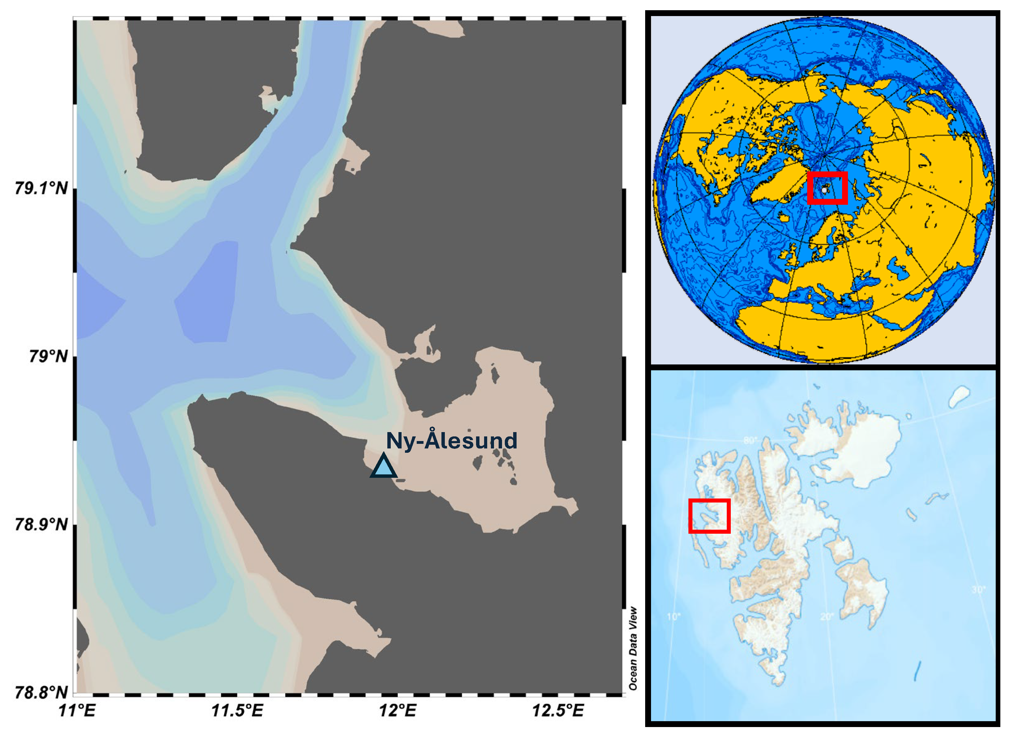
Figure 1. Map of the sampling area (Schlitzer and Reiner, Ocean Data View, https://odv.awi.de/, 2023).
Jmse 12 02245 g001
Figure 2. Length frequency distribution of female and male shorthorn sculpins from Kongsfjorden (Svalbard).
Jmse 12 02245 g002
Figure 3. Principal component analysis. (a) Score plot and (b) loading plot of PC1 vs. PC2. F = female; I = immature; M = male.
Jmse 12 02245 g003
Figure 4. Box plots showing the concentrations of (a) Cu, (b) Se, and (c) Pb in different tissues of shorthorn sculpins. “F mus” = female muscle; “M mus” = male muscle; “F gon” = female gonads; “M gon” = male gonads; “F liv” = female liver; “M liv” = male liver; “F gil” = female gills; “M gil” = male gills.
Jmse 12 02245 g004
Figure 5. Relationships between elemental concentration and body weight of shorthorn sculpins. (a) As in muscle; (b) Mn in muscle; (c) Se in gonads; and (d) P in gonads.
Jmse 12 02245 g005
Figure 6. Box plots showing the concentrations of (a) Mg and (b) Zn in the different tissues of shorthorn sculpins, highlighting the differences between the female and male specimens. “F mus” = female muscle; “M mus” = male muscle; “F gon” = female gonads; “M gon” = male gonads; “F liv” = female liver; “M liv” = male liver; “F gil” = female gills; “M gil” = male gills.
Jmse 12 02245 g006
Table 1. Biometrics of shorthorn sculpins collected in the Kongsfjorden. Data are means ± standard deviation, with minimum and maximum reported in brackets. TL = total length; TWW = total wet weight; Fulton’s CI = Fulton’s condition index.
Sexn TL (cm)TWW (g)Fulton’s CI
Female1922.3 ± 3.1 (17–30)142.1 ± 64.4 (46–328)1.21 ± 0.12 (0.94–1.42)
Male1319.1 ± 1.7 (16.5–23)84.9 ± 22.2 (54–138)1.21 ± 0.19 (0.93–1.74)
Immature114381.38
Table 2. Descriptive statistics of the elemental concentrations in the fish samples.
Tissue AlAsBaCaCdCoCrCuFeHgKMgMn
mg/kgmg/kgµg/kgmg/kgµg/kgmg/kgmg/kgmg/kgvmg/kgmg/kgmg/kgmg/kgmg/kg
MuscleMin<1.204.7<16493<250.0330.1500.890.08111,00110210.75
Mean89.7641904<250.0730.8861.5260.27612,44411491.27
Median48.3321601<250.0690.6121.5240.21012,48611511.22
Max4725.92365817<250.1364.4362.2550.92313,36013892.24
LiverMin<1.205.4<16611990.137<0.1013.899-61924631.90
Mean612.7323128480.2940.28415.1356-86417543.64
Median49.8323186130.2670.16712.4400-87667413.50
Max2031.03566424670.4460.99038.2739-10,91314156.28
GonadsMin<1.203.5<16210<250.186<0.1013.260-98066961.48
Mean186.5441460570.5580.5065.9106-11,49811243.41
Median66.0441445520.6240.2086.0108-11,51210513.22
Max13010.8441896950.7622.0727.4148-13,59017145.12
GillsMin276.01471420720.1791.7159.24930.08793168314.98
Mean427.43251862990.2173.01512.35700.12497798866.12
Median386.72581868970.1972.35911.95520.13496978945.79
Max669.756822331360.2734.97015.26440.14310,2949427.75
Tissue MoNaNiPPbSbSeSiSnSrVZnMeHg
mg/kgmg/kgµg/kgmg/kgmg/kgmg/kgmg/kgmg/kgmg/kgmg/kgµg/kgmg/kgmg/kg
MuscleMin<0.0052590<1026708<0.014<0.0070.6013<0.0530.9<40210.037
Mean0.022407621780450.0370.0240.85280.8279.0143350.164
Median0.013382219779950.0310.0220.85270.3866.9119340.117
Max0.080622048897560.0890.0411.24553.60938.3407520.679
LiverMin0.22528581385865<0.014<0.0071.7112<0.053<0.033<4081-
Mean0.355441425487520.088<0.0072.69280.1212.6267143-
Median0.356447424891020.067<0.0072.82240.1212.1147141-
Max0.548578839711,2820.260<0.0073.70770.1216.4739223-
GonadsMin0.0574769<1028436<0.014<0.0071.3716<0.0531.4<4091-
Mean0.083616324810,8930.0400.0143.55320.1504.7211115-
Median0.080601222410,6860.0360.0143.47300.1504.7158119-
Max0.110804051114,7260.0700.0166.27590.1508.8806129-
GillsMin0.082852918175320.4400.0073.93600.32511.0198575<0.016
Mean0.086910731076930.4520.0324.131200.79017.12380840.030
Median0.085867233177700.4490.0124.151110.67517.22376850.030
Max0.0911050040577800.4660.0774.301901.37023.42710870.041
Disclaimer/Publisher’s Note: The statements, opinions and data contained in all publications are solely those of the individual author(s) and contributor(s) and not of MDPI and/or the editor(s). MDPI and/or the editor(s) disclaim responsibility for any injury to people or property resulting from any ideas, methods, instructions or products referred to in the content.