Brief Report

Effect of Biologger Attachment on the Stress and Health State of the Spotted Sea Bass Lateolabrax maculatus

1
Marine Biotechnology & Bioresource Research Department, Korea Institute of Ocean Science & Technology, Busan 49111, Republic of Korea
2
Environmental Assessment Group, Korea Environment Institute, Sejong 30147, Republic of Korea
3
Department of Marine Environment, MS BioLab, Daejeon 34576, Republic of Korea
*
Author to whom correspondence should be addressed.
J. Mar. Sci. Eng. 2024, 12(5), 793; https://doi.org/10.3390/jmse12050793
Submission received: 28 March 2024 / Revised: 4 May 2024 / Accepted: 4 May 2024 / Published: 9 May 2024
(This article belongs to the Section Marine Biology)

Abstract

:
The biologger is a widely used tool in biotelemetry for investigating marine fish. However, studies reported that the attachment of biologgers has a negative effect on the target organisms. We assessed the health status of spotted sea bass with attached biologgers by analyzing changes in their serum biochemistry and biomarker gene expression at varying biologger/fish body weight ratios. Especially, the assessment of the health status using biomarker genes offers the rapid evaluation of the condition of an individual. The genes bax, hsp70-2, and Cx32.7, associated with apoptosis, stress, and immunity, were selected as biomarker genes to assess the fish stress levels and overall health. The experimental groups included a control group without the biologger (C) and biologger-equipped groups, each carrying a biologger whose weight was 2.0–3.0% (W2), 5.0–6.0% (W5), and 10.0–12.0% (W10) of the fish body weight. Blood samples were collected on days 0, 1, 7, 14, and 21 post-attachment. The expression of bax, hsp70-2, and Cx32.7 was analyzed in the liver and muscle on day 21. The W10 group showed significantly higher levels of superoxide dismutase on day 1 and of glutamic oxaloacetic transaminase and glutamic pyruvic transaminase on day 7 than the other groups. On day 21 post-attachment, regardless of the biologger/fish body weight ratio, the liver and muscle tissues from groups W2, W5, and W10 showed significantly higher expression of bax, hsp70-2, and Cx32.7 than those from group C, respectively. However, there was no significant change in blood parameters depending on the weight of the attached biologger on day 21 post-attachment. These results indicate that the spotted sea bass gradually adapted to the attached biologgers of weights up to 10–12% of their body weight under our experimental conditions, providing clues to determine the timing of biologger release for biotelemetry studies.

1. Introduction

The pop-up satellite archival tag (PSAT) biologger is a widely used tool in biotelemetry for investigating marine fish, allowing the assessment of their habitats, spawning grounds, resources, and horizontal and vertical movements [1,2]. Recently, the applications of the PSAT biologger have expanded to include the examination of farmed fish responses to environmental changes, feed intake, and behavioral patterns in rearing environments, as well as physiological monitoring [3,4,5]. The advantage of PSAT is its ability to collect diverse data once attached to marine fish, monitoring them for a specific period and then automatically detaching from the fish on a predetermined date to transmit the collected data to the Argos system [6,7,8,9]. Owing to its design, PSAT can only be externally attached to the fish body, typically below the dorsal fin muscles, using tethers made of medical nylon, stainless steel, cable ties, steel wires/strings, or nylon bolts and nuts [4,10,11].
However, previous studies reported disadvantages associated with the external attachment of biologgers to fish, including muscle and integument damage caused by the attachment wire, secondary dislodgement of the biologger, and reduced swimming ability [4,12,13]. The weight of the biologger can also cause epidermal damage and impact the fish swimming ability due to its external attachment [4]. Although there are no universally established rules governing the relation between biologger weight and fish weight to avoid adverse effects, a few researchers contend that the weight of the biologger must ideally be below 2% of the fish body weight to prevent any undue impact on research outcomes [4,14]. However, it is necessary to note that these claims may vary depending on the fish species [15], highlighting the need to assess the impact of the biologger weight on the physiological status of fish before conducting biotelemetry research [4].
Blood biochemistry factors are widely recognized as indicators for evaluating and understanding the effects of stress, including those caused by the external or internal attachment of biologgers, on fish physiology and health [13,16,17,18,19]. In recent years, the development and utilization of biomarker genes to assess the health status of organisms has garnered significant interest. The assessment of the health status using biomarker genes offers the advantage of a rapid and cost-effective evaluation of the condition of an individual. In marine organisms, stress induced by environmental changes can disrupt homeostasis and lead to poor health. Consequently, many studies have employed biomarker genes to investigate the stress-induced effects on the physiology and health of marine organisms [20,21,22]
The spotted sea bass (Lateolabrax maculatus) is a highly economically valuable species in aquaculture [23] and has a wide distribution range, from the coasts of China to Vietnam and the Korea [24,25]. However, limited research has been conducted regarding its geographical distribution and ecology. Biotelemetry research is crucial for assessing the potential utilization of fish resources and acquiring scientific information. However, no previous studies investigated this aspect. Accordingly, this study serves as a preliminary investigation to gather geo-ecological information on spotted sea bass using biologgers. In this study, we aimed to assess the impact of the ratio of biologger weight to fish body weight in spotted sea bass equipped with biologgers by analyzing changes in serum biochemical markers and the expression of specific biomarker genes involved in apoptosis, stress, and immunity over time. The execution of this study necessitated the collection of samples such as blood, liver, and muscle. Consequently, this research had to be conducted in indoor rearing tanks. The environmental conditions required for such research imposed constraints on the experimental setup. To conduct the study, a suitable stocking density was maintained in the rearing tanks to ensure a stress-free environment for the experimental subjects.

2. Materials and Methods

2.1. Experimental Fish and Attachment of the Biologgers

Spotted sea bass (n = 12, mean body weight: 2399.2 ± 212.3 g, mean total length: 64.7 ± 3.3 cm, mean ± SD) were used as the experimental fish. These fish were reared in the biological laboratory at the Korea Institute of Ocean Science and Technology. Throughout the experiments, the fish were maintained in a recirculating filtration system, which included a biological filtration tank, a foam separator, and a rearing tank. They were fed a commercial feed (Aller Aqua, Christiansfeld, Denmark) with a protein content of 49.0% at a dosage of 1.0–1.5% of the fish body weight before the experiments. The biologgers employed in the study included a dummy mark-report satellite tag (mrPAT; Wildlife Computers Inc., Redmond, WA, USA) weighing 40 g and the MET CATS dummy (Marin EQ & Technology Solutions, Busan, Republic of Korea) weighing 140 g (Figure 1).
The experimental groups consisted of a control group without an attached biologger (C) and three biologger-equipped groups with biologgers weighing 2.0–3.0% of the fish body weight (W2), 5.0–6.0% of the fish body weight (W5), and 10.0–12.0% of the fish body weight (W10). To the spotted sea bass of the W2 group, one dummy mrPAT was attached, whereas to those of the W5 and W10 groups, one and two MET CATS dummies, respectively, were attached to the muscle beneath the dorsal fin. Each experimental group, including the control group, consisted of three individuals. Before the attachment of the biologger, all experimental fish were subjected to a 2-day fasting period to reduce stress. The fish were anesthetized with 150 mg/L of 2-phenoxyethanol (Junsei Chemical Co., Ltd., Koshigaya, Saitama, Japan), their body weights were measured, and subsequently, the biologgers were attached. Following the attachment, the attachment sites in all experimental fish were disinfected with a povidone–iodine solution, following which, the fish were immersed for 1 min in seawater containing 200 mg/L of the antibiotic oxytetracycline [26]. The fish were then randomly distributed into two rearing tanks (1.5 × 1.5 × 0.6 m), with six fish in each tank. The number of experimental fish was determined according to the size of the indoor rearing tanks installed for the study. This is because the stocking density can affect experimental fish. The rearing tanks maintained a salinity of 31.5–33.5 psu, a water temperature of 16.0–20.0 °C, dissolved oxygen levels of 6.8–8.1 mg/L, a pH of 7.8–8.1, and ammonia, nitrite, and nitrate nitrogen levels of 0.12–0.15 mg/L, 0.002–0.003 mg/L, and 1.2–4.5 mg/L, respectively. The experiments were conducted over a total period of 21 days.

2.2. Blood Biochemistry

To investigate the internal physiological responses of spotted sea bass to the biologgers at varying ratios of biologger weight/fish body weight, blood samples were collected from both control and experimental groups at various time points (days 0, 1, 7, 14, and 21) following the biologger attachment. The collected blood samples were used to analyze changes in serum biochemical markers and serum antioxidant levels, specifically, in superoxide dismutase (SOD). Before blood collection, each experimental fish was anesthetized with 2-phenoxyethanol (150 mg/L) for 1 min to minimize stress. Blood on each sampling day was collected from the caudal blood vessel using a syringe treated with heparin, and then the fish were immediately returned to the rearing tank. The blood samples were centrifuged at 12,000 rpm for 5 min at 4 °C to separate the plasma, subsequently analyzed for glutamic oxaloacetic transaminase (GOT), glutamic pyruvic transaminase (GPT), total protein (TP), total cholesterol (TCHO), and triglyceride levels. The measurements were performed using a blood analyzer (DRI-CHEM 4000i; FUJIFILM, Tokyo, Japan). In addition, the SOD levels in the plasma were determined using the competitive–inhibition enzyme-linked immunosorbent assay (ELISA) technique performed with an ELISA kit (CUSABIO, Wuhan, China).

2.3. Total RNA Extraction from Liver and Muscle

The liver and muscle were selected to assess the fish health status and stress levels using biomarker gene expression. Total RNA was extracted from these tissue samples using RNA Isoplus (TaKaRa, Shiga, Japan), following the manufacturer’s extraction protocol. The extracted total RNA was then quantified using a spectrophotometer (NanoVue, GE Healthcare, Buckinghamshire, UK). A quality check was performed to ensure the quality of the RNA, which confirmed that the 260/280 ratio fell within the range of 1.8–2.0. The extracted total RNA was subjected to DNase I treatment to remove any remaining genomic DNA and obtain pure total RNA free from genomic DNA contamination.

2.4. cDNA Synthesis Using Reverse Transcriptase

A reverse transcriptase (RT) reaction was conducted to generate a complementary DNA (cDNA) strand using only the mRNA present in the extracted total RNA. The RT reaction involved the usage of reverse transcriptase and 1 μg of total RNA. The synthesis of cDNA was performed using the CycleScript RT PreMix (dT) kit from Bioneer (Daejeon, Republic of Korea). To prepare the final reaction solution, 1 μg of total RNA and DEPC water were added to the premix provided in the kit, resulting in a total volume of 20 μL. The reaction was then carried out in a thermocycler (TaKaRa, Shiga, Japan), with the following temperature cycles repeated 12 times: 20 °C for 30 s, 42 °C for 4 min, and 55 °C for 30 s. Finally, the reaction was terminated by heating at 95 °C for 5 min, resulting in cDNA synthesis.

2.5. Assessment of the Expression of Biomarker Genes Using the Real-Time Polymerase Chain Reaction

The expression of biomarker genes was assessed using the real-time polymerase chain reaction (real-time PCR or qPCR). For the qPCR reaction, the SFC green 1 fast qPCR 2× master mix (SFC, Hongseong, Republic of Korea) was used. The reaction mix consisted of 1.0 μL of cDNA, 1.0 μL of each primer (20 pmol), 10.0 μL of master mix, and D-water, resulting in a final reaction volume of 20.0 μL. The qPCR reaction was performed using a qPCR system (CFX96 Optics Module; BIO-RAD, Singapore), and the fluorescence of the amplification product was monitored. The amplification conditions included an initial denaturation step at 95 °C for 5 min, followed by 40 cycles of denaturation at 95 °C for 20 s, annealing at 58 °C for 20 s, and extension at 72 °C for 20 s. A melting curve analysis was conducted by gradually increasing the temperature from 65 °C to 95 °C at 0.5 °C intervals. The comparative Ct method was employed to determine the relative expression levels of the target genes, with the expression levels normalized to that of the housekeeping gene rpl7. The primer sequences used for the qPCR analysis of the biomarker genes are shown in Table 1.

2.6. Statistical Analyses

An analysis of variance (ANOVA) was conducted to analyze the blood samples by examining the experimental groups using the SPSS 11.5 program (SPSS, Chicago, IL, USA). In the cases where a significant difference was observed in the ANOVA results, Tukey’s multiple range test was used to determine the significance between the means at a 95% confidence level.
For the analysis of biomarker genes, Student’s t-test was used to assess the significance between the control and the experimental groups. A p-value of 0.05 or lower was considered statistically significant.

3. Results and Discussion

3.1. Blood Biochemistry Analysis for Assessing the Health of Spotted Sea Bass in Response to Stress Induced by the Attached Biologgers at Different Biologger/Fish Body Weight Ratios

Figure 2 illustrates the changes in the plasma concentrations of GOT, GPT, TP, TCHO, and triglycerides in spotted sea bass based on the ratio of biologger weight to fish body weight and the duration of the biologger attachment. On day 0 and days 1, 7, 14, and 21 after the attachment, the concentrations of serum TP, TCHO, and triglycerides were not significantly affected by the biologger weight (p > 0.05). However, on day 7 post-attachment, the concentrations of GOT and GPT in the W10 group were significantly higher than in the other experimental groups (p < 0.05). The serum TP concentration is an indicator that reflects the immune function of fish, and changes in this concentration can be clinically relevant [31]. However, the biologger/fish body weight ratio did not affect the TP concentration in this study. TCHO and triglycerides are critical components and energy substrates derived from fat that meet the energy demands of fish [32,33,34]. Although TCHO and triglycerides are closely related indicators [31,35], the biologger/fish body weight ratio did not affect their concentrations, indicating that it did not impact body homeostasis and metabolism in spotted sea bass. Notably, GOT and GPT are non-specific plasma enzymes implicated in sugar, lipid, and protein metabolism [36]. Their activity increases when fish are in poor physiological conditions or when tissues such as the liver, heart, and muscles are damaged, which leads to their elevated blood levels [37]. They are important indicators for evaluating the stress response in fish [38]. The significantly higher levels of GOT and GPT observed in the W10 group than in the groups C, W2, and W5 on day 7 post-attachment suggested that a high biologger/fish body weight ratio can act as a stress factor, potentially causing muscle damage in spotted sea bass and affecting physiological activities. Park and Oh [37] conducted a similar study on rockfish (Sebastes schlegelii) and red sea bream (Pagrus major), attaching dummy mrPATs at biologger/fish body weight ratios of 4.1–4.9% and 3.1–3.2%. They reported no significant effects on the TP, TCHO, and triglyceride levels, which aligns with the findings obtained in the present study [37]. Oh and Jeong [26] reported no effects on the plasma GOT and GPT concentrations in yellowtails (Seriola quinqueradiata) after attaching dummy mrPATs at a biologger/fish body weight ratio of 0.7–0.9% for 28 days. However, they observed an effect on the TP and TCHO concentrations [26]. Oh [39] reported no significant changes in the serum concentrations of GPT, GOT, TP, and TCHO 35 days after attaching a dummy mrPAT to spotted sea bass with an average weight of 2356.7 g at a biologger/fish body weight ratio of 1.5–2.0% compared with their concentrations in spotted sea bass without attached biologgers [39]. These varying results suggest that the effects of the biologger weight/fish body weight ratio may differ depending on the fish species.
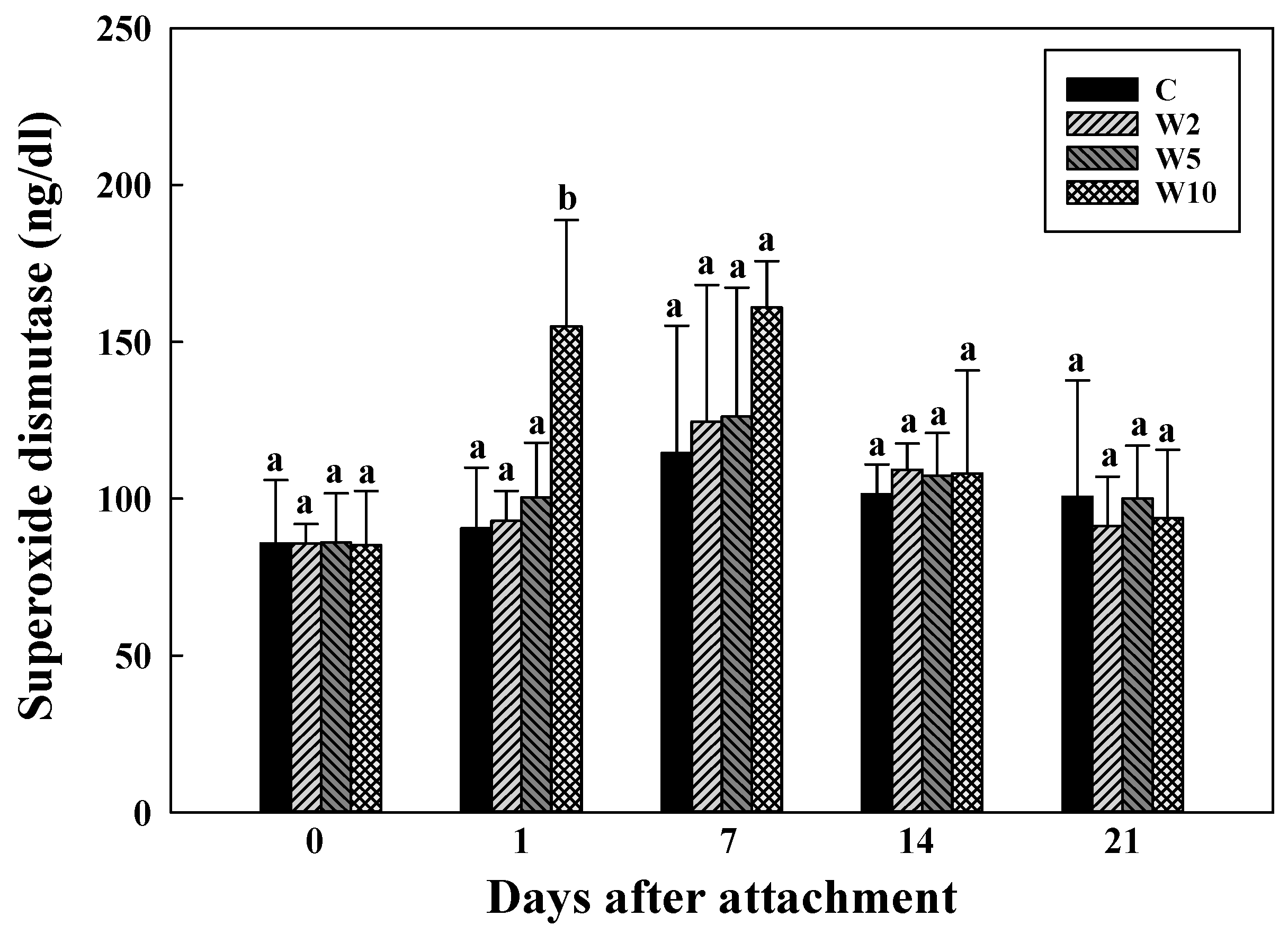
Figure 3 illustrates the changes in SOD concentrations in the plasma of spotted sea bass over time based on the ratio of biologger weight to fish body weight. As shown in Figure 2, the SOD concentrations were similar in the four experimental groups before the biologger attachment (day 0) and were not significantly different on days 1, 7, 14, and 21 after the biologger attachment, except for a significant increase (p < 0.05) on day 1 post-attachment in the W10 group. Stress can lead to the generation of reactive oxygen species in the body of fish, causing damage to cell and organ membranes and resulting in a functional decline. Notably, SOD is a representative immune indicator and acts as an antioxidant enzyme in the body, protecting against damage to cellular functions caused by the generation of free radicals [40]. The significant temporary increase in SOD concentrations on day 1 post-attachment in the W10 group suggests that the stress levels were the highest immediately post-attachment and then gradually decreased over time as the fish adapted, indicating a decrease in the stress levels over time. These findings are consistent with the results observed for GOT and GPT, which showed a significant increase in the levels of these markers on the seventh day after the biologger attachment in the W10 group compared to the control, W2, and W5 groups. The levels of GOT and GPT increase in the blood, in addition to their enhanced activation, during physiological stress and tissue damage in fish [37]. Therefore, the attachment of biologgers with a high biologger/fish body weight ratio of 10.0–12.0% (i.e., W10) appeared to induce stress in spotted sea bass. In addition, the stress caused by the biologger attachment was most prominent in the early days post-attachment, specifically on days 1 and 7, after which the stress level decreased, and the fish entered an adaptation phase. It is important to note that not only the weight of the biologger but also factors such as attachment location, attachment method, fish capture, and anesthesia during the attachment can induce stress in fish and cause physiological changes in the body [17,18,39,41,42,43]. Therefore, the impact of these factors needs to be considered and further investigated in the future.

3.2. Analysis of the Expression of Health Biomarker Genes Associated with Stress in Spotted Sea Bass Equippe with Biologgers at Different Biologger/Fish Body Weight Ratios

The expression levels of genes associated with apoptosis, stress, and immunity were analyzed to evaluate the stress levels and assess the health status of the spotted sea bass equipped with biologgers. Specifically, the genes bax, hsp70-2, and Cx32.7 were selected as biomarker genes to assess the fish stress levels and overall health. This analysis was conducted after 21 days following the attachment of the biologger.
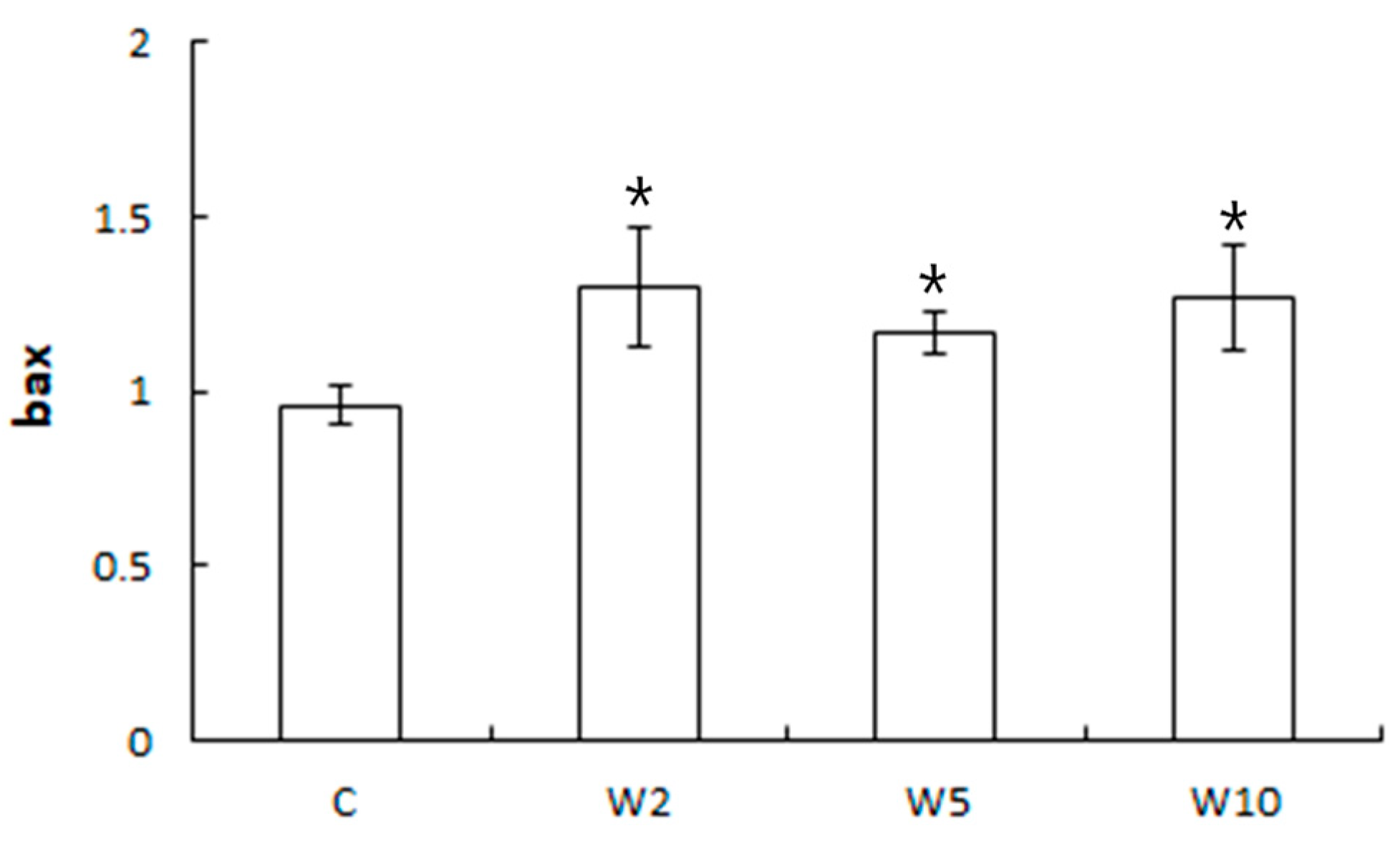
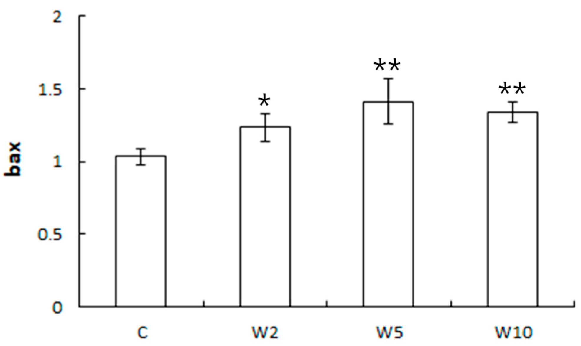
The expression of the bax gene in the liver tissue was significantly higher in the experimental groups with spotted sea bass equipped with biologgers than in the control group. However, there was no significant difference in the expression of the bax gene in the liver tissue based on the weight of the biologger (Figure 4). Similarly, the expression of the bax gene in the muscle tissue was significantly higher in the experimental groups equipped with biologgers than in the control group. However, there was no significant difference in the bax expression levels in the muscle tissue between the three groups W2, W5, and W10, equipped with biologgers (Figure 5).
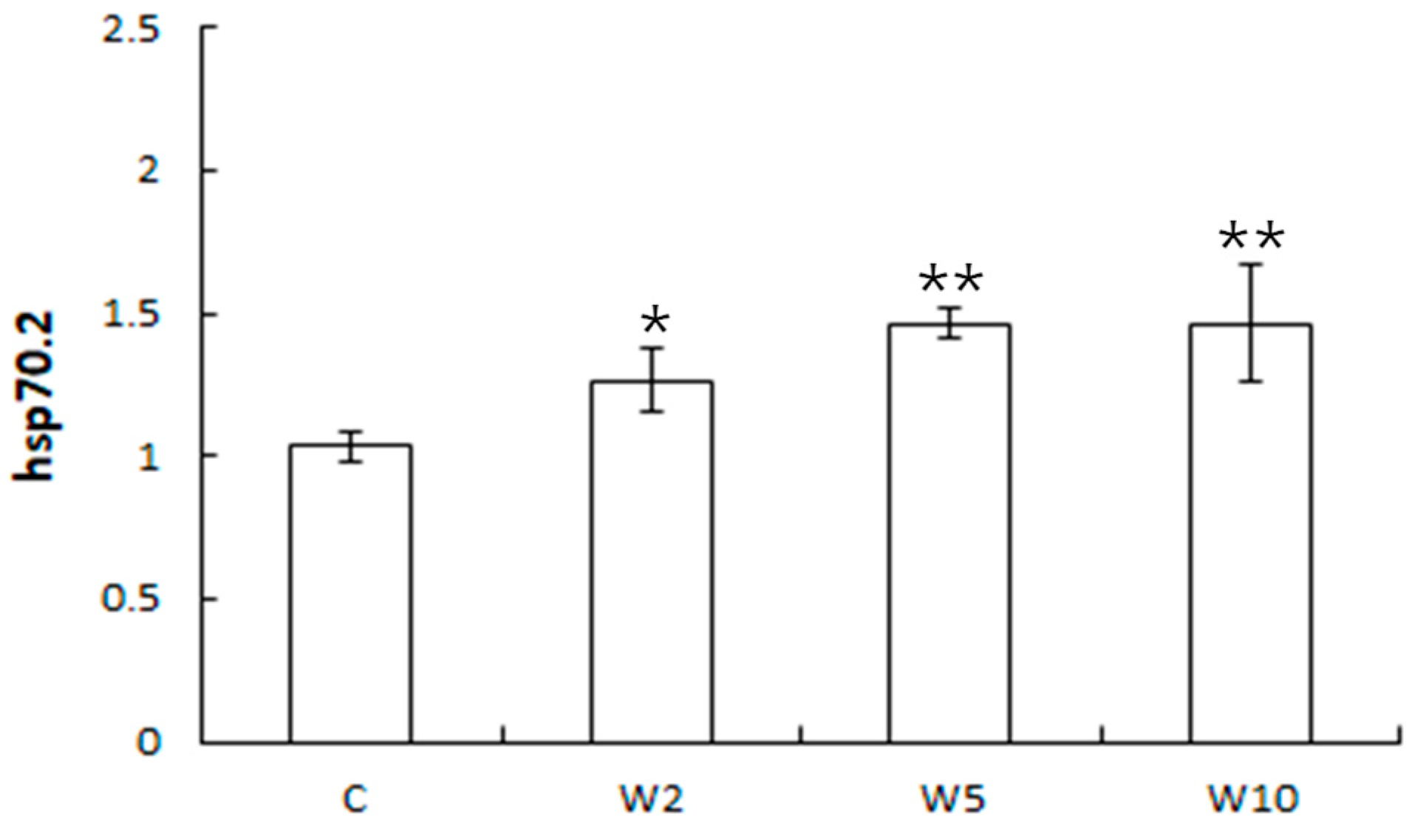
After 21 days from attaching the biologger to the back muscles, the expression levels of the bax gene in both liver and muscle tissues increased compared with that in the control group without the biologger. However, the difference in the expression levels was relatively small, within 1.5-fold. The bax gene encodes a member of the bcl-2 family, which acts as an anti-apoptotic or pro-apoptotic regulator in various cellular activities. It is activated by several factors, such as stress, heat, hydrogen peroxide, and pH [44]. The activation of the bax gene involves the interaction with p53, a tumor suppressor protein, and its upregulation is associated with apoptosis [45]. The expression of the bax gene tends to increase in response to stress and inflammation [46]. For example, the exposure of largemouth bass to the pesticide chlorpyrifos leads to increased bax gene expression due to oxidative stress and apoptosis in the fish body [47]. The flavonoid rutin, which reduces oxidative stress and inflammation, decreases bax expression in silver catfish [47]. Feeding restriction in male zebrafish leads to increased bax gene expression and a decreased bcl-2/bax ratio, accompanied by decreased sperm count, testicular tissue apoptosis, and inflammation [48]. These findings indicate that bax can serve as a biomarker gene in fish to evaluate the individual health status in terms of stress, apoptosis, and inflammation. In this study, the experimental group with the attached biologger showed an increase in bax gene expression compared to the control group without the biologger. However, the difference in the expression levels was relatively small, and there was no significant difference based on the weight of the biologger. These results suggest that the stress response in living organisms decreases significantly after 21 days from biologger attachment. Therefore, it is important to ensure that the biologger remains attached for a sufficient period without falling off to minimize the impact on the organism. After 21 days from attaching the biologger, the expression of the hsp70-2 gene in the liver tissue was significantly higher in the experimental groups with the attached biologger compared to the control group. There was no significant difference in the expression of the hsp70-2 gene based on the weight of the biologger in the experimental groups (Figure 6).
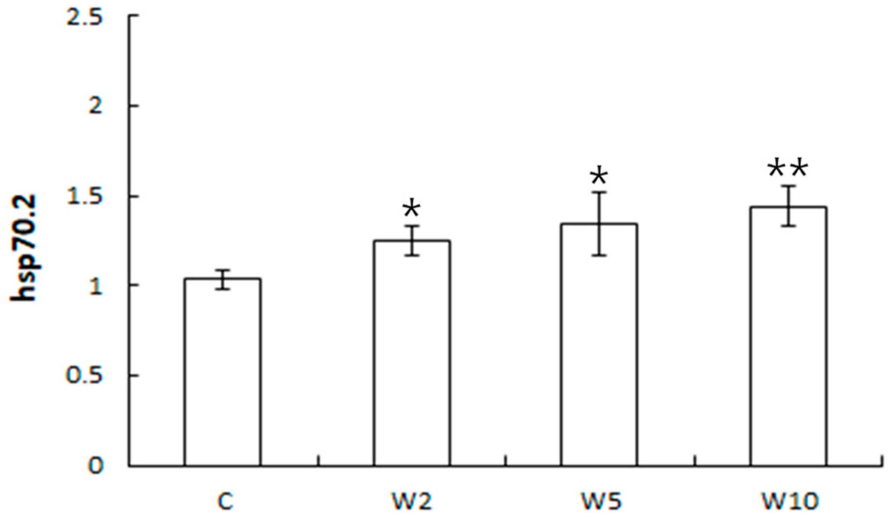
Similarly, the expression of the hsp70-2 gene in muscle tissue was higher in the experimental groups with the attached biologger compared to the control group. There was no significant difference in the expression of the hsp70-2 gene depending on the weight of the biologger (Figure 7).
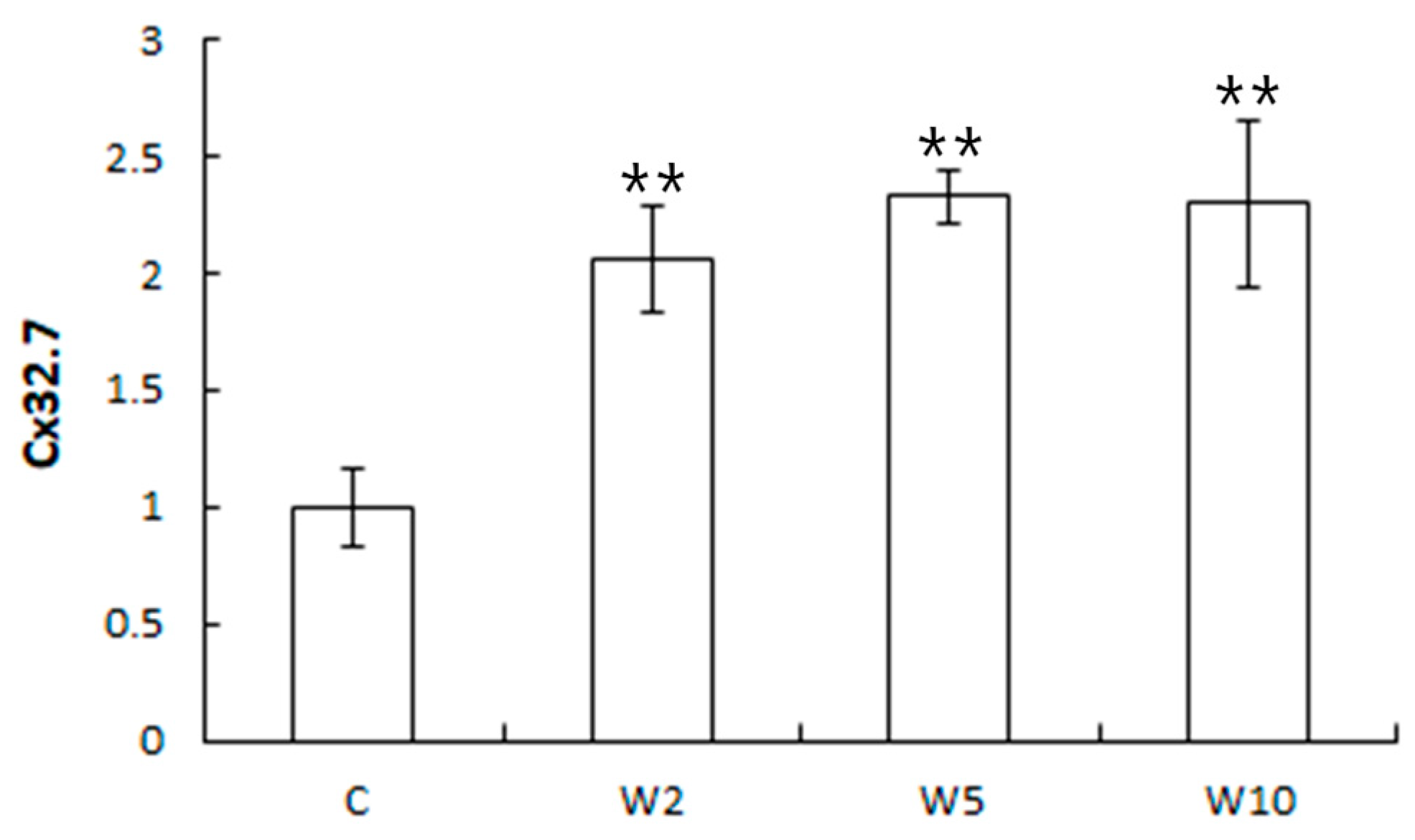
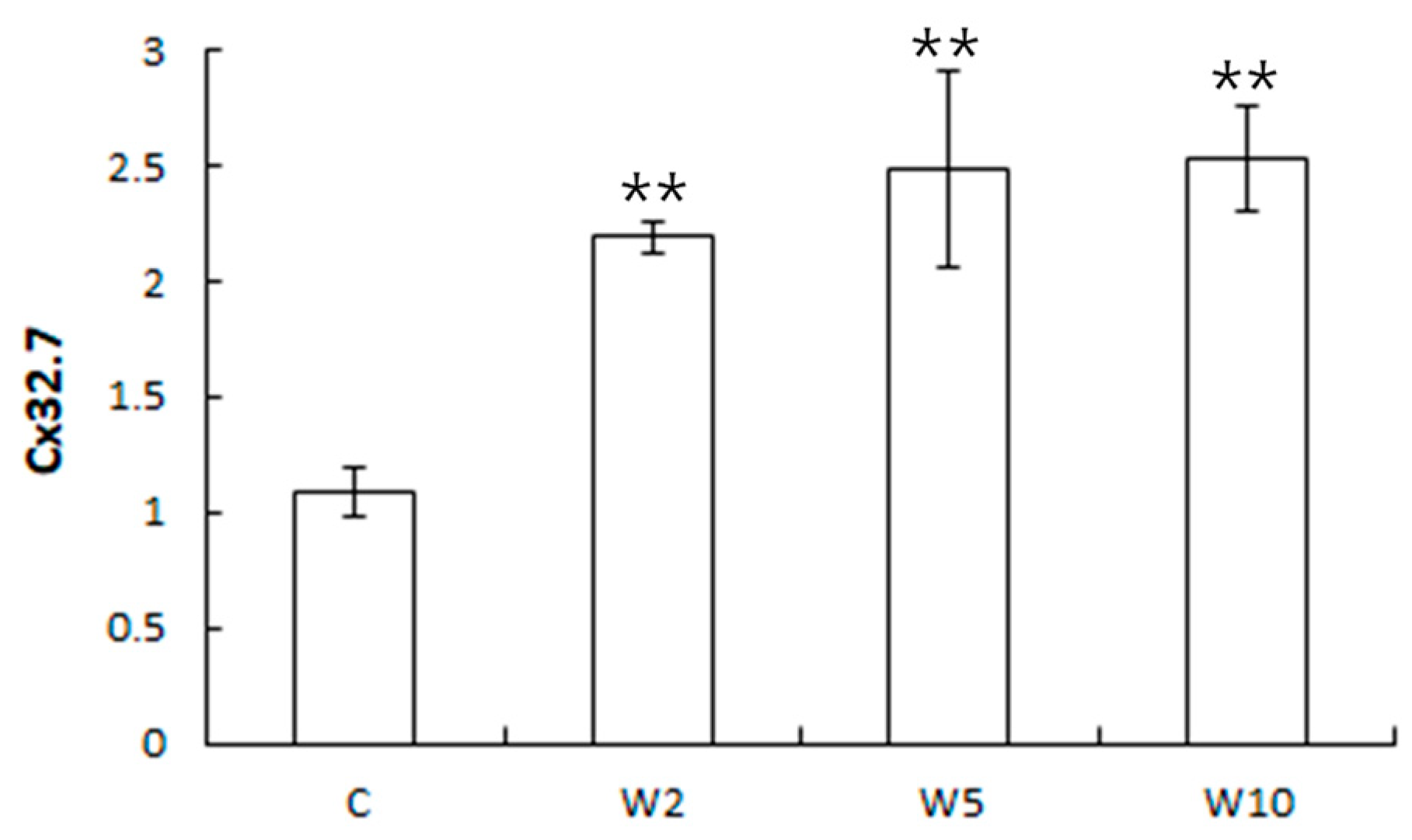
The approximately 1.5-fold difference in the expression levels of the hsp70-2 gene was also relatively small. Hsp70 proteins play a role in protecting cells from heat or oxidative stress [28]. They are involved in various biological processes, including signaling, apoptosis, protein homeostasis, and cell growth and differentiation and are implicated in diseases such as cancer, neurodegenerative diseases, cellular aging, and inflammatory diseases [49,50]. Hsp70-2 is a specific isoform of Hsp70 that is involved in maintaining homeostasis against stress [28]. In the liver tissue, the expression of the Cx32.7 gene was significantly higher in the experimental groups with the attached biologger compared to the control group after 21 days from the biologger attachment. The difference in the expression was also higher than two-fold. However, there was no significant difference in the expression of the Cx32.7 gene based on the weight of the biologger in the experimental groups (Figure 8). Similarly, the expression of the Cx32.7 gene in muscle tissue was higher in the experimental groups with the attached biologger compared to the control group, with an approximately 2.5-fold difference. There was no significant difference in the expression of the Cx32.7 gene in the muscle tissue depending on the weight of the biologger (Figure 9). The expression levels of the Cx32.7 gene were relatively higher than those of the bax and hsp70-2 genes in both liver and muscle tissues. Connexin32 (Cx32) is a protein involved in regulating signals through cell membranes, primarily in the liver and peripheral nervous system, and is expressed in various organs. It plays a role in the immune function via the release of extracellular ATP during inflammation, injury, or apoptosis [29,51]. Cx proteins have various immune functions beyond ATP release. In sea bass, Cx32.7, a specific isoform of Cx32, is known to be involved in pro-inflammatory ATP release and the immune response [29].
The biomarker genes selected for evaluation are associated with apoptosis, stress, and immunity. The expression of these genes is associated with the maintenance of homeostasis when the body encounters stress and immune imbalances. By analyzing the expression patterns of these genes, we can detect levels of stress and immunity, which, in turn, provides insights into the overall health status of an organism. A high expression of these genes indicates the body’s effort to maintain homeostasis, and the level of gene activity can be measured as an indicator of their related functions.
In this study, the level of stress in experimental fish with attached biologgers was assessed by analyzing the expression of the biomarker genes bax, hsp70-2, and Cx32.7. The results showed that the expression of these biomarker genes in the experimental fish with biologgers was slightly higher compared to that in the control fish without biologgers. However, the overall difference in gene expression based on the weight of the biologgers was not significant. Among the biomarker genes, Cx32.7 exhibited higher sensitivity and significance in terms of expression. The evaluation of biomarker genes is known to be highly sensitive and can provide more sensitive assessments compared to protein-level evaluations. In this study, the Cx32.7 gene, which is primarily associated with inflammation, showed significant expression changes. We hypothesized that the main source of stress in the spotted sea bass was inflammation in the tissues following the attachment of the biologgers. Analyzing the expression of biomarker genes in the tissues of experimental fish is an important method for evaluating the health of organisms in response to stress.
The use of biologgers in biological telemetry typically involves attaching them to the target organisms. However, depending on the attachment location or surgical method, this process can potentially cause injury to the target fish and negatively affect their normal swimming, growth, food intake, and survival. Previous studies examined these potential negative impacts [4,9,15].
This study aimed to assess the impact on experimental animals of biologger attachment by conducting a blood biochemistry analysis and examining biomarker genes related to stress, cell death, and immunology up to 21 days after biologger attachment. The findings suggest that variations may arise depending on the size of the target species and the method used to attach the biologger. However, the results of this study indicate that the level of stress experienced by the organisms during the 21 days following the biologger attachment was minimal. This suggests that the organisms were able to adapt to the presence of the biologger. Therefore, researchers interested in collecting data for eco-physiological research using biologgers should prioritize the selection of biologgers with high performance and efficient attachment capabilities.
This study required research methods such as the collection of blood, liver, and muscle from experimental fish for 21 days. Because it is difficult to carry out these procedures in the ocean, it was conducted in an indoor rearing tank (1.5 × 1.5 × 0.6 m). In addition, the 12 experimental fish used for the study were evenly divided into a control group and 3 experimental groups after weighing them, considering the size and the rearing density in the rearing tanks. In terms of reliability of the research results, it is believed that the number of experimental fish used could be low. However, due to the nature of this research, it was difficult to conduct it using a large number of animals. We tried to improve the reliability of the study through blood biochemistry analysis and the analysis of biomarker genes to ensure trust in the research results. The results of this study are believed to be valuable as basic data for related research.

4. Conclusions

A high ratio of biologger weight to fish body weight (i.e., 10–12%) affected blood biochemistry in spotted sea bass early after the biologger attachment (i.e., up to day 7), but had no effect thereafter. The experimental fish equipped with the biologger showed slightly higher expression levels of biomarker genes compared to the experimental fish not equipped with the biologger. These expression patterns of genes are believed to be because genetic analysis is more sensitive compared to blood biochemistry. These results indicate that spotted sea bass adapts over time after the attachment of biologgers of up to 10–12% of their body weight, as also indicated by the examined blood biochemistry under our experimental conditions, which provides clues to determine the best timing of the release of individuals with biologger attached for future biotelemetry studies of marine fish. Biologgers are very useful equipment for studying fish ecology, migratory routes, and behavior, but the weight of the biologger can have a physiological effect on the target fish. Therefore, it is important to determine the timing of release of fish with biologger attached by considering the weight of the attached biologger and the time after the biologger attachment during which the target fish’s physiology is not affected.

Author Contributions

Conceptualization, S.-Y.O. and J.-H.M.; methodology, S.-Y.O. and H.-S.K.; formal analysis, S.-Y.O. and H.-S.K.; investigation, S.-Y.O. and H.-S.K.; data curation, S.-Y.O. and H.-S.K.; writing—original draft preparation, S.-Y.O. and H.-S.K.; writing—review and editing, S.-Y.O. and H.-S.K.; funding acquisition, S.-Y.O. and J.-H.M. All authors have read and agreed to the published version of the manuscript.

Funding

This research was supported by the research project “Environmental Impact Analysis on Offshore Wind Farm and Database System Development”, which was driven by the Korea Environment Institute (KEI) and funded by the Korea Institute of Energy Technology Evaluation and Planning (KETEP) and the Ministry of Trade, Industry, and Energy (MOTIE) of Korea (Project No. 20203030020080, PN91870).

Institutional Review Board Statement

All experiments were conducted in compliance with the guidelines of the Institutional Animal Care and Experimental Committee of the Korea Institute of Ocean Science and Technology (KIOST), which approved the experimental protocol (1 November 2022, No. 2022-13).

Informed Consent Statement

Not applicable.

Data Availability Statement

Data are contained within the article.

Acknowledgments

The authors thank the Korea Institute of Energy Technology Evaluation and Planning (KETEP) and Ministry of Trade, Industry, and Energy (MOTIE) of the Republic of Korea (Project No. 20203030020080, PN91870). Finally, we thank the editor and the anonymous reviewers whose comments have greatly improved the manuscript.

Conflicts of Interest

The authors declare no conflict of interest.

References

  1. Eddy, C.; Brill, R.; Bernal, D. Rates of at-vessel mortality and post-release survival of pelagic sharks captured with tuna purse seines around drifting fish aggregating devices (FADs) in the Equatorial eastern Pacific Ocean. Fish. Res. 2016, 174, 109–117. [Google Scholar] [CrossRef]
  2. Lynch, S.D.; Marcek, B.J.; Marshall, H.M.; Bushnell, P.G.; Bernal, D.; Brill, R.W. The effects of pop-up satellite archival tags (PSAT) on the metabolic rate and swimming kinematics of juvenile sandbar shark Carcharhinus plumbeus. Fish. Res. 2017, 186, 205–215. [Google Scholar] [CrossRef]
  3. Bridger, C.J.; Booth, R.K. The effects of biotelemetry transmitter presence and attachment procedures on fish physiology and behavior. Rev. Fish. Sci. 2003, 11, 13–34. [Google Scholar] [CrossRef]
  4. Thorstad, E.B.; Rikardsen, A.H.; Alp, A.; Økland, F. The use of electronic tags in fish research—An overview of fish telemetry methods. Turk. J. Fish. Aquat. Sci. 2013, 13, 881–896. [Google Scholar]
  5. Macaulay, G.; Warren-Myers, F.; Barrett, L.T.; Oppedal, F.; Fore, M.; Dempster, T. Tag use to monitor fish behaviour in aquaculture: A review of benefits, problems and solutions. Rev. Aquac. 2021, 13, 1565–1582. [Google Scholar] [CrossRef]
  6. Wilson, S.G.; Polovina, J.J.; Stewart, B.S.; Meekan, M.G. Movements of whale sharks Rhincodon typus tagged at ningaloo reef, Western Australia. Mar. Biol. 2006, 148, 1157–1166. [Google Scholar] [CrossRef]
  7. Hazen, E.L.; Maxwell, S.M.; Bailey, H.; Bograd, S.J.; Hamann, M.; Gaspar, P.; Godley, B.J.; Shillinger, G.L. Ontogeny in marine tagging and tracking science: Technologies and data gaps. Mar. Ecol. Prog. Ser. 2012, 457, 221–240. [Google Scholar] [CrossRef]
  8. Block, B.A.; Dewar, H.; Farwell, C.; Prince, E.D. A new satellite technology for tracking the movements of Atlantic bluefin tuna. Proc. Natl. Acad. Sci. USA 1998, 95, 9384–9389. [Google Scholar] [CrossRef] [PubMed]
  9. Jepsen, N.; Thorstad, E.B.; Havn, T.; Lucas, M.C. The use of external electronic tags on fish: An evaluation of tag retention and tagging effects. Anim. Biotelem. 2015, 3, 49. [Google Scholar] [CrossRef]
  10. Domeier, M.L.; Dewar, H.; Nasby-Lucas, N. Mortality rate of striped marlin (Tetrapturus audax) caught with recreational tackle. Mar. Freshw. Res. 2003, 54, 435–445. [Google Scholar] [CrossRef]
  11. Gilly, W.F.; Markaida, U.; Baxter, C.H.; Block, B.A.; Boustany, A.; Zeidberg, L.; Reisenbichler, K.; Robinson, B.; Bazzino, G.; Salinas, C. Vertical and horizontal migrations by the jumbo squid Dosidicus gigas revealed by electronic tagging. Mar. Ecol. Prog. Ser. 2006, 324, 1–17. [Google Scholar] [CrossRef]
  12. Counihan, T.D.; Frost, C.N. Influence of externally attached transmitters on the swimming performance of juvenile white sturgeon. Trans. Am. Fish. Soc. 1999, 128, 965–970. [Google Scholar] [CrossRef]
  13. Thorstad, E.B.; Økland, F.; Finstad, B. Effects of telemetry transmitters on swimming performance of adult Atlantic salmon. J. Fish Biol. 2000, 57, 531–535. [Google Scholar] [CrossRef]
  14. Smircich, M.G.; Kelly, J.T. Extending the 2% rule: The effects of heavy internal tags on stress physiology, swimming performance, and growth in brook trout. Anim. Biotelem. 2014, 2, 16. [Google Scholar] [CrossRef]
  15. Jepsen, N.; Schreck, C.; Clements, S.; Thorstad, E.B. A brief discussion on the 2% tag/bodymass rule of thumb. In Aquatic Telemetry: Advances and Applications, Proceedings of the Fifth Conference on Fish Telemetry held in Europe, Ustica, Italy, 9–13 June 2003; Spedicato, M.T., Lembo, G., Marmulla, G., Eds.; FAO/COISPA: Rome, Italy, 2005; pp. 255–259. [Google Scholar]
  16. Makiguchi, Y.; Ueda, H. Effects of external and surgically implanted dummy radio transmitters on mortality, swimming performance and physiological status of juvenile masu salmon Oncorhynchus masou. J. Fish Biol. 2009, 74, 304–311. [Google Scholar] [CrossRef] [PubMed]
  17. Rożyński, M.; Kapusta, A.; Demska-Zakęś, K.; Hopko, M.; Sikora, A.; Zakęś, Z. The effects of surgically implanted dummy tags on the survival, growth performance, and physiology of pikeperch Sander lucioperca. Fish Physiol. Biochem. 2017, 43, 999–1010. [Google Scholar] [CrossRef] [PubMed]
  18. Zakęś, Z.; Rożyński, M.; Demska-Zakęś, K. Effect of PIT tagging on hematology and plasma composition of juvenile pikeperch (Sander lucioperca (L.)). Aquacult. Int. 2019, 27, 971–981. [Google Scholar] [CrossRef]
  19. Mohan, J.A.; Jones, E.R.; Hendon, J.M.; Falterman, B.; Boswell, K.M.; Hoffmayer, E.R.; Wells, R.J.D. Capture stress and post-release mortality of blacktip sharks in recreational charter fisheries of the Gulf of Mexico. Conserv. Physiol. 2020, 8, coaa041. [Google Scholar] [CrossRef] [PubMed]
  20. Múgica, M.; Sokolova, I.M.; Izagirre, U.; Marigómez, I. Seasondependent effects of elevated temperature on stress biomarkers, energy metabolism and gamete development in mussels. Mar. Environ. Res. 2015, 103, 1–10. [Google Scholar] [CrossRef]
  21. Balbi, T.; Fabbri, R.; Montagna, M.; Camisassi, G.; Canesi, L. Seasonal variability of different biomarkers in mussels (Mytilus galloprovincialis) farmed at different sites of the Gulf of La Spezia, Ligurian sea, Italy. Mar. Pollut. Bull. 2017, 116, 348–356. [Google Scholar] [CrossRef]
  22. Parisi, M.G.; Mauro, M.; Sarà, G.; Cammarata, M. Temperature increases, hypoxia, and changes in food availability affect immunological biomarkers in the marine mussel Mytilus galloprovincialis. J. Comp. Physiol. B 2017, 187, 1117–1126. [Google Scholar] [CrossRef] [PubMed]
  23. Cheng, Y.; Li, X.; Wang, L.; Lu, K.; Song, K.; Ai, Q.; Mai, K.; Zhang, C. Effects of dietary arginine levels on growth, immune function of physical barriers and serum parameters of spotted seabass (Lateolabrax maculatus) reared at different water temperatures. Aquaculture 2021, 541, 736812. [Google Scholar] [CrossRef]
  24. Yokogawa, K.; Seki, S. Morphological and genetic differences between Japanese and Chinese sea bass of the genus Lateolabrax. Jpn. J. Ichthyol. 1995, 41, 437–445. [Google Scholar]
  25. Kim, Y.U.; Myoung, J.G.; Kim, Y.S.; Han, K.H.; Kang, C.B.; Kim, J.G. The Marines Fishes of Korea; Hanguel: Busan, Korea, 2001; Volume 222. (In Korean) [Google Scholar]
  26. Oh, S.Y.; Jeong, Y.K. Effects of external pop-up satellite archival tag (PSAT) tagging method on blood indices and PSAT attachment efficiency of yellowtail Seriola quinqueradiata. Korean J. Fish. Aquat. Sci. 2021, 54, 38–45. (In Korean) [Google Scholar]
  27. Yan, L.; Wang, P.; Zhao, C.; Fan, S.; Lin, H.; Guo, Y. Toxic responses of liver in Lateolabrax maculatus during hypoxia and re-oxygenation. Aquat. Toxicol. 2021, 236, 105841. [Google Scholar] [CrossRef]
  28. Sun, Y.; Wen, H.; Tian, Y.; Mao, X.; Li, X.; Li, J.; Hu, Y.; Liu, Y.; Li, J.; Li, Y. HSP90 and HSP70 families in Lateolabrax mac ulatus: Genome-wide identification, molecular characterization, and expression profiles in response to various environmental stressors. Front. Physiol. 2021, 12, 784803. [Google Scholar]
  29. Sun, Z.; Xu, C.; Chen, Y.; Liu, D.; Wu, P.; Gao, Q. Characterization of Pannexin1, Connexin32, and Connexin43 in spotted sea bass (Lateolabrax maculatus): They are important neuro-related immune response genes involved in inflammation-induced ATP release. Front. Immunol. 2022, 13, 870679. [Google Scholar] [CrossRef]
  30. Wang, H.; Wen, H.; Yun, L.; Zhang, K.; Lin, Y. Evaluation of potential reference genes for quantitative RT-PCR analysis in spotted sea bass (Lateolabrax maculatus) under normal and salinity stress conditions. PeerJ 2018, 6, e5631. [Google Scholar] [CrossRef] [PubMed]
  31. Peres, H.; Costas, B.; Perez-Jimenez, A.; Guerreiro, I.; Oliva-Teles, A. Reference values for selected hematological and serum biochemical parameters of Senegalese sole (Solea senegalensis Kaup, 1858) juveniles under intensive aquaculture conditions. J. Appl. Ichthyol. 2015, 31, 65–71. [Google Scholar] [CrossRef]
  32. Cho, C.Y.; Cowey, C.B. Rainbow trout, Oncorhynchus mykiss. In Handbook of Nutritional Requirements of Finfish; Wilson, R.P., Ed.; CRS Press: Boca Raton, FL, USA, 1991; pp. 131–143. [Google Scholar]
  33. Hardy, R.W. Rainbow trout, Oncorhynchus mykiss. In Nutritional Requirements and Feeding of finfish for Aquaculture; Webster, C.D., Lim, C.E., Eds.; CABI Publishing: Wallingford, UK, 2002; pp. 184–202. [Google Scholar]
  34. Nabi, N.; Ahmed, I.; Wani, G.B. Hematological and serum biochemical reference intervals of rainbow trout, Oncorhynchus mykiss cultured in Himalayan aquaculture: Morphology, morphometrics and quantification of peripheral blood cells. Saudi J. Biol. Sci. 2022, 29, 2942–2957. [Google Scholar] [CrossRef] [PubMed]
  35. Caruso, G.; Denaro, M.G.; Caruso, R.; Genovese, L.; Mancari, F.; Maricchiolo, G. Short fasting and refeeding in red porgy (Pagrus pagrus, Linnaeus 1958): Response of some haematological, biochemical and non-specifc immune parameters. Mar. Environ. Res. 2012, 81, 18–25. [Google Scholar] [CrossRef]
  36. Davis, K.B.; Torrance, P.; Parker, N.C. Physiological stress in striped bass: Effect of acclimation temperature. Aquaculture 1990, 91, 349–358. [Google Scholar] [CrossRef]
  37. Park, J.W.; Oh, S.Y. Physiological responses of marine fish to external attachment of pop-up satellite archival tag (PSAT). Ocean Pol. Res. 2018, 40, 169–176. [Google Scholar] [CrossRef]
  38. Pan, C.H.; Chien, Y.H.; Hunter, B. The resistance to ammonia stress of Penaeus monodon Fabricius juvenile fed diets supplemented with astaxanthin. J. Exp. Mar. Biol. Ecol. 2003, 297, 107–118. [Google Scholar] [CrossRef]
  39. Oh, S.Y. Effect of bio-logger attachment location on blood characteristics and bio-logger attachment efficiency in spotted sea bass Lateolabrax maculatus. Korean J. Fish. Aquat. Sci. 2023, 56, 651–659. (In Korean) [Google Scholar]
  40. Wendel, A.; Feuerstein, S. Drug-induced lipid peroxidation in mice–I. Modulation by monooxygenase activity, glutathione and selenium status. Biochem. Pharmacol. 1981, 30, 2513–2520. [Google Scholar] [CrossRef] [PubMed]
  41. Cooke, S.J.; Woodley, C.M.; Eppard, M.B.; Brown, R.S.; Nielsen, J.L. Advancing the surgical implantation of electronic tags in fish: A gap analysis and research agenda based on a review of trends in intracoelomic tagging effects studies. Rev. Fish Biol. Fish. 2011, 21, 127–151. [Google Scholar] [CrossRef]
  42. Dick, M.; Eliason, E.J.; Patterson, D.A.; Robinson, K.A.; Hinch, S.G.; Cooke, S.J. Short-term physiological response profiles of tagged migrating adult sockeye salmon: A comparison of gastric insertion and external tagging methods. Trans. Am. Fish. Soc. 2018, 147, 300–315. [Google Scholar] [CrossRef]
  43. Runde, B.J.; Buckel, J.A.; Bacheler, N.M.; Tharp, R.M.; Rudershausen, P.J.; Harms, C.A.; Ben-Horin, T. Evaluation of six methods for external attachment of electronic tags to fish: Assessment of tag retention, growth and fish welfare. J. Fish Biol. 2022, 101, 419–430. [Google Scholar] [CrossRef] [PubMed]
  44. Liu, E.; Zhao, X.; Li, C.; Wang, Y.; Li, L.; Zhu, H.; Ling, Q. Effects of acute heat stress on liver damage, apoptosis and inflammation of pikeperch (Sander lucioperca). J. Therm. Biol. 2022, 106, 103251. [Google Scholar] [CrossRef]
  45. Shen, Y.; White, E. p53-dependent apoptosis pathways. Adv. Cancer Res. 2001, 82, 55–84. [Google Scholar]
  46. Zhao, L.; Tang, G.; Xiong, C.; Han, S.; Yang, C.; He, K.; Liu, Q.; Luo, J.; Luo, W.; Wang, Y.; et al. Chronic chlorpyrifos exposure induces oxidative stress, apoptosis and immune dysfunction in largemouth bass (Micropterus salmoides). Environ. Pollut. 2021, 282, 117010. [Google Scholar] [CrossRef] [PubMed]
  47. da Rosaa, V.M.; Ariottia, K.; Bressana, C.A.; da Silvaa, E.G.; Dallaportab, M.; Júniora, G.B.; da Costab, S.T.; de Vargasc, A.C.; Baldisserottoa, B.; Finamora, I.A.; et al. Dietary addition of rutin impairs inflammatory response and protects muscle of silver catfish (Rhamdia quelen) from apoptosis and oxidative stress in Aeromonas hydrophila-induced infection. Comp. Biochem. Physiol. C 2019, 226, 108611. [Google Scholar] [CrossRef] [PubMed]
  48. Fan, X.; Cui, L.; Hou, T.; Xue, X.; Zhang, S.; Wang, Z. Stress responses of testicular development, inflammatory and apoptotic activities in male zebrafish (Danio rerio) under starvation. Dev. Comp. Immunol. 2021, 114, 103833. [Google Scholar] [CrossRef] [PubMed]
  49. Guha, D.; Lorenz, D.R.; Misra, V.; Chettimada, S.; Morgello, S.; Gabuzda, D. Proteomic analysis of cerebrospinal fluid extracellular vesicles reveals synaptic injury, inflammation, and stress response markers in HIV patients with cognitive impairment. J. Neuroinflamm. 2019, 16, 254. [Google Scholar] [CrossRef]
  50. Xu, B.; Wang, J.; Liu, S.; Liu, H.; Zhang, X.; Shi, J.; Ji, L.; Li, J. HSP70 alleviates spinal cord injury by activating the NF-kB pathway. J. Musculoskelet. Neuronal Interact. 2021, 21, 542–549. [Google Scholar]
  51. Li, S.; Wang, N.; Zhang, T.; Feng, Y.; Wang, L.; Sun, J. Characterization of three connexin32 genes and their role in inflammation-induced ATP release in the Japanese flounder Paralichthys olivaceus. Fish Shellfish Immunol. 2020, 106, 181–189. [Google Scholar] [CrossRef]
Figure 1. The biologgers (a) dummy mrPAT and (b) dummy MET CATS used in the experiments, and a fish in the experimental group W10 carrying 2 dummy MET CATSs (c).
Jmse 12 00793 g001
Figure 2. Changes in serum glutamic oxaloacetic transaminase (GOT), glutamic pyruvic transaminase (GPT), total protein, total cholesterol, and triglyceride concentrations in spotted sea bass equipped with biologgers at different biologger/fish body weight ratios (C: unattached, W2: 2–3% of fish weight, W5: 5–6% of fish weight, W10: 10–12% of fish weight) over time. Values (mean ± SD, n = 3) with a different letter for data acquired on the same day indicate a significant difference (p < 0.05).
Jmse 12 00793 g002
Figure 3. Changes in serum superoxide dismutase concentrations in spotted sea bass equipped with biologgers at different biologger/fish body weight ratios (C: unattached, W2: 2–3% of fish body weight, W5: 5–6% of fish body weight, W10: 10–12% of fish body weight) over time. Values (mean ± SD, n = 3) with a different letter for data acquired on the same day indicate a significant difference (p < 0.05).
Jmse 12 00793 g003
Figure 4. Expression of bax mRNA in the liver of spotted sea bass, L. maculatus, analyzed by real-time reverse transcription PCR (qRT-PCR). C: control; W2, W5, and W10: biologger weight of 2–3%, 5–6%, and 10–12% of fish body weight, respectively. * indicate significant differences from the values of the control group (C), as assessed by Student’s t-test (n = 3, * p < 0.05).
Jmse 12 00793 g004
Figure 5. Expression of bax mRNA in the muscle of spotted sea bass, L. maculatus, analyzed by real-time reverse transcription PCR (qRT-PCR). C: control; W2, W5, and W10: biologger weight of 2–3%, 5–6%, and 10–12% of fish body weight, respectively. * and ** indicates a significant difference from the value in the control group (C), as assessed by Student’s t-test (n = 3, * p < 0.05, ** p < 0.01).
Jmse 12 00793 g005
Figure 6. Expression of hsp70-2 mRNA in the liver of spotted sea bass, L. maculatus, analyzed by real-time reverse transcription PCR (qRT-PCR). C: control; W2, W5, and W10: biologger weight of 2–3%, 5–6%, and 10–12% of fish body weight, respectively. * and ** indicates a significant difference from the value in the control group (C), as assessed by Student’s t-test (n = 3, * p < 0.05, ** p < 0.01).
Jmse 12 00793 g006
Figure 7. Expression of hsp70-2 mRNA in the muscle of spotted sea bass, L. maculatus, analyzed by real-time reverse transcription PCR (qRT-PCR). C: control; W2, W5, and W10: biologger weight of 2–3%, 5–6%, and 10–12% of fish body weight, respectively. * and ** indicate significant differences from the values in the control group (C), as assessed by Student’s t-test (n = 3, * p < 0.05, ** p < 0.01).
Jmse 12 00793 g007
Figure 8. Expression of Cx32.7 mRNA in the liver of spotted sea bass, L. maculatus, analyzed by real-time reverse transcription PCR (qRT-PCR). C: control; W2, W5, and W10: biologger weight of 2–3%, 5–6%, and 10–12% of fish body weight, respectively. ** indicates a significant difference from the value in the control group (C), as assessed by Student’s t-test (n = 3, ** p < 0.01).
Jmse 12 00793 g008
Figure 9. Expression of Cx32.7 mRNA in the muscle of spotted sea bass, L. maculatus, analyzed by real-time reverse transcription PCR (qRT-PCR). C: control, W2, W5, and W10: biologger weight of 2–3%, 5–6%, and 10–12% of fish body weight, respectively. ** indicates a significant difference from the value in the control group (C), as assessed by Student’s t-test (n = 3, ** p < 0.01).
Jmse 12 00793 g009
Table 1. Sequences of the primers used for biomarker gene expression analysis.
Biomarker GenesPrimer SequenceAmplicon
Size (bp)
Reference
bax5-TTCATCCGTCTGCTCTTCACAAAC-3
5-GGTGGCTGGGAGGGTATTCG-3
108Yan et al. (2021) [27]
hsp70-25-CCTCATCCAGGTCTACG-3
5-CTGCTCATCCTCGCTAA-3
388Sun et al. (2021) [28]
Cx32.75-CCCTCGTCCTCAGTCTGGTTG-3
5-TGTTCTCAGGCGATACGTTCTTG-3
252Sun et al. (2022) [29]
rpl75-ACCCCAACCTGAAGTCTGTG-3
5-ATGCCATATTTGCCAAGAGC-3
121Wang et al. (2018) [30]
Disclaimer/Publisher’s Note: The statements, opinions and data contained in all publications are solely those of the individual author(s) and contributor(s) and not of MDPI and/or the editor(s). MDPI and/or the editor(s) disclaim responsibility for any injury to people or property resulting from any ideas, methods, instructions or products referred to in the content.