Article

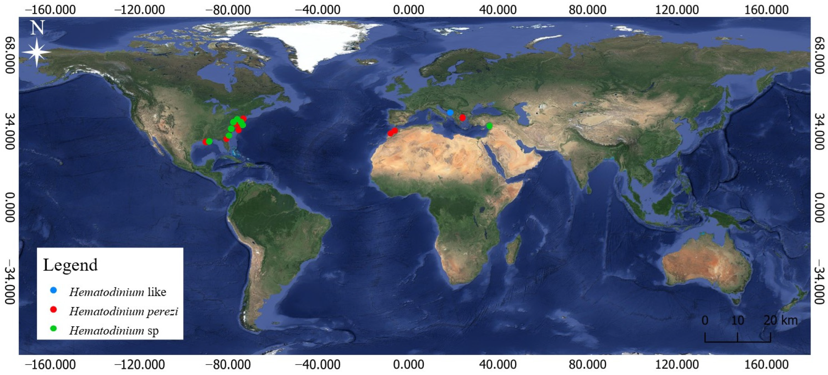
Hematodinium perezi (Dinophyceae: Syndiniales) in Morocco: The First Record on the African Atlantic Coast and the First Country Record of a Parasite of the Invasive Non-Native Blue Crab Callinectes sapidus

1
Laboratory of Biodiversity, Ecology and Genome, Faculty of Sciences, Mohammed V University in Rabat, 4 Avenue Ibn Battouta, B.P. 1014 RP, Rabat 10100, Morocco
2
Laboratoire Santé et Environnement, Faculté des Sciences Aïn Chock, Hassan II University of Casablanca, B.P. 5366 Maârif, Casablanca 20100, Morocco
3
Research Group Zoology: Biodiversity and Toxicology, Centre for Environmental Sciences, Hasselt University, Agoralaan Gebouw D, 3590 Diepenbeek, Belgium
4
Aquatic and Terrestrial Ecology, Operational Directorate Natural Environment, Royal Belgian Institute of Natural Sciences, Vautierstraat 29, 1000 Brussels, Belgium
5
Natural Sciences and Environment Research Hub, University of Gibraltar, Europa Point Campus, Gibraltar GX11 1AA, Gibraltar
*
Author to whom correspondence should be addressed.
J. Mar. Sci. Eng. 2024, 12(7), 1045; https://doi.org/10.3390/jmse12071045
Submission received: 15 May 2024 / Revised: 7 June 2024 / Accepted: 11 June 2024 / Published: 21 June 2024
(This article belongs to the Section Marine Ecology)

Abstract

:
Dinoflagellates belonging to the genus Hematodinium are key parasites of marine crustaceans, primarily decapods. In this study, we document the first report of H. perezi Chatton & Poisson, 1930 on the African Atlantic coast. This is also the first parasite record in the invasive non-native Atlantic blue crab Callinectes sapidus Rathbun, 1896 in Morocco. Specimens of C. sapidus were sampled in winter 2023 from two Ramsar sites on the Moroccan Atlantic, namely Merja Zerga and Oualidia Lagoons, and were screened to detect the presence of parasites in their hemolymph. Based on staining fresh hemolymph smears, we did not detect Hematodinium in any of the 36 investigated individuals (20 and 16 from Merja Zerga and Oualidia Lagoons, respectively), probably due to methodological artifacts. The PCR-based method was revealed to be more accurate in diagnosing the Hematodinium parasite. It showed that at Merja Zerga Lagoon, 13 individuals of C. sapidus were infected by the parasite (prevalence: 65%) in comparison to four at Oualidia Lagoon (25%). Genetic analysis, based on the ITS1 rDNA gene from Hematodinium, confirmed the sequences as being those of Hematodinium perezi.

1. Introduction

Biological invasions are considered to be a severe threat to marine biodiversity and the functioning of invaded coastal and marine ecosystems [1]. In addition, their ability to carry invasive and/or enhance native parasites can induce a loss of native biodiversity and an increase in disease and mortality in native species, which may pose risks to human health and the economy [2,3,4]. The establishment of introduced hosts and their parasites may also affect the life cycles of native parasites. On the other hand, native hosts can be infected by parasites associated with introduced species, i.e., ‘host switching’ (more specifically, spill-over) [3,5,6,7,8]. For example, the invasive nematode Spirocamallanus istiblenni Noble, 1966, was introduced to the Hawaiian archipelago along with Lutjanus kasmira Forsskål, 1775 from French Polynesia, and this introduction led to the spread of the nematode into native hosts [9]. The monogenean Nitzschia sturionis (Abildgaard, 1794) Krøyer, 1852 was co-introduced with Acipenser stellatus Pallas, 1771 from the Caspian Sea to the Aral Sea and induced severe mass mortalities of the native bastard sturgeon Acipenser nudiventris Lovetsky, 1828. Once infested by the parasite in its gills, the fish ends up dying on the beach [10]. The eel nematode Anguillicola crassus Kuwahara, Niimi & Itagaki, 1974 has infected seven different eel species on four different continents via the global eel trade, causing severe damage to the swim bladder of the eels, resulting in significant mortality in most eel species [11,12,13,14]. The rhizocephalan barnacle Loxothylacus panopaei Gissler, 1884, which infects native flatback mud crabs Eurypanopeus depressus Smith, 1869, was probably co-introduced with infected mud crabs (species unknown) in batches of oysters transferred from the Gulf of Mexico to Chesapeake Bay (North America) [15].
The Atlantic blue crab Callinectes sapidus Rathbun, 1896, belonging to Portunidae Rafinesque, 1815, is native to the western Atlantic Ocean [16,17]. Its natural geographical distribution ranges from Nova Scotia, Canada, down the eastern coast of the United States, and as far south as Northern Argentina [16,18,19]. This species was accidentally (with water ballast being the most likely introduction vector [16,20]) or intentionally introduced into Asia, Europe and Africa, and this widened its current worldwide biogeographic distribution. The first record from European Seas occurred in 1900 along the Atlantic coast of France [21]. In the Mediterranean Sea, the first presence of C. sapidus was reported in Italy in 1949 [20]. Now, the species is present all over the Mediterranean and the Black Sea [16,22,23,24], where it is ranked among the most invasive species [25]. It was recorded in 2022 in a freshwater ecosystem in the Mediterranean area, in Sicilian inland waters [26]. In Morocco, C. sapidus was first reported on its Mediterranean coast in the Marchica Lagoon in 2017 [27], while its first record in the Moroccan Atlantic was in the Merja Zerga Lagoon in 2019 [28]. The species is now established in many localities along the Mediterranean and Atlantic coasts of Morocco [28,29,30]. In native habitats, Callinectes sapidus is known to host a wide range of pathogens, including viral, bacterial and microalgal agents [31], which have been implicated in causing diseases and mortalities or reducing fecundity [32]. While C. sapidus constitutes a relevant model for studying invasions related to the spread of microorganisms along invaded Mediterranean and north-eastern Atlantic coasts, scientific research on the species concerns mostly its population dynamics, structure, and fisheries [33]. The occurrence of parasites in the species has been explored very little, even though it is an appreciated shellfish product in many countries such as Greece, Turkey, Italy, and Egypt [34].
Among pathogens of Callinectes sapidus, the parasite Hematodinium perezi (Dinophyceae: Syndiniales: Syndiniaceae) is an important disease-causing agent infecting over 40 species of crustaceans worldwide [35,36,37]. This parasite causes the so-called bitter crab disease in a number of species of crab such as Chionoecetes opilio Fabricius, 1788 and Chionoecetes bairdi Rathbun, 1924 [38], resulting in a crab meat flavour resembling bitterness with an aspirin-like taste due to biochemical alterations [35]. Also, the Hematodinium-infected hosts generally exhibit tissue or organ dysfunction or failure, as well as mortality in the later stages of infection, due in part to the enormous multiplication of parasites in the hemolymph of affected tissues [39,40,41]. In the Atlantic blue crab, H. perezi does not directly induce discernible biochemical alterations, but it can severely impact host health by inducing critical tissue or organ dysfunctions, ultimately resulting in high mortality rates [42].
Hematodinium perezi is currently the only confirmed Hematodinium species known to be infecting Callinectes sapidus [43]. The other representative of this genus, namely Hemtodinium sp., was reported as a parasite of C. sapidus from many coastal areas in USA) [44,45,46,47,48,49,50,51,52,53], and in Turkey [54]. This one, infecting boreal hosts, particularly Chionoecetes opilio and Nephrops norvegicus (Linnaeus, 1758), has not been identified at the species level yet (Shields J.D., pers. comm.).
Hematodinium perezi was first described from Carcinus maenas Linnaeus, 1758 and Liocarcinus depurator Linnaeus, 1758 on the French coastline [55]. Since then, records on the number of host species and distribution have notably increased [56]. Gallien [57] reported its spread in the French host Portunus latipes Pennant, 1777. In the Mid-Atlantic, it showed rare infections in Cancer irroratus Say, 1817 and Cancer borealis Stimpson, 1859, and in Ovalipes oceallatus Herbst, 1799 from the New York Bight area of the Northeastern United States [58]. Hematodinium perezi was documented in Callinectes sapidus for the first time in coastal Maryland and Virginia, USA [59], and afterwards in many habitats in the USA [49,60,61,62,63,64]. In the Mediterranean and north-eastern Atlantic, regions where C. sapidus invaded, H. perezi was reported as an endoparasite of C. sapidus in the Eastern Mediterranean, Greece, [34] and now also on the African Atlantic coast (this study) (Figure 1).
In this study, the hemolymph of Callinectes sapidus specimens from Merja Zerga and Oualidia Lagoons, located on the Atlantic coast of Morocco, was screened to detect the presence of parasites. Here, we document the first detection of dinoflagellates belonging to Hematodinium in C. sapidus on the African Atlantic coast.

2. Materials and Methods
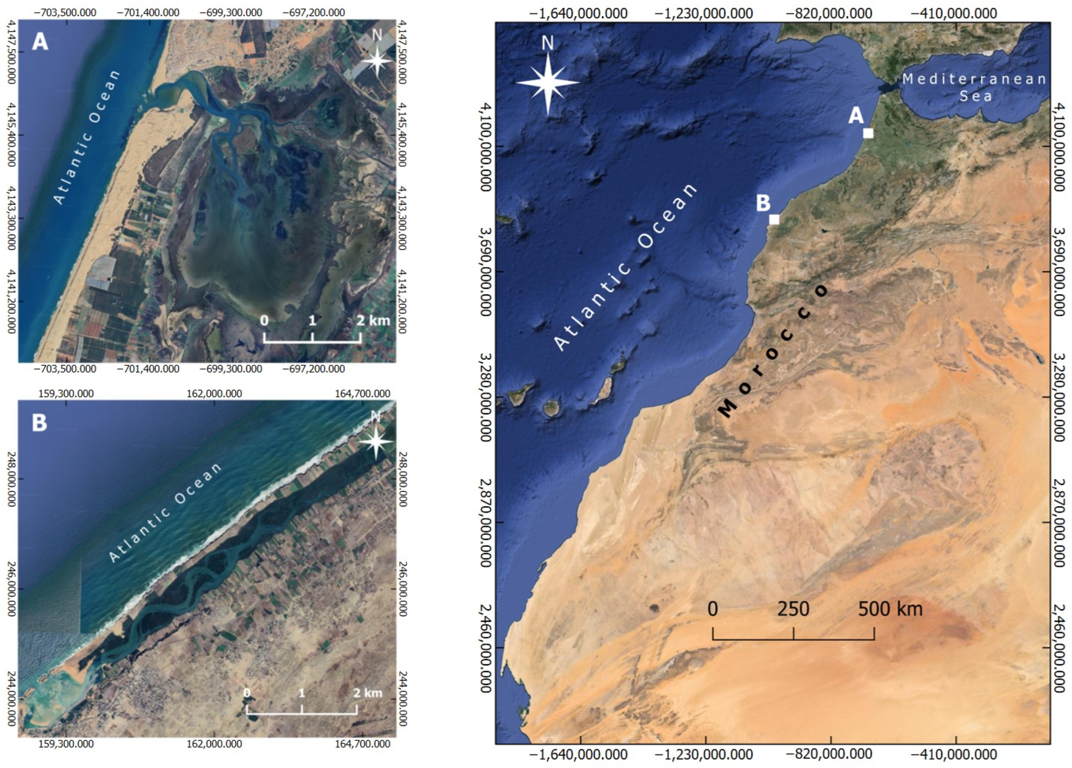
2.1. Study Sites

Merja Zerga and Oualidia Lagoons are two semi-enclosed coastal systems (SECS) situated along Morocco’s Atlantic coast (Figure 2). Both sites are recognized as Sites of Biological and Ecological Interest (SIBEs) [66] and Ramsar sites as Wetlands of International Importance.
The Merja Zerga Lagoon (34°47′ N, 6°13′ W) is an elliptically shaped lagoon that is 9 km long and 5 km wide, with a depth from 0.50 to 1.50 m and a total surface of 35 km2 [67]. The lagoon is connected to the ocean through a relatively deep gully (up to 6 m), and the circulation of sea water during flood and ebb is ensured by shallow subtidal channels. The freshwater supply is provided by Oued Drader and Canal of Nador [68]. Tides are semi-diurnal, with an average amplitude of 0.15 to 1.50 m [69]. Salinity in this lagoon fluctuates between 8 PSU and 36 PSU, with the mean water temperature varying between 14.6 °C and 24.15 °C [68].
The Oualidia Lagoon (32°74′ N, 9°03′ W) is over 7 km long and 1 km wide, with a mean depth of 2 m and a total surface of 3 km2 [67]. This coastal basin takes the form of an elongated depression oriented east–north-west, bordered by a coastal consolidated dune ridge and a continental cliff [70]. Tides are semi-diurnal, with amplitudes ranging from 0.8 to 3.6 m [71]. The average water temperature ranges from 16.1 °C to 21.1 °C, and the lagoon’s salinity varies between 20 PSU and 35 PSU at low tide, while at high tide, it can reach 30 PSU to 36 PSU throughout the year [72].

2.2. Sampling and Microscopic Analysis

Specimens of Callinectes sapidus were sampled from Merja Zerga (February 2023) and Oualidia (March 2023) Lagoons using a seine net. Collected crabs were transferred to the laboratory in refrigerated containers. Before dissection, we soaked each crab individually in ice water for about 15 to 20 min, depending on the size of the individuals, to induce anaesthesia. The captured crabs were identified based on morphological criteria (shape and colour of the carapace) according to Williams [18] and numbered. For each crab, sex and maturity were determined, and then, carapace length (CL), carapace width (CW) and fresh weight (W) were measured.
The hemolymph was extracted from each specimen (based on dorsal view) at the uncalcified joint of the right swimming leg near the carapace. For complete sterilisation, the leg was sterilized twice with a 70% ethanol-soaked cotton swab. A disposable 1 mL syringe coupled to a 26 g needle was inserted into the leg. The hemolymph of each specimen was analysed by the preparation of wet smears; one drop of hemolymph was mixed (1:1) with 0.3% neutral red solution on a glass slide and directly observed under an optical microscope Leica® DM 2500 (sourced from Leica Microsystems, Wetzlar, Germany). Hemolymph (0.1 mL) was also collected and placed in EDTA tubes containing 1 mL of 95% ethanol and frozen at −20 °C for DNA extraction to detect the presence of Hematodinium by polymerase chain reaction (PCR).

2.3. DNA Extraction, Amplification and Sequencing

Before extracting DNA, 200 µL of ethanol-preserved hemolymph was centrifuged at 1500× g for 1 min to eliminate excess ethanol [44]. In order to allow residual ethanol to evaporate, samples were dried for at least 30 min at 55 °C [44]. The Invitrogen TM Kit Blood and Tissue kit (sourced from Thermo Fisher Scientific, Waltham, MA, USA) was used to extract DNA as recommended by the manufacturer, with an overnight lysis of the hemolymph samples and two 5 min elution incubations and two 50 µL elutions.
For amplification, HITS1F (5′CATTCACCGTGAACCTTAGCC3′) and HITS1R (5′CTAGTCATACGTTTGAAGAAAGCC3′) primers that target the ITS1 rDNA were used according to Gruebl [45], with the expected length of 299 bp [46].
Each amplification was carried out in a final volume of 25 µL containing 5X standard Taq (Gquence) reaction buffer, 1.5 mM MgCl2, 0.1 mM dNTPs, 0.5 µM HITS1F, 0.5 µM HITS1R, 1 unit of Platinum™ Taq DNA Polymerase, 50 ng of extracted DNA [44] and 16.2 µL of ddH2O water. Amplification reactions were performed in a thermal gradient PCR MultiGene OptiMax Thermal Cycler (sourced from Labnet International, Edison, NJ, USA) according to the following program: 95.0 °C for 10 min; 40 cycles of 94.0 °C (30 s), 56.0 °C (30 s), 72.0 °C (1 min), and a final extension at 72.0 °C for 10 min. Amplified products were separated by 1% agarose gel electrophoresis stained with ethidium bromide. Positive PCR products were sent to the National Centre for Scientific and Technical Research (CNRST) in Rabat for purification and sequencing. The sequencing was carried out using a Genomix sequencer (MGX) with identical primers as used in the initial PCR.

2.4. Sequence Analysis

Using MEGA version XI [73], the obtained sequences were manually cleaned and aligned with the software ClustalW version 2.1 [74]. The resulting sequences were compared to genetic data previously published by the Basic Local Alignment Search Tool (BLAST) (sourced from the National Center for Biotechnology Information (NCBI), Bethesda, MD, USA) [75]. The uncorrected genetic p-distance between Moroccan sequences and all published sequences downloaded from GenBank were calculated using MEGA version XI (sourced from the MEGA Development Team, Tempe, AZ, USA). Using the same software, the optimal model of molecular evolution based on the Akaike information criterion (AIC) was the Jukes–Cantor model [76]. Maximum likelihood (ML) with Nearest-Neighbour Interchange (NNI) as a branch swapping algorithm and neighbour-joining (NJ) phylogenetic trees based on the unique haplotypes of ITS1 rDNA were constructed with 1000 bootstrap replicates using MEGA software version XI.
Sequences generated from this study were deposited in GenBank under accession numbers PP928476–PP928480 and PP933794–PP933803.

3. Results

3.1. Biometric Characteristics of Analysed Specimens of Callinectes sapidus

Overall, 36 specimens of Callinectes sapidus were collected in winter 2023. The twenty specimens from Merja Zerga Lagoon comprised five adult females, five adult males, five female juveniles and five male juveniles. Among the sixteen specimens from Oualidia Lagoon, there were four adult females, three adult males, five female juveniles and four male juveniles. Their biometric data are summarized in Table 1.

3.2. The Hemolymph Smear Assay with Neutral Red

Based on staining fresh hemolymph smears, we did not detect Hematodinium in any of the 36 investigated individuals (20 and 16 from Merja Zerga and Oualidia Lagoons, respectively) sampled in winter 2023.

3.3. PCR-Based Method and Sequence Analysis

Overall, 17 samples out of the 36 individuals investigated were successfully amplified, from which 13 specimens were revealed to be infected by the parasite at Merja Zerga Lagoon (prevalence 65%) and 4 at Oualidia Lagoon (25%).
The 15 ITS1 rDNA sequences that were generated (13 sequences from Merja Zerga Lagoon and 2 from Oualidia Lagoon) produced an alignment of a 295 bp long fragment. The ITS1 rDNA sequences compared, using BLAST search, to existing ones available in the GenBank database confirmed the identification of our parasites’ sequences as being those of Hematodinium perezi.
The uncorrected p-distance between Moroccan sequences varied between 0% and 0.6%, and the uncorrected p-distances between Moroccan sequences and published sequences downloaded from GenBank [34,43,77] (Table 2) varied between 0.2% from Hematodinium perezi in Callinectes sapidus collected in Greece and 4% from H. perezi in C. sapidus from the United States of America (Table 3).
Maximum likelihood (ML) and neighbour-joining (NJ) phylogenetic trees were topologically identical. Statistical support for most nodes was low, though the topology suggests that the Moroccan sequences were more related to those from Greece (Figure 3).
The measurements of specimens of Callinectes sapidus parasitized by Hematodinium perezi (as identified using PCR) from the Merja Zerga and Oualidia Lagoons of Morocco are reported in Table 4. All four groups (male adult, female adult, male juveniles and female juveniles) were parasitized. Moreover, females (adults and juveniles) were most likely affected in the Merja Zerga Lagoon, while in the Oualidia Lagoon, the infected crabs were mostly juveniles.

4. Discussion

The present study documents the first detection of Hematodinium perezi (Dinophyceae: Syndiniales) on the African Atlantic coast and also represents the first report of this (or any) parasite in the invasive non-native crab Callinectes sapidus in Morocco, namely in the Merja Zerga and Oualidia Lagoons.
Seasonal, sex- and size-related relationships or correlations have been reported be-tween Hematodinium species and their hosts ([78] and the references herein). In this study, specimens of Callinectes sapidus were collected in winter 2023, and females and juveniles seemed more likely to test positive for Hematodinium perezi, respectively, in the Merja Zerga and Oualidia Lagoons. Notwithstanding, the sampling effort in this study was small in terms of both time and space. Our ongoing in-depth research will enable us to better define the key drivers of C. sapidus infection by H. perezi in its area of introduction, in particular on the Mediterranean and Atlantic coasts of Morocco.
Because of its wide host range and capacity to transition between different host species, Hematodinium is regarded as a generalist parasite [35,43,56]. This trait enables it to persist in the environment, even in situations when its preferred host may become rare. Positive infections of Hematodinium were reported in 13 crustacean species belonging to two orders, Decapoda and Amphipoda [65]. The epidemiology of Hematodinium is influenced by a number of variables, including environmental factors (salinity and temperature). It is well known that this parasite prefers to infect hosts in highly saline waters [79]. For example, in Europe (Wadden Sea), no detection of Hematodinium in 1252 individuals of eight crustacean species from six sites was reported due to lower salinity [65]. Epidemics of these parasites have damaged commercial stocks of Nephrops norvegicus, Chionoecetes opilio, Chionoecetes bairdi Rathbun, 1924, C. sapidus and Necora puber (Linnaeus, 1767) [35]. Moreover, their impact on fisheries and host populations is thought to be similar to that of viral diseases of crustaceans [43], resulting in significant mortality in the host [80]. In the present research, the diagnosis of infection by representatives of Hematodinium in Callinectes sapidus was performed using the fresh hemolymph smear essay with neutral red and molecular analysis (PCR-based method and sequencing). The hemolymph smear assay with neutral red was used as an initial assessment tool due to its cost-effective and time-efficient diagnostic method for the detection of members of Hematodinium [65] as well as its specificity and sensitivity [81]. Hematodinium lysosomes actively absorb neutral red, producing a distinctive stain that visually contrasts with host hemocytes [35]. In our case, based on staining fresh hemolymph smears, we did not detect Hematodinium in any of the 36 investigated individuals (20 and 16 from the Merja Zerga and Oualidia Lagoons, respectively), probably due to methodological artifact. Indeed, as smears are rated positive when abnormal cells (i.e., cells that cannot be identified as crab hemocytes, but have certain characteristics corresponding to those of Hematodinium) are observed, expertise in parasite identification is required [82]. Some pathogens of crustaceans such as parasitic dinoflagellates and rhizocephalans may be more difficult to identify for the non-specialist [83]. Moreover, certain stages of parasites belonging to Hematodinium can be very difficult to detect in fresh hemolymph smears because the trophic stages resemble hemocytes. The vermiform plasmodium (cf. filamentous trophont) is the most straightforward form to identify, while the most frequently observed form (the vegetative, amoeboid stage) is easily confused with a hemocyte by the uninitiated, and they may be present at relatively low densities, making microscopic diagnosis difficult [35,50].
The use of molecular analysis is increasingly widespread in disease diagnosis, pathogen identification and monitoring, as well as the detection of cryptic organisms such as dinoflagellates and parasitic stages [45,84,85,86]. The PCR-based method offers 1000 times higher sensitivity compared to histology approaches [45]. The sensitivity of PCR diagnosis has been estimated at 1 parasite cell in 300,000 crab hemocytes [45]. Use of the PCR test eliminates the need for the visual identification of cells with ambiguous characteristics. Overall, a combination of morphological and molecular characterization is often used to ensure the accurate detection and monitoring of Hematodinium infections in crustacean populations, because PCR results simply indicate the presence of parasite genetic material. To confirm active infection or disease, it would be necessary to detect live parasite cells or clinical signs of infection by morphological characterization.
The PCR assay, adopted in our study to detect the Hematodinium infecting Callinectes sapidus, based on the amplification of the parasite’s first internal transcribed spacer region (ITS1), was developed by Small et al. [50]. The difference in parasite prevalence between the Merja Zerga (65%) and Oualidia Lagoons (25%) could be explained by environmental factors [47,65,80] or biological factors [53,87]. According to Barbosa et al. [80], several studies have shown that temperature and salinity conditions favour the invasion of Hematodinium. The prevalence of Hematodinium decreases at lower temperatures [80], and lower salinity can also limit the distribution of the parasite [65]. For example, the highest prevalence (69%) of the parasite in C. sapidus collected from the USA was found at salinities of 26 PSU to 30 PSU and a water temperature >25 °C, and no infected crabs were found below 11 PSU salinities [45]. Hematodinium may also be more prevalent because of the wide range of environmental reservoirs and high densities of hosts [87], and the absence of host immunological response can also be the reason of the high prevalence of Hematodinium in crustaceans [87]. Furthermore, according to Parmenter et al. [53], additional factors showing temporal or geographical variation may contribute to varying levels of Hematodinium infection in C. sapidus.
Calculated pairwise uncorrected p-distances from sequences of Hematodinium perezi parasitizing Callinectes sapidus in Morocco and all published sequences of H. perezi obtained from GenBank show that Moroccan sequences are closely similar to that from the host C. sapidus, collected from Greece (A.N., PP056127), and to those from Licocarcinus depurator collected from the South Coast of England (A.N., EF065716, EF065711, EF065708), and differ more from the sequence from C. sapidus collected from the USA (A.N., KX758132). The mean uncorrected p-distances between Moroccan sequences and sequences having an A.N of KX244637, KX244644, or KX244641 from Portunus trituberculatus Miers, 1876 from China is 1.8% (Table 3). According to Small et al. [43], the mean interspecific genetic distances between H. perezi from L. depurator and the Hematodinium sp. infecting P. trituberculatus and Scylla serrata Forskål, 1775 is 2.5% and 4.6% between Hematodinium sp. from C. sapidus and H. perezi from L. depurator. In our study, the mean intraspecific genetic distances between H. perezi from C. sapidus collected from Morocco and all published sequences of H. perezi from GenBank is 1.1%. In comparison with the above-mentioned interspecific distances, this confirms that our sequences belong to parasites that are conspecific with H. perezi.
Ultimately, in-depth studies are desirable for understanding the interactions between the invasive non-native blue crab and its parasites in the coastal areas of Morocco and for assessing their effects on native biodiversity, associated marine diseases and risks to human health. In addition, whole genome-based analyses of native and introduced populations of the Atlantic blue crab, sampled at a large scale, along with its associated Hematodinium parasites, will contribute to understanding the invasion history of the Atlantic blue crab in Morocco.

Author Contributions

Conceptualization, A.L., I.R., N.K., M.P.M.V. and H.B.; methodology, A.L., I.R., N.K., M.P.M.V. and H.B.; software, A.L and I.R.; formal analysis, A.L., I.R. and H.B.; N.K. and M.P.M.V.; investigation, A.L., I.R., M.S. and A.H.; resources, A.L., M.S.; A.H. and H.B.; data curation, A.L., I.R., N.K., M.P.M.V. and H.B.; writing—original draft preparation, A.L.; writing—review and editing, A.L., I.R., H.B., N.K. and M.P.M.V.; supervision, I.R., H.B., N.K. and M.P.M.V.; funding acquisition, A.L., I.R., M.S., N.K., M.P.M.V. and H.B. All authors have read and agreed to the published version of the manuscript.

Funding

This research was supported by the Special Research Fund of Hasselt University (BOF23BL07 to A.L.; BOF21PD01 to N.K.; BOF20TT06 to M.P.M.V.; BOF21INCENT09), by a research grant 1513419N from the Research Foundation—Flanders (FWO-Vlaanderen), and by a grant N 36 UM5R2022 from the National Centre for Scientific and Technical Research (CNRST), Morocco.

Institutional Review Board Statement

Not applicable.

Informed Consent Statement

Not applicable.

Data Availability Statement

The genetic data are available on GenBank under the accession numbers PP928476–PP928480 and PP933794–PP933803.

Acknowledgments

We thank Jeffrey Shields at the Virginia Institute of Marine Science for his helpful suggestions and advice on preparing neutral red dye, and for sharing with us a video on crab bleeding and smear preparation, as well as valuable documentation on the topic. The authors would like to thank the National Centre of Scientific and Technical Research (CNRST) of Morocco for putting at their disposal the technical facilities of the UATRS Division.

Conflicts of Interest

The authors declare no conflicts of interest.

References

  1. Katsanevakis, S.; Wallentinus, I.; Zenetos, A.; Leppäkoski, E.; Çinar, M.E.; Oztürk, B.; Grabowski, M.; Golani, D.; Cardoso, A.C. Impacts of marine invasive alien species on ecosystem services and biodiversity: A pan-European review. Aquat. Invasions 2014, 9, 391–423. [Google Scholar] [CrossRef]
  2. Crowl, T.A.; Crist, T.O.; Parmenter, R.R.; Belovsky, G.; Lugo, A.E. The spread of invasive species and infectious disease as drivers of ecosystem change. Front. Ecol. Environ. 2008, 6, 238–246. [Google Scholar] [CrossRef]
  3. Goedknegt, M.A.; Feis, M.E.; Wegner, K.M.; Luttikhuize, P.C.; Buschbaum, C.; Camphuysen, K.C.J.; Vander Meer, J.; Thieltges, D.W. Parasites and marine invasions: Ecological and evolutionary perspectives. J. Sea Res. 2016, 113, 11–27. [Google Scholar] [CrossRef]
  4. Bojko, J.; Burgess, A.L.; Baker, A.G.; Orr, C.H. Invasive Non-Native Crustacean Symbionts: Diversity and Impact. J. Invertebr. Pathol. 2021, 186, 107482. [Google Scholar] [CrossRef]
  5. Hall, S.R.; Becker, C.R.; Simonis, J.L.; Duffy, M.A.; Tessier, A.J.; Cáceres, C.E. Friendly competition: Evidence for a dilution effect among competitors in a planktonic host–parasite system. Ecology 2009, 90, 791–801. [Google Scholar] [CrossRef]
  6. Kelly, D.W.; Paterson, R.A.; Townsend, C.R.; Poulin, R.; Tompkins, D.M. Parasite spillback: A neglected concept in invasion ecology? Ecology 2009, 90, 2047–2056. [Google Scholar] [CrossRef]
  7. Ortega, N.; Roznik, E.A.; Surbaugh, K.L.; Cano, N.; Price, W.; Campbell, T.; Rohr, J.R. Parasite spillover to native hosts from more tolerant, supershedding invasive hosts: Implications for management. J. Appl. Ecol. 2021, 59, 39–51. [Google Scholar] [CrossRef]
  8. Goedknegt, M.A.; Havermans, J.; Waser, A.M.; Luttikhuizen, P.C.; Vellila, E.; Camphuysen, K.C.J.; Vander Meer, J.; Thieltges, D.W. Cross-species comparison of parasite richness, prevalence, and intensity in a native compared to two invasive brachyuran crabs. Aquat. Invasions 2017, 12, 201–212. [Google Scholar] [CrossRef]
  9. Gaither, M.R.; Aeby, G.; Vignon, M.; Meguro, Y.I.; Rigby, M.; Runyon, C.; Bowen, B.W. An invasive fish and the time-lagged spread of its parasite across the Hawaiian archipelago. PLoS ONE 2013, 8, e56940. [Google Scholar] [CrossRef]
  10. Dogiel, V.A.; Lutta, A.S. On the mortality of Acipenser nudiventris of the Aral Sea in 1936. J. Rybn. Choz. 1937, 12, 25–27. [Google Scholar]
  11. Barse, A.M.; McGuire, S.A.; Vinores, M.A.; Eierman, L.E.; Weeder, J.A. The swimbladder nematode Anguillicola crassus in American eels (Anguilla rostrata) from middle and upper regions of Chesapeake Bay. J. Parasitol. 2001, 87, 1366–1370. [Google Scholar] [CrossRef]
  12. Køie, M. Swimbladder nematodes (Anguillicola spp.) and gill monogeneans (Pseudoactylogyrus spp.) parasitic on the European eel (Anguilla anguilla). ICES J. Mar. Sci. 1991, 47, 391–398. [Google Scholar] [CrossRef]
  13. Kvach, Y.; Skóra, K.E. Metazoan parasites of the invasive round goby Apollonia melanostoma (Neogobius melanostomus) (Pallas) (Gobiidae: Osteichthyes) in the Gulf of Gdansk, Baltic Sea, Poland: A comparison with the Black Sea. Parasitol. Res. 2007, 100, 767–774. [Google Scholar] [CrossRef]
  14. Sasal, P.; Taraschewski, H.; Valade, P.; Grondin, H.; Wielgoss, S.; Moravec, F. Parasite communities in eels of the Island of Reunion (Indian Ocean): A lesson in parasite introduction. Parasitol. Res. 2008, 102, 1343–1350. [Google Scholar] [CrossRef]
  15. Van Engel, W.A.; Dillon, W.A.; Zwerner, D.; Eldridge, D. Loxothylacus panopaei (Cirripedia, Sacculinidae) an introduced parasite on a xanthid crab in Chesapeake Bay, USA. Crustacean 1966, 10, 110–112. [Google Scholar]
  16. Nehring, S. Invasion history and success of the American blue crab Callinectes sapidus in European and adjacent waters. In The Wrong Place-Alien Marine Crustaceans: Distribution, Biology and Impacts; Springer: Dordrecht, The Netherlands, 2011; pp. 607–624. [Google Scholar] [CrossRef]
  17. Nehring, S. Callinectes sapidus. In NOBANIS—Invasive Alien Species Fact Sheet. Online Database of the European Network on Invasive Alien Species; NOBAMIS: Akureyri, Iceland, 2012; Available online: http://www.nobanis.org (accessed on 16 February 2024).
  18. Williams, A.B. The swimming crabs of the genus Callinectes (Decapoda: Portunidae). Fish. Bull. 1974, 72, 685–798. [Google Scholar]
  19. Castriota, L.; Andaloro, F.; Costantini, R.; De Ascentiis, A. First record of the Atlantic crab Callinectes sapidus Rathbun, 1896 (Crustacea: Brachyura: Portunidae) in Abruzzi waters, central Adriatic Sea. Acta Adriat. 2012, 53, 467–471. [Google Scholar]
  20. Giordani Soika, A. II Neptunus pelagicus (L.) nell’Alto Adriatico. Natura 1951, 42, 18–20. [Google Scholar]
  21. Bouvier, E.L. Sur un Callinectes sapidus M. Rathbun trouvé à Rocheford. Bull. Mus. Natl. Hist. Nat. 1901, 7, 16–17. [Google Scholar]
  22. Pashkov, A.N.; Reshetnikov, S.I.; Bondarev, K.B. The capture of the blue crab (Callinectes sapidus, Decapoda, Crustacea) in the Russian sector of the Black Sea. Russ. J. Biol. Invasions 2012, 3, 22–28. [Google Scholar] [CrossRef]
  23. Castejón, D.; Guerao, G. A new record of the American blue crab, Callinectes sapidus Rathbun, 1896 (Decapoda: Brachyura: Portunidae), from the Mediterranean coast of the Iberian Peninsula. BioInvasions Rec. 2013, 2, 141–143. [Google Scholar] [CrossRef]
  24. Öztürk, R.Ç.; Terzi, Y.; Feyzioğlu, A.M.; Şahin, A.; Aydın, M. Genetic Characterization of the Invasive Blue Crab, Callinectes sapidus (Rathbun 1896), in the Black Sea. Reg. Stud. Mar. Sci. 2020, 39, 101412. [Google Scholar] [CrossRef]
  25. Streftaris, N.; Zenetos, A. Alien marine species in the Mediterranean—The 100 ‘Worst Invasives’ and their impact. Mediterr. Mar. Sci. 2006, 7, 87–118. [Google Scholar] [CrossRef]
  26. Vecchioni, L.; Russotto, S.; Arculeo, M.; Marrone, F. On the occurrence of the invasive Atlantic blue crab Callinectes sapidus Rathbun 1896 (Decapoda: Brachyura: Portunidae) in Sicilian inland waters. Nat. Hist. Sci. 2022, 9, 43–46. [Google Scholar] [CrossRef]
  27. Chartosia, N.; Anastasiadis, D.; Bazairi, H.; Crocetta, F.; Deidun, A.; Despalatović, M.; Di Martino, V.; Dimitriou, N.; Dragičević, B.; Dulčić, J.; et al. New Mediterranean biodiversity records. Mediterr. Mar. Sci. 2018, 19, 398–415. [Google Scholar] [CrossRef]
  28. Oussellam, M.; Benhoussa, A.; Pariselle, A.; Rahmouni, I.; Salmi, M.; Agnèse, J.F.; Selfati, M.; El Ouamari, N.; Bazairi, H. First and southern-most records of the American blue crab Callinectes sapidus Rathbun, 1896 (Decapoda, Portunidae) on the African Atlantic coast. Bioinvasions Rec. 2023, 12, 403–416. [Google Scholar] [CrossRef]
  29. Chaouti, A.; Belattmania, Z.; Nadri, A.; Serrão, E.; Encarnação, J.; Teodósio, A.; Reani, A.; Sabour, B. The invasive Atlantic blue crab Callinectes sapidus Rathbun 1896 expands its distributional range southward to Atlantic African shores: First records along the Atlantic coast of Morocco. BioInvasions Rec. 2022, 11, 227–237. [Google Scholar] [CrossRef]
  30. Chairi, H.; González-Ortegón, E. Additional records of the blue crab Callinectes sapidus Rathbun, 1896 in the Moroccan Sea, Africa. BioInvasions Rec. 2022, 11, 776–784. [Google Scholar] [CrossRef]
  31. Flowers, E.M.; Simmonds, K.; Messick, G.A.; Sullivan, L.; Schott, E.J. PCR-based prevalence of a fatal reovirus of the blue crab, Callinectes sapidus (Rathbun) along the northern Atlantic coast of the USA. J. Fish Dis. 2016, 39, 705–714. [Google Scholar] [CrossRef] [PubMed]
  32. Messick, G.A.; Sindermann, C.J. Synopsis of Principal Diseases of the Blue Crab, Callinectes sapidus. 1992. Available online: https://repository.library.noaa.gov/view/noaa/6066 (accessed on 16 February 2024).
  33. Kampouris, T.E.; Kouroupakis, E.; Batjakas, I.E. Morphometric relationships of the global invader Callinectes sapidus Rathbun,1896 (Decapoda, Brachyura, Portunidae) from Papapouli lagoon, NW Aegean Sea, Greece. with notes on its ecological preferences. Fishes 2020, 5, 5. [Google Scholar] [CrossRef]
  34. Lattos, A.; Papadopoulos, D.K.; Giantsis, I.A.; Stamelos, A.; Karagiannis, D. Histopathology and phylogeny of the dinoflagellate Hematodinium perezi and the epibiotic peritrich ciliate Epistylis sp. infecting the blue crab Callinectes sapidus in the Eastern Mediterranean. Microorganisms 2024, 12, 456. [Google Scholar] [CrossRef] [PubMed]
  35. Stentiford, G.D.; Shields, J.D. A review of the parasitic dinofagellates Hematodinium species and Hematodinium-like infections in marine crustaceans. Dis. Aquat. Org. 2005, 66, 47–70. [Google Scholar] [CrossRef] [PubMed]
  36. Shields, J.D. The impact of pathogens on exploited populations of decapod crustaceans. J. Invertebr. Pathol. 2012, 110, 211–224. [Google Scholar] [CrossRef] [PubMed]
  37. Small, H.J. Advances in our understanding of the global diversity and distribution of Hematodinium spp. significant pathogens of commercially exploited crustaceans. J. Invertebr. Pathol. 2012, 110, 234–246. [Google Scholar] [CrossRef] [PubMed]
  38. Shields, J.D.; Overstreet, R. Diseases, parasites, and others Symbionts. In The Blue Crab Callinectes sapidus; Kennedy, V., Cronin, L., Eds.; Maryland Sea Grant: College Park, MD, USA, 2007; Volume 1, pp. 299–417. [Google Scholar]
  39. Field, R.; Appleton, P. A Hematodinium-like dinoflagellate infection of the Norway lobster Nephrops norvegicus: Observations on pathology and progression of infection. Dis. Aquat. Org. 1995, 22, 115–128. [Google Scholar] [CrossRef]
  40. Taylor, A.; Field, R.; Parslow-Williams, P. The effects of Hematodinium sp.-infection on aspects of the respiratory physiology of the Norway lobster, Nephrops norvegicus (L.). J. Exp. Mar. Biol. Ecol. 1996, 207, 217–228. [Google Scholar] [CrossRef]
  41. Wheeler, K.; Shields, J.D.; Taylor, D.M. Pathology of Hematodinium infections in snow crabs (Chionoecetes opilio) from Newfoundland. Canada. J. Invertebr. Pathol. 2007, 95, 93–100. [Google Scholar] [CrossRef] [PubMed]
  42. Albalat, A.; Collard, A.; Cadam, B.; Coates, C.J.; Fox, C.J. Physiological condition, short-term survival, and predator avoidance behavior of discraded Norway lobsters (Nephrops norvegicus). J. Shellfish Res. 2012, 35, 1053–1065. [Google Scholar] [CrossRef]
  43. Small, H.J.; Shields, J.D.; Reece, K.S.; Bateman, K.; Stentiford, G.D. Morphological and molecular characterization of Hematodinium perezi (Dinophyceae: Syndiniales), a dinoflagellate parasite of the harbour crab, Liocarcinus depurator. J. Eukaryot. Microbiol. 2012, 59, 54–66. [Google Scholar] [CrossRef]
  44. Lohan, K.M.P.; Reece, K.S.; Miller, T.L.; Wheeler, K.N.; Small, H.J.; Shields, J.D. The role of alternate hosts in the ecology and life history of Hematodinium sp., a parasitic dinoflagellate of the blue crab (Callinectes sapidus). J. Parasitol. 2012, 98, 73–84. [Google Scholar] [CrossRef]
  45. Gruebl, T.; Frischer, M.E.; Sheppard, M.; Neumann, M.A.; Maurer, A.N.; Lee, R.F. Development of an 18S rRNA gene-targeted PCR-based diagnostic for the blue crab parasite Hematodinium sp. Dis. Aquat. Org. 2002, 49, 61–70. [Google Scholar] [CrossRef] [PubMed]
  46. Newman, M.W.; Johnson, C.A. A disease of blue crabs (Callinectes sapidus) caused by a parasitic dinofagellate Hematodinium Sp. J. Parasitol. 1975, 61, 554–557. [Google Scholar] [CrossRef]
  47. Messick, G.A.; Shields, J.D. Epizootiology of the parasitic dinofagellate Hematodinium sp. in the American blue crab Callinectes sapidus. Dis. Aquat. Org. 2000, 43, 139–152. [Google Scholar] [CrossRef] [PubMed]
  48. Sheppard, M.; Walker, A.; Frischer, M.E.; Lee, R.F. Histopathology and prevalence of the parasitic dinofagellate, Hematodinium sp, in crabs (Callinectes sapidus, Callinectes similis, Neopanopesayi, Libinia emarginata, Menippe mercenaria) from a Georgia estuary. J. Shellfsh Res. 2003, 22, 873–880. [Google Scholar]
  49. Frischer, M.; Lee, R.; Sheppard, M.; Mauer, A.; Rambow, F.; Neumann, M.; Broft, J.; Wizenmann, T.; Danforth, J. Evidence for a freeliving life stage of the blue crab parasitic dinofagelate Hematodinium sp. Harmful Algae 2006, 5, 548–557. [Google Scholar] [CrossRef]
  50. Small, H.J.; Shields, J.D.; Hudson, K.L.; Reece, K.S. Molecular detection of Hematodinium sp infecting the blue crab Callinectes sapidus. J. Shellfish Res. 2007, 26, 131–139. [Google Scholar] [CrossRef]
  51. Troedsson, C.; Lee, R.F.; Walters, T.; Stokes, V.; Brinkley, K.; Naegele, V.; Frischer, M.E. Detection and discovery of crustacean parasites in blue crabs (Callinectes sapidus) by using 18S rRNA gene-targeted denaturing high-performance liquid chromatography. Appl. Environ. Microbiol. 2008, 74, 4346–4353. [Google Scholar] [CrossRef] [PubMed]
  52. Nagle, L.; Place, A.; Schott, E.; Jagus, R.; Messick, G.; Pitula, J. Real-time PCR-based assay for quantitative detection of Hematodinium sp. in the blue crab Callinectes sapidus. Dis. Aquat. Org. 2009, 84, 79–87. [Google Scholar] [CrossRef] [PubMed]
  53. Parmenter, K.J.; Vigueira, P.A.; Morlok, C.K.; Micklewright, J.A.; Smith, K.M.; Paul, K.S.; Childress, M.J. Seasonal prevalence of Hematodinium sp. infections of blue crabs in three South Carolina (USA) rivers. Estuar. Coast 2013, 36, 174–191. [Google Scholar] [CrossRef]
  54. Aldik, R.; Cengizler, İ. The investigation of bacteria, parasite and fungi in blue crabs (Callinectes sapidus, Rathbun 1896) caught from Akyatan Lagoon in east Mediterranean Sea. J. VetBio. Sci. Technol. 2017, 2, 11–17. [Google Scholar]
  55. Chatton, E.; Poisson, R. Sur l’existence, dans le sang des crabs, de péridiniens parasites: Hematodinium perezi n.g., n.sp. (Syndinidae). C.R. Séances. Soc. Biol. Paris. 1931, 105, 553–557. [Google Scholar]
  56. Li, M.; Huang, Q.; Lv, X.; Song, S.; Li, C. The parasitic dinolagellate Hematodinium infects multiple crustaceans in the polyculture systems of Shandong Province, China. J. Invertebr. Pathol. 2021, 178, 107523. [Google Scholar] [CrossRef] [PubMed]
  57. Gallien, L. Sur la presence dans le sang de Platyonychus latipes Penn. d’un Peridinien parasite Hematodinium perezi Chatton et Poisson. Bull. Biol. Fr. Belg. 1938, 72, 1–7. [Google Scholar]
  58. MacLean, S.A.; Ruddell, C.L. Three new crustacean hosts for the parasitic dinofagellate Hematodinium perezi (Dinofagellata: Syndinidae). J. Parasitol. 1978, 64, 158–160. [Google Scholar] [CrossRef]
  59. Messick, G.A. Hematodinium perezi infections in adult and juvenile blue crabs Callinectes sapidus from coastal bays of Maryland and Virginia, USA. Dis. Aquat. Org. 1994, 19, 77–82. [Google Scholar] [CrossRef]
  60. Shields, J.D.; Squyars, C.M. Mortality and hematology of blue crabs, Callinectes sapidus, experimentally infected with the parasitic dinofagellate Hematodinium perezi. Fish. Bull. 2000, 98, 139. [Google Scholar]
  61. Hanif, A.W.; Dyson, W.D.; Bowers, H.A.; Pitula, J.S.; Messick, G.A.; Jagus, R.; Schott, E.J. Variation in spatial and temporal incidence of the crustacean pathogen Hematodinium perezi in environmental samples from Atlantic Coastal Bays. Aquat. Biosyst. 2013, 9, 11. [Google Scholar] [CrossRef] [PubMed]
  62. Butler, M.J.; Tiggelaar, J.M.; Shields, J.D. Effects of the parasitic dinofagellate Hematodinium perezi on blue crab (Callinectes sapidus) behavior and predation. J. Exp. Mar. Biol. Ecol. 2014, 461, 381–388. [Google Scholar] [CrossRef]
  63. Lycett, K.A.; Chung, J.S.; Pitula, J.S. The relationship of blue crab (Callinectes sapidus) size class and molt stage to disease acquisition and intensity of Hematodinium perezi infections. PLoS ONE 2018, 13, e0192237. [Google Scholar] [CrossRef]
  64. Small, H.J.; Huchin-Mian, J.; Reece, K.; Lohan, K.P.; Butler, M.J.; Shields, J.D. Parasitic dinofagellate Hematodinium perezi prevalence in larval and juvenile blue crabs Callinectes sapidus from coastal bays of Virginia. Dis. Aquat. Org. 2019, 134, 215–222. [Google Scholar] [CrossRef]
  65. Huang, Q.; Waser, A.M.; Li, C.; Thieltges, D.W. Lack of Hematodinium microscopic detection in crustaceans at the northern and southern ends of the Wadden Sea and an update of its distribution in Europe. Mar. Biol. 2024, 171, 63. [Google Scholar] [CrossRef]
  66. PDAPM. Plan directeur des Aires protégées du Maroc. Les sites d’intérêt biologique et écologique de domaine littoral. 1996; p. 166. [Google Scholar]
  67. Bououarour, O.; El Kamcha, R.; Boutoumit, S.; Pouzet, P.; Maanan, M.; Bazairi, H. Effects of the Zostera noltei meadows on benthic macrofauna in North Atlantic coastal ecosystems of Morocco: Spatial and seasonal patterns. Biologia 2021, 76, 2263–2275. [Google Scholar] [CrossRef]
  68. Boutoumit, S.; Bououarour, O.; El Kamcha, R.; Pouzet, P.; Zourarah, B.; Benhoussa, A.; Maanan, M.; Bazairi, H. Spatial patterns of macrozoobenthos assemblages in a sentinel coastal Lagoon: Biodiversity and environmental drivers. J. Mar. Sci. Eng. 2021, 9, 461. [Google Scholar] [CrossRef]
  69. Gam, M. Dynamique des Systèmes Parasites-Hôte, Entre Trématodes Digènes et Coque Cerastoderma edule: Comparaison de la Lagune de Merja Zerga Avec le Bassin D’arcachon. Ph.D. Thesis, Université Bordeaux, Bordeaux, France, 2008. [Google Scholar]
  70. Maanan, M.; Landesman, C.; Maanan, M.; Zourarah, B.; Fattal, P.; Sahabi, M. Evaluation of the anthropogenic influx of metal and metalloid contaminants into the Moulay Bousselham lagoon, Morocco, using chemometric methods coupled to geographical information systems. Environ. Sci. Pollut. Res. 2013, 20, 4729–4741. [Google Scholar] [CrossRef]
  71. Hilmi, K.; Orbi, A.; Lakhdar Idrissi, J. Hydrodynamisme de la lagune de Oualidia (Maroc) durant l’été et l’automne 2005. Bull. Inst. Sci. Rabat Sect. Sci. Terre 2009, 31, 29–34. [Google Scholar]
  72. Makaoui, A.; Idrissi, M.; Agouzouk, A.; Larissi, J.; Baibai, T.; El Ouehabi, Z.; Laamal, M.A.; Bessa, I.; Ettahiri, O.; Hilmi, K. Etat océanographique de la lagune de Oualidia, Maroc (2011–2012). Eur. Sci. J. 2018, 14, 93. [Google Scholar] [CrossRef]
  73. Kumar, S.; Stecher, G.; Li, M.; Knyaz, C.; Tamura, K. MEGA X: Molecular Evolutionary Genetics Analysis across Computing Platforms. Mol. Biol. Evol. 2018, 35, 1547–1549. [Google Scholar] [CrossRef]
  74. Thompson, J.D.; Higgins, D.G.; Gibson, T.J. CLUSTAL W: Improving the sensitivity of progressive multiple sequence alignment through sequence weighting, position-specific gap penalties and weight matrix choice. Nucleic Acids Res. 1994, 22, 4673–4680. [Google Scholar] [CrossRef]
  75. Altschul, S.F.; Gish, W.; Miller, W.; Myers, E.W.; Lipman, D.J. Basic local alignment search tool. J. Mol. Biol. 1990, 215, 403–410. [Google Scholar] [CrossRef]
  76. Erickson, K. The Jukes-Cantor Model of Molecular Evolution. PRIMUS 2010, 20, 438–445. [Google Scholar] [CrossRef]
  77. Xiao, J.; Miao, X.; Li, C.; Xu, W.; Zhang, X.; Wang, Z. Genetic variations of the parasitic dinoflagellate Hematodinium infecting cultured marine crustaceans in China. Protist 2016, 167, 597–609. [Google Scholar] [CrossRef]
  78. Molto-Martin, I.; Neil, D.M.; Coates, C.J.; MacKenzie, S.A.; Bass, D.; Stentiford, G.D.; Albalat, A. Infection of Norway lobster (Nephrops norvegicus) by the parasite Hematodinium sp.: Insights from 30 years of field observations. R. Soc. Open Sci. 2024, 11, 231147. [Google Scholar] [CrossRef]
  79. Alimin, A.W.F.; Yusoff, N.A.H.; Kadriah, I.A.K.; Anshary, H.; Abdullah, F.; Jabir, N.; Hassan, M. Parasitic dinoflagellate Hematodinium in marine decapod crustaceans: A review on current knowledge and future perspectives. Parasitol. Res. 2024, 123, 49. [Google Scholar] [CrossRef]
  80. Barbosa, H.; Soares, A.M.; Pereira, E.; Freitas, R. Are the consequences of lithium in marine clams enhanced by climate change? Environ. Pollut. 2023, 326, 121416. [Google Scholar] [CrossRef]
  81. Shields, J.D.; Sullivan, S.E.; Small, H.J. Overwintering of the parasitic dinoflagellate Hematodinium perezi in dredged blue crabs (Callinectes sapidus) from Wachapreague Creek, Virginia. J. Invertebr. Pathol. 2015, 130, 124–132. [Google Scholar] [CrossRef]
  82. Morado, J.F.; Jensen, P.; Hauzer, L.; Lowe, V.; Califf, K.; Roberson, N.; Shavey, C.; Woodby, D. Species Identity and Life History of Hematodinium, the Causitive Agent of Bitter Crab Syndrome in North East Pacific Snow, Chionoecetes opilio, and Tanner, C. bairdi, Crabs; Project 0306 Final Report; North Pacific Research Board: Anchorage, AK, USA, 2005. [Google Scholar]
  83. Shields, J.D. Collection techniques for the analyses of pathogens in crustaceans. J. Crus. Biol. 2017, 37, 753–763. [Google Scholar] [CrossRef]
  84. Cunningham, C.O. Molecular diagnosis of fish and shellfish diseases: Present status and potential use in disease control. Aquaculture 2002, 206, 19–55. [Google Scholar] [CrossRef]
  85. Lee, R.F.; Frischer, M.E. The decline of the blue crab. Am. Sci. 2004, 92, 548–553. [Google Scholar] [CrossRef]
  86. Small, H.J.; Neil, D.M.; Taylor, A.C.; Atkinson, R.J.A.; Coombs, G.H. Molecular detection of Hematodinium spp. in Norway lobster Nephrops norvegicus and other crustaceans. Dis. Aquat. Org. 2006, 69, 185–195. [Google Scholar] [CrossRef]
  87. Davies, C.E.; Thomas, J.E.; Malkin, S.H.; Batista, F.M.; Rowley, A.F.; Coates, C.J. Hematodinium sp. infection does not drive collateral disease contraction in a crustacean host. eLife 2022, 11, e70356. [Google Scholar] [CrossRef]
Figure 1. Map showing the distribution of reported representatives of Hematodinium infecting Callinectes sapidus (see also [34,65]).
Jmse 12 01045 g001
Figure 2. Map showing the localization of Merja Zerga (A) and Oualidia (B) Lagoons on the Moroccan Atlantic.
Jmse 12 01045 g002
Figure 3. Phylogram constructed using maximum likelihood (ML) and neighbour-joining (NJ) methods based on ITS1 sequences of Hematodinium perezi. Bootstrap support from 1000 replicates is shown based on the ML method (before slash) and on the NJ method (behind slash). ML and NJ trees are topologically identical, and it is the ML tree that is shown here (midpoint rooted). The scale bar represents the number of expected substitutions per site.
Jmse 12 01045 g003
Table 1. Biometric measurements of specimens of Callinectes sapidus from Merja Zerga and Oualidia Lagoons on the Moroccan Atlantic. CL: carapace length; CW: carapace width; W: body weight; SD: standard deviation.
LagoonSampling PeriodNumber of IndividualsW (g)CW (cm)CL (cm)
Min–MaxMean ± SDMin–MaxMean ± SDMin–MaxMean ± SD
Merja ZergaFebruary 2023204.6–237.588.9 ± 78.73.1–14.89.2 ± 4.12.2–6.64.6 ± 1.8
OualidiaMarch 20231618.8–20757.0 ± 52.45.8–14.98.6 ± 2.63.1–7.44.5 ± 1.4
Table 2. List of Hematodinium perezi sequences used in the present study, their GenBank accession numbers, host species, localities and references.
GenBank Accession NumberHostLocalityReferences
PP056127Callinectes sapidus (Portunidae)Greece[34]
EF065716Liocarcinus depurator (Polybiidae)South Coast of England[43]
EF065708Liocarcinus depuratorSouth Coast of England[43]
EF065711Liocarcinus depuratorSouth Coast of England[43]
KX244637Portunus trituberculatus (Portunidae)China[77]
KX244644Portunus trituberculatusChina[77]
KX244641Portunus trituberculatusChina[77]
KX244634Callinectes sapidusUSA[77]
Table 3. Range of uncorrected pairwise genetic distances (p-distances in %) between ITS1 rDNA sequences of Hematodinium perezi infecting Callinectes sapidus collected from Morocco and all published sequences from GenBank.
Merja ZergaOualidiaSouth Coast of EnglandChinaUSAGreece
Merja Zerga Lagoon------
Oualidia Lagoon0–0.6%-----
South Coast of England
EF065716, EF065708, EF065711
0.6–1.3%0.6–1.3%----
China
KX244637, KX244644, KX244641
1.7–2.3%1.7–2.3%1–1.7%---
USA
KX244634
3.7–4%3.7–4%3.6–4%3.7–4%--
Greece
PP056127
0.2–1.3%0.2–1.3%0.3–0.6%1.3–1.7%3.4%-
Table 4. Biometric measurements of specimens of Callinectes sapidus parasitized by Hematodinium perezi from Merja Zerga and Oualidia Lagoons on the Moroccan Atlantic. CL: carapace length; CW: carapace width; W: body weight; SD: standard deviation.
LagoonGroupsTotal IndividualsW (g)CW (cm)CL (cm)
Min–MaxMean ± SDMin–MaxMean ± SDMin–MaxMean ± SD
Merja Zergamale adult2159.2–175.7167.4 ± 11.611.7–12.111.9 ± 0.26.2–6.56.3 ± 0.2
female adult5109.9–153.1131 ± 18.212.5–13.312.9 ± 0.35.8–6.36.1 ± 0.2
male juveniles14.6 3.12.2
female juveniles510.4–22.217.0 ± 5.04.3–6.45.4 ± 0.92.6–3.23.0 ± 0.3
Oualidiamale adult170.19.75.0
female adult0------
male juveniles221.6–30.525.8 ± 6.66.2–7.16.6 ± 0.63.3–3.73.5 ± 0.3
female juveniles118.76.13.1
Disclaimer/Publisher’s Note: The statements, opinions and data contained in all publications are solely those of the individual author(s) and contributor(s) and not of MDPI and/or the editor(s). MDPI and/or the editor(s) disclaim responsibility for any injury to people or property resulting from any ideas, methods, instructions or products referred to in the content.

