Article

A Study of the EH36 Surface Sediment Layer under Joint Protection from Seawater Electrolysis Antifouling and Impressed Current Cathode Protection (ICCP) in a Marine Environment

1
College of Mechanical Engineering, Guangdong Ocean University, Zhanjiang 524088, China
2
Zhanjiang Key Laboratory of Corrosion and Protection of Ocean Engineering Equipment, Zhanjiang 524088, China
3
Guangdong Provincial Ocean Equipment and Manufacturing Engineering Technology Research Center, Zhanjiang 524088, China
4
College of Chemistry and Environment, Guangdong Ocean University, Zhanjiang 524088, China
*
Author to whom correspondence should be addressed.
J. Mar. Sci. Eng. 2024, 12(7), 1155; https://doi.org/10.3390/jmse12071155
Submission received: 25 June 2024 / Revised: 5 July 2024 / Accepted: 8 July 2024 / Published: 10 July 2024

Abstract

:
A joint protection device for seawater electrolysis antifouling and ICCP was constructed, and comparative experiments were conducted to study the composition of the EH36 surface deposition layer under joint protection in a marine environment. Surface morphology analysis, energy-dispersive spectroscopy (EDS) imaging analysis, and X-ray diffraction (XRD) composition analysis were performed on the surface deposition layers of the experimental samples. The experimental results showed that under joint protection, a sedimentary layer was rapidly formed on the surface of EH36 to isolate the seawater medium, and this layer was mainly composed of Mg(OH)2 and a small amount of CaCO3. There was no corrosion on the surface of the EH36 substrate. When only ICCP was used, a relatively thin layer of calcium magnesium was deposited on the surface of EH36. Marine fouling organisms adhere to the surface of calcium and magnesium sedimentary layers and the EH36 substrate, and their attachment affects the formation of calcium and magnesium sedimentary layers. Moreover, marine fouling organisms cause corrosion on the surface of the EH36 substrate. The joint protection of seawater electrolysis antifouling and ICCP can simultaneously prevent electrochemical corrosion and marine biological fouling corrosion on the surface of EH36.

1. Introduction

Since the onset of the 21st century, the marine economy has seen a sharp rise in demand for marine vessels. Metal materials are a primary material in modern marine shipbuilding, with their robust construction capable of enduring high-speed and heavy-load operations. However, ship working environments are particularly harsh, requiring metal hulls to withstand not only the chemical and electrochemical corrosion from seawater but also the corrosion caused by marine organisms and microbial growth. Currently, humans have developed various electrochemical protection technologies to slow down the corrosion of ship metal hulls, yet none of these technologies can effectively prevent both electrochemical corrosion and marine biological fouling simultaneously.
ICCP is an electrochemical protection technology whose basic principle is to apply an external current to the surface of a corroded metal structure, making the protected metal structure a cathode, thereby suppressing electron migration during metal corrosion and preventing or weakening corrosion [1]. ICCP can effectively control metal corrosion and is widely used in marine industries, such as offshore platforms, submarine pipelines, and marine engineering equipment [2,3]. When ICCP is carried out in seawater, a calcium magnesium deposition layer is formed on the surface of the metal materials [4]. Over the years, many studies have been conducted on calcium magnesium deposition layers, and these layers have been found to limit the diffusion of dissolved oxygen in seawater to the surface of steel structures, reduce the current density, and enhance cathodic protection effects [5]. However, due to the complexity of the marine environment, various marine fouling organisms can influence the cathodic protection effect. The formation of biofilms can lead to a reduction in the amount of calcium and magnesium deposits on the metal surface, weakening the protective properties on the EH36 carbon steel substrate and accelerating local corrosion [6]. The technology of seawater electrolysis antifouling uses special electrodes to electrolyze seawater without a diaphragm to produce available chlorine (HClO). The strong oxidizing nature of available chlorine is utilized to kill the larvae or spores of marine fouling organisms, thereby achieving the goal of preventing biological fouling [7,8].
The technology of seawater electrolysis antifouling can effectively prevent the formation of biofilms on metal surfaces, preventing marine biological fouling on metal surfaces. ICCP and seawater electrolysis antifouling technology are very similar in structure, and they can be combined to protect marine metal materials. This form of joint application of anti-corrosion technology has precedence, such as the technology of joining ICCP with coatings [9,10,11] and the anti-corrosion measures of joining ICCP with sacrificial anode cathodic protection (SACP) [12,13,14]. The formation of a deposition layer on the surface of EH36 carbon steel under the joint application of seawater electrolysis antifouling and ICCP in marine environments is studied in this article. Our research findings help to elucidate the effect of this joint protection method on the composition of the calcium magnesium deposition layer on the surface of EH36 carbon steel. At the same time, a comparison is made with ICCP to verify whether the joint application of seawater electrolysis antifouling and ICCP can achieve better protection effects.

2. Materials and Methods

2.1. Materials

The cathode-protected material used was EH36 (S32760) low-alloy structural steel with the following chemical composition (mass fraction, %): 0.084 C, 1.326 Mn, 0.281 Si, 0.012 P, 0.0151 Al, 0.009 S, 0.0406 Mo, 0.0352 Cu, 0.225 Cr, 0.372 Ni, margin Fe.

2.2. Production of Protected Body Samples

The sample was an EH36 carbon steel sample with a size of 10 × 10 × 11 mm. An insulated wire was welded on the surface of the sample, and the sample was then wrapped in epoxy resin to leave a working area of 10 × 10 mm. A total of 8 samples were made. The surface of the sample working area was polished with water sandpaper according to models P200, P600, P1000, and P1500 until the surface was smooth. Then, the working area was rinsed with distilled water and dried with ethanol to remove residual water. The cleaning process was carried out quickly to avoid premature corrosion, and the experiments were conducted immediately.

2.3. A Joint Protection Device for Seawater Electrolysis Antifouling and ICCP

The device (Figure 1) included a DC stabilized power supply (UTP1305S, 30 V/5 A, Shenzhen Junhai Zhongyi Technology Co., Ltd., Shenzhen, China), two variable resistors (resistance value of 2 kΩ), two saturated calomel electrodes (SCEs), and two potentiometers (DLX-UA9233B, Fuyang Feile Technology Co., Ltd., Guangzhou, China). The cathodes comprised the EH36 samples, and the anodes consisted of the DSA electrodes.
DSA (Dimensionally Stable Anode) electrodes in the device (commercially available S001-type DSA electrode produced by Suzhou Shuertai Industrial Technology Co., Ltd., Suzhou, China) can be divided into two types: DSA chlorine evolution electrodes [15] and DSA oxygen evolution electrodes. The DSA chlorine evolution electrode uses Ti as the substrate and is coated with RuO2 and IrO2 coatings with high catalytic activity on the surface [16]. The DSA oxygen evolution electrode uses Ti as the substrate and is coated with a MnO2 coating with high catalytic activity on the surface. The DSA electrode size was 50 × 100 × 1 mm, with a 5 cm long handle. It was connected to an insulated wire and then welded, and the welded area was wrapped with epoxy resin. When using the device shown in Figure 1 for joint protection, DSA chlorine evolution electrode was used to undergo CER reaction and precipitate chlorine gas; when only ICCP was performed using the device shown in Figure 1, a DSA oxygen evolution electrode was used to undergo OER reaction and precipitate oxygen for comparison and reference.
Prior to the formal experiment, EH36 samples were individually subjected to ICCP for one day at various cathodic protection potentials. Subsequently, the AC impedance of the EH36 samples was measured using a three-electrode system. Initially, potentials of −0.8 V, −1 V, −1.1 V, and −1.2 V were applied, as depicted in Figure 2a. The radius of the arc in the AC impedance plot decreased sharply at −1.2 V, suggesting that the optimal cathodic protection potential lied between −1.1 V and −1.2 V. Subsequently, potentials of −1.135 V, −1.155 V, −1.170 V, and −1.190 V were applied, as shown in Figure 2b. The arc radius of the AC impedance reached its maximum at −1.170 V. A larger arc radius in AC impedance indicates a more effective protective effect of ICCP on the sample. Therefore, the potential of the EH36 sample was set to −1.17 V (vs. SCE). The potential of the chlorine evolution DSA electrode was set to 1~1.75 V (vs. SCE) [17,18]. The potential of the oxygen evolution DSA electrode was also set to 1~1.75 V (vs. SCE). The EH36 samples were collected at 7 d (Figure 3a,e and Figure 4a,e), 15 d (Figure 3b,f and Figure 4b,f), 30 d (Figure 3c,g and Figure 4c,g), and 60 d (Figure 3d,h and Figure 4d,h) for observation and testing.

2.4. Analysis of the Sedimentary Layer Morphology

The epoxy resin was removed, and the EH36 sample was dried. A super depth of field microscope (Leica, DVM6, Wetzlar, Germany) and a scanning electron microscope (SEM, Hitachi TM-4000plus, Tokyo, Japan) were used to observe the morphological characteristics of the layer deposited on the surface of the sample. Magnifications of 100× and 250× were used.

2.5. Analysis of Sedimentary Layer Components

The elemental composition of the deposited layer on the surface of the sample was obtained using an energy-dispersive spectrometer (EDS, Model 550i, Austin, TX, USA) in the range of 0 to 15 kV. XRD patterns of the layer deposited on the surface of the sample were obtained using an X-ray diffractometer (XRD, 6100, Shimadzu, Japan) with a two-theta scanning range of 10–90°. Furthermore, the MDI Jade 6 program and PDF 2002 database were used for crystal phase identification to determine the specific composition of the sedimentary layer.

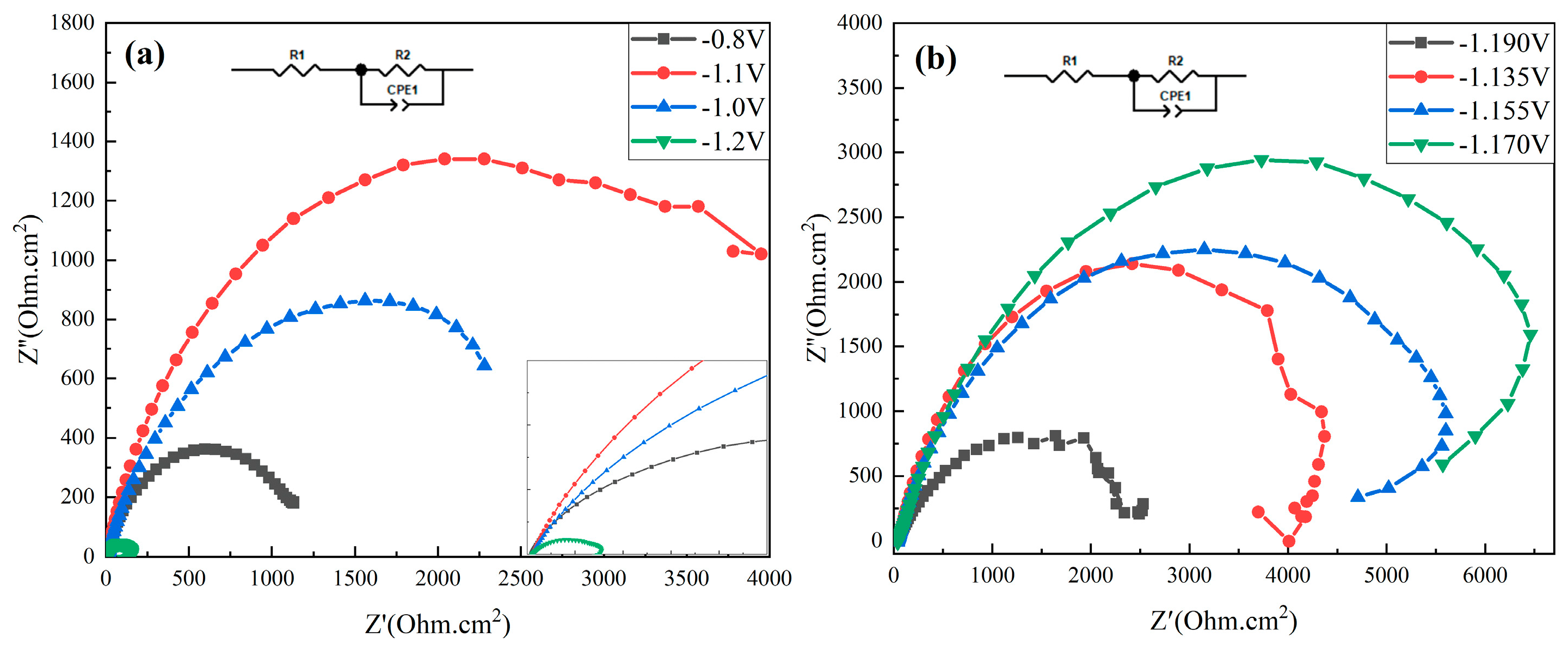
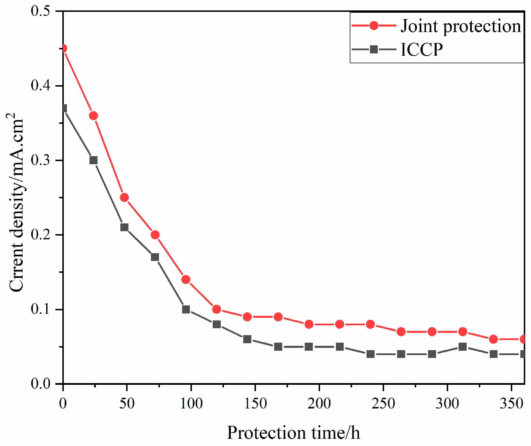
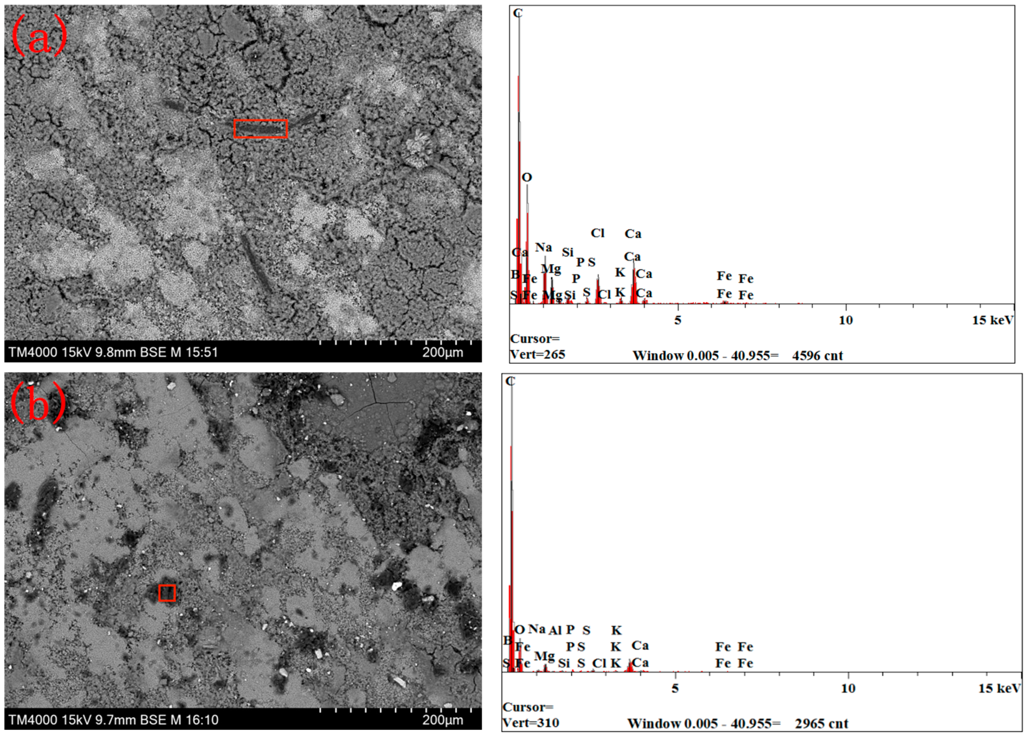
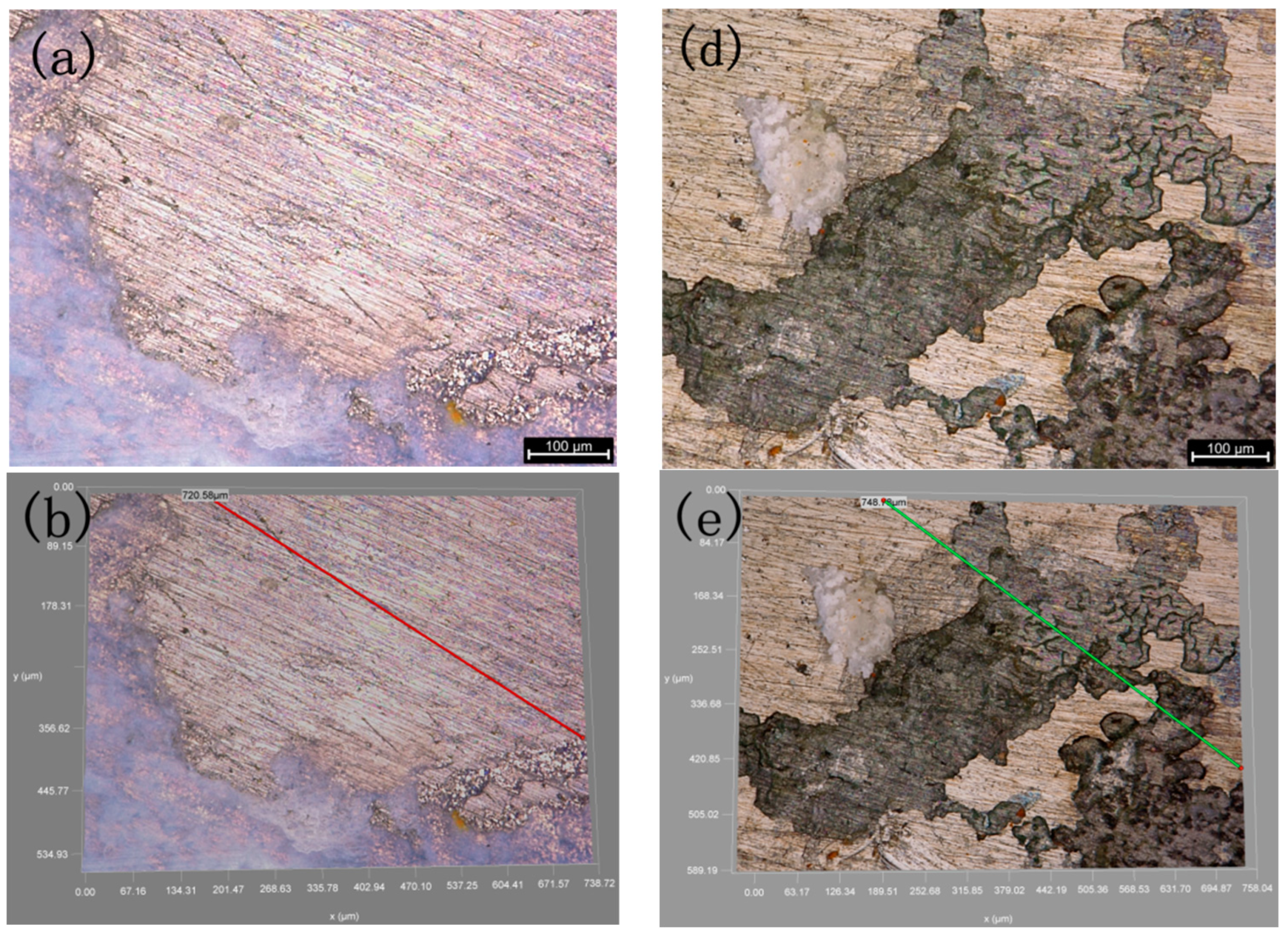
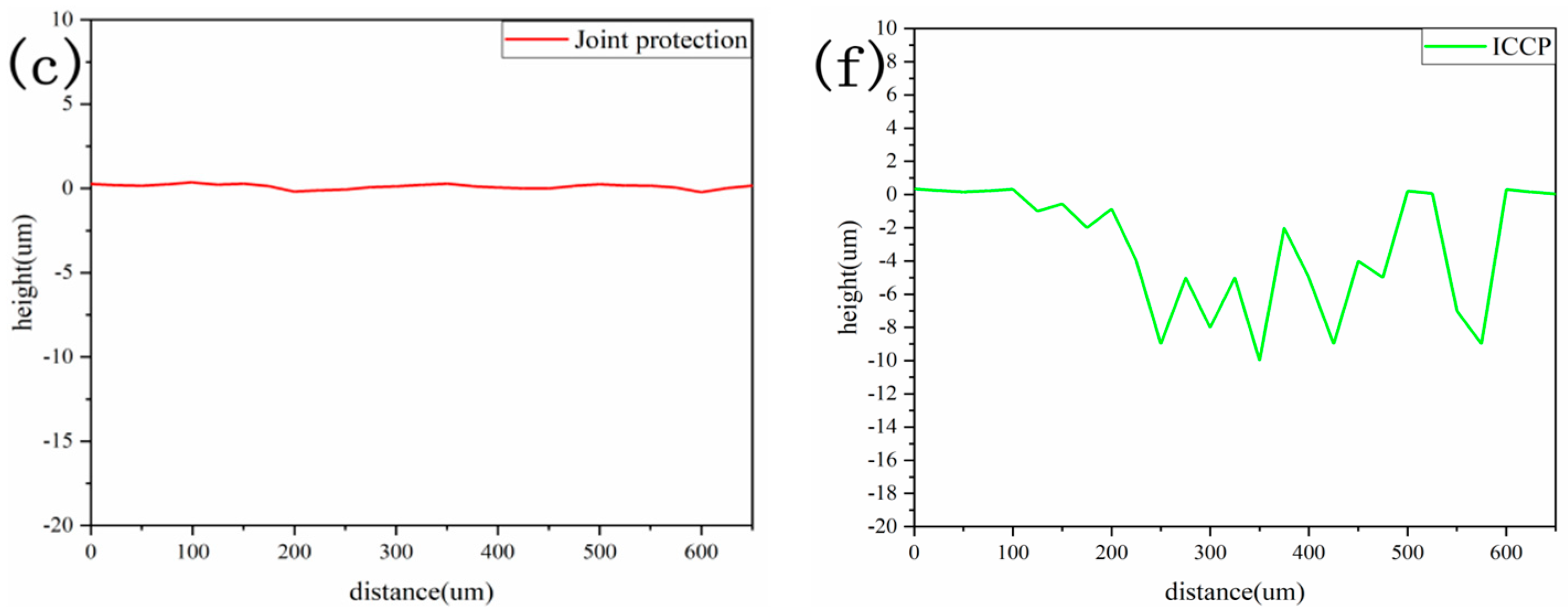
3. Results and Discussion

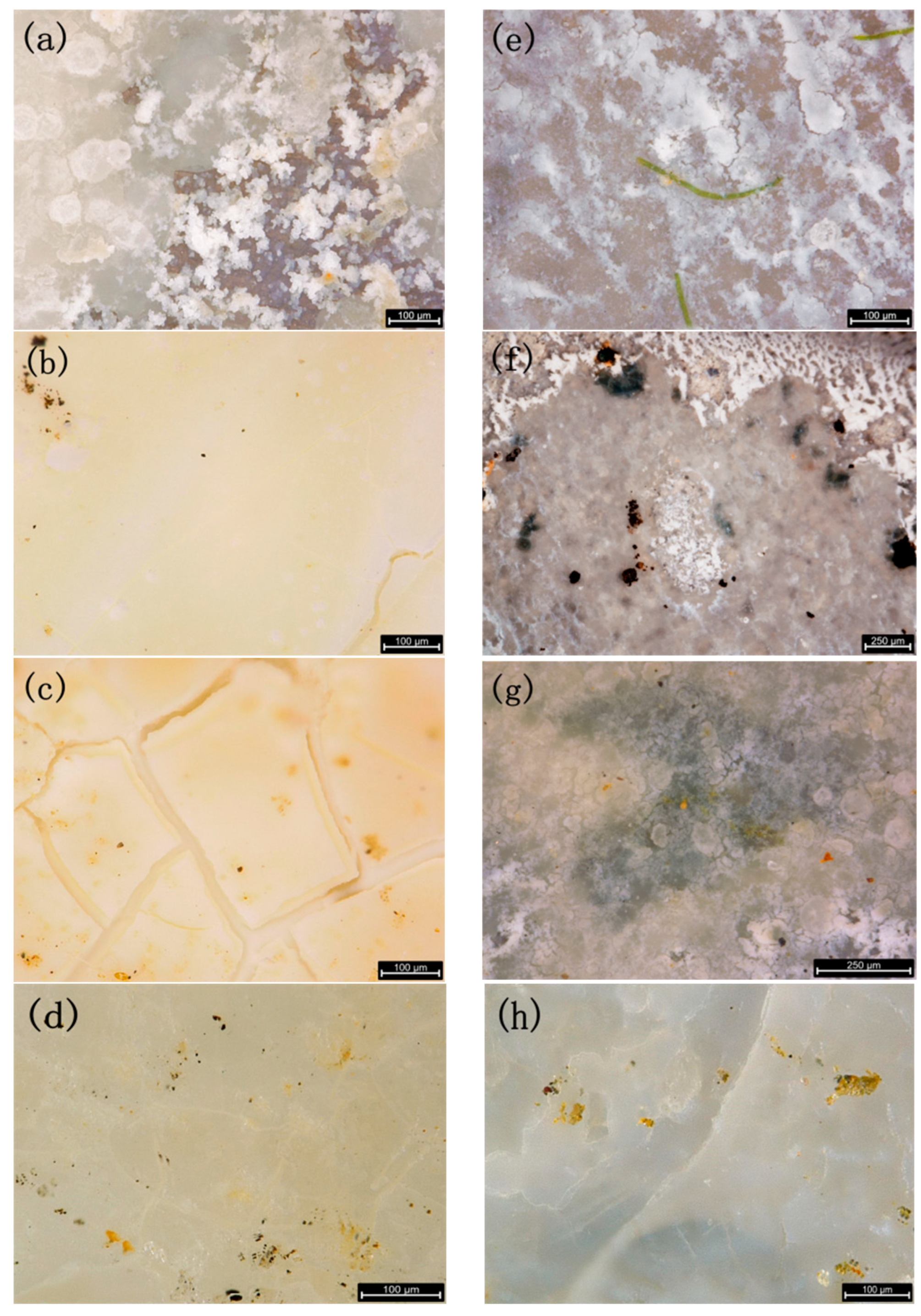
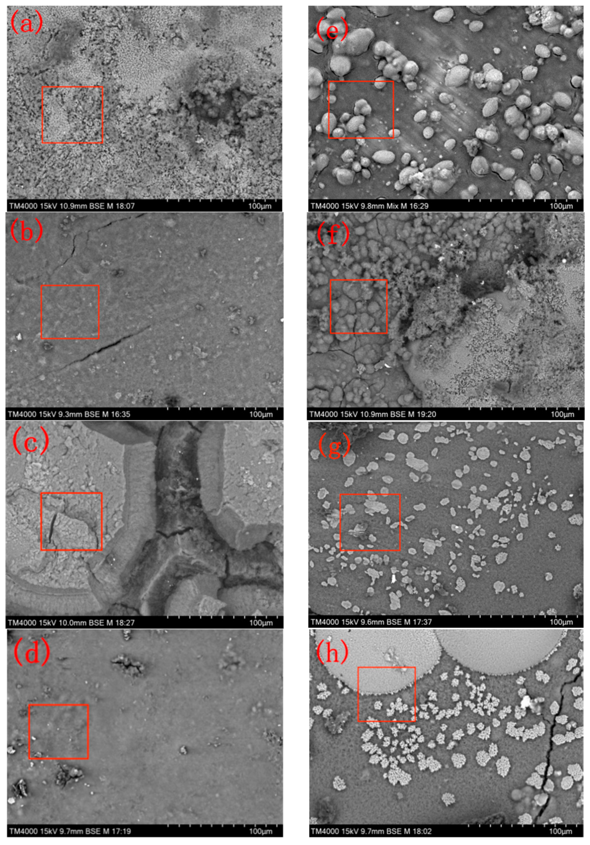
3.1. Comparison of the Sedimentary Layers’ Surface Morphology

Figure 3 shows optical microscopy images of the EH36 specimen under two different protection methods. After 7 days of joint protection, the sediment adhered to the surface of the EH36 sample, but the substrate of the EH36 sample could still be seen. At 15 days, the sedimentary layer completely covered the EH36 substrate with a small number of cracks. Between 15 and 30 days, a large number of network cracks appeared in the sedimentary layer. At 60 days, the network cracks in the sedimentary layer were filled with new sediment. At 7 days, a deposition layer also formed on the surface of the EH36 sample under ICCP, and green striped substances also appeared. Between 7 and 15 days, the thickness of the sedimentary layer increased, and multiple black substances were found below the sedimentary layer. At 30 days, the sedimentary layer completely covered the EH36 substrate, and black substances under the sedimentary layer could still be seen. At 60 days, the thickness of the sedimentary layer further increased, and black substances under the sedimentary layer could still be seen with large cracks.
By comparison, the surface deposition layer thickness of the EH36 sample under ICCP is significantly thinner than that under joint protection. It is speculated that the adhesion of black matter under ICCP inhibits the formation of a surface deposition layer on the EH36 sample, which is consistent with the conclusion of Li et al. [19] and others that marine fouling organisms can inhibit the formation of a surface deposition layer under ICCP.
Figure 4 shows SEM surface morphology images of the EH36 specimen under two different protection methods. As shown in Figure 4, after 7 days, the surface of the sedimentary layer of the EH36 sample under joint protection showed two color differences, white and grey, with irregular pores in the black area. At 15 days, the color difference on the surface of the sedimentary layer is no longer significant, with a small number of cracks. At 30 days, there were also multiple slender fissures in the large cracks on the surface of the sedimentary layer. At 60 days, the large cracks were filled with new sediment, forming a dense sedimentary layer. The EH36 sample under ICCP showed a large number of white particles on the surface after 7 days, and the sample substrate was clearly visible. At 15 days, white particles were still visible, with a color difference similar to that in Figure 4a, and there were numerous cracks. At 30 days, a large number of white blocky areas appeared on the surface of the sedimentary layer. At 60 days, the white blocky area continued to expand and concentrate, with significant cracks appearing.
From Figure 4a,b, we can observe the changes in the white and grey areas and then compare these changes with the changes in the white and grey areas in Figure 4e–h. The joint protection method accelerates the formation of the surface sedimentary layer and increases the density of the surface of the sedimentary layer. The rapid accumulation of excessive sediment can easily lead to large cracks, as shown in Figure 4c; however, excessive sediment can also quickly fill the cracks without affecting the density of the sedimentary layer or the protective effect on the EH36 carbon steel substrate.
For the large amount of white particles deposited on the surface layer of EH36 under ICCP after 7 days, Luo et al.’s [20] analysis of this phenomenon suggested that the factors affecting the deposition and morphological changes of white particles are most likely the initially formed thin film, which is the grey area in Figure 4e. Moreover, as can be clearly seen from Figure 3e, when white particles deposited on the surface of the EH36 substrate, a thin film was formed around the white particles. The thickness of the film increases with increasing experimental time. When the film does not fully cover the white particles (Figure 4f), the presence of white particles can easily lead to the generation of cracks. When the thin film completely covers the white particles (Figure 4g), a dense sedimentary layer is formed. The large white areas appearing on the surface of the sedimentary layer (Figure 4h) may be caused by the accumulation of a large number of white particles. In their study, Yang et al. [21] found that the deposition layer on the surface deposition layer of carbon steel under ICCP is a layered structure composed of an internal magnesium layer and an external calcium layer. Therefore, it is speculated that if the experiment continues, the white area will completely cover the grey area and ultimately form a sedimentary layer with a double-layer structure.

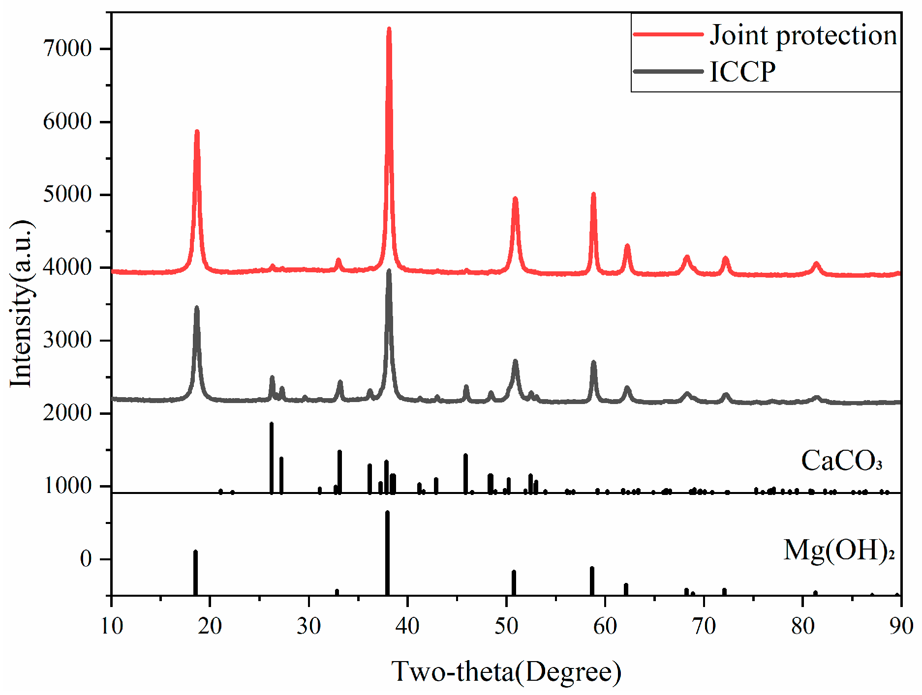
3.2. Composition of the Sedimentary Layer

In addition to the microstructure of the surface deposition layer, the surface chemical composition is also an important factor affecting the physical and chemical properties of the surface deposition layer. The EDS data in Table 1 show that the elemental composition of the surface deposition layer of the sample under joint protection is mainly Mg and O, with very little Ca content, indicating that the main components of the surface deposition layer are compounds composed of Mg and O. At the same time, the Fe content was 6.72% and 3.43% at 7 and 30 days, respectively, being significantly greater. This may be due to the incomplete coverage of the EH36 matrix by the surface deposition layer at 7 days and the appearance of large cracks in the surface deposition layer at 30 days.
The EDS data in Table 2 show that the elemental composition of the surface sediment of the sample under ICCP is mainly O, Mg, and Ca. The Ca/Mg ratio in sedimentary layers is an important measure to characterize their protective performance, and sedimentary layers with higher Ca/Mg ratios have better protective performance [22]. During the 60-day experiment, the mass fraction of O did not change significantly, and the Ca/Mg ratio increased from 0.4 (7 days) to 4.6 (60 days). Combined with the surface microstructure in Figure 4e–h, the Ca/Mg ratio is correlated with the area ratio of the white and grey areas on the surface of the sedimentary layer. As the Ca/Mg ratio increases, the area of the white area also increases. It can be inferred that the main components of the white area are Ca and O, while the main components of the grey area are Mg and O.
The XRD pattern in Figure 5 confirms the analysis results of the EDS data in Table 1 and Table 2. The main diffraction peaks of the surface deposition layer on the EH36 sample subjected to joint protection are basically consistent with those on the EH36 sample subjected to ICCP. Multiple clear Mg(OH)2 diffraction peaks are located at 18.51°, 32.83°, 37.92°, 50.77°, 58.61°, etc. However, the XRD pattern of the EH36 sample subjected to ICCP shows a significantly lower intensity of the main diffraction peaks, with multiple clear and low CaCO3 diffraction peaks located at 26.21°, 27.21°, 33.12°, 36.17°, 37.88°, 45.85°, etc., appearing in the pattern. According to the PDF standard cards of Mg(OH)2 and CaCO3 in Figure 5, the surface sediment components of the EH36 sample subjected to joint protection mainly comprise Mg(OH)2 and a very small amount of CaCO3, while the surface sediment components of the EH36 sample subjected to cathodic protection are mainly Mg(OH)2 and a large amount of CaCO3 [23].
The potential of the EH36 cathode sample is set to −1.17 V (vs. SCE), which is a relatively low protective potential and can cause a large amount of negative charge to accumulate on the surface of the EH36 sample. A strong oxygen absorption reaction occurs on the surface of the EH36 sample [24], producing a large amount of OH: O2 + 2H2O + 4e → 4OH. The surface pH of the EH36 sample rapidly increases. When the pH increases to 9.5 [25], Mg2+ combines with a large amount of OH to produce Mg(OH)2 precipitates, which attach to the surface of the EH36 sample. Therefore, the XRD pattern reflects that the surface deposition layer of the EH36 sample under both protection methods has a higher Mg(OH)2 content and a lower CaCO3 content.
Figure 6 shows that the initial current density of the EH36 sample under joint protection is 0.08 mA.cm2 greater than that of the EH36 sample under ICCP. After the current density is basically stable, the current density of the EH36 sample under joint protection is also 0.02 mA.cm2 greater. Combining the Mg(OH)2 and CaCO3 content in the surface deposition layer of the EH36 sample under joint protection and ICCP in Figure 5, it is found that there is a corresponding relationship between the current density and the content of Mg(OH)2 and CaCO3 in the surface deposition layer of the EH36 sample under both protection methods. That is, there is a clear correlation between the Ca/Mg ratio in the calcium magnesium deposition layer of the EH36 sample and the current density: when the current density is high, the content of Mg(OH)2 is greater, the content of CaCO3 is lower, and the Ca/Mg ratio decreases; if the current density is low, the opposite is true. This regular pattern is consistent with the study by Sharker et al. [26], which revealed that under low-current-density (55 µA/cm2) and low-temperature (7 °C) conditions, a greater proportion of aragonite (CaCO3) was deposited on the carbon steel cathode. Wen et al. [22] noted that a high cathode current leads to an increased rate of cathode reactions, higher pH levels, faster calcium layer formation, and a subsequent decrease in the Ca/Mg ratio. A higher Ca/Mg ratio results in greater density of the calcium/magnesium deposition layer. In their investigation of stainless steel polarization behavior, Wang et al. [27] observed that as the polarization current density initially decreased and then increased, the density of the calcium/magnesium deposition layer followed a pattern of an initial increase followed by a decrease, indirectly indicating a linear relationship between the Ca/Mg ratio and current density. It was also found that the electrochemical deposition of calcium magnesium compounds has great potential for reducing the cathodic protection current density and promoting low-cost cathodic protection (CP) design.

3.3. Biological Fouling of Surface Sedimentary Layers

Figure 7 shows the analysis of the special areas on the surface of the EH36 sample under ICCP. The SEM image in Figure 7a shows the green striped substance shown in Figure 3e, and the SEM image in Figure 7b shows the black substance not completely covered by the surface deposition layer shown in Figure 3f. The EDS spectrum shows that the amount of C in the red marked areas of Figure 7a,b is much greater than that of other elements, such as Mg, Ca, and O. It is clear that the strip-shaped green substance appearing on the surface sediment of the EH36 sample in the experiment is marine algae, the black substance is marine microorganisms, and that the microorganisms show aggregation.
Permeh et al. [28] evaluated the effectiveness of cathodic protection (CP) in marine fouling environments, and the test results showed that the presence of various fouling forms and crevices promoted the growth of microorganisms, such as sulfate-reducing bacteria (SRB), while the presence of surface fouling and crevices reduced the effectiveness of cathodic protection. Figure 6 shows that regardless of the level of cathodic protection implemented, a large number of microbial populations, such as sulfate-reducing bacteria, can grow and reproduce in the gaps between the carbon steel substrate and the sedimentary layer. The crack environment provides a low oxygen area and sufficient nutrient supply, allowing SRB to continue to grow in the gaps.
After removing the sedimentary layer from the surface of the EH36 sample that was protected for 60 days, as shown in Figure 8, there was no substance present on the surface of the EH36 sample under joint protection. There were traces of microbial attachment on the surface of the EH36 sample under ICCP. According to the surface height changes of the EH36 sample under the two protection methods, the surface height fluctuations of the EH36 sample under joint protection were not significant. However, according to the surface microbial attachment area in Figure 8e, there is a consistent pattern with the surface height fluctuation of the EH36 sample under ICCP, as shown in Figure 8f. This observation indicates that there is a significant depression on the surface of the EH36 sample under ICCP, with the maximum depression depth reaching −10 µm. This indicates that the attachment of marine microorganisms under ICCP causes corrosion on the surface of the EH36 sample. Vitor et al. [29] (2021) described similar corrosion phenomena, and this research study also revealed that a protective potential of −1000 mV (vs. Ag/AgCl) actually stimulates the hydrogenase activity of SRB, promoting the critical density and depth of the pits. In this experiment, we employed a cathodic protection potential of −1.17 V (vs. SCE) to steer clear of unsuitable values. Meanwhile, a higher potential ensures enhanced protection efficacy, offering insights for the future selection and application of cathodic protection potentials in ICCP systems.
On the surface of the EH36 sample with marine fouling organisms, cathodic polarization exhibits nonuniformity, resulting in significant differences in the cathodic protection ability in various areas of the EH36 carbon steel substrate surface. In this case, corrosion on the substrate surface will accelerate. Moreover, the extensive proliferation of microorganisms will further accelerate the process of corrosion.
By comparing the corrosion of the EH36 substrate surface under two protection methods, it can be seen that the application of a chlorine evolution anode in joint protection can effectively kill marine microorganisms, such as SRB, preventing microorganisms from adhering to the metal surface and preventing microbial corrosion. Compared to ICCP, joint protection can simultaneously prevent electrochemical corrosion and marine fouling corrosion on EH36 carbon steel samples, achieving dual protection.

4. Conclusions

Under two protection conditions, joint protection and ICCP, a 60-day study was conducted on the surface sedimentary layer of EH36 in natural seawater. By observing the morphological characteristics of the EH36 surface deposition layer and analyzing its composition, the following conclusions were drawn:
The surface calcium and magnesium deposition layer of EH36 subjected to joint protection was thicker and denser, while the surface calcium and magnesium deposition layer of EH36 subjected to ICCP was significantly thinner, indicating that marine fouling organisms, such as microorganisms, affected the formation of calcium and magnesium deposition layers on the cathode surface.
Compared with ICCP, the specific composition of the calcium magnesium deposition layer on the surface of EH36 under joint protection had a relatively high Mg(OH)2 content and a relatively low CaCO3 content. This is because the changes in the surface pH and current density of EH36 had an impact on the content of various components in the surface deposition layer.
By comparing the corrosion situation on the surface of the EH36 substrate, it can be seen that joint protection not only provides ICCP for EH36 but also effectively prevents marine fouling organisms from adhering to the surface of EH36 by electrolyzing seawater to produce chlorine, preventing marine fouling corrosion.

Author Contributions

J.H.: investigation, writing—original draft preparation, and writing—review and editing; P.W.: investigation, visualization, methodology, and writing—original draft preparation; P.D.: investigation, visualization, and data curation; B.G.: investigation and visualization; J.Z.: investigation; X.H.: visualization. All authors have read and agreed to the published version of the manuscript.

Funding

This research was funded by the Natural Science Foundation of China (51801033) and the Natural Science Foundation of Guangdong Province China (2021A1515012129).

Institutional Review Board Statement

Not applicable.

Informed Consent Statement

Not applicable.

Data Availability Statement

The raw/processed data required to reproduce these findings cannot be shared at this time due to technical or time limitations. However, they can be made available upon request.

Conflicts of Interest

The authors declare that they have no known competing financial interests or personal relationships that could have appeared to influence the work reported in this paper.

References

  1. Larsen, R.K. Designing and Managing an Offshore Cathodic Protection System. Mater. Perform. 2019, 58, 26–30. [Google Scholar]
  2. Hartt, H.W. 2012 Frank Newman Speller Award: Cathodic Protection of Offshore Structures-History and Current Status. Corros. J. Sci. Eng. 2012, 68, 1063–1075. [Google Scholar] [CrossRef]
  3. Szabó, S.; Bakos, I. Impressed current cathodic protection. Corros. Rev. 2006, 24, 39–62. [Google Scholar] [CrossRef]
  4. Barchiche, C.; Deslouis, C.; Festy, D.; Gil, O.; Refait, P.; Touzain, S.; Tribollet, B. Characterization of cal-careous deposits in artificial seawater by impedance techniques 3-Deposit of CaCO3 in the presence of Mg(II). Electrochim. Acta 2003, 48, 1645–1654. [Google Scholar] [CrossRef]
  5. Gabrielli, C.; Keddam, M.; Khalil, A.; Rosset, R.; Zidoune, M. Study of calcium carbonate scales by electro-chemical impedance spectroscopy. Electrochim. Acta 1997, 42, 1207–1218. [Google Scholar] [CrossRef]
  6. Li, X.; Liu, T.; Wang, H.; Sun, X. Mixed Fouling Growth Process-Microbial and CaCO3 Fouling in Water Systems. J. Chem. Ind. Eng. 2002, 53, 1247–1252. [Google Scholar] [CrossRef]
  7. Li, F.; An, M.; Liu, G.; Duan, D. Effects of sulfidation of passive film in the presence of SRB on the pitting corrosion behaviors of stainless steels. Mater. Chem. Phys. 2008, 113, 971–976. [Google Scholar] [CrossRef]
  8. Christine, R.; PhilipS, S. Removal and inactivation of Staphylococcus epidermidis biofilms by electrolysis. Appl. Environ. Microbiol. 2006, 72, 6364–6366. [Google Scholar] [CrossRef]
  9. Quej-Ake, L.; Nava, N.; Espinosa-Medina, M.A.; Liu, H.B.; Alamilla, J.L.; Sosa, E. Characterisation of soil/pipe interface at a pipeline failure after 36 years of service under impressed current cathodic protection. Corros. Eng. Sci. Technol. 2015, 50, 311–319. [Google Scholar] [CrossRef]
  10. Deshpande, P.; Kolekar, A. Impressed Current Cathodic Protection of Low Carbon Steel in Conjunction with Conducting Polyaniline based Paint Coating. Prot. Met. Phys. Chem. Surf. 2019, 55, 1236–1241. [Google Scholar] [CrossRef]
  11. Pravin, D.; Aniket, K.; Abhijit, B.; Kalendova, A.; Kohl, M. Impressed current cathodic protection (ICCP) of mild steel in association with zinc based paint coating. Mater. Today Proc. 2022, 50, 1660–1665. [Google Scholar] [CrossRef]
  12. Jiang, Z.; Zhang, C.; Du, J.; Shen, W.; Tang, S.; Ma, L.; He, M. A pipeline Was Protected By Impressed Current Cathodic Protection and Sacrificial Anode External Corrosion Factors and Control Measures. Compr. Corros. Control 2021, 35, 105–109. [Google Scholar] [CrossRef]
  13. Xiao, Y. A Case Study of Combined Use of Cathodic Protection of Sacrificial Anode and Impressed Current. Corros. Prot. 2021, 42, 66–70. [Google Scholar] [CrossRef]
  14. Liao, Z.; Li, H.; Luo, X.; Shi, Y.; Li, N.; Wang, C.; Liu, Y.; Yu, X.L. Joint cathodic protection of impressed current and sacrificial anode for long-distance pipelines under special conditions. Oil Gas Storage Transp. 2023, 42, 320–327. [Google Scholar] [CrossRef]
  15. Trasatti, S. Electrocatalysis: Understanding the success of DSA. Electrochim. Acta 2000, 45, 2377–2385. [Google Scholar] [CrossRef]
  16. Chi, M.; Yun, X.; Luo, B.; Guo, C.; Wang, S.; Min, D. Research progress on preparation and application of DSA electrode. Appl. Chem. Ind. 2021, 50, 498–503. [Google Scholar] [CrossRef]
  17. Liu, X.; Geng, J.; Liao, D.; Bai, B. Electrocatalytic chlorine cvolution performance of Ti/RuO2-IrO2 mesh anode. Chem. Eng. (China) 2023, 51, 61–65. [Google Scholar] [CrossRef]
  18. Qi, H.; Gao, K.; You, J.; Sun, W.; Wang, L.; Liu, G. Preparation and properties of highly active low iridium-doping Ti/IrRuSnSbOx electrode for chloride evolution. Mod. Chem. Ind. 2023, 43, 94–98. [Google Scholar] [CrossRef]
  19. Li, X.; Zhang, J.; Wang, J.; Duan, J.; Hou, B. Effects of Sulfate Reducing Bacteria Adhesion Formation of Calcareous Deposits under Method Protection Condition And Its Protection for Metals. Corros. Prot. 2018, 39, 265–269. [Google Scholar] [CrossRef]
  20. Luo, J.; Lee, R.; Chen, T.; Hartt, W.; Smith, S. Formation of Calcareous Deposits under Different Modes of Cathodic Polarization. Corrosion 1991, 47, 189–196. [Google Scholar] [CrossRef]
  21. Yang, Y.; Scantlebury, J.; Koroleva, E. A Study of Calcareous Deposits on Cathodically Protected Mild Steel in Artificial Seawater. Met. Open Access Metall. J. 2015, 5, 439–456. [Google Scholar] [CrossRef]
  22. Wen, G.; Zheng, F. The formation and application of calcium deposits during cathodic protection in seawater. Corros. Prot. 1995, 16, 50–53. [Google Scholar]
  23. Karoui, H.; Riffault, B.; Jeannin, M.; Kahoul, A.; Gil, O.; Amor, M.B.; Tlili, M.M. Electrochemical scaling of stainless steel in artificial seawater: Role of experimental conditions on CaCO3 and Mg(OH)2 formation. Desalination 2013, 311, 234–240. [Google Scholar] [CrossRef]
  24. Du, M.; Sun, Z. Study on the Cathodic Polarization Behavior of Stainless Steel 410 in Seawater. Period. Ocean Univ. China 2010, 40, 91–95. [Google Scholar] [CrossRef]
  25. Lee, R.U.; Ambrose, J.R. Influence of Cathodic Protection Parameters on Calcareous Deposit Formation. Corrosion 2012, 44, 887–891. [Google Scholar] [CrossRef]
  26. Sharker, T.; Simonsen, K.R.; Margheritini, L.; Kucheryavskiy, S.V.; Simonsen, M.E. Optimisation of electrochemical deposition of calcareous material during cathodic protection by implementing response surface methodology (RSM). Electrochim. Acta 2023, 444, 141960. [Google Scholar] [CrossRef]
  27. Wang, X.; Bai, S.F.; Guo, Y.F.; Huang, Z.H.; Li, X.B.; Hou, J.; Zhang, H.X. Study on cathodic protection behavior of high-strength stainless steel in seawater environment. Equip. Environ. Eng. 2023, 20, 45–52. [Google Scholar]
  28. Perme, S.; Lau, K.; Boan, M.; Tansel, B.; ASCE, F.; Duncan, M. Cathodic Polarization Behavior of Steel with Different Marine Fouling Morphologies on Submerged Bridge Elements with Cathodic Protection. J. Mater. Civ. Eng. 2020, 32, 04020184. [Google Scholar] [CrossRef]
  29. Vitor, L.; Mariana, G.; Simone, B.; Servulo, E. SRB-mediated corrosion of marine submerged AISI 1020 steel under impressed current cathodic protection. Colloids Surf. B Biointerfaces 2021, 202, 111701. [Google Scholar] [CrossRef]
Figure 1. Schematic diagram of a joint protection device for seawater electrolysis antifouling and ICCP (the chlorine evolution reaction is carried out when the DSA electrode is a chlorine evolution electrode, and the oxygen evolution reaction is carried out when the DSA electrode is an oxygen evolution electrode).
Jmse 12 01155 g001
Figure 2. Electrochemical impedance spectra (EIS) of EH36 samples under various cathodic protection potentials ((a) displays the AC impedance at potentials of −0.8 V, −1 V, −1.1 V, and −1.2 V, and (b) displays the AC impedance at potentials of −1.135 V, −1.155 V, −1.170 V, and −1.190 V).
Jmse 12 01155 g002
Figure 3. Comparison of optical microscopy images of EH36 samples under joint protection and ICCP at 7 d, 15 d, 30 d, and 60 d: parts (ad) show the surface morphology of samples under joint protection at 7 d, 15 d, 30 d, and 60 d; parts (eh) show the surface morphology of the samples at 7 d, 15 d, 30 d, and 60 d under ICCP.
Jmse 12 01155 g003
Figure 4. Comparison of the SEM surface morphology of the EH36 samples under joint protection and ICCP at 7 d, 15 d, 30 d, and 60 d: parts (ad) show the surface morphology of the samples under joint protection at 7 d, 15 d, 30 d, and 60 d; parts (eh) show the surface morphology of the samples at 7 d, 15 d, 30 d, and 60 d under ICCP.
Jmse 12 01155 g004
Figure 5. XRD patterns of the surface deposition layer on the EH36 sample under joint protection and ICCP.
Jmse 12 01155 g005
Figure 6. Time variation curve of the current density of the EH36 sample under joint protection and ICCP.
Jmse 12 01155 g006
Figure 7. SEM and EDS images of special areas on the surface of the EH36 sample under ICCP ((a) displays shows the green striped substance shown in Figure 3e, and (b) displays the black substance not completely covered by the surface deposition layer shown in Figure 3f).
Jmse 12 01155 g007
Figure 8. Comparison of optical microscopy images and profile height fluctuations of the EH36 sample after surface deposition layer removal under joint protection and ICCP: joint protection (ac); ICCP (df) (The red line in (b) is identical to the red line in (c), and similarly, the green line in (e) matches the green line in (f)).
Jmse 12 01155 g008aJmse 12 01155 g008b
Table 1. EDS analysis results of the deposition layer on the surface of the EH36 sample under joint protection (red marked area in Figure 4a–d).
Time (day)CONaMgAlSiPSClKCaFe
7 d1.98%45.13%4.68%36.55%-3.47%----1.47%6.72%
15 d2.60%52.74%-42.16%0.01%1.20%--0.78%0.25%-0.25%
30 d-48.74%2.43%34.42%0.05%4.80%-0.96%3.69%0.76%0.71%3.43%
60 d-47.54%1%46.27%0.39%0.06%0.16%2.74%--1.39%0.45%
Table 2. EDS analysis results of the deposition layer on the surface of the EH36 sample under ICCP (red marked area in Figure 4e–h).
Time (day)CONaMgAlSiPSClKCaFe
7 d-50.67%1.50%26.65%0.04%2.76%0.21%0.42%6.36%0.64%10.75%-
15 d1.10%55.79%0.84%25.54%0.01%2.27%0.17%0.26%1.56%0.14%11.87%0.45%
30 d5.98%50.10%0.81%23.50%-0.01%0.21%0.21%1.44%-17.59%0.15%
60 d8.75%53.16%1.05%6.36%0.32%0.34%0.01%0.10%0.45%-29.38%0.09%
Disclaimer/Publisher’s Note: The statements, opinions and data contained in all publications are solely those of the individual author(s) and contributor(s) and not of MDPI and/or the editor(s). MDPI and/or the editor(s) disclaim responsibility for any injury to people or property resulting from any ideas, methods, instructions or products referred to in the content.

