Article

Aquaporin 12 Is Expressed in the Stomach and Liver of the Spiny Dogfish (Squalus acanthias)

Biology Department, Georgia Southern University, P.O. Box 8042, Statesboro, GA 30460, USA
*
Author to whom correspondence should be addressed.
J. Mar. Sci. Eng. 2025, 13(1), 161; https://doi.org/10.3390/jmse13010161
Submission received: 27 December 2024 / Revised: 10 January 2025 / Accepted: 17 January 2025 / Published: 18 January 2025
(This article belongs to the Special Issue Nutrition and Physiology of Marine Fish)

Abstract

:
The sequence of Aquaporin 12 (AQP12) cDNA was amplified from spiny dogfish (Squalus acanthias) cDNAs using degenerate PCR, followed by 5′ and 3′ RACE PCR. The AQP12 nucleotide sequence had an open reading frame of 300 amino acids, which included one or more N-glycosylation sites. Degenerate and tissue PCRs revealed that AQP12 is expressed at the highest levels in the liver, followed by the pyloric stomach and the esophagus/cardiac stomach, with a small amount potentially present in the eye. A polyclonal antibody was made using a peptide from the derived amino acid sequence. Western blotting with the antibody showed faint banding around the size expected (33 kDa) by the 300 amino acid protein. A few more intense bands were seen at around 40 kDa and larger sizes. Immunohistochemistry in cardiac stomach tissue sections showed staining in a few sporadic paneth-like secretory cells along the surface of the epithelium. High-magnification imaging showed that the AQP12 staining was located in the membrane of secretory granules in the apical pole of the cells. This localization is reminiscent of the AQP12 localization in pancreatic acinar cells, where it is found in the membrane of zymogen granules containing digestive enzymes.

1. Introduction

Aquaporin 12 (AQP12) is a member of a large family of membrane channel proteins, which in mammals includes 13 members (AQP0-12) [1] and in wider vertebrate animals extends to 17 members (AQP0-16) [2]. Aquaporin 12 was originally described as AQP X2 [3], but this name was subsequently changed to AQP12 [4,5]. Aquaporins 11 and 12 are termed unorthodox or superaquaporins and are the most divergent members of the family with relatively low levels (around 20%) of homology to other family members [1,3,4,5,6]. Humans have two highly similar (around 95%) duplicate copies of AQP12 (AQP12A and AQP12B) [7]. Aquaporins are generally viewed as water and small-solute channels [1]; however, although Aquaporin 11 (the closest family member) exhibits water permeability, the sole study investigating mouse AQP12 showed no water permeability [8,9]. Some functional evidence suggests that AQP12 may be permeable to water [9]. Both AQP11 and AQP12 may also represent peroxiporins transporting hydrogen peroxide (H2O2) [9]. For reviews of aquaporins in vertebrates, see [1,9,10,11,12,13,14].
In most mammals, the small number of studies that have been carried out concerning AQP12 show that it is exclusively expressed in the pancreas [1,4,5,8,9]. However, one study showed that in mice, AQP12 is additionally expressed in the stomach and tongue [15]. Additionally, in the teleost zebrafish, AQP12 is ubiquitously expressed in all tissues studied, including the liver [16]. In the mammalian pancreas, AQP12 is expressed in acinar cells, where its expression localizes to the endoplasmic reticulum (ER) and zymogen granules containing digestive enzymes [5,9]. AQP12 may also be expressed in the β-cells of the rat pancreas [17]. AQP12-null mutant mice developed severe acute pancreatitis after secretagogue stimulation and exhibited large vacuoles in acinar cells [8,9]. Possible explanations for this include the absence of AQP12’s peroxiporin function, leading to cell damage, or the absence of AQP12’s role in zymogen granule fusion and exocytosis due to the lack of water permeability [9]. The other possibility suggested was that “AQP12 may be needed to reduce the size of zymogen granules by exporting water to concentrate the digestive enzyme therein and the absence of AQP12 may expand zymogen granules observed as vacuoles” [9]. This situation would also require AQP12 channels to exhibit a significant level of water permeability.
Outside of mammals and teleost fish, almost nothing is known about AQP12 besides the presence of AQP12 genes in the genome. It is known that many other animals including elasmobranchs, such as sharks, more primitive lampreys, and even invertebrates, possess a copy of the AQP12 gene in their genomes [2]. In recent years, several shark genomes have been sequenced, and AQP12 sequences from these genomes enabled this study to clone and sequence AQP12 transcripts from the spiny dogfish (Squalus acanthias) using a degenerate PCR approach utilized many times by this lab [18,19,20,21,22,23,24,25,26,27,28]. Despite the limited resources available, this study yielded new information concerning the role of AQP12 in sharks.

2. Materials and Methods

2.1. Animals

Three spiny dogfish were taken at random from the stock tank at the Mount Desert Island Biological Laboratory (MDIBL), one for the preparation of tissue total RNA samples, another for the preparation of protein samples for Western blots, and a third for the production of paraffin tissue blocks for immunohistochemistry. Stock tank fish were maintained outdoors at ambient temperatures and lighting and were fed daily with pieces of squid. All fish were treated in accordance with the prevailing IACUC regulations/procedures that had been approved by both MDIBL and Georgia Southern University IACUC committees.

2.2. Polymerase Chain Reactions (PCRs) and DNA Cloning

Total RNA samples were extracted from multiple tissues using methods outlined in [29]. Single-stranded cDNAs from gill*, rectal gland, kidney, esophagus/cardiac stomach*, pyloric stomach*, spiral valve intestine*, brain, skeletal muscle, eye and liver (no pancreas) tissue (* = epithelial scrapes used from the tissue) total RNA samples (1 μg) were produced using the method outlined in [30]. As on other occasions [18,19,20,21,22,23,24,25,26,27,28], deoxy-inosine (I) containing degenerate PCR primers (see Table 1) were designed to target two regions of conserved amino acids in the AQP12 sequences from various elasmobranchs (see Figure 1) and were used in 20 μL degenerate PCR reactions at a concentration of 5 μM. This was carried out to increase the concentration of each version of the primer present.
The conditions for the degenerate PCR were 40 cycles of 94 °C for 15 s, 55 °C for 15 s, and 72 °C for 20 s. Reactions used 1.25 units of Taq polymerase with standard buffer (New England Biolabs [NEB], Ipswich, MA USA) and 0.2 mM dNTPs. PCRs were run on a 1.2% agarose electrophoresis gel. DNA bands obtained were excised with a No. 11 scalpel blade and purified using a Monarch DNA purification kit (NEB) and then cloned into a plasmid vector using a pCR4-TOPO TA cloning kit for sequencing (Invitrogen/Thermofisher, Carlsbad, CA, USA). The insert region of plasmid cloning vector DNA was amplified (colony PCR) using M13XL Forward and M13XL Reverse primers and purified using a Quickstep 2 PCR purification kit (Edge Biosystems, Gaitherburg, MD, USA). DNA fragments were quantified using agarose gel electrophoresis and logic DNA ladder (Lamda Biotech, St Louis, MO, USA). DNA fragments (20 ng/kb) were prepared for sequencing in 8 μL with 4 pmol of T3XL primer and sent to Eton Biosciences (Durham, NC, USA) for sanger DNA sequencing. Agarose gels (Figure 1) were imaged using a Syngene (Cambridge, UK) GBox gel documentation system.
For RACE PCR, cardiac stomach 5′ and 3′ RACE cDNAs had previously been produced using a Smarter RACE kit (Takara Bio USA, San Jose, CA, USA) [27]. Two rounds of nested RACE PCRs using Phusion DNA polymerase (NEB) were performed. The first round (using 0.5 μM R1 primers) used the kit manufacturer’s protocol for touchdown PCR. The second-round nested PCRs (using 0.5 μM R2 primers and 0.5 μL from the initial reactions as a template) used 40 cycles of 2-step PCR, at 98 °C for 10 s, and at 72 °C for 3 min. DNA fragments produced were cloned and sequenced as above.

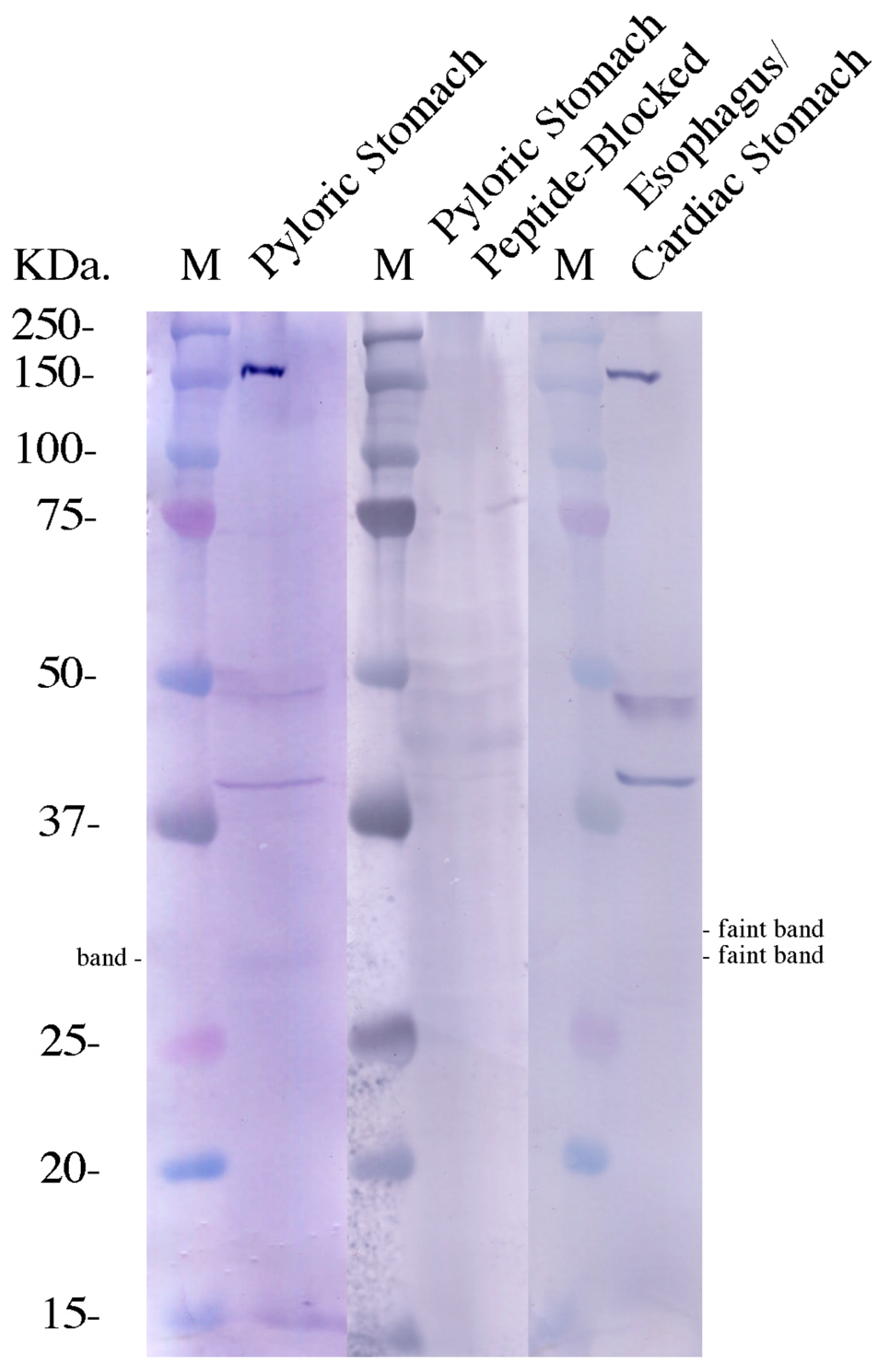
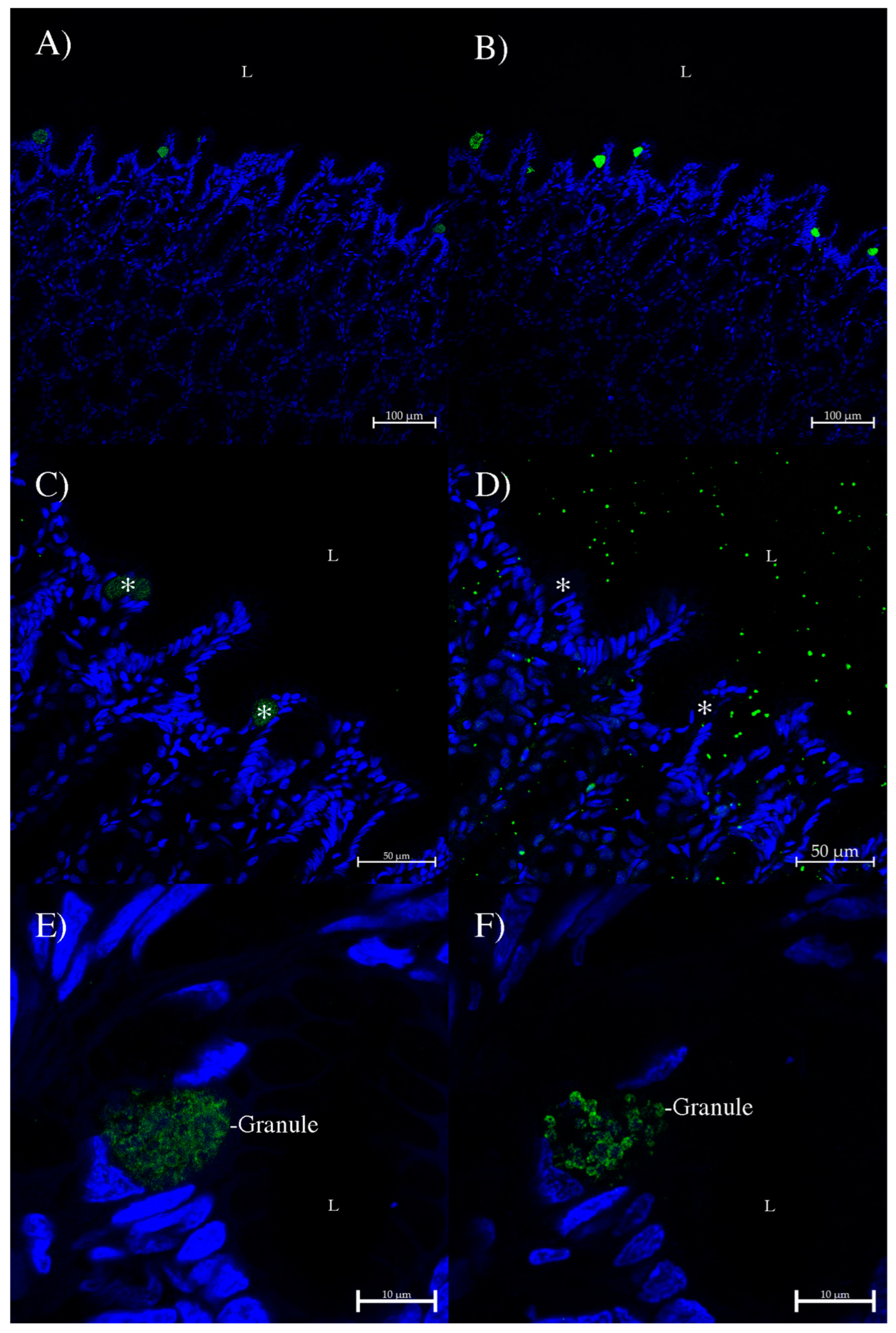
2.3. Western Blotting and Immunohistochemistry
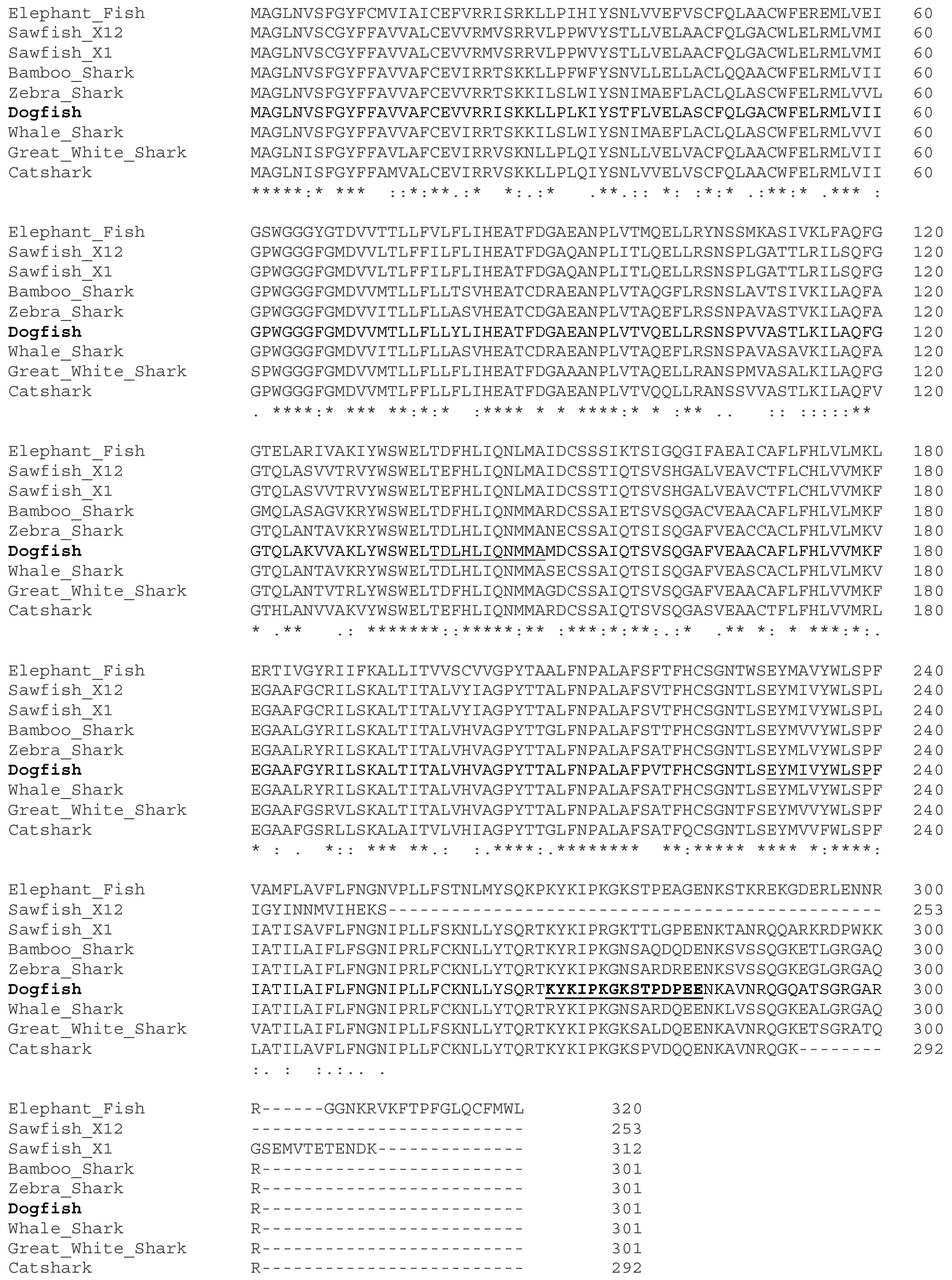
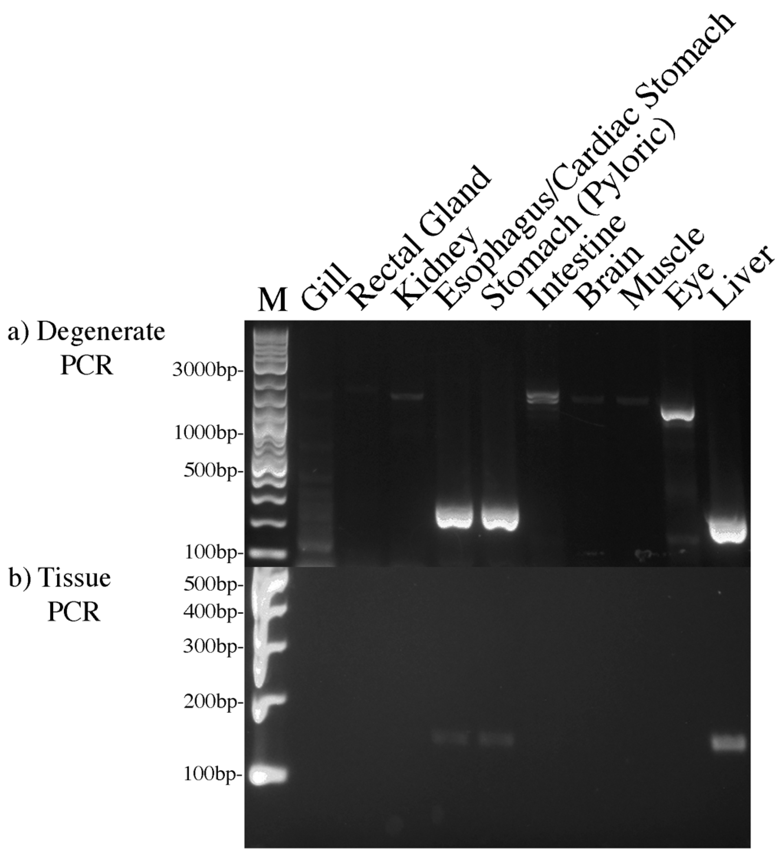
The Western blot protein samples utilized in this study were purified plasma membranes used in previous studies, where the methodology has already been outlined [26]. Western blotting was otherwise carried out as in [31]. The purified plasma membrane protein samples were limited in availability (no liver or pancreas, only 160 μg of esophagus/cardiac stomach samples), and so the main blotting was carried out using 300 μg of pyloric stomach purified plasma membranes. The rabbit anti-spiny dogfish AQP12 custom-made affinity-purified polyclonal antibody was produced against a peptide sequence (NH2-CKYKIPKGKSTPDPEE-COOH) from the derived amino sequence determined for AQP12. An extra N-terminal cysteine amino acid was added to the sequence to facilitate attachment to the Keyhole Limpet Hemocyanin (KLH) carrier protein used. The antibody was made by Genosphere Biotechnologies (Clamart, Paris, France). Genosphere affinity purified the antibodies from blood plasma from two rabbits independently, producing A1 and A2 versions of the antibody. The antibodies were used for both Western blotting and immunohistochemistry. Immunohistochemistry was performed on sections of spiny dogfish cardiac stomach using the same methodology as in [29,32]. Unfortunately, no paraffin-embedded tissue blocks of pyloric stomach, liver, or pancreas were available. The rabbit anti-dogfish AQP12 polyclonal antibodies were detected using a donkey anti-rabbit IgG highly cross-absorbed Alexa 488 plus secondary antibody (Thermofisher). Western blots were imaged using an Epson Perfection 4990 computer photo scanner, and immunohistochemistry images were produced using a Zeiss (White Plains, NY, USA) LSM710 laser-scanning confocal microscope.

3. Results

The initial degenerate PCR amplifications generated strong bands in the pyloric stomach, esophagus/cardiac stomach, and liver. There was also potentially a very faint band in the eye (Figure 2a). The expected size of AQP12 bands was around 305 bp; however, degenerate PCR DNA fragments are known to run somewhat smaller than expected, probably due to buffer differences between the DNA marker and PCR buffers. The bands in the liver and eye seemed to be somewhat smaller than those in the pyloric stomach and esophagus/cardiac stomach.
The degenerate PCR band from the esophagus/cardiac stomach was cloned and sequenced and confirmed to represent a segment of AQP12. Primers for 5′ and 3′ RACE were then designed (see Table 1) using the sequence information and used with available cardiac stomach RACE cDNAs. The 5R1 and 3R1 primers were used in initial RACE reactions, followed by 5R2 and 3R2 in subsequent nested RACE PCRs. The PCRs resulted in bands of around 1100 bp in both the 5′ and 3′ RACE PCRs. These were also cloned and sequenced, and the resulting sequences were combined in the complete AQP12 nucleotide sequence (Acc. No PQ807107) using clustal omega.
To confirm the expression profile of AQP12 in spiny dogfish tissues, further tissue PCRs were carried out at high stringency using AQP12-specific primers (see Table 1). The primers were also designed to span expected exon–intron boundaries to prevent genomic DNA amplification. The tissue PCRs were carried out using fewer cycles (30 rather than 40 in the degenerate PCR) to better distinguish between the level of expression in each tissue. These PCRs resulted in a similar profile to the degenerate PCR (Figure 2b). The liver DNA band was again slightly smaller than those in the pyloric stomach and esophagus/cardiac stomach and also more intense. There was no visible amplification in the eye cDNA sample.
The nucleotide sequence of AQP12 was translated using the Expasy translate tool (https://web.expasy.org/translate/) accessed on 16 August 2024, and the amino acid sequence (Figure 1) was used to design a peptide antigen for polyclonal antibody production. The subsequent antibody was used for Western blotting and immunohistochemistry experiments.
The tissue PCR amplifications suggested that the level of AQP12 expression was fairly low, and so it was not clear that a Western blot signal would be obtainable. Only 160 μg of purified esophagus/cardiac stomach purified plasma membrane protein was available, and only a couple of miniscule faint bands of around the right size (33 kDa) were seen on the Western blot (Figure 3). With 300 μg of pyloric stomach purified plasma membrane protein, a much stronger (although still faint) band was seen. On both blots, several much stronger higher-molecular-weight bands were also seen. These seem to be a common feature of blots with AQP antibodies, as seen many times previously [25,26,29]. The faint and stronger bands were diminished or missing on the control blot carried out using pyloric stomach protein with an antibody blocked by its peptide antigen. The peptide incubated with the antibody did lead to some background on the blot.
As with Western blotting, it was also not clear whether any staining would be apparent in immunohistochemistry unless the expression was localized and concentrated in a few cells, which is what occurred. As the AQP12 mRNA expression occurred in the pyloric stomach, it was assumed that the expression in the esophagus/cardiac stomach PCRs was most likely to be from the cardiac stomach. However, there were paraffin-embedded tissue blocks for the cardiac stomach but not for the pyloric stomach (or the liver and pancreas). So, immunohistochemistry was performed on sections of the cardiac stomach (see Figure 4).
With the use of the antibodies in the immunohistochemical localization of the AQP12 protein in tissue sections, of the two antibodies (A1 and A2; Figure 4A,B), the A2 antibody produced stronger fluorescent signals and so all subsequent work was performed using that antibody. However, the two antibodies were essentially staining the same cells in serial sections of the surface epithelium of the cardiac stomach. The cells stained were sporadically located along the cardiac stomach epithelium. The staining of these AQP12-positive cells was blocked out when the antibody was pre-blocked by its peptide antigen (Figure 4D compared to Figure 4C). High-resolution imaging of the staining (Figure 4E,F) showed that the staining appeared to be in the membrane of granules in what appear to be paneth-like secretory cells.

4. Discussion

The original degenerate PCR amplification (Figure 2) was interesting, in that without pancreatic tissue cDNAs available, it was not guaranteed that AQP12 would amplify anywhere else. As in most mammals, AQP12 is exclusively expressed in the pancreas [1,5,7,8,17]. However, of course, the timeframe for the divergence of vertebrates during evolution is long and allows the possibility that other roles could have developed for AQP12 in other taxonomic groups. This is easily illustrated by the situation in teleost fish, where AQP12 is ubiquitously expressed [16]. The fact that the AQP12 bands in spiny dogfish liver and eye appear to be smaller than the other bands produced suggests that there may be a splice variant version of AQP12 present in these tissues, and that is something to possibly be explored in the future. The closest species (to sharks) in the genebank (ensembl.org) suggests that the chimera elephant fish (Callorhincus milii) is predicted to have five splice variants of the AQP12 gene with protein-encoded sizes predicted to be between 293 and 320 amino acids. Likewise, the human AQP12B gene has multiple protein-encoding splice variants predicted, with products between 41 and 307 amino acids. There is no ensembl genomic sequence for the spiny dogfish, Squalus acanthias, so it is not possible to know what its AQP12 gene would or would not have regarding splice variants.
The fact the Western blots with the AQP12 antibody only gave faint bands was not too surprising given the level of amplification in the tissue PCRs in the pyloric stomach and esophagus/cardiac stomach. However, the amino acid sequence derived from the completed nucleotide sequence includes a single orthodox N-glycosylation site and at least two additional unorthodox sites (see https://research.bidmc.org/ncfg/blog/facts-about-protein-glycosylation#) accessed on 12 December 2024. These modifications, if they are utilized and remain unchanged, would increase the expected molecular weight of the protein from 33 kDa, based on the protein sequence alone, to ~40 kDa, as observed in the strong protein bands on the Western blots of both pyloric stomach and esophagus/cardiac stomach proteins. Essentially, glycosylations and other protein modifications mean the molecular weight of proteins is impossible to determine just from the amino acid sequence and could potentially be significantly larger than expected.
The AQP 12 antibody immunohistochemistry showed that the AQP12 protein expression was concentrated in what appear to be paneth-like secretory cells. While it is not impossible that these cells could represent mucus-secreting goblet cells, they are much smaller than the easily identifiable mucus/goblet cells found elsewhere in the gastrointestinal tract. Studies in the mammalian pancreas have shown that AQP12 is present in the membrane of zymogen granules containing digestive enzymes of acinar cells [5,9], which correlates well with the immunohistochemistry results observed here, and suggests that the AQP12-containing granules in these paneth-like cells might also contain digestive enzymes. As in mammals, the role of AQP12 in the granule membranes remains undetermined. The reasons might be as speculated for mammals [9], which is that AQP12 may play a role in granule fusion and exocytosis, or may be used to export water to concentrate the digestive enzyme inside the granules. However, another possibility also exists, which is that AQP12 is part of a mechanism to burst open the granules after their release into the gastric lumen (there is some evidence granules may be released intact). All that would be required would be a different osmotic environment leading to water uptake through AQP12 and granule swelling and bursting, or possibly the activation of some kind of metabolic process inside the granule to produce osmolytes that attract water to enter through AQP12. Whatever the role of AQP12, it is clearly of importance in mammals at least, as its absence in mice can lead to acute pancreatitis [8,9].
The expression of AQP12 in the spiny dogfish liver is also of interest. Initial studies in spiny dogfish suggest that AQP11 may be a pseudogene, as no expression was found for AQP11 in any of the tissues studied, including in the liver [28]. AQP11 in the mammalian liver is thought to play a role as a peroxyporin and a regulator of endoplasmic reticulum (ER) redox homeostasis and signaling via the export of hydrogen peroxide out of the ER to alleviate ER oxidative stress [9]. In spiny dogfish, without AQP11 expression in the liver, AQP12 may instead be fulfilling the role that AQP11 does in the mammalian liver. Mammalian AQP12 is thought to potentially be a peroxiporin and is also known to be expressed in the ER [9].

5. Conclusions

While the current study adds significantly to the literature concerning the role of AQP12 outside of mammalian species, there are still some avenues to be further investigated, the most important of which would be to determine if AQP12 is expressed in spiny dogfish pancreas. Secondarily, the role of splice variant versions of AQP12 in the liver and the cellular localization of AQP12 in liver cells (ER?) would also be important to determine. These would both require further animal work to gain suitable tissue samples that are currently beyond the scope of this lab to obtain. Consequently, this lab would make the AQP12 antibodies available to anyone wishing to further these studies.

Author Contributions

C.P.C. was responsible for writing this article and was involved in laboratory experimental work. J.B., S.C., and E.O. carried out significant critical laboratory experimental work. All authors have read and agreed to the published version of the manuscript.

Funding

Funding was provided by Georgia Southern University and the corresponding author, C.P.C.

Institutional Review Board Statement

Animal samples were collected under Georgia Southern IACUC protocol approval number I04023.

Informed Consent Statement

None required.

Data Availability Statement

The data are contained within this article. Figure images can be provided upon request.

Acknowledgments

The authors would like to thank the former staff of the Mount Desert Island Biological Laboratory, Maine, for their assistance of many kinds from 2004 to 2016.

Conflicts of Interest

The authors declare no conflicts of interest.

Abbreviations

AQPAquaporin
cDNAComplementary DNA
RACERapid Amplification of cDNA Ends
PCRPolymerase Chain Reaction
ElasElasmobranch
dNTPDe-oxy Nucleotide Tri-phosphates
TaqThermus aquatus
EREndoplasmic Reticulum
mRNAMessenger RNA
MDIBLMount Desert Island Biological Laboratory
DNADe-oxyribo Nucleic Acid
IUPACInternational Union of Pure and Applied Chemistry
RNARibo Nucleic Acid
KLHKeyhole Limpet Hemocyanin

References

  1. Ishibashi, K.; Hara, S.; Kondo, S. Aquaporin water channels in mammals. Clin. Exp. Nephrol. 2009, 13, 107–117. [Google Scholar] [CrossRef] [PubMed]
  2. Finn, R.N.; Chauvigné, F.; Hlidberg, J.B.; Cutler, C.P.; Cerdà, J. The lineage-specific evolution of aquaporin gene clusters facilitated tetrapod terrestrial adaptation. PLoS ONE 2014, 9, e113686. [Google Scholar] [CrossRef] [PubMed]
  3. Ishibashi, K.; Kuwahara, M.; Kageyama, Y.; Sasaki, S.; Suzuki, M.; Imai, M. Molecular cloning of a new aquaporin superfamily in mammals. In Molecular Biology and Physiology of Water and Solute Transport; Hohmann, S., Neilsen, S., Eds.; Kluwer Academic/Plenum Publishers: New York, NY, USA, 2000; pp. 123–126. [Google Scholar]
  4. Morishita, Y.; Sakube, Y.; Sasaki, S.; Ishibashi, K. Molecular mechanisms and drug development in aquaporin water channel diseases: Aquaporin superfamily (superaquaporins): Expansion of aquaporins restricted to multicellular organisms. J. Pharmacol. Sci. 2004, 96, 276–279. [Google Scholar] [CrossRef]
  5. Itoh, T.; Rai, T.; Kuwahara, M.; Ko, S.B.; Uchida, S.; Sasaki, S.; Ishibashi, K. Identification of a novel aquaporin, AQP12, expressed in pancreatic acinar cells. Biochem. Biophys. Res. Commun. 2005, 330, 832–838. [Google Scholar] [CrossRef]
  6. Ishibashi, K. Aquaporin subfamily with unusual NPA boxes. Biochim. Biophys. Acta 2006, 1758, 989–993. [Google Scholar] [CrossRef]
  7. Eisler, K.; Dropmann, L.M.; Bugert, P.; Ewers, M.; Witt, H. Genetic analysis of the aquaporin water channels AQP12A and AQP12B in patients with chronic pancreatitis. Pancreatology 2022, 22, 1079–1083. [Google Scholar] [CrossRef] [PubMed]
  8. Ohta, E.; Itoh, T.; Nemoto, T.; Kumagai, J.; Ko, S.B.; Ishibashi, K.; Ohno, M.; Uchida, K.; Ohta, A.; Sohara, E.; et al. Pancreas-specific aquaporin 12 null mice showed increased susceptibility to caerulein-induced acute pancreatitis. Am. J. Physiol. 2009, 297, C1368–C1378. [Google Scholar] [CrossRef] [PubMed]
  9. Ishibashi, K.; Tanaka, Y.; Morishita, Y. The role of mammalian superaquaporins inside the cell: An update. Biochim. Biophys. Acta 2021, 1863, 183617. [Google Scholar] [CrossRef] [PubMed]
  10. Ishibashi, K.; Kondo, S.; Hara, S.; Morishita, Y. The evolutionary aspects of aquaporin family. Am. J. Physiol. 2011, 300, R566–R576. [Google Scholar] [CrossRef] [PubMed]
  11. Ishibashi, K.; Kondo, S.; Hara, S.; Morishita, Y. The role of mammalian superaquaporins inside the cell. Biochim. Biophys. Acta 2014, 1840, 1507–1512. [Google Scholar] [CrossRef]
  12. Ishibashi, K.; Tanaka, Y.; Morishita, Y. Chapter One-Perspectives on the evolution of aquaporin superfamily. Vitam. Horm. 2020, 112, 1–27. [Google Scholar] [CrossRef] [PubMed]
  13. Adeoye, A.; Odugbemi, A.; Ajewole, T. Structure and function of aquaporins: The membrane water channel protein. Biointerface Res. Appl. Chem. 2022, 12, 690–705. [Google Scholar] [CrossRef]
  14. Markou, A.; Ungera, L.; Abir-Awan, M.; Saadallah, A.; Halsey, A.; Balklava, Z.; Conner, M.; T¨ornroth-Horsefield, S.; Greenhill, S.D.; Conner, A.; et al. Molecular mechanisms governing aquaporin relocalisation. Biochim. Biophys. Acta 2022, 1864, 183853. [Google Scholar] [CrossRef] [PubMed]
  15. Isokpehi, R.D.; Rajnarayanan, R.V.; Jeffries, C.D.; Oyeleye, T.O.; Cohly, H.H.P. Integrative sequence and tissue expression profiling of chicken and mammalian aquaporins. BMC Genom. 2009, 10 (Suppl. 2), S7. [Google Scholar] [CrossRef]
  16. Tingaud-Sequeira, A.; Calusinska, M.; Finn, R.N.; Chauvigné, F.; Lozano, J.; Cerda, J. The zebrafish genome encodes the largest vertebrate repertoire of functional aquaporins with dual paralogy and substrate specificities similar to mammals. BMC Evol. Biol. 2010, 10, 38. [Google Scholar] [CrossRef]
  17. Da Silva, I.V.; Cardoso, C.; Méndez-Giménez, L.; Camoes, S.P.; Frühbeck, G.; Rodríguez, A.J.; Miranda, P.; Soveral, G. Aquaporin-7 and aquaporin-12 modulate the inflammatory phenotype of endocrine pancreatic beta-cells. Arch. Biochem. Biophys. 2020, 691, 108481. [Google Scholar] [CrossRef] [PubMed]
  18. Cutler, C.P.; Sanders, I.L.; Hazon, N.; Cramb, G. Primary sequence, tissue specificity and expression of the Na+, K+-ATPase β1 subunit in the European eel (Anguilla anguilla). Fish Physiol. Biochem. 1995, 14, 423–429. [Google Scholar] [CrossRef] [PubMed]
  19. Cutler, C.P.; Sanders, I.L.; Cramb, G. Expression of Na+, K+-ATPase subunit isoforms in the European eel (Anguilla anguilla). Fish Physiol. Biochem. 1997, 17, 371–376. [Google Scholar] [CrossRef]
  20. Cutler, C.P.; Brezillon, S.; Bekir, S.; Sanders, I.L.; Hazon, N.; Cramb, G. Expression of a duplicate Na+, K+-ATPase β1 isoform in the European eel (Anguilla anguilla). Am. J. Physiol. 2000, 279, R222–R229. [Google Scholar] [CrossRef] [PubMed]
  21. Cutler, C.P.; Cramb, G. Branchial expression of an aquaporin 3 (AQP-3) homologue is downregulated in the European eel Anguilla anguilla following seawater acclimation. J. Exp. Biol. 2002, 205, 2643–2651. [Google Scholar] [CrossRef] [PubMed]
  22. Cutler, C.P.; Cramb, G. Two isoforms of the Na+/K+/2Cl cotransporter (NKCC1) are expressed in the European eel (Anguilla anguilla). Biochim. Biophys. Acta 2002, 1566, 92–103. [Google Scholar] [CrossRef] [PubMed]
  23. Martinez, A.-S.; Cutler, C.P.; Wilson, G.; Phillips, C.; Hazon, N.; Cramb, G. Regulation of expression of two aquaporin homologues in the intestine of the European eel: Effects of seawater acclimation and cortisol treatment. Am. J. Physiol. 2005, 288, R1733–R1743. [Google Scholar]
  24. Cutler, C.P.; Cramb, G. Differential expression of absorptive cation-chloride cotransporters in the intestinal and renal tissues of the European eel (Anguilla anguilla). Comp. Biochem. Physiol. 2008, 149, 63–73. [Google Scholar] [CrossRef] [PubMed]
  25. Cutler, C.P.; MacIver, B.; Cramb, G.; Zeidel, M.L. Aquaporin 4 is a ubiquitously expressed isoform in the dogfish (Squalus acanthias) shark. Front. Physiol. 2012, 2, 107. [Google Scholar] [CrossRef] [PubMed]
  26. Cutler, C.P.; Murray, D.; Ojo, T.; Harmon, S.; MacIver, B.; Cramb, G.; Zeidel, M.L. Aquaporin (AQP) channels in the spiny dogfish, Squalus acanthias I: Characterization of AQP3 and AQP15, and localization of their proteins in the gill and spiral valve intestine. Comp. Biochem. Physiol. 2022, 258, 110702. [Google Scholar] [CrossRef]
  27. Cutler, C.P.; Mainer, S.; Ojo, T. The aquaporin 8 (AQP8) membrane channel gene is present in the elasmobranch dogfish (Squalus acanthias) genome and is expressed in brain but not in gill, kidney or intestine. Comp. Biochem. Physiol. 2022, 260, 110730. [Google Scholar] [CrossRef] [PubMed]
  28. Cutler, C.P.; Canicatti, M.E.; Omoregie, E. Evidence that Aquaporin 11 (AQP11) in the spiny dogfish (Squalus acanthias) may represent a pseudogene. Int. J. Mol. Sci. 2024, 25, 2028. [Google Scholar] [CrossRef] [PubMed]
  29. Cutler, C.P.; Ojo, T. Immunohistochemical localization and expression of aquaporin 3-2 (AQP3C1) in the spiny dogfish, Squalus acanthias. Hydrobiology 2024, 3, 378–391. [Google Scholar] [CrossRef]
  30. Cutler, C.P.; Omoregie, E.; Ojo, T. UT-1 transporter expression in the spiny dogfish (Squalus acanthias): UT-1 protein shows a different localization in comparison to that of other sharks. Biomolecules 2024, 14, 1151. [Google Scholar] [CrossRef]
  31. Cutler, C.P.; Harmon, S.; Walsh, J.; Burch, K. Characterization of aquaporin 4 protein expression and localization in tissues of the dogfish (Squalus acanthias). Front. Physiol. 2012, 3, 21. [Google Scholar] [CrossRef] [PubMed]
  32. Cutler, C.P.; Kurt, K.; Campbell, K.E.; Ojo, T. Aquaporin (AQP) channels in the spiny dogfish, Squalus acanthias II: Localization of AQP3, AQP4 and AQP15 in the kidney. Comp. Biochem. Physiol. 2022, 258, 110701. [Google Scholar] [CrossRef] [PubMed]
Figure 1. Amino acid alignment of the spiny dogfish (Dogfish) AQP12 sequence determined in this study, aligned with AQP12 amino acid sequences from other cartilaginous fish, including those of elephant fish (Callorhinchus milii; XM_042339256), Sawfish (Pristis pectinata; X1, XM_052018964; X12, XM_052018965), bamboo shark (Chiloscyllium plagiosum; XM_043702897), zebra shark (Stegostoma fasciatum; XM_048542632), whale shark (Rhincodon typus; XM_020511124), great white shark (Carcharodon carcharias; XM_041205846), and catshark (Scyliorhinus canicula; XM_038816463). The sequences were initially aligned using Clustal Omega (https://www.ebi.ac.uk/jdispatcher/msa/clustalo accessed on 16 August 2024). Underlined sequences represent the sites used to design degenerate primers. The bold and underlined sequence is the amino acid sequence used to produce the spiny dogfish AQP12 custom-made affinity-purified polyclonal antibody. Numbers indicate the number of the end amino acid in the sequence. * indicates amino acids completely conserved in every species. : and . indicate chemically similar amino acid substitutions. – indicates gaps inserted into the alignment.
Jmse 13 00161 g001
Figure 2. Agarose electrophoresis gel images of AQP12 DNA fragment PCR amplifications. (a) Degenerate PCRs (40 cycles) from different spiny dogfish tissue cDNAs as indicated (see also Table 1 for degenerate PCR primers). (b) Subsequent high-stringency PCR amplifications (30 cycles) of AQP12 in the same tissue cDNAs (see also Table 1 for tissue PCR primers). M = 2-log ladder DNA marker (NEB).
Jmse 13 00161 g002
Figure 3. Western blots performed using the AQP12 A2 custom-made affinity-purified polyclonal antibody. Three blots were produced using 300 μg of pyloric stomach purified plasma membrane protein or 160 μg of esophagus/cardiac stomach purified plasma membrane protein. One blot was incubated with an antibody blocked by its peptide antigen (peptide-blocked). M = Precision Plus Protein Kaleidoscope marker (Biorad, Santa Rosa, CA, USA).
Jmse 13 00161 g003
Figure 4. Localization of the spiny dogfish AQP12 A1 and A2 antibodies in cells in the surface epithelium of the cardiac stomach. (A) is an image of the epithelial tissue stained with the AQP12 A1 antibody (green). (B) is a serial section (to (A)) of the epithelial tissue stained with the AQP12 A2 antibody (green). (C) is an image of the epithelial tissue stained with the AQP12 A2 antibody (green). (D) is a control serial section (to (C)) of the epithelial tissue stained with the AQP12 A2 antibody, which was blocked by its peptide antigen (green). * marks the same cells in (D) compared to (C). (E) High-magnification image through the center of a single cell stained with the AQP12 A2 antibody. (F) A second focal plane of the cell in (E) through the edge of the cell. Images (AD) were produced with a 20× lens zoom factor of 0.6 (A/B) or 1.5 (C/D). (E,F) were produced with 100× oil lens using a zoom factor of 1.5. L = cardiac stomach lumen. Sections counterstained with DAPI (blue) nuclear stain.
Jmse 13 00161 g004
Table 1. PCR primers utilized.
Degenerate PCR (expected product 305 bp):- I* = deoxy-inosine
                                                                                     IUPAC Codes: Y = C/T
                                                                                                                R = A/G
                                                                                                               H = A/C/T
Elas AQP12 Sense 2ACI* GAY YTI* CAY YTI* ATH CAR AAY ATG ATG GC
Elas AQP12 Anti 2GG RCT I*AR CCA RTA I*AC I*AI* CAT RTA YTC
5′ (5R) and 3′ (3R) RACE
Squal AQP12 5R 1CAGCTACGTG CACCAGGGCA GT
Squal AQP12 5R 2AGGGCTTTGG ACAGGATCCT GTATCCA
Squal AQP12 3R 1 CCTAGTTGTG ATGAAGTTTG AGGGAGCAGC T
Squal AQP12 3R 2GATACAGGAT CCTGTCCAAA GCCCTAACCA
Tissue PCR (expected product 152 bp)
AQP12 senseGGTGCACGTA GCTGGCCCAT AT
AQP12 antiACAGAAAGAT GGCCAGAATT GTTGCAATGA ATGG
Disclaimer/Publisher’s Note: The statements, opinions and data contained in all publications are solely those of the individual author(s) and contributor(s) and not of MDPI and/or the editor(s). MDPI and/or the editor(s) disclaim responsibility for any injury to people or property resulting from any ideas, methods, instructions or products referred to in the content.

