Article

Detection of the Benthic Dinoflagellates, Ostreopsis cf. ovata and Amphidinium massartii (Dinophyceae), Using Loop-Mediated Isothermal Amplification

Environment and Resource Convergence Center, Advanced Institute of Convergence Technology, Suwon 16229, Korea
*
Author to whom correspondence should be addressed.
J. Mar. Sci. Eng. 2021, 9(8), 885; https://doi.org/10.3390/jmse9080885
Submission received: 12 June 2021 / Revised: 10 August 2021 / Accepted: 13 August 2021 / Published: 17 August 2021
(This article belongs to the Special Issue Taxonomy and Ecology of Marine Algae)

Abstract

:
For the in situ and sensitive detection of benthic dinoflagellates, we have established an integrated loop-mediated isothermal amplification (LAMP) assay based on Ostreopsis cf. ovata and Amphidinium massartii. To detect the two species, a set of species-specific primers was constructed between the ITS gene and D1–D6 LSU gene, and the reaction temperature, time, and buffer composition were optimized to establish this method. In addition, the specificity of the LAMP primers was verified both in strains established in the laboratory and in field samples collected from the Jeju coastal waters, Korea. With the LAMP assay, the analysing time was within 45 to 60 min, which may be shorter than that with the conventional PCR. The detection sensitivity of the LAMP assay for O. cf. ovata or A. massartii was comparable to other molecular assays (PCR and quantitative PCR (qPCR)) and microscopy examination. The detection limit of LAMP was 0.1 cell of O. cf. ovata and 1 cell of A. massartii. The optimized LAMP assay was successfully applied to detect O. cf. ovata and A. massartii in field samples. Thus, this study provides an effective method for detecting target benthic dinoflagellate species, and could be further implemented to monitor phytoplankton in field surveys as an altenative.

1. Introduction

The genus Ostreopsis Johannes Schmidt and most of the species of Amphidinium Claparède & J. Lachmann are benthic dinoflagellates that grow on macrophytes or are attached to sand or coral rubble [1,2,3]. Their occurrence has been generally reported in tropical and subtropical seas [4,5]. The global occurrence of some species in these genera has significantly increased over the last decade and is expected to expand to temperate regions. The main harmful effects of some species of benthic dinoflagellates are related to the fact that they not only affect marine life and the aquaculture industry, but also pose a threat to human health [6,7,8,9,10]. Ostreopsis species have a particularly rich mucilaginous matrix, and some of them have thus far been reported to produce several toxins [1,11,12,13,14,15]. Moreover, Ostreopsis species are potentially toxic and can affect marine organisms and humans through the food web [16]. Their toxins can cause severe irritation to human skin and respiratory problems through aerosolization. They can also cause vomiting, kidney problems, and even death in severe cases [17,18].
Over the past several decades, blooms of benthic dinoflagellates have been observed in temperate to tropical coastal waters, in both the southern and northern hemispheres, whereas the proliferation of Ostreopsis cf. ovata was found in temperate regions during the summer [5]. The expansion of toxin-producing Ostreopsis spp. to temperate regions can potentially occur due to ballast water discharge by cargo ships, and, mainly due to the marginal dispersal associated with global warming, can induce bloom formations [19].
The cosmopolitan dinoflagellate genus Amphidinium has been found in pelagic and mainly in benthic environments with frequent occurrence [20,21,22,23]. Some Amphidinium species, particularly A. caterae, have been known to produce a number of bioactive compounds with cytotoxic or hemolytic activity to marine organisms [24,25,26,27,28]. Moreover, the cytotoxicity of A. cf. massartii affecting Artemia salina mortality has been revealed [29].
Several benthic dinoflagellate species, including Ostreopsis, have been reported as potential causative agents of toxic poisoning in Korean coastal waters [30,31,32,33,34,35]. Recently, a rapid increase in the O. cf. ovata biomass in Jeju coastal waters has been reported [36]. Most of these reports on the occurrence of benthic dinoflagellates in Korean coastal waters were based on conventional microscopic analysis methods [37,38,39,40]. Because blooms in coastal and oceanic waters with negative impacts on environmental health have the possibility of occurrence, several toxigenic dinoflagellates belonging to the genera Amphidinium, Coolia, Gambierdiscus, and Ostreopsis (including O. cf. ovata and A. cf. massartii) have been seriously considered as harmful organism candidates in Korea [41].
Following the development of molecular techniques, a number of modifications of polymerase chain reaction (PCR) methods have been established [42,43]. However, whereas these PCR-based assays have provided a reliable, sensitive, and specific tool to detect potentially harmful dinoflagellates, their economic limitations such as dependence on an expensive apparatus, practical limitations such as low amplification efficiencies, and long reaction times ultimately restrict their widespread application. Therefore, the development of rapid, simple, and cost-effective detection methods is still necessary to effectively detect harmful dinoflagellates.
Since Loop-mediated Isothermal Amplification (LAMP) was first introduced by Notomi et al. in 2000 [44], more than 100 LAMP detection methods have been developed for animal pathogens, including humans, and are ideally used due to the advantages of low cost and high sensitivity [45,46,47]. Because LAMP is performed under isothermal conditions, this method facilitates the amplification of only a few copies of initial DNA to obtain approximately 109 copies in less than 1 h, which can be visualized after the reaction using SYBR Green dye. The development of a LAMP assay, therefore, enhances the detection of various dinoflagellate species [48,49].
Owing to the recent increase in the abundance of benthic dinoflagellates in Korean coastal waters, their rapid detection and extensive monitoring are required. In this study, we developed a highly specific LAMP assay for the sensitive detection of two species of benthic dinoflagellates, O. cf. ovata and A. massartii. Moreover, we established the proper LAMP conditions for each species and applied them to the field samples to verify the sensitivity.

2. Materials and Methods

2.1. Sampling Site and Establishment of Dinoflagellate Strains

Macrophytes samples were collected by scuba divers in May 2018 within a water depth of 10 m at Seongsan, Jeju Island (33°27.35′ N, 126°56.01′ E). The seawater temperature and salinity were recorded as 17.4 °C and 33.2, respectively, at that time. The collected macrophytes were transferred into a 1 L bottle, which was filled with filtered seawater, and then shaken vigorously to detach the attached dinoflagellates. The samples were filtered by a 100 μm mesh to separate the macrophyte from seawater, and then transferred to the laboratory.
To establish a single cell strain, 5 mL of the sample was placed in a six-well plate, and single-cell isolation was performed under a dissecting microscope (SZX10, Olympus, Tokyo, Japan). After the clonal cultures of O. cf. ovata and A. massartii were established, the strains were transferred into 30 mL flasks containing fresh f/2-Si medium. The cultures were placed under white fluorescent lights at 22 °C with a continuous illumination of 20 μE·m−2·s−1.

2.2. DNA Extraction and Species Identification

The DNA sequences of these cells were analysed when the concentration of each strain was more than 103 cells mL−1. The dense culture (10 mL) was centrifuged at 10,000× g for 3 min at room temperature and the pellet was used for DNA extraction. Genomic DNA was extracted using an AccuPrep Genomic DNA Extraction Kit (BIONEER, Daejeon, Korea). The quality and purity of the DNA were assessed using agarose gel electrophoresis and spectrophotometry. For species identification, the SSU, ITS1, 5.8S, ITS2, and LSU rDNA sequences were amplified using universal eukaryotic primers [50,51]; the obtained DNA sequences were confirmed using BLAST (https://blast.ncbi.nlm.nih.gov/Blast.cgi, NCBI (accessed on 3 September 2018). The rDNA sequences of the two species were identical to those of the Korean strain (Ostreopsis cf. ovata [52]; Amphidinium massartii [53]).

2.3. Construction of LAMP Primers

Primers for LAMP assay were designed based on the internal transcribed spacer region (ITS gene) of O. cf. ovata (HE793379.2) and the D1–D6 region of large subunit rRNA (LSU gene) of A. massartii (AY455670.1) using Primer Explorer V5 (http://primerexplorer.jp/lampv5e/index.html (accessed on 17 September 2018) software. A total of six distinct sequences (B1, B2, B3, F1, F2, and F3) in the target DNA were designed for LAMP assay (B3, F3, backward inner primer (BIP), and forward inner primer (FIP)). Primer details are listed in Table 1. The primer sequences and their respective binding sites are shown in Figure 1. Although LAMP reaction could be accelated using additional primers (termed loop primers), no suitable loop primer was found within the target gene. The LAMP reactions of the designed primer sets were confirmed as typical ladder-like patterns by gel electrophoresis as well as direct color change by SYBR Green dye [44].

2.4. Optimization of LAMP Reaction Conditions

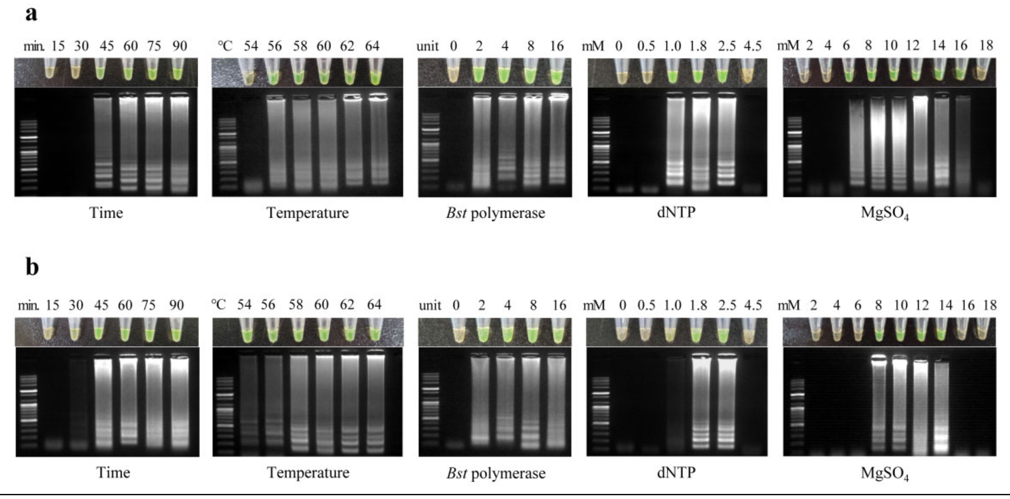
To determine the optimal conditions of LAMP assay for detecting O. cf. ovata and A. massartii, experiments were performed in which the different variables known to affect this assay, such as reaction times, temperatures, and buffer composition concentrations (dNTP, Bst polymerase, and MgSO4), were altered (Table 2). The reaction conditions and buffer compositions were varied to include ranges of 0–16 units of Bst DNA polymerase, 0–18 mM of MgSO4, 0–4.5 mM of dNTP each, 54–64 °C of temperatures, and 15–90 min of reaction time. After that, the LAMP reaction mixture contained 20 mM tris-HCl, 50 mM KCl, 10 mM (NH4)2SO4, 1.0 μM of each FIP and BIP, 0.4 μM of each of the outer F3 and B3 primers, and 4 μL of genomic DNA extracted from 100 cells using syringe filter set, which resulted in a final reaction volume of 20 μL.
The detectable ranges for the species (O. cf. ovata or A. massartii) included the concentraions of compositon in the commercial LAMP mix (2× LAMP Maeter mix, NEB E1700L): 8 U Bst polymerase, 1.4 mM dNTPs, and 8 mM MgSO4. Therefore, the following tests (sensitivity, specific-specificity, and field samples application) were performed with the commercial LAMP mix, at 62 °C in O. cf. ovata and 60 °C A. massartii for 60 min. The reaction temperature was determined to the median between the minimum reaction temperature and the manufacturer’s recommended temperature.
By adding 1 μL of fluorescent dye SYBR Green I (×1000, Thermo Fisher Scientific, Waltham, MA, USA) after completing the LAMP reaction, the positive reactions could be confirmed with the naked eye. The colour of positive reactions turned orange to yellow-green, while negative reactions remained orange. Moreover, by performing the gel electrophoresis, the LAMP products could be double-checked. The positive sample of LAMP showed a band in the ladder pattern, while the negative sample had no band in the gel. The photographs of the agarose gel were taken using an Azure c200 gel imaging workstation (Azure Biosystems, Dublin, CA, USA).

2.5. Sensitivity of LAMP

2.5.1. Sensitivity Test from Extracted DNA

The sensitivity tests were evaluated using DNA templates extracted from the clonal cultures of O. cf. ovata or A. massartii each (3 × 10³ cells of O. cf. ovata and 104 cells of A. massartii). The cells were concentrated with GF/C filters (Whatman® glass microfiber filters, Little Chalfont, UK) using a syringe filter, and then the genomic DNAs were extracted using an AccuPrep Genomic DNA Extraction Kit (BIONEER, Daejeon, Korea). The extracted DNA was diluted serially.

2.5.2. Sensitivity Test from 10 Cells Directly

To evaluate the detection limit more precisely, we used the cells isolated from the clonal cultures as the DNA templates directly without the column-based DNA extraction processing. Ten cells were isolated each from the culture of O. cf. ovata or A. massartii using a sterile micropipette under a dissecting microscope (SZX10, Olympus, Tokyo, Japan). Individual cells were transferred to sterile seawater to remove any contaminants and then suspended in approximately 1 μL of TE buffer. The ten cells were lysed by freeze-thawing process and diluted ten-fold serially, by adding sterile distilled water (Invitrogen, Carlsbad, CA, USA), from 1 to 10−4. All samples were frozen at −20 ℃ in preparation for the test.

2.5.3. Comparison of the Detection Sensitivity with Other Molecular Assays

We carried out the conventional PCR and qPCR (quantitative PCR) for comparison with LAMP. The PCR was performed on a Mastercycler Nexus (Eppendorf, Hamburg, Germany) in a 20 μL PCR mixture containing 4 μL of HiPi PCR premix, 1 μL of 10 μM each forward/reverse primers, 10 μL of sterile distilled water, and 4 μL of DNA template. PCR conditions of O. cf. ovata and A. massartii were as follows: an initial denaturation step of 95 °C for 15 min, followed by 35 cycles of denaturation at 94 °C for 30 s, and then an annealing step at 55 °C for 30 s, and elongation at 72 °C for 50 s, followed by a final extension step 72 °C for 7 min. The PCR products were analysed on a 1.5% agarose gel using gel electrophoresis.
Quantitative PCR (qPCR) was performed in duplicates with a PCR max Eco 48 real-time PCR system (PCR max, Stone, UK) using qPCRBIO probe Mix No-ROX (PCR biosystms, London, England) following the manufacturer’s guidelines: 10 μL of 2× qPCRBIO Probe Mix, 1 μL of 10 μM each of forward/reverse primers, 0.5 μL of 10 μM probe labelled at the 5′ and 3′ ends with the fluorescent dye 6-FAM and BHQ-1, 4.5 μL of distilled water, and 3 μL of DNA template. The primers and TaqMan probes (coupled to for qPCR were also designed to amplify the ITS gene (Table 3). Thermal cycling was performed under the following conditions: 95 °C for 3 min of initial denaturation, then 40 cycles of amplification at 95 °C for 10 s and 60 °C for 30 s. The standard templates (3 × 10³ cells of O. cf. ovata) were concentrated with GF/C filters using a syringe filter. Following DNA extraction and qPCR methods were as described above. The eluted genomic DNA was diluted to the equivalent of 1000, 300, 100, 30, and 10 cells. In the application of the field samples, the relatively quantified cell concentrations were calculated according to Park et al. [36].

2.6. Confirmation of Species-Specific LAMP Primers

To confirm species specificity and exclude the possibility of false positives about the two sets of LAMP primers (O. cf. ovata and A. massartii), DNAs of eleven dinoflagellate species (Alexandrium tamarense (Lebor) Balech; Amphidinium carterae Hulbert; Coolia malayensis Leaw, P.-T. Lim & Usup; Gambierdiscus jejuensis S. H. Jang & H. J. Jeong; Gymnodinium aureolum (Hulburt) G. Hansen; Heterocapsa steinii Tillmann, Gottschling, Hoppenrath, Kusber & Elbrächter; Ostreopsis lenticularis Y. Fukuyo; Prorocentrum minimum (Pavillard) Schiller; Prorocentrum koreanum M.-S. Han, S. Y. Cho & P. Wang; Scrippsiella acuminata (Ehrenberg) Kretschmann, Elbrächter, Zinssmeister, S. Soehner, Kirsch, Kusber & Gottschling); and Symbiodinium voratum Jeong, Lee, Kang, LaJeunesse) were extracted and used as a template for the LAMP reaction (Table 3). All strains, mentioned above, have been established and maintained in our cell culture laboratory (at 22 °C under continuos illumination of 20 μE·m−2·s−1) of the Advanced Institute of Convergence Technology (AICT, Suwon, Korea), excluding Amphidinium carterae strain (CCMP1314). Two thousand cells from each species were isolated from the dense culture media of the dinoflagellate strains and mixed in a tube. The mixture was concentrated with GF/C filters using a syringe filter. Then, the genomic DNA was extracted using an AccuPrep Genomic DNA extraction kit (Bioneer, Daejeon, Korea). The positive mixture including either O. cf. ovata or A. massartii and the negative mixture without O. cf. ovata and A. massartii were used as the controls. The LAMP reaction was conducted at 62 °C in O. cf. ovata and 60 °C in A. massartii for 60 min.
All DNA templates tested in this work were from fresh samples only that had not been stored for more than two days. The amplification results were obtained following electrophoresis, and gels were stained to verify specificity.

2.7. Testing of Field Samples

To test the O. cf. ovata LAMP primers using field samples, four different macroalgal species were randomly collected from two sampling sites on Jeju Island (Sasu and Seongsan) in 2019 (January, March, and June) (Figure 2). Macroalgae living within 10 m of water depth were collected by scuba divers and pooled in a 1 L bottle with ambient seawater. The bottle was shaken vigorously to detach the benthic dinoflagellates. After that, macroalgae were frozen on dry ice immediately and the water sample was filtered using a 100 μm mesh to remove macroalgal particles and zooplankton. Each water sample (50 mL) was concentrated with a GF/C filter using a syringe filter. The GF/C filter was placed in a 1.5 mL tube and kept frozen immediately until DNA extraction. Genomic DNAs were extracted with a Bioneer AccuPrep® Genomic DNA Extraction kit, and a LAMP assay was performed. The LAMP reaction was conducted at 62 °C for 60 min.
To compare microscopic detection, 300 mL of water sample was fixed with formalin (final concentration, 1%). The fixed samples were concentrated for microscopic observation. Fixed samples were left overnight to allow the cells to sink, and the supernatant was removed to adjust the final volume to 50 mL. A solution of Calcofluor (Sigma-Aldrich, St. Louis, MO, USA) was added at a final concentration of 10 μg mL1 for 1–2 min in the dark before observation. Then, 1 mL samples were observed in a Sedgewick-Rafter chamber (SPI Supplies, West Chester, PA, USA) at 100× magnification using an epifluorescence microscope (BX 53, Olympus, Tokyo, Japan) to confirm the presence of cells. For the same reason, dense plankton samples were collected at each sampling sites by towing a 20 μm mesh sized plankton net along the water column for 1 min. Collected samples were fixed with formalin and observed with light and epifluorescence microscopes (BX 53, Olympus, Tokyo, Japan).

3. Results

3.1. Optimization of LAMP Conditions

LAMP reaction in O. cf. ovata required at least 45 min and occurred at a temperature of 56 °C or higher. We confirmed that LAMP reactions occurred even at two units of Bst polymerase per reaction tube. To find an optimal dNTP concentration, each 0–4.5 mM of dNTPs were tested in 20 μL of the reaction mixture. LAMP reaction occurred in 1.0–2.5 mM dNTPs, but it did not occur in 0.5 and 4.5 mM dNTPs. In the concentration test of MgSO4 per reaction tube, LAMP reaction occurred from 6 to 16 mM of MgSO4 (Figure 3a). LAMP reaction in A. massartii showed that the reaction was initiated at least after 30 min and at all temperatures from 54 to 64 °C. The required concentration of dNTP per reaction tube was from 1.0 (very weak) to 2.5 mM. Compared to what was confirmed in O. cf. ovata LAMP reactions, LAMP reactions in A. massartii were in a narrow range of MgSO4 concentration, from 8 to 14 uM per reaction tube (Figure 3b). Too low or too high dNTP and MgSO4 concentrations resulted in false-negative results in both O. cf. ovata and A. massartii. These composition (Bst polymerase, dNTPs, MgSO4) concentration tests were performed at 62 °C in O. cf. ovata and 60 °C A. massartii for 60 min.

3.2. Comparison of the Sensitivity of LAMP and Other Molecular Assays

3.2.1. Ostreopsis. cf. ovata

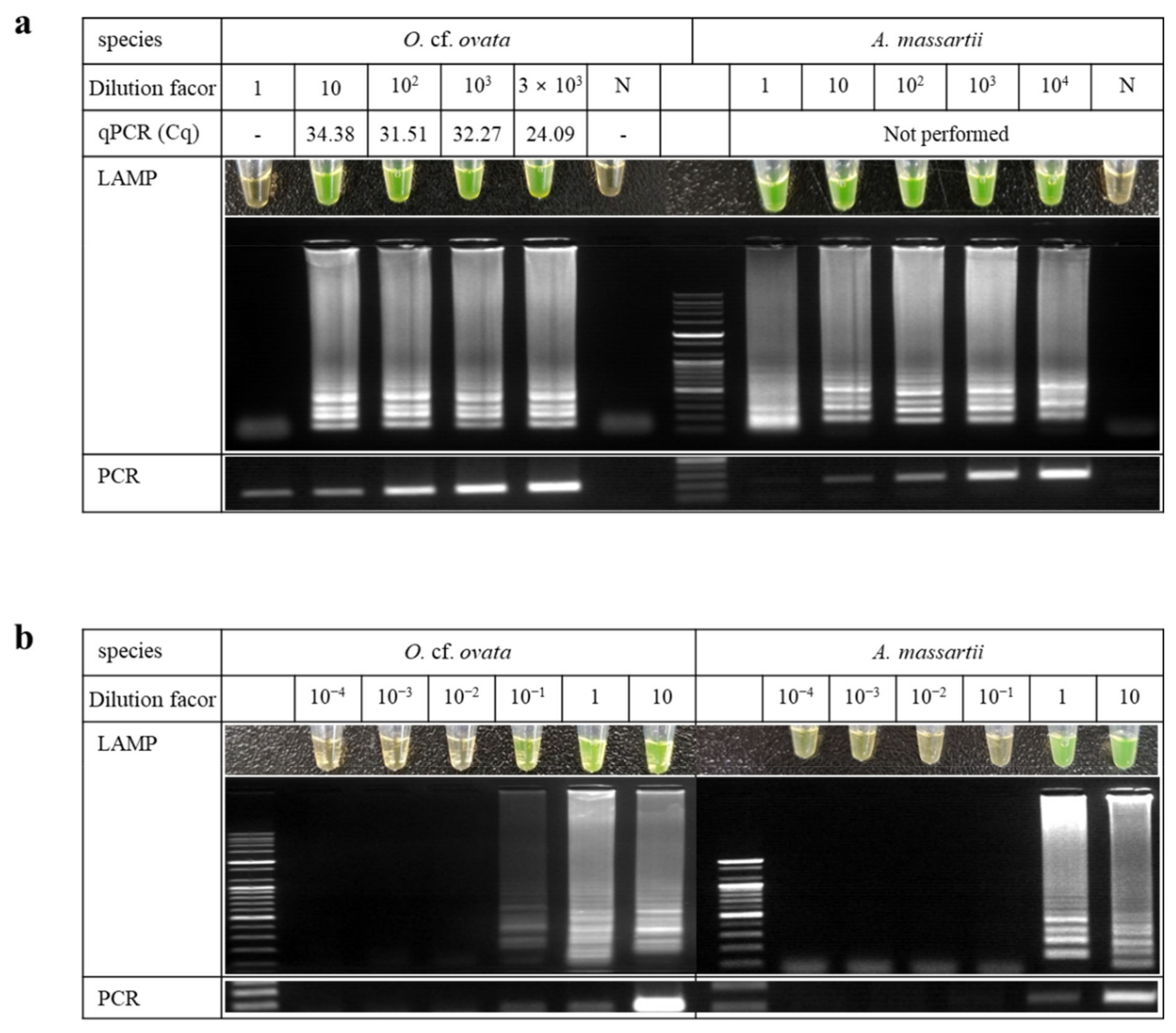
When tested with extracted DNA from 3 × 10³ cells concentrated with GF/C filter, the detection limit of LAMP was 10 cells (Figure 4a). Meanwhile, when we tested using lysed DNA from 10 cells isolated directly, sensitivity was increased as 0.1 cell of O. cf. ovata (Figure 4b). The sensitivity was higher when performing LAMP with isolated cells than with extracted DNA. The sensitivity of qPCR was similar to the LAMP result while the PCR was ten-fold more sensitive than the LAMP assay (Figure 4a). The qPCR was only performed for O. cf. ovata as suitable primers and probes, but could not be designed for A. massarti.

3.2.2. Amphidinium Massartii

The detecton limit of A. massartii was 1 cell in both DNA extracted from 104 cells concentrated with GF/C filter (Figure 4a) and DNA lysed from 10 cells isolated directly (Figure 4b). The PCR detection limit of A. massartii was similar to that of the LAMP results.

3.3. Confirmation of Species-Specific LAMP Primers

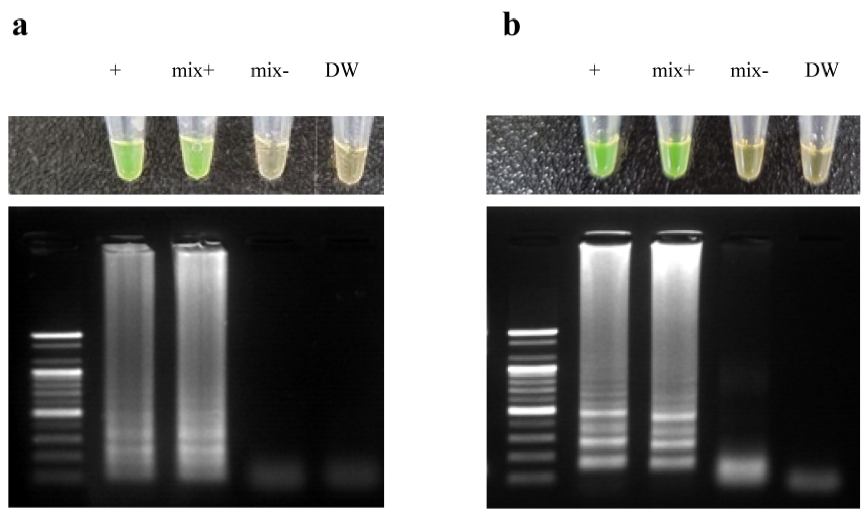
By performing LAMP reactions on the DNA mixture containing eleven strains of dinoflagellates (A. tamarense, A. carterae, C. malayensis, G. jejuensis, G. aureolum, H. steinii, O. lenticularis, P. minimum, P. koreanum, S. acuminata, and S. voratum), we confirmed that the positive LAMP reactions were only observed for target species and positive control mixtures containing either O. cf. ovata or A. massartii, while no products were amplified in both negative control and negative control mixture (Figure 5). Therefore, the results showed that the designed LAMP primers for the two species react specifically.

3.4. Application to Field Samples

The LAMP assay for O. cf. ovata was applied using the field samples collected from Jeju, Korea. PCR and qPCR were also performed to confirm the accuracy and the reliability of LAMP results. The result of LAMP showed that O. cf. ovata was detected in both Sasu and Seongsan in January and April, but detected only in Seongsan in June. Specifically, the positive LAMP results of O. cf. ovata were obtained only in samples estimated to be more than 10 cells mL−1 in qPCR assay, while PCR results showed that they were amplified even at less than 10 cells mL1. In the microscopy examination results, O. cf. ovata was not observed in some of the samples (Seongsan no.4 in Jan. and no.1, 2 in Jun.) in which it was confirmed by all molecular assays, including LAMP (Figure 6).

4. Discussion

Traditional PCR and qPCR assays using Taq polymerase have already been used to detect or quantitatively evaluate many dinoflagellates with high sensitivity and specificity. However, these assays require special and expensive devices, such as a thermal cycler. LAMP assay using Bst polymerase is a simple and fast molecular diagnostic tool that does not require an expensive thermocycler because Bst polymerase amplifies nucleic acids under isothermal conditions [54]. Until now, attempts have been made to apply LAMP to some dinoflagellates related to harmful algal blooms, but no studies have been conducted on the early detection of benthic dinoflagellates. Novel and highly specific LAMP assays for the sensitive detection of benthic dinoflagellates O. cf. ovata and A. massartii were established in this study. Moreover, another purpose of this work was to evaluate the detection capabilities of LAMP compared to other molecular assays. The LAMP assay was sensitive enough to detect for O. cf. ovata and A. massartii, similar to both PCR and qPCR results.
The LAMP assay provides several advantages over a conventional PCR assay in that it can amplify target DNA sequence under isothermal conditions faster (≤1 h) and does not require the use of sophisticated or expensive equipment. Thus, in laboratories or isolated areas where equipment is minimal, the entire reaction process can still be performed using only a heat block or temperature-controlled water tank. However, the most substantial feature is the ability to visually detect amplification through the addition of fluorescent dyes such as SYBR Green I [55], making it suitable for implementation in rapid field trials.
Recently, LAMP has been applied in the detection of marine dinoflagellates such as Alexandrium sp., Karenia mikimotoi, and Prorocentrum sp., and many studies have been conducted for the rapid detection of harmful dinoflagellates [56,57,58]. To date, most LAMP methods for detecting dinoflagellates have been limited to planktonic dinoflagellates. In the present study, we successfully detected benthic dinoflagellates and confirmed that the detection threshold of LAMP was similar to that of PCR and qPCR assays. When the LAMP method was used, we completed the analysis and obtained the results in situ with high accuracy within 60 min to detect the occurrence of O. cf. ovata and A. massartii. Generally, LAMP positive products could be confirmed in the ladder pattern band through the gel electrophoresis. However, we found that the samples kept frozen for a long time (more than a month) or repeated freezing-thawing several times caused false-positive results with a smear band rather than a ladder pattern. This is why we only used the fresh samples to avoid false-positive results.
In the field samples detached from macrophytes, we observed not only O. cf. ovata and A. massartii but also diverse benthic dinoflagellates (such as Ostreopsis lenticularis, Coolia spp., Gambierdiscus spp., Prorocentrum spp.) with high abundances, but there was no LAMP reaction from the samples without O. cf. ovata and A. massartii. The LAMP sets in this study have been proven to react only with O. cf. ovata or A. massartii.

5. Conclusions

In this study, we developed a LAMP method for in situ and precise detection of benthic dinoflagellate species, O. cf. ovata and A. massartii. This provides a useful detection technique for field research, owing to the use of simple reaction conditions and inexpensive equipment and the lack of a need for an expert phycologist for algal identification by microscopy. Thus, the LAMP method can be applied to monitor the occurrence and distribution of benthic dinoflagellates in large-scale environments such as the coastal area of Korea.

Author Contributions

Data curation, formal analysis, writing—original draft preparation, E.S.L., J.H.; field investigation, methodology, J.-H.H.; conceptualization, supervision, project administration, writing—review and editing, J.P. All authors have read and agreed to the published version of the manuscript.

Funding

This research was a part of the project titled “Improvement of management strategies on marine disturbing and harmful organisms (No. 20190518)” funded by the Ministry of Oceans and Fisheries, Korea and supported by the National Research Foundation of Korea (NRF) grant funded by the Korean government (MSIT) (No. NRF-2021R1A2C1005943).

Institutional Review Board Statement

Not applicable.

Informed Consent Statement

Not applicable.

Data Availability Statement

Not applicable.

Acknowledgments

We thank E. J. Kim and S. W. Kim for providing technical support and field sampling. We thank the reviewers for their comments.

Conflicts of Interest

The authors declare no conflict of interest.

References

  1. Mangialajo, L.; Chiantore, M.; Cattaneo-Vietti, R. Loss of fucoid algae along a gradient of urbanisation, and structure of benthic assemblages. Mar. Ecol. Prog. Ser. 2008, 358, 63–74. [Google Scholar] [CrossRef] [Green Version]
  2. Shah, M.M.R.; An, S.-J.; Lee, J.-B. Seasonal abundance of epiphytic dinoflagellates around coastal waters of Jeju Island, Korea. J. Mar. Sci. Technol. 2013, 21, 156–165. [Google Scholar] [CrossRef]
  3. Accoroni, S.; Romagnoli, T.; Penna, A.; Capellacci, S.; Ciminiello, P.; Dell′Aversano, C.; Tartaglione, L.; Abboud-Abi Saab, M.; Giussani, V.; Asnaghi, V.; et al. Ostreopsis fattorussoi sp. nov. (Dinophyceae), a new benthic toxic Ostreopsis species from the eastern Mediterranean Sea. J. Phycol. 2016, 52, 1064–1084. [Google Scholar] [CrossRef] [PubMed]
  4. Murray, S.; Patterson, D. The benthic dinoflagellate genus Amphidinium in south-eastern Australian waters, including three new species. Eur. J. Phycol. 2002, 37, 279–298. [Google Scholar] [CrossRef]
  5. Rhodes, L. World-wide occurrence of the toxic dinoflagellate genus Ostreopsis Schmidt. Toxicon 2011, 57, 400–407. [Google Scholar] [CrossRef] [PubMed]
  6. Delgado, G.; Lechuga-Devéze, C.H.; Popowski, G.; Troccoli, L.; Salinas, C.A. Epiphytic dinoflagellates associated with ciguatera in the northwestern coast of Cuba. Rev. Biol. Trop. 2006, 54, 299–310. [Google Scholar] [CrossRef]
  7. Friedman, M.A.; Fleming, L.E.; Fernandez, M.; Bienfang, P.; Schrank, K.; Dickey, R.; Bottein, M.-Y.; Backer, L.; Ayyar, R.; Weisman, R.; et al. Ciguatera fish poisoning: Treatment, prevention and management. Mar. Drugs 2008, 6, 456–479. [Google Scholar] [CrossRef]
  8. Pagliara, P.; Caroppo, C. Toxicity assessment of Amphidinium carterae, Coolia cfr. monotis and Ostreopsis cfr. ovata (Dinophyta) isolated from the northern Ionian Sea (Mediterranean Sea). Toxicon 2012, 60, 1203–1214. [Google Scholar] [CrossRef]
  9. Berdalet, E.; Fleming, L.E.; Gowen, R.; Davidson, K.; Hess, P.; Backer, L.C.; Moore, S.K.; Hoagland, P.; Enevoldsen, H. Marine harmful algal blooms, human health and wellbeing: Challenges and opportunities in the 21st century. J. Mar. Biol. Assoc. UK 2016, 96, 61–91. [Google Scholar] [CrossRef] [Green Version]
  10. Berdalet, E.; Tester, P.A.; Chinain, M.; Fraga, S.; Lemée, R.; Litaker, W.; Penna, A.; Usup, G.; Vila, M.; Zingone, A. Harmful algal blooms in benthic systems: Recent progress and future research. Oceanography 2017, 30, 36–45. [Google Scholar] [CrossRef] [Green Version]
  11. Lenoir, S.; Ten-Hage, L.; Turquet, J.; Quod, J.-P.; Bernard, C.; Hennion, M.-C. First evidence of palytoxin analogues from an Ostreopsis mascarenensis (Dinophyceae) benthic bloom in southwestern Indian Ocean. J. Phycol. 2004, 40, 1042–1051. [Google Scholar] [CrossRef]
  12. Mohammad-Noor, N.; Daugbjerg, N.; Moestrup, Ø.; Anton, A. Marine epibenthic dinoflagellates from Malaysia—A study of live cultures and preserved samples based on light and scanning electron microscopy. Nord. J. Bot. 2007, 24, 629–690. [Google Scholar] [CrossRef]
  13. Ciminiello, P.; Dell′Aversano, C.; Iacovo, E.; Fattorusso, E.; Forino, M.; Tartaglione, L.; Yasumoto, T.; Battocchi, C.; Giacobbe, M.; Amorim, A.; et al. Investigation of toxin profile of Mediterranean and Atlantic strains of Ostreopsis cf. siamensis Dinophyceae) by liquid chromatography–high resolution mass spectrometry. Harmful Algae 2013, 23, 19–27. [Google Scholar] [CrossRef]
  14. Ben-Gharbia, H.; Yahia, O.K.; Amzil, Z.; Chomérat, N.; Abadie, E.; Masseret, E.; Sibat, M.; Triki, Z.H.; Nouri, H.; Laabir, M. Toxicity and growth assessments of three thermophilic benthic Dinoflagellates (Ostreopsis cf. ovata, Prorocentrum lima and Coolia monotis) developing in the Southern Mediterranean Basin. Toxins 2016, 8, 297. [Google Scholar] [CrossRef] [PubMed]
  15. Lassus, P.; Chomérat, N.; Hess, P.; Nézan, E. Toxic and Harmful Microalgae of the World Ocean/Micro-Algues Toxiques et Nuisibles de l’Océan Mondial; International Society for the Study of Harmful Algae, United Nations Educational, Scientific and Cultural Organisation: Copenhagen, Denmark, 2016; pp. 111–170. ISBN 978-87-990827-6-6. [Google Scholar]
  16. Aligizaki, K.; Katikou, P.; Nikolaidis, G.; Panou, A. First episode of shellfish contamination by palytoxin-like compounds from Ostreopsis species (Aegean Sea, Greece). Toxicon 2008, 51, 418–427. [Google Scholar] [CrossRef] [PubMed]
  17. Deeds, J.R.; Schwartz, M.D. Human risk associated with palytoxin exposure. Toxicon 2010, 56, 150–162. [Google Scholar] [CrossRef] [Green Version]
  18. Tester, P.A.; Litaker, R.W.; Berdalet, E. Climate change and harmful benthic microalgae. Harmful Algae 2020, 91, 101655. [Google Scholar] [CrossRef]
  19. Shears, N.T.; Ross, P.M. Blooms of benthic dinoflagellates of the genus Ostreopsis; an increasing and ecologically important phenomenon on temperate reefs in New Zealand and worldwide. Harmful Algae 2009, 8, 916–925. [Google Scholar] [CrossRef]
  20. López-Flores, R.; Boix, D.; Badosa, A.; Brucet, S.; Quintana, X.D. Is Mirtox toxicity related to potentially harmful algae proliferation in Mediterranean salt marshes? Limnetica 2010, 29, 257–268. [Google Scholar] [CrossRef]
  21. Murray, S.A.; Kohli, G.S.; Farrell, H.; Spiers, Z.B.; Place, A.R.; Dorantes-Aranda, J.J.; Ruszczyk, J. A fish kii associated with a bloom of Amphidinium carterae in a coastal lagoon in Sydney, Australia. Harmful Algae 2015, 49, 19–28. [Google Scholar] [CrossRef]
  22. Yong, H.L.; Mustapa, N.I.; Lee, L.K.; Lim, Z.F.; Tan, T.H.; Usup, G.; Gu, H.; Litaker, R.W.; Tester, P.; Lim, P.T.; et al. Habitat complexity affects benthic harmful dinoflagellate assemblages in the fringing reef of Rawa Island, Malaysia. Harmful Algae 2018, 78, 56–68. [Google Scholar] [CrossRef]
  23. Lee, L.K.; Lim, Z.F.; Gu, H.; Chan, L.L.; Litaker, R.W.; Tester, P.A.; Leaw, C.P.; Lim, P.T. Effects of substratum and depth on benthic harmful dinoflagellate assemblages. Sci. Rep. 2020, 10, 11251. [Google Scholar] [CrossRef]
  24. Kobayashi, J.; Ishibashi, M.; Nakamura, H.; Ohizumi, Y. Amphidinolide-A, a novel antineoplastic macrolide from the marine dinoflagellate Amphidinium sp. Tetrahedron Lett. 1986, 27, 5755–5758. [Google Scholar] [CrossRef]
  25. Yasumoto, T.; Seino, N.; Murakami, Y.; Murata, M. Toxins produced by benthic dinoflagellates. Biol. Bull. 1987, 172, 129–131. [Google Scholar] [CrossRef]
  26. Kobayashi, J.; Ishibashi, M.; Wälchli, M.R.; Nakamura, H.; Hirata, Y.; Sasaki, T.; Ohizumi, Y. Amphidinolide C: The first 25-membered macrocyclic lactone with potent antineoplastic activity from the cultured dinoflagellte Amphidinium sp. J. Am. Chem. Soc. 1988, 110, 490–494. [Google Scholar] [CrossRef]
  27. Murray, S.A.; Garby, T.; Hoppenrath, M.; Neilan, B.A. Genetic diversity, morphological uniformity and polyketide production in dinoflagellates (Amphidinium, Dinoflagellata). PLoS ONE 2012, 7, e38253. [Google Scholar] [CrossRef] [Green Version]
  28. Moreira-González, A.R.; Fernandes, L.F.; Uchida, H.; Uesugi, A.; Suzuki, T.; Chomérat, N.; Bilien, G.; Pereira, T.A.; Mafra, L.L., Jr. Morphology, growth, toxin production, and toxicity of cultured marine benthic dinoflagellates from Brazil and Cuba. J. Appl. Phycol. 2019, 31, 3699–3719. [Google Scholar] [CrossRef]
  29. Karafas, S.; Teng, S.T.; Leaw, C.P.; Alves-de-Souza, C.A. An evaluation of the genus Amphidinium (Dinophyceae) combining evidence from morphology, phylogenetics, and toxin production, with the introduction of six novel species. Harmful Algae 2017, 68, 128–151. [Google Scholar] [CrossRef] [PubMed]
  30. Kim, H.S.; Yih, W.; Kim, J.H.; Myung, G.; Jeong, H.J. Abundance of epiphytic dinoflagellates from coastal waters off Jeju Island, Korea during autumn 2009. Ocean Sci. J. 2011, 46, 205–209. [Google Scholar] [CrossRef] [Green Version]
  31. Hwang, B.S.; Yoon, E.Y.; Kim, H.S.; Yih, W.; Park, J.Y.; Jeong, H.J.; Rho, J.-R. Ostreol A: A new cytotoxic compound isolated from the epiphytic dinoflagellate Ostreopsis cf. ovata from the coastal waters of Jeju Island, Korea. Bioorg. Med. Chem. Lett. 2013, 23, 3023–3027. [Google Scholar] [CrossRef]
  32. Lee, K.-W.; Kang, J.-H.; Baek, S.-H.; Choi, Y.-U.; Lee, D.-W.; Park, H.-S. Toxicity of the dinoflagellate Gambierdiscus sp. toward the marine copepod Tigriopus japonicus. Harmful Algae 2014, 37, 62–67. [Google Scholar] [CrossRef]
  33. Yang, A.R.; Lee, S.; Yoo, Y.D.; Kim, H.S.; Jeong, E.J.; Rho, J.-R. Limaol: A polyketide from the benthic marine dinoflagellate Prorocentrum lima. J. Nat. Prod. 2017, 80, 1688–1692. [Google Scholar] [CrossRef]
  34. Lee, S.; Yang, A.R.; Yoo, Y.D.; Jeong, E.J.; Rho, J.-R. Relative configurational assignment of 4-hydroxyprorocentrolide and prorocentrolide C isolated from a benthic Dinoflagellate (Prorocentrum lima). J. Nat. Prod. 2019, 82, 1034–1039. [Google Scholar] [CrossRef]
  35. Lim, A.S.; Jeong, H.J. Benthic dinoflagellates in Korean waters. Algae 2021, 36, 91–109. [Google Scholar] [CrossRef]
  36. Park, J.; Hwang, J.; Hyung, J.; Yoon, E.Y. Temporal and spatial distribution of the toxic epiphytic dinoflagellate Ostreopsis cf. ovata in the coastal waters off Jeju Island, Korea. Sustainability 2020, 12, 5864. [Google Scholar] [CrossRef]
  37. Baek, S.H. First report for appearance and distribution patterns of the epiphytic dinoflagellates in the Korean Peninsula. Korean J. Environ. Biol. 2012, 30, 355–361. [Google Scholar] [CrossRef]
  38. Jeong, H.J.; Lim, A.S.; Jang, S.H.; Yih, W.H.; Kang, N.S.; Lee, S.Y.; Yoo, Y.D.; Kim, H.S. First report of the epiphytic dinoflagellate Gambierdiscus caribaeus in the temperate waters off Jeju Island, Korea: Morphology and molecular characterization. J. Eukaryot. Microbiol. 2012, 59, 637–650. [Google Scholar] [CrossRef]
  39. Shah, M.M.R.; An, S.-J.; Lee, J.B. Occurrence of sand-dwelling and epiphytic dinoflagellates including potentially toxic species along the coast of Jeju Island, Korea. J. Fish. Aquat. Sci. 2014, 9, 141–156. [Google Scholar] [CrossRef] [Green Version]
  40. Kang, S.-M.; Lee, J.-B. New records of genus Dinophysis, Gonyaulax, Amphidinium, Heterocapsa (Dinophyceae) from Korean waters. Korean J. Environ. Biol. 2018, 36, 260–270. [Google Scholar] [CrossRef]
  41. Ministry of Oceans and Fisheries. Improvement of Management Strategies on Marine Ecosystem Disturbing and Harmful Organisms (MoMHO); Advanced Institute of Convergence Technology: Suwon, Korea, 2021; p. 751.
  42. Bolch, C.J.; de Salas, M.F. A review of the molecular evidence for ballast water introduction of the toxic dinoflagellates Gymnodinium catenatum and the Alexandriumtamarensis complex” to Australasia. Harmful Algae 2007, 6, 465–485. [Google Scholar] [CrossRef]
  43. Battocchi, C.; Totti, C.; Vila, M.; Masó, M.; Capellacci, S.; Accoroni, S.; Reñé, A.; Scardi, M.; Penna, A. Monitoring toxic microalgae Ostreopsis (dinoflagellate) species in coastal waters of the Mediterranean Sea using molecular PCR-based assay combined with light microscopy. Mar. Pollut. Bull. 2010, 60, 1074–1084. [Google Scholar] [CrossRef]
  44. Notomi, T.; Okayama, H.; Masubuchi, H.; Yonekawa, T.; Watanabe, K.; Amino, N.; Hase, T. Loop-mediated isothermal amplification of DNA. Nucleic Acids Res. 2000, 28, e63. [Google Scholar] [CrossRef] [Green Version]
  45. Karanis, P.; Ongerth, J. LAMP—A powerful and flexible tool for monitoring microbial pathogens. Trends Parasitol. 2009, 25, 498–499. [Google Scholar] [CrossRef]
  46. Deb, R.; Chakraborty, S. Trends in veterinary diagnostics. J. Vet. Sci. Technol. 2012, 3, 1. [Google Scholar] [CrossRef] [Green Version]
  47. Li, Y.; Fan, P.; Zhou, S.; Zhang, L. Loop-mediated isothermal amplification (LAMP): A novel rapid detection platform for pathogens. Microb. Pathog. 2017, 107, 54–61. [Google Scholar] [CrossRef]
  48. Wang, L.; Li, L.; Alam, M.; Geng, Y.; Li, Z.; Yamasaki, S.; Shi, L. Loop-mediated isothermal amplification method for rapid detection of the toxic dinoflagellate Alexandrium, which causes algal blooms and poisoning of shellfish. FEMS Microbiol. Lett. 2008, 282, 15–21. [Google Scholar] [CrossRef]
  49. Picón-Camacho, S.M.; Thompson, W.P.; Blaylock, R.B.; Lotz, J.M. Development of a rapid assay to detect the dinoflagellate Amyloodinium ocellatum using loop-mediated isothermal amplification (LAMP). Vet. Parasitol. 2013, 196, 265–271. [Google Scholar] [CrossRef]
  50. Medlin, L.; Elwood, H.J.; Stickel, S.; Sogin, M.L. The characterization of enzymatically amplified eukaryotic 16S-like rRNA-coding regions. Gene 1988, 71, 491–499. [Google Scholar] [CrossRef] [Green Version]
  51. Litaker, R.W.; Vandersea, M.W.; Kibler, S.R.; Reece, K.S.; Stokes, N.A.; Steidinger, K.A.; Millie, D.F.; Bendis, B.J.; Pigg, R.J.; Tester, P.A. Identification of Pfiesteria piscicida (Dinophyceae) and Pfiesteria-like organisms using internal transcribed spacer-specific PCR assays. J. Phycol. 2003, 39, 754–761. [Google Scholar] [CrossRef]
  52. Kang, N.S.; Jeong, H.J.; Lee, S.Y.; Lim, A.S.; Lee, M.J.; Kim, H.S.; Yih, W. Morphology and molecular characterization of the epiphytic benthic dinoflagellate Ostreopsis cf. ovata in the temperate waters off Jeju Island, Korea. Harmful Algae 2013, 27, 98–112. [Google Scholar] [CrossRef]
  53. Lee, K.H.; Jeong, H.J.; Park, K.; Kang, N.S.; Yoo, Y.D.; Lee, M.J.; Lee, J.; Lee, S.; Kim, T.; Kim, H.S.; et al. Morphology and molecular characterization of the epiphytic dinoflagellate Amphidinium massartii, isolated from the temperate waters off Jeju Island, Korea. Algae 2013, 28, 213–231. [Google Scholar] [CrossRef]
  54. Fernandez-Soto, P.; Mvoulouga, P.O.; Akue, J.P.; Abán, J.L.; Santiago, B.V.; Sánchez, M.C.; Muro, A. Development of a highly sensitive loop-mediated isothermal amplification (LAMP) method for the detection of Loa loa. PLoS ONE 2014, 9, e94664. [Google Scholar] [CrossRef]
  55. Hendel, R.C.; Patel, M.R.; Kramer, C.M.; Poon, M.; Carr, J.C.; Gerstad, N.A.; Gillam, L.D.; Hodgson, J.M.; Kim, R.J.; Lesser, J.R. ACCF/ACR/SCCT/SCMR/ASNC/NASCI/SCAI/SIR 2006 appropriateness criteria for cardiac computed tomography and cardiac magnetic resonance imaging/A report of the american college of cardiology foundation quality strategic directions committee appropriateness criteria working group, american college of radiology, society of cardiovascular computed tomography, society for cardiovascular magnetic resonance, american society of nuclear cardiology, north american society for cardiac imaging, society for cardiovascular aangiography and interventions, and society of interventional radiology. J. Am. Coll. Cardiol. 2006, 48, 1475–1497. [Google Scholar] [CrossRef] [Green Version]
  56. Zhang, F.; Ma, L.; Xu, Z.; Zheng, J.; Shi, Y.; Lu, Y.; Miao, Y. Sensitive and rapid detection of Karenia mikimotoi (Dinophyceae) by loop-mediated isothermal amplification. Harmful Algae 2009, 8, 839–842. [Google Scholar] [CrossRef]
  57. Nagai, S.; Itakura, S. Specific detection of the toxic dinoflagellates Alexandrium tamarense and Alexandrium catenella from single vegetative cells by a loop-mediated isothermal amplification method. Mar. Genom. 2012, 7, 43–49. [Google Scholar] [CrossRef]
  58. Zhang, F.; Shi, Y.; Jiang, K.; Song, W.; Ma, C.; Xu, Z.; Ma, L. Rapid detection and quantification of Prorocentrum minimum by loop-mediated isothermal amplification and real-time fluorescence quantitative PCR. J. Appl. Phycol. 2014, 26, 1379–1388. [Google Scholar] [CrossRef]
Figure 1. Nucleotide sequence used to design the primers for loop-mediated isothermal amplification (LAMP). The square boxes indicate the recognition sequences of the primers. The right arrow indicates that a sense sequence was used for the primers. The left arrow indicates that a complementary sequence was used for the primer. (a) Primer sequences for Ostreopsis cf. ovata. (b) Primer sequences for Amphidinium massartii. (c) Schematic representation of the primers used in this study. Construction of two outer (F3 and B3) and two inner (forward inner primer (FIP) and backward inner primer (BIP)) primers for loop-mediated isothermal amplification (LAMP).
Jmse 09 00885 g001
Figure 2. Map of Korea and Jeju Island indicating the sampling sites.
Jmse 09 00885 g002
Figure 3. Optimization of the loop-mediated isothermal amplification (LAMP) assay. LAMP reactions were performed with different reaction times (15–90 min), temperatures (54–64 °C), concentrations of Bst DNA polymerase (0–16 unit), dNTP (0–4.5 mM), and MgSO4 (2–18 mM). (a) Ostreopsis cf. ovata. (b) Amphidinium massartii.
Jmse 09 00885 g003
Figure 4. Comparison of the detection sensitivity with loop-mediated isothermal amplification (LAMP) and conventional PCR. (a) Samples in each line were serially diluted (from 3 × 10³ cells in O. cf. ovata and 104 cells in A. massartii) from genomic DNA which was extracted from the sample concentrated with GF/C filter. (b) Samples in each line were serially diluted from directly isolated ten cells without the column-based DNA extraction processing. cq = quantification cycle.
Jmse 09 00885 g004
Figure 5. Species-specificity of loop-mediated isothermal amplification (LAMP) primers. LAMP reactions were performed by visual inspection with diluted SYBR Green I and electrophoresis with EtBr. (a) Ostreopsis cf. ovata. (b) Amphidinium massartii.
Jmse 09 00885 g005
Figure 6. Field sample detection using loop-mediated isothermal amplification (LAMP) primers. Detection of each species was confirmed using a microscope, LAMP, PCR, and qPCR in seawater samples from Sasu and Seongsan of Jeju Island, 2019. The numbers (1 to 4) in the table indicate the randomly collected macrophytes (unidentified).
Jmse 09 00885 g006
Table 1. Oligonucleotide primers developed for detecting Ostreopsis cf. ovata and Amphidinium massartii using loop-mediated isothermal amplification (LAMP), PCR, and qPCR.
Species NamePrimersSequence (5′-3′)
Ostreopsis cf. ovataLAMP F3 AAGAGTGCAGCCAAATGC
B3GTTGACATGTTTACACACTGTA
FIP (F1c-F2)TGGCCCAAGAACATGCCTACATTGCAAATCGCAGAATTACG
BIP (B1c-B2)GACATCATTCATTCAGTGCACCAGTATATACTACATCACACACTCAC
PCRForwardTGATGTGTACAACTCCCTT
ReverseGAATGATGTCCTTAGGAATGG
qPCRForwardGGCCATTCCTAAGGACATCA
ReverseTGGCCATATACAGCATGTTGAC
Probe6-FAM-ATCATGCATTGTGTGAGTGTGTGATGT-BHQ-1
Amphidinium massartiiLAMP F3TGACAGCTCTAAGTGGGCC
B3CCATCCATTTTCGGGGCTAG
FIP (F1c-F2)TGAGCGAGCAGTCAAGCACTTTGGTAAGCAGAACTGGCGATG
BIP (B1c-B2)CATACTGACAGCAGGACGGTGGATTCGGCAGGTGAGTTGTT
PCRForwardAGTGGGTGGAATGACAGCT
ReverseCATCCATTTTCGGGGCTAGT
Table 2. Conditions and tested ranges of each factor for optimizing LAMP.
ConditionsTested Ranges
Temperature (℃)545658606264
Reaction Time (min.)153045607590
dNTP (mM)00.51.01.82.54.5
Bst DNA polymerase (units)0248
MgSO4 (mM)0246810121416
Table 3. List of dinoflagellate species used to confirm the species specificity of the two sets of LAMP primers (O. cf. ovata and A. massartii).
Species NameStrainRemarks
Alexandrium tamarenseS041118-JSPBusan, Korea
Amphidinium carteraeCCMP1314USA
Coolia malayensisJSGP-CMSeoguipo, Korea
Gambierdiscus jejuensisJSGP201117-GJSeoguipo, Korea
Gymnodinium aureolumGASH1103Kunsan, Korea
HeterocapsasteiniiSMS-MSJMasan bay, Korea
Ostreopsis lenticularisJSGP201117-OLSeoguipo, Korea
Prorocentrum minimumWKS-BJHKunsan, Korea
Prorocentrum koreanumJSS-KEJJeju, Korea
ScrippsiellaacuminataUS1-G6Ulsan, Korea
Symbiodinium voratumJSGP201117-SVSeoguipo, Korea
Ostreopsis cf. ovataJSS200917-OOSeongsan, Korea
Amphidinium massartiiJ-MSJ-AMJeju, Korea
Publisher’s Note: MDPI stays neutral with regard to jurisdictional claims in published maps and institutional affiliations.