Article

Conditional Mitigation of Dental-Composite Material-Induced Cytotoxicity by Increasing the Cure Time

1
Division of Regenerative and Reconstructive Sciences and Weintraub Center for Reconstructive Biotechnology, UCLA School of Dentistry, Los Angeles, CA 90095, USA
2
Department of Periodontology, Graduate School of Medical and Dental Sciences, Tokyo Medical and Dental University, Tokyo 113-8510, Japan
*
Author to whom correspondence should be addressed.
J. Funct. Biomater. 2023, 14(3), 119; https://doi.org/10.3390/jfb14030119
Submission received: 28 January 2023 / Revised: 14 February 2023 / Accepted: 18 February 2023 / Published: 22 February 2023
(This article belongs to the Special Issue Feature Papers in Dental Biomaterials)

Abstract

:
Light-cured composite resins are widely used in dental restorations to fill cavities and fabricate temporary crowns. After curing, the residual monomer is a known to be cytotoxic, but increasing the curing time should improve biocompatibility. However, a biologically optimized cure time has not been determined through systematic experimentation. The objective of this study was to examine the behavior and function of human gingival fibroblasts cultured with flowable and bulk-fill composites cured for different periods of time, while considering the physical location of the cells with regard to the materials. Biological effects were separately evaluated for cells in direct contact with, and in close proximity to, the two composite materials. Curing time varied from the recommended 20 s to 40, 60, and 80 s. Pre-cured, milled-acrylic resin was used as a control. No cell survived and attached to or around the flowable composite, regardless of curing time. Some cells survived and attached close to (but not on) the bulk-fill composite, with survival increasing with a longer curing time, albeit to <20% of the numbers growing on milled acrylic even after 80 s of curing. A few cells (<5% of milled acrylic) survived and attached around the flowable composite after removal of the surface layer, but attachment was not cure-time dependent. Removing the surface layer increased cell survival and attachment around the bulk-fill composite after a 20-s cure, but survival was reduced after an 80-s cure. Dental-composite materials are lethal to contacting fibroblasts, regardless of curing time. However, longer curing times mitigated material cytotoxicity exclusively for bulk-fill composites when the cells were not in direct contact. Removing the surface layer slightly improved biocompatibility for cells in proximity to the materials, but not in proportion to cure time. In conclusion, mitigating the cytotoxicity of composite materials by increasing cure time is conditional on the physical location of cells, the type of material, and the finish of the surface layer. This study provides valuable information for clinical decision making and novel insights into the polymerization behavior of composite materials.

1. Introduction

Light-cured composite resin materials are tooth-colored materials widely used in dental restorative treatment to fill cavities and fabricate temporary crowns [1,2]. They are mainly composed of inorganic filler, photoinitiator, and matrix monomer such as bisphenol A glycidyl methacrylate (bis-GMA) and urethane dimethacrylate (UDMA) [3,4,5]. Depending on the ratio of components, they are classified into flowable composites, which have low viscosity, and bulk-fill composites, which have high viscosity. These different properties make them suitable for different purposes [6,7].
The polymerization reaction occurs when the photoinitiator is activated by visible-light wavelengths between 450 and 490 nm, followed by the generation of free radicals and the formation of polymers [8]. Like other polymer-based materials, their chemical composition alters the biological properties and responses of soft tissues [9,10,11,12,13,14,15,16,17,18]. Many studies have reported cytotoxicity of monomer components, which varies with chemical composition, type, and amount of residual monomer leached [19,20,21,22,23,24,25,26,27,28,29,30,31]. In addition, the free radicals generated during and after polymerization cause significant cellular damage [11,13,14,15,32,33]. Even after photo-polymerization, bis-GMA is eluted as residual monomer for over a month [34]. It is also known that light-cured composites do not polymerize in areas in contact with oxygen, with an unpolymerized layer formed at the surface [35].
Since composite materials are often used in direct contact with, or close to, the gingival tissue, their cytocompatibility is important. Nevertheless, materials are usually selected based on user preference, user friendliness, or esthetics. Cytocompatibility is almost never considered when selecting the material.
For many composite materials, the manufacturers recommend a cure time of 20 s, but it is unclear whether this is biologically sufficient. Both clinically and theoretically, extending the cure time is expected to improve cytocompatibility by reducing the amount of residual monomer. A few studies have reported that extending the cure time improves cytocompatibility by reducing residual monomer [24,36]. However, the optimized cure time from the biological perspective has not been determined through systematic experimentation.
We have established an experimental method that simultaneously evaluates the behavior of cells in contact with, and close to, the test material [37,38]. Exploiting this approach, here we examined the behavior and function of human gingival fibroblasts cultured with two different composite materials (flowable and bulk-fill) cured for different periods of time, with a consideration of the physical location of the cells in relation to the materials: in direct contact with, and in close proximity to, each material. In addition, the effect of removing the unpolymerized layer on cytocompatibility was investigated. The null hypotheses are that cytocompatibility of composite materials is dependent on the curing time and the removal of the unpolymerized layer significantly reduces the adverse effects on fibroblasts.

2. Materials and Methods

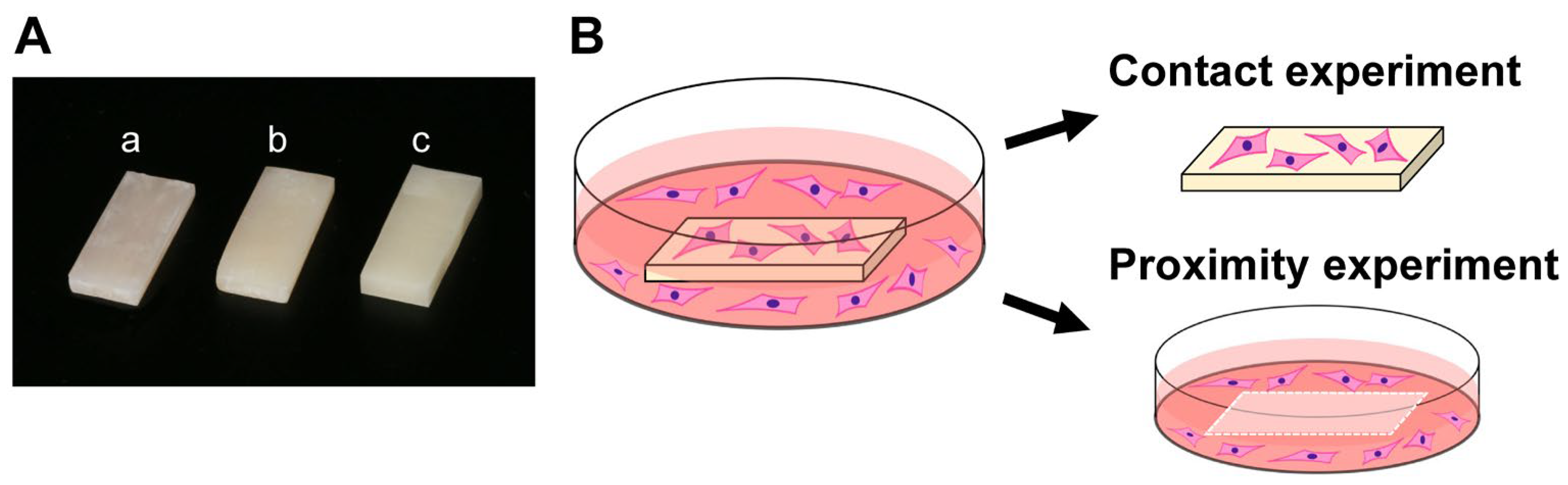
2.1. Material Preparation and Characterization

Flowable and bulk-fill composites were prepared in rectangular plate form (6 mm × 14 mm, 2 mm thickness) for evaluation (Figure 1A). Four different curing times were tested: 20, 40, 60, and 80 s. A light curing device (Coltolux LED; Coltène, Altstätten, Switzerland) was used to polymerize the samples with a wavelength of 450–470 nm and an intensity of 1275 mW/cm2. Milled-acrylic plates were designed using CAD software (123D Design, Hyperdent®, Synergy Health, Sydney, Australia) and manufactured from poly(methyl methacrylate) (PMMA) disks with a milling machine (Versamill 5 × 200, Axsys Dental Solutions, Wixom, MI, USA). These materials and their principal constituents are shown in Table 1 and Figure 1A. After preparation, all plates were washed with a steam cleaner and disinfected with 75% ethanol. Each test plate was placed on the center of each culture well, to standardize the physical distance between the plate and the cells in close proximity to the plate.

2.2. Cell Culture

Human gingival fibroblasts were purchased from ScienCell Research Laboratories (Carlsbad, CA, USA) and grown in fibroblast medium supplemented with 5% fetal bovine serum (FBS), 1% fibroblast growth supplement-2, and 1% penicillin/streptomycin solution. At 80% confluence, the cells were detached using 0.05% trypsin-EDTA solution, and seeded onto culture plates. Passage 5–8 cells were seeded onto test material placed in each well (20 mm diameter) of 12-well culture plates at a density of 4 × 10⁴ cells/well. The culture medium was renewed every three days. The UCLA Institutional Biosafety Committee (BUA-2-22-036-001) approved the study protocol.

2.3. Quantification of Attached and Propagated Cells

The number of attached fibroblasts was counted, to quantify the contact effect and proximity effect. The contact effect was defined as the quantification of fibroblasts attached to test materials, while the proximity effect was defined as the quantification of fibroblasts attached to the well of the culture dish around the material (Figure 1B). Attached fibroblasts were measured two days after seeding, and propagated fibroblasts were measured four and six days after seeding. The water-soluble tetrazolium salt (WST-1)-based colorimetric assay was used to quantify the number of cells, as reported elsewhere [39,40]. The amount of formazan product was measured at an absorbance of 450 nm, using a microplate reader (Synergy H1, BioTek Instruments, Winooski, VT, USA).

2.4. Fluorescent Microscopy

The cell structure on and around test materials was visualized by fluorescence microscopy (DMI6000B, Leica Microsystems, Wetzlar, Germany) two days after seeding. For this experiment, fibroblasts were cultured on glass-bottom 35 mm dishes, to stain cells around the test materials. Fibroblasts were dual stained with fluorescent dyes: 4′,6-diamidino-2-phenylindole (DAPI) to identify nuclei, and rhodamine-phalloidin for actin filaments. Fibroblast density was quantified by counting the cells in the images.

2.5. Collagen Production

Fibroblast collagen production was determined by Picrosirius-red staining (Picrosirius Red Staining Kit, Polysciences Inc., Warrington, PA, USA). As reported elsewhere [41,42,43], Picrosirius red stains collagen by reacting with basic groups present in the collagen molecule, via its sulfonic acid groups. Four days after seeding, cells were fixed in 10% formaldehyde. After binding Picrosirius red to produced collagen, 0.1 N sodium hydroxide was added and left for 60 min, to elute the binding dye. Then, the supernatant was measured at an absorbance of 550 nm, using a microplate reader.

2.6. Statistical Analysis

All cell-culture experiments were conducted in triplicate (n = 3). Results are expressed as mean ± standard deviations (SD). The test materials were compared using one-way analysis of variance (ANOVA) followed by the Tukey–Kramer post hoc test. p-values less than 0.05 were deemed statistically significant.

3. Results

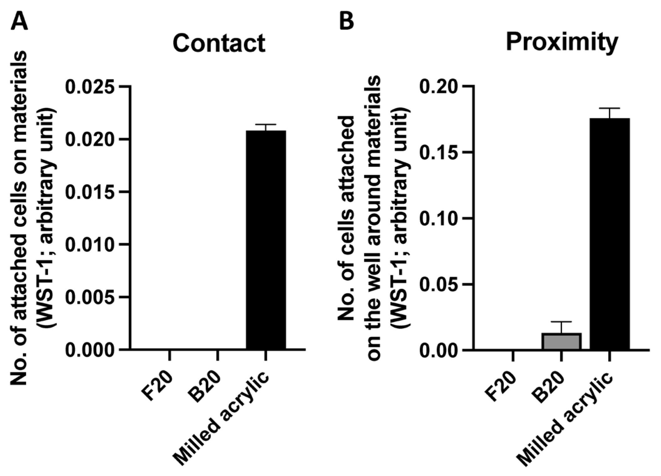
3.1. Initial Cell Attachment

To evaluate the successful settlement of human fibroblasts after seeding, fibroblasts attached on or around the test material were counted, using the WST-1 assay, two days after seeding. There was no cell attachment in the contact experiment for either composite cured at 20 s, the manufacturer’s recommended time (Figure 2A). Similarly, in the proximity experiment, no cells attached around the flowable composite. Some fibroblasts attached around the bulk-fill composite, but only <10% of the number attaching around the milled-acrylic controls (Figure 2B).
Next, both materials were cured for 40, 60, or 80 s, and the same assay performed. No cells attached to either composite, regardless of the cure time in contact experiments (Figure 3A). As in the contact experiment, no cells attached around the d flowable composite in the proximity experiment at any timepoint. However, some cells attached around the bulk-fill composite, with the number increasing with a longer cure time, although the number was <20% of the number of cells attaching around the milled acrylic after 80 s of curing (Figure 3B). The number of cells around B80 was approximately three times higher than that of B20 (p < 0.05). The number of cells around B20, B40, B60, B80 and the milled acrylic were 0.013 ± 0.009, 0.022 ± 0.006, 0.029 ± 0.005, 0.034 ± 0.004 and 0.161 ± 0.012 arbitrary units, respectively.

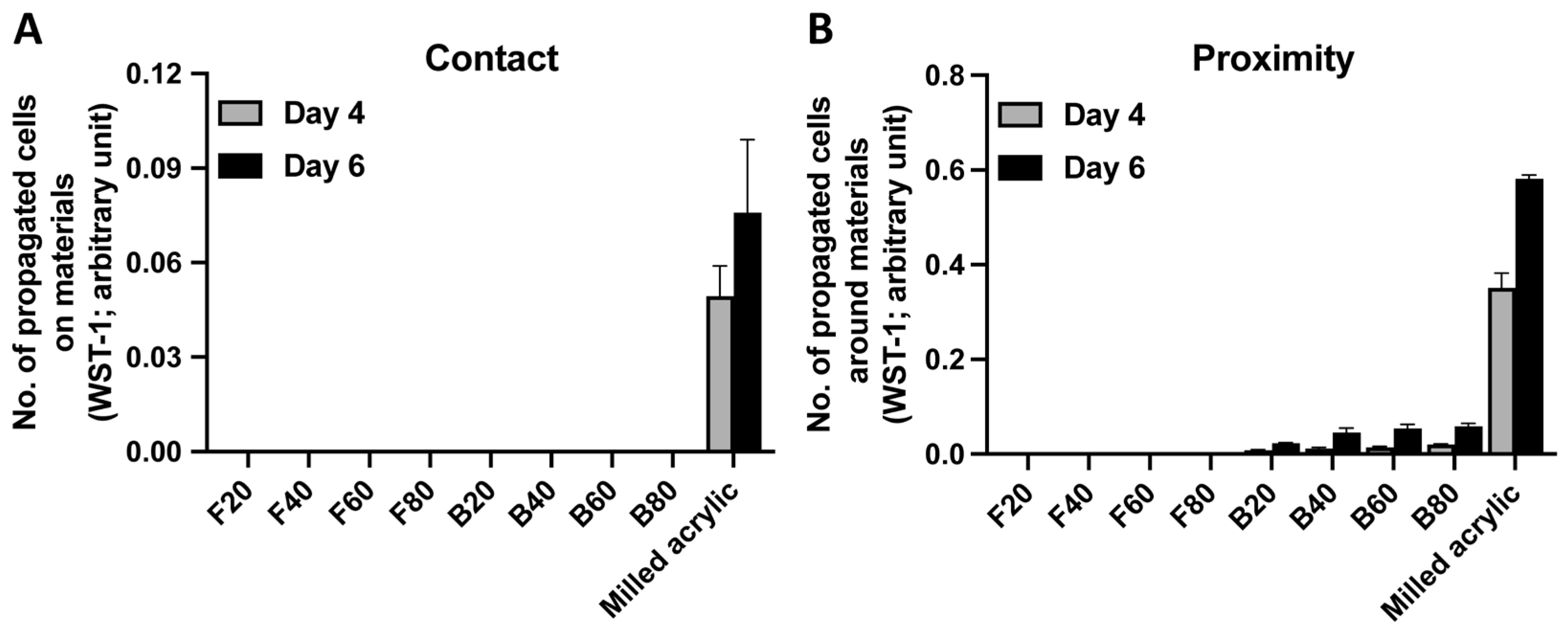
3.2. Cell Proliferation

Cell proliferation was evaluated by measuring the number of propagated cells four and six days after seeding. No cells attached to either composite on day 4 (Figure 4A). There were no propagated cells around flowable composite in the proximity experiment at either timepoint. The number of cells propagating around bulk-fill composite increased with cure time, although to levels < 15% of milled-acrylic controls (Figure 4B). The number of propagated cells around B20, B40, B60, B80 and milled acrylic were 0.023 ± 0.001, 0.046 ± 0.009, 0.055 ± 0.009, 0.059 ± 0.006 and 0.582 ± 0.008 arbitrary units, respectively.

3.3. Cell Visualization

Fibroblasts around test materials showing cell adherence were visualized on culture day two by dual staining with DAPI, to identify nuclei and rhodamine-phalloidin to stain actin filaments. The flowable composite was excluded because the fibroblasts did not adhere. The abundant propagated cells around milled acrylic were spindle shaped with positive cytoskeletal and outline staining. Some cells with small cell outlines were present around the bulk-fill composite (Figure 5A). Similar to the WST-1 assay results, cell density increased with cure time, albeit to <15% of the milled-acrylic controls (Figure 5B). The cell density of B80 was significantly higher than that of B20 (p < 0.001) and B40 (p < 0.01). B60 showed higher cell density than B40 (p < 0.05). The cell density of B20, B40, B60, B80 and milled acrylic were 27.78 ± 6.94, 36.67 ± 5.77, 48.89 ± 8.39, 66.67 ± 6.67 and 315.56 ± 25.24 cells/mm2, respectively.

3.4. Collagen Production

To evaluate fibroblast function around test materials, Picrosirius-red staining was performed to measure collagen production. The flowable composite was excluded because the fibroblasts did not adhere. Fibroblast collagen production around the bulk-fill composite was <15% of that around the milled acrylic, with production slightly increasing in proportion to cure time (Figure 6). The collagen production of both B60 and B80 was significantly higher than that of B20 (p < 0.05). The collagen production of B20, B40, B60, B80 and milled acrylic was 0.028 ± 0.001, 0.033 ± 0.002, 0.035 ± 0.002, 0.035 ± 0.004 and 0.271 ± 0.024 arbitrary units, respectively.

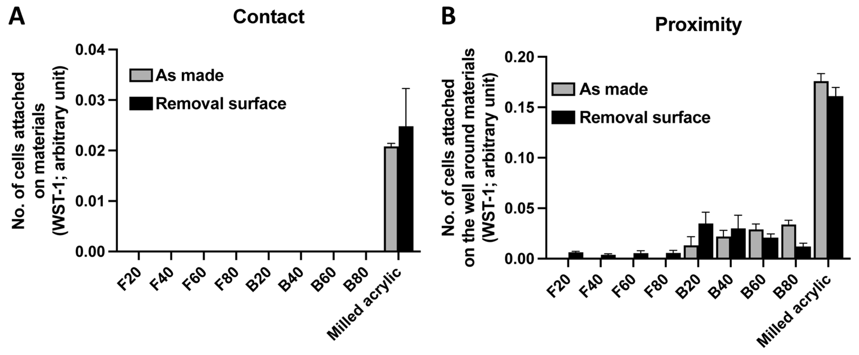
3.5. Improvement in Cell Attachment after Surface Removal

To determine whether removal of the unpolymerized layer increases the number of attached fibroblasts 2 days after seeding, the WST-1 assay was performed. Regardless of removal, no cells attached to either composite (Figure 7A). A limited number of cells attached around the flowable composite in the proximity experiment. The number of cells around F20, F40, F60, F80 with surface removed were 0.006 ± 0.001, 0.004 ± 0.001, 0.006 ± 0.002, 0.006 ± 0.003 arbitrary units, respectively. An increased number of cells were observed around B20 and B40 and a decreased number of cells around B60 and B80 (Figure 7B). The number of cells around B20, B40, B60, B80 and milled acrylic with surface removed were 0.035 ± 0.011, 0.030 ± 0.013, 0.021 ± 0.004, 0.012 ± 0.003 and 0.161 ± 0.009 arbitrary units, respectively.

4. Discussion

Here, we investigated the behavior and function of human gingival fibroblasts cultured with two different composite materials cured for different amounts of time. To model the physical location of cells in relation to the material, the properties of human gingival fibroblasts were evaluated on and around the materials. This allowed us to determine whether the cure time of the materials altered the compatibility of fibroblasts, both in direct contact with and in close proximity to, the materials. In addition, the effect of removal of the unpolymerized surface layer of the materials on initial fibroblast attachment was determined.
In culture experiments, fibroblasts attach to or around the material, and those that do not attach may undergo cell death. We used an indirect method to measure the viability of cells exposed to material cytotoxicity by quantifying the number of cells attached to either the test material (“contact experiments”) or to the culture wells around the material (“proximity experiments”). We studied two types of composite material (flowable composite and bulk-fill composite), with milled acrylic as a control. The milled acrylic was made from a poly(methyl methacrylate) (PMMA) block/disc pre-polymerized at a high temperature and pressure [44,45]. Milled acrylic has the lowest amount of residual monomer and the highest cytocompatibility with fibroblasts of the resin-based materials, so was considered suitable as a positive control [26,46].
In contact experiments, fibroblasts did not adhere to the flowable or the bulk-fill composites after 20 s of curing (recommended by the manufacturer). Both composites had detrimental effects on cell growth. In the proximity experiments, fibroblasts did not adhere in proximity to the flowable composite, but there was some minimal cell attachment around the bulk-fill composite. This suggest that even though composite materials may seemingly have the same clinical properties, apart from viscosity, the cytocompatibility with cells in proximity was substantially different.
Several studies have shown that the cytotoxicity of a composite material depends on its monomer, photoinitiator, and inorganic-filler-particle components [22,47]. In any polymer-based materials, unreacted monomers have negative effects on various cells [10,12,13,16,17,28,33,48,49,50]. Bis-GMA and UDMA are the main components of composites, the former eluting at higher concentrations than the latter [51]. Both monomers cause DNA-strand breaks in fibroblasts, providing a plausible cytotoxic mechanism [52,53]. Flowable composite has a lower filler content and a higher percentage of bis-GMA to reduce viscosity, which might explain our observations of differing cytotoxic effects of the monomers.
We evaluated the behavior and function of fibroblasts cultured with composites for variable curing times of 40, 60, and 80 s. Interestingly, no fibroblasts attached to the flowable composite, regardless of the cure time or physical location. In the contact experiments with the bulk-fill composite, there was similarly no fibroblast adhesion, regardless of the cure time. In the proximity experiments, however, the number of fibroblasts around the bulk-fill composite increased proportionately with the cure time. However, the number of attaching/growing cells was considerably less than that around the milled acrylic. As expected, the number of cells attaching around the bulk-fill composite increased proportionately with the cure time. However, unexpectedly, there was only 20% cell attachment around the bulk-fill composite at a cure time of 80 s, so mitigating cytotoxicity with increased curing is insufficient to solve the cytotoxicity problem.
When composite materials thicker than 2 mm are required, laminate filling is used to compensate for shrinkage during polymerization [54]. We evaluated this up to 80 s, because, depending on the tooth size, the composite resin filled at first may be exposed to light 3–4 times longer than the manufacturer’s recommended time. Further extension of cure time might improve cytocompatibility and reach a plateau, but such long cure times for an intraoral procedure may not be tolerated in practice.
Cells attached around the bulk-fill composite on day 2 had a less well-developed cytoskeleton and were less numerous than those around the milled acrylic, but the number of cells increased on day 4 and day 6. Collagen production also increased in proportion to the curing time. Fibroblasts close to the material were adversely affected, but the curing time did seem to have at least some positive effect in cell proliferation ability and function.
The test materials used in this study were 2 mm thick, and a single irradiation was considered sufficient for polymerization [55,56]. The distance between the light-curing device and the material and its position were fixed, for consistency. Thus, the degree of polymerization at the bottom of the material, furthest from the tip of the light-curing device, was considered to depend on the curing time. As a result, the amount of monomer eluted into the medium varied in proportion to the cure time, and the eluted monomer might have acted on adjacent cells.
The surface layer of composite material does not polymerize when in contact with oxygen, forming an unpolymerized layer due to its polymerization mechanism [13,24,33,35]. We therefore compared composite material with and without the unpolymerized surface layer. Surprisingly, the contact toxicity of both composites did not decrease, despite removing the layer. In proximity experiments, however, removal of the surface layer mitigated the cytotoxicity, but only a few cells attached around the flowable composite. With respect to the bulk-fill composite, the number of cells increased at 20 and 40 s, but decreased at 60 and 80 s. Therefore, removing the surface layer slightly improved the biocompatibility with cells in proximity, but the effect was not proportional to the cure time.
From a clinical point of view, composite materials are often used in the treatment of caries and tooth defect close to the gingival margin. Gingival inflammation and recession of the cervical gingiva are generally considered to be caused by plaque accumulation on the composite materials [57,58]. Hence, differences in bacterial adhesion and biofilm formation due to various curing times and surface treatments should be investigated. In addition, the results of this study suggested that the deleterious effect of composite materials filled in the cervical area may cause cell damage, gingival inflammation, and gingival recession. However, how this cytotoxicity affects the gingival tissue is unclear. Therefore, it is necessary to evaluate host mechanisms or responses to composite materials, for instance by measuring the inflammatory cytokine expression of gingival tissues in direct and close contact with composite materials, in vivo.
The results of this study showed that composite materials are highly harmful to human gingival fibroblasts. A previous study reported that the resistance to negative effects varied among cell types, such as dental pulp stem cells or periodontal ligament cells [59]. There is also a difference between fibroblasts and osteoblasts in terms of their proliferation and differentiation [38]. In addition, many studies have used the experimental method of adding the eluate from the test materials to the culture medium [27,60,61]. The eluate from the material initially releases a large amount of residual components, and then the amount gradually decreases. In our experiment, on the other hand, the test materials were added directly to the culture medium, resulting in the continuous elution of higher concentrations of residual components. Thus, cells could not survive as in the previously reported experiment, due to differences in the resistance of the cells to the material components and to the effects of the different methods of eluting the residual components. Chromatographic analysis is necessary to measure the elution of components over time [18,60].
This study showed that currently used dental-composite materials are non-negligibly cytotoxic. It would now be interesting to assess the host mechanisms or response to the composites, for instance, by the inflammatory cytokine expression of the gingival tissues in direct and close contact with the composite materials, in vivo. The mitigation of composite cytotoxicity with increasing cure time depended on the physical location of the cells, the type of material, and the surface-layer treatment. In light of other potential methods to improve composite materials, N-acetyl cysteine, a precursor of glutathione, the most potent antioxidant in the body, effectively scavenges polymerization radicals and neutralizes chemicals that cause oxidative stress [9,10,15,17,62,63,64,65,66,67,68,69,70,71]. Tri-n-butyl borane, a polymerization initiator, also reduces cytotoxicity, due to the suppressive role of polymerization radicals [11,13,14,18,33,72]. These data provide design rules for the use of monomers and initiators to create novel composite materials with higher biocompatibility. Although this study focused on the effect on gingival fibroblasts, the effects on other cell types routinely exposed to composite materials, such as epithelial cells, whose origin is ectoderm and different from fibroblasts [39,73], and osteoblasts, differentiating cells that uniquely respond to oxidative stress [9,11,74,75,76,77,78], remain to be studied in order to comprehensively assess the biocompatibility of composite materials.

5. Conclusions

Here, we examined the behavior and function of human gingival fibroblasts cultured on or around two different composite materials which were cured for different lengths of time. In addition, we examined the effect of removal of the unpolymerized surface layer on cytocompatibility. No cells survived and attached to or around the flowable composite, regardless of cure time. Although no cells attached to the bulk-fill composite, regardless of cure time, some cells survived around the material, and the number increased with longer cure times. Removal of the surface layer slightly improved biocompatibility for cells in proximity, but the effect was not proportional to cure time. In conclusion, the mitigation of cytotoxicity in composites due to an increase in the curing time depends on the physical location of the cells, the type of material, and the finish of the surface layer. These results provide valuable information for clinical decision making and new insights into the polymerization behavior of composite materials. Further in vivo studies are needed to explore changes in biocompatibility at the tissue level.

Author Contributions

Conceptualization, T.M. and T.O.; methodology, T.M., T.-L.C. and D.C.; validation, T.S. and J.C.; formal analysis, T.M. and K.K.; investigation, T.M., K.C. and D.C.; resources, T.-L.C.; data curation, T.M.; writing—original draft preparation, T.M.; writing—review and editing, K.K. and T.O.; visualization, T.M. and K.C.; supervision, T.O.; project administration, T.M. All authors have read and agreed to the published version of the manuscript.

Funding

This research received no external funding.

Data Availability Statement

The data presented in this study are available on request from the corresponding author.

Conflicts of Interest

The authors declare no conflict of interest.

References

  1. Ferracane, J.L. Resin composite—State of the art. Dent. Mater. 2011, 27, 29–38. [Google Scholar] [CrossRef]
  2. Studenikin, R.; Niftaliev, S. Fabrication and Use of a Customized Provisional Composite Abutment in Dental Practice. Int. J. Dent. 2021, 2021, 9929803. [Google Scholar] [CrossRef]
  3. Pratap, B.; Gupta, R.K.; Bhardwaj, B.; Nag, M. Resin based restorative dental materials: Characteristics and future perspectives. Jpn. Dent. Sci. Rev. 2019, 55, 126–138. [Google Scholar] [CrossRef]
  4. Peutzfeldt, A. Resin composites in dentistry: The monomer systems. Eur. J. Oral Sci. 1997, 105, 97–116. [Google Scholar] [CrossRef]
  5. Nicolae, L.C.; Shelton, R.M.; Cooper, P.R.; Martin, R.A.; Palin, W.M. The Effect of UDMA/TEGDMA Mixtures and Bioglass Incorporation on the Mechanical and Physical Properties of Resin and Resin-Based Composite Materials. In Hindawi Publishing Corporation Conference Papers in Science; Hindawi: London, UK, 2014; pp. 1–5. [Google Scholar]
  6. Boruziniat, A.; Gharaee, S.; Sarraf Shirazi, A.; Majidinia, S.; Vatanpour, M. Evaluation of the efficacy of flowable composite as lining material on microleakage of composite resin restorations: A systematic review and meta-analysis. Quintessence Int. 2016, 47, 93–101. [Google Scholar]
  7. Van Ende, A.; De Munck, J.; Lise, D.P.; Van Meerbeek, B. Bulk-Fill Composites: A Review of the Current Literature. J. Adhes. Dent. 2017, 19, 95–109. [Google Scholar]
  8. Stansbury, J.W. Curing dental resins and composites by photopolymerization. J. Esthet. Dent. 2000, 12, 300–308. [Google Scholar] [CrossRef]
  9. Aita, H.; Tsukimura, N.; Yamada, M.; Hori, N.; Kubo, K.; Sato, N.; Maeda, H.; Kimoto, K.; Ogawa, T. N-acetyl cysteine prevents polymethyl methacrylate bone cement extract-induced cell death and functional suppression of rat primary osteoblasts. J. Biomed. Mater. Res. A 2010, 92, 285–296. [Google Scholar] [CrossRef]
  10. Att, W.; Yamada, M.; Kojima, N.; Ogawa, T. N-Acetyl cysteine prevents suppression of oral fibroblast function on poly(methylmethacrylate) resin. Acta Biomater. 2009, 5, 391–398. [Google Scholar] [CrossRef]
  11. Hamajima, K.; Ozawa, R.; Saruta, J.; Saita, M.; Kitajima, H.; Taleghani, S.R.; Usami, D.; Goharian, D.; Uno, M.; Miyazawa, K.; et al. The Effect of TBB, as an Initiator, on the Biological Compatibility of PMMA/MMA Bone Cement. Int. J. Mol. Sci. 2020, 21, 4016. [Google Scholar] [CrossRef]
  12. Kojima, N.; Yamada, M.; Paranjpe, A.; Tsukimura, N.; Kubo, K.; Jewett, A.; Ogawa, T. Restored viability and function of dental pulp cells on poly-methylmethacrylate (PMMA)-based dental resin supplemented with N-acetyl cysteine (NAC). Dent. Mater. 2008, 24, 1686–1693. [Google Scholar] [CrossRef]
  13. Nakagawa, K.; Saita, M.; Ikeda, T.; Hirota, M.; Park, W.; Lee, M.C.; Ogawa, T. Biocompatibility of 4-META/MMA-TBB resin used as a dental luting agent. J. Prosthet. Dent. 2015, 114, 114–121. [Google Scholar] [CrossRef]
  14. Sugita, Y.; Okubo, T.; Saita, M.; Ishijima, M.; Torii, Y.; Tanaka, M.; Iwasaki, C.; Sekiya, T.; Tabuchi, M.; Mohammadzadeh Rezaei, N.; et al. Novel Osteogenic Behaviors around Hydrophilic and Radical-Free 4-META/MMA-TBB: Implications of an Osseointegrating Bone Cement. Int. J. Mol. Sci. 2020, 21, 2405. [Google Scholar] [CrossRef] [Green Version]
  15. Tsukimura, N.; Yamada, M.; Aita, H.; Hori, N.; Yoshino, F.; Chang-Il Lee, M.; Kimoto, K.; Jewett, A.; Ogawa, T. N-acetyl cysteine (NAC)-mediated detoxification and functionalization of poly(methyl methacrylate) bone cement. Biomaterials 2009, 30, 3378–3389. [Google Scholar] [CrossRef]
  16. Yamada, M.; Kojima, N.; Att, W.; Hori, N.; Suzuki, T.; Ogawa, T. N-Acetyl cysteine restores viability and function of rat odontoblast-like cells impaired by polymethylmethacrylate dental resin extract. Redox Rep. 2009, 14, 13–22. [Google Scholar] [CrossRef] [Green Version]
  17. Yamada, M.; Kojima, N.; Paranjpe, A.; Att, W.; Aita, H.; Jewett, A.; Ogawa, T. N-acetyl cysteine (NAC)-assisted detoxification of PMMA resin. J. Dent. Res. 2008, 87, 372–377. [Google Scholar] [CrossRef]
  18. Komatsu, K.; Hamajima, K.; Ozawa, R.; Kitajima, H.; Matsuura, T.; Ogawa, T. Novel Tuning of PMMA Orthopedic Bone Cement Using TBB Initiator: Effect of Bone Cement Extracts on Bioactivity of Osteoblasts and Osteoclasts. Cells 2022, 11, 3999. [Google Scholar] [CrossRef]
  19. Yoshii, E. Cytotoxic effects of acrylates and methacrylates: Relationships of monomer structures and cytotoxicity. J. Biomed. Mater. Res. 1997, 37, 517–524. [Google Scholar] [CrossRef]
  20. Haugen, H.J.; Marovic, D.; Par, M.; Thieu, M.K.L.; Reseland, J.E.; Johnsen, G.F. Bulk Fill Composites Have Similar Performance to Conventional Dental Composites. Int. J. Mol. Sci. 2020, 21, 5136. [Google Scholar] [CrossRef]
  21. Schweikl, H.; Spagnuolo, G.; Schmalz, G. Genetic and cellular toxicology of dental resin monomers. J. Dent. Res. 2006, 85, 870–877. [Google Scholar] [CrossRef]
  22. Kraus, D.; Wolfgarten, M.; Enkling, N.; Helfgen, E.H.; Frentzen, M.; Probstmeier, R.; Winter, J.; Stark, H. In-vitro cytocompatibility of dental resin monomers on osteoblast-like cells. J. Dent. 2017, 65, 76–82. [Google Scholar] [CrossRef]
  23. Ergun, G.; Mutlu-Sagesen, L.; Karaoglu, T.; Dogan, A. Cytotoxicity of provisional crown and bridge restoration materials: An in vitro study. J. Oral Sci. 2001, 43, 123–128. [Google Scholar] [CrossRef] [Green Version]
  24. Knezevic, A.; Zeljezic, D.; Kopjar, N.; Tarle, Z. Cytotoxicity of composite materials polymerized with LED curing units. Oper. Dent. 2008, 33, 23–30. [Google Scholar] [CrossRef] [Green Version]
  25. Goncalves, F.P.; Alves, G.; Guimaraes, V.O.J.; Gallito, M.A.; Oliveira, F.; Scelza, M.Z. Cytotoxicity Evaluation of Two Bis-Acryl Composite Resins Using Human Gingival Fibroblasts. Braz. Dent. J. 2016, 27, 492–496. [Google Scholar] [CrossRef] [Green Version]
  26. Campaner, M.; Takamiya, A.S.; Bitencourt, S.B.; Mazza, L.C.; de Oliveira, S.H.P.; Shibayama, R.; Barao, V.A.R.; Sukotjo, C.; Pesqueira, A.A. Cytotoxicity and inflammatory response of different types of provisional restorative materials. Arch. Oral Biol. 2020, 111, 104643. [Google Scholar] [CrossRef]
  27. Tsitrou, E.; Kelogrigoris, S.; Koulaouzidou, E.; Antoniades-Halvatjoglou, M.; Koliniotou-Koumpia, E.; van Noort, R. Effect of extraction media and storage time on the elution of monomers from four contemporary resin composite materials. Toxicol. Int. 2014, 21, 89–95. [Google Scholar] [CrossRef] [Green Version]
  28. Minamikawa, H.; Yamada, M.; Iwasa, F.; Ueno, T.; Deyama, Y.; Suzuki, K.; Yawaka, Y.; Ogawa, T. Amino acid derivative-mediated detoxification and functionalization of dual cure dental restorative material for dental pulp cell mineralization. Biomaterials 2010, 31, 7213–7225. [Google Scholar] [CrossRef]
  29. Yamada, M.; Ogawa, T. Chemodynamics underlying N-acetyl cysteine-mediated bone cement monomer detoxification. Acta Biomater. 2009, 5, 2963–2973. [Google Scholar] [CrossRef]
  30. Collado-Gonzalez, M.; Pecci-Lloret, M.R.; Tomas-Catala, C.J.; Garcia-Bernal, D.; Onate-Sanchez, R.E.; Llena, C.; Forner, L.; Rosa, V.; Rodriguez-Lozano, F.J. Thermo-setting glass ionomer cements promote variable biological responses of human dental pulp stem cells. Dent. Mater. 2018, 34, 932–943. [Google Scholar] [CrossRef]
  31. Lopez-Garcia, S.; Pecci-Lloret, M.P.; Pecci-Lloret, M.R.; Onate-Sanchez, R.E.; Garcia-Bernal, D.; Castelo-Baz, P.; Rodriguez-Lozano, F.J.; Guerrero-Girones, J. In Vitro Evaluation of the Biological Effects of ACTIVA Kids BioACTIVE Restorative, Ionolux, and Riva Light Cure on Human Dental Pulp Stem Cells. Materials 2019, 12, 3694. [Google Scholar] [CrossRef] [Green Version]
  32. Lefeuvre, M.; Amjaad, W.; Goldberg, M.; Stanislawski, L. TEGDMA induces mitochondrial damage and oxidative stress in human gingival fibroblasts. Biomaterials 2005, 26, 5130–5137. [Google Scholar] [CrossRef]
  33. Nakagawa, K.; Ikeda, T.; Saita, M.; Hirota, M.; Tabuchi, M.; Park, W.; Lee, M.; Ogawa, T. Biological and biochemical characterization of 4-META/MMA-TBB resin. J. Dent. Oral Disord. Ther. 2015, 3, 1–7. [Google Scholar]
  34. Polydorou, O.; Trittler, R.; Hellwig, E.; Kummerer, K. Elution of monomers from two conventional dental composite materials. Dent. Mater. 2007, 23, 1535–1541. [Google Scholar] [CrossRef]
  35. Vallittu, P.K. Oxygen inhibition of autopolymerization of polymethylmethacrylate-glass fibre composite. J. Mater. Sci. Mater. Med. 1997, 8, 489–492. [Google Scholar] [CrossRef]
  36. Fujioka-Kobayashi, M.; Miron, R.J.; Lussi, A.; Gruber, R.; Ilie, N.; Price, R.B.; Schmalz, G. Effect of the degree of conversion of resin-based composites on cytotoxicity, cell attachment, and gene expression. Dent. Mater. 2019, 35, 1173–1193. [Google Scholar] [CrossRef]
  37. Matsuura, T.; Komatsu, K.; Ogawa, T. N-Acetyl Cysteine-Mediated Improvements in Dental Restorative Material Biocompatibility. Int. J. Mol. Sci. 2022, 23, 15869. [Google Scholar] [CrossRef]
  38. Matsuura, T.; Komatsu, K.; Chao, D.; Lin, Y.C.; Oberoi, N.; McCulloch, K.; Cheng, J.; Orellana, D.; Ogawa, T. Cell Type-Specific Effects of Implant Provisional Restoration Materials on the Growth and Function of Human Fibroblasts and Osteoblasts. Biomimetics 2022, 7, 243. [Google Scholar] [CrossRef]
  39. Nakhaei, K.; Ishijima, M.; Ikeda, T.; Ghassemi, A.; Saruta, J.; Ogawa, T. Ultraviolet Light Treatment of Titanium Enhances Attachment, Adhesion, and Retention of Human Oral Epithelial Cells via Decarbonization. Materials 2020, 14, 151. [Google Scholar] [CrossRef]
  40. Okubo, T.; Tsukimura, N.; Taniyama, T.; Ishijima, M.; Nakhaei, K.; Rezaei, N.M.; Hirota, M.; Park, W.; Akita, D.; Tateno, A.; et al. Ultraviolet treatment restores bioactivity of titanium mesh plate degraded by contact with medical gloves. J. Oral Sci. 2018, 60, 567–573. [Google Scholar] [CrossRef] [Green Version]
  41. Att, W.; Yamada, M.; Ogawa, T. Effect of titanium surface characteristics on the behavior and function of oral fibroblasts. Int. J. Oral Maxillofac. Implant. 2009, 24, 419–431. [Google Scholar]
  42. Saruwatari, L.; Aita, H.; Butz, F.; Nakamura, H.K.; Ouyang, J.; Yang, Y.; Chiou, W.A.; Ogawa, T. Osteoblasts generate harder, stiffer, and more delamination-resistant mineralized tissue on titanium than on polystyrene, associated with distinct tissue micro- and ultrastructure. J. Bone Miner. Res. 2005, 20, 2002–2016. [Google Scholar] [CrossRef]
  43. Takeuchi, K.; Saruwatari, L.; Nakamura, H.K.; Yang, J.M.; Ogawa, T. Enhanced intrinsic biomechanical properties of osteoblastic mineralized tissue on roughened titanium surface. J. Biomed. Mater. Res. A 2005, 72A, 296–305. [Google Scholar] [CrossRef]
  44. Nguyen, J.F.; Migonney, V.; Ruse, N.D.; Sadoun, M. Resin composite blocks via high-pressure high-temperature polymerization. Dent. Mater. 2012, 28, 529–534. [Google Scholar] [CrossRef]
  45. Hada, T.; Kanazawa, M.; Iwaki, M.; Katheng, A.; Minakuchi, S. Comparison of Mechanical Properties of PMMA Disks for Digitally Designed Dentures. Polymers 2021, 13, 1745. [Google Scholar] [CrossRef]
  46. Yilmaz, M.N.; Gul, P. Monomer release from dental restorative materials containing dimethacrylate resin after bleaching. Clin. Oral Investig. 2022, 26, 4647–4662. [Google Scholar] [CrossRef]
  47. Lang, O.; Kohidai, L.; Kohidai, Z.; Dobo-Nagy, C.; Csomo, K.B.; Lajko, M.; Mozes, M.; Keki, S.; Deak, G.; Tian, K.V.; et al. Cell physiological effects of glass ionomer cements on fibroblast cells. Toxicol. Vitr. 2019, 61, 104627. [Google Scholar] [CrossRef]
  48. Ogawa, T.; Aizawa, S.; Tanaka, M.; Matsuya, S.; Hasegawa, A.; Koyano, K. Effect of water temperature on the fit of provisional crown margins during polymerization. J. Prosthet. Dent. 1999, 82, 658–661. [Google Scholar] [CrossRef]
  49. Ogawa, T.; Hasegawa, A. Effect of curing environment on mechanical properties and polymerizing behaviour of methyl-methacrylate autopolymerizing resin. J. Oral Rehabil. 2005, 32, 221–226. [Google Scholar] [CrossRef]
  50. Ogawa, T.; Tanaka, M.; Matsuya, S.; Aizawa, S.; Koyano, K. Setting characteristics of five autopolymerizing resins measured by an oscillating rheometer. J. Prosthet. Dent. 2001, 85, 170–176. [Google Scholar] [CrossRef]
  51. Sideridou, I.D.; Achilias, D.S. Elution study of unreacted Bis-GMA, TEGDMA, UDMA, and Bis-EMA from light-cured dental resins and resin composites using HPLC. J. Biomed. Mater. Res. B Appl. Biomater. 2005, 74, 617–626. [Google Scholar] [CrossRef]
  52. Ciapetti, G.; Granchi, D.; Savarino, L.; Cenni, E.; Magrini, E.; Baldini, N.; Giunti, A. In vitro testing of the potential for orthopedic bone cements to cause apoptosis of osteoblast-like cells. Biomaterials 2002, 23, 617–627. [Google Scholar] [CrossRef]
  53. De Angelis, F.; Mandatori, D.; Schiavone, V.; Melito, F.P.; Valentinuzzi, S.; Vadini, M.; Di Tomo, P.; Vanini, L.; Pelusi, L.; Pipino, C.; et al. Cytotoxic and Genotoxic Effects of Composite Resins on Cultured Human Gingival Fibroblasts. Materials 2021, 14, 5225. [Google Scholar] [CrossRef]
  54. Santin, D.C.; Velo, M.; Camim, F.D.S.; Brondino, N.C.M.; Honorio, H.M.; Mondelli, R.F.L. Effect of thickness on shrinkage stress and bottom-to-top hardness ratio of conventional and bulk-fill composites. Eur. J. Oral Sci. 2021, 129, e12825. [Google Scholar] [CrossRef]
  55. Prati, C.; Chersoni, S.; Montebugnoli, L.; Montanari, G. Effect of air, dentin and resin-based composite thickness on light intensity reduction. Am. J. Dent. 1999, 12, 231–234. [Google Scholar]
  56. Cidreira Boaro, L.C.; Pereira Lopes, D.; de Souza, A.S.C.; Lie Nakano, E.; Ayala Perez, M.D.; Pfeifer, C.S.; Goncalves, F. Clinical performance and chemical-physical properties of bulk fill composites resin -a systematic review and meta-analysis. Dent. Mater. 2019, 35, e249–e264. [Google Scholar] [CrossRef]
  57. Larato, D.C. Influence of a composite resin restoration on the gingiva. J. Prosthet. Dent. 1972, 28, 402–404. [Google Scholar] [CrossRef]
  58. Sirajuddin, S.; Narasappa, K.M.; Gundapaneni, V.; Chungkham, S.; Walikar, A.S. Iatrogenic Damage to Periodontium by Restorative Treatment Procedures: An Overview. Open Dent. J. 2015, 9, 217–222. [Google Scholar] [CrossRef] [Green Version]
  59. Rodriguez-Lozano, F.J.; Serrano-Belmonte, I.; Perez Calvo, J.C.; Coronado-Parra, M.T.; Bernabeu-Esclapez, A.; Moraleda, J.M. Effects of two low-shrinkage composites on dental stem cells (viability, cell damaged or apoptosis and mesenchymal markers expression). J. Mater. Sci. Mater. Med. 2013, 24, 979–988. [Google Scholar] [CrossRef]
  60. Herrera-Gonzalez, A.M.; Perez-Mondragon, A.A.; Cuevas-Suarez, C.E. Evaluation of bio-based monomers from isosorbide used in the formulation of dental composite resins. J. Mech. Behav. Biomed. Mater. 2019, 100, 103371. [Google Scholar]
  61. Wedekind, L.; Guth, J.F.; Schweiger, J.; Kollmuss, M.; Reichl, F.X.; Edelhoff, D.; Hogg, C. Elution behavior of a 3D-printed, milled and conventional resin-based occlusal splint material. Dent. Mater. 2021, 37, 701–710. [Google Scholar] [CrossRef]
  62. Minamikawa, H.; Yamada, M.; Deyama, Y.; Suzuki, K.; Kaga, M.; Yawaka, Y.; Ogawa, T. Effect of N-acetylcysteine on Rat Dental Pulp Cells Cultured on Mineral Trioxide Aggregate. J. Endod. 2011, 37, 637–641. [Google Scholar] [CrossRef]
  63. Sato, N.; Ueno, T.; Kubo, K.; Suzuki, T.; Tsukimura, N.; Att, W.; Yamada, M.; Hori, N.; Maeda, H.; Ogawa, T. N-Acetyl cysteine (NAC) inhibits proliferation, collagen gene transcription, and redox stress in rat palatal mucosal cells. Dent. Mater. 2009, 25, 1532–1540. [Google Scholar] [CrossRef]
  64. Ueno, T.; Yamada, M.; Igarashi, Y.; Ogawa, T. N-acetyl cysteine protects osteoblastic function from oxidative stress. J. Biomed. Mater. Res. A 2011, 99, 523–531. [Google Scholar] [CrossRef]
  65. Yamada, M.; Kubo, K.; Ueno, T.; Iwasa, F.; Att, W.; Hori, N.; Ogawa, T. Alleviation of commercial collagen sponge- and membrane-induced apoptosis and dysfunction in cultured osteoblasts by an amino acid derivative. Int. J. Oral Maxillofac. Implant. 2010, 25, 939–946. [Google Scholar]
  66. Yamada, M.; Minamikawa, H.; Ueno, T.; Sakurai, K.; Ogawa, T. N-acetyl cysteine improves affinity of beta-tricalcium phosphate granules for cultured osteoblast-like cells. J. Biomater. Appl. 2012, 27, 27–36. [Google Scholar] [CrossRef]
  67. Yamada, M.; Tsukimura, N.; Ikeda, T.; Sugita, Y.; Att, W.; Kojima, N.; Kubo, K.; Ueno, T.; Sakurai, K.; Ogawa, T. N-acetyl cysteine as an osteogenesis-enhancing molecule for bone regeneration. Biomaterials 2013, 34, 6147–6156. [Google Scholar] [CrossRef]
  68. Yamada, M.; Ueno, T.; Minamikawa, H.; Sato, N.; Iwasa, F.; Hori, N.; Ogawa, T. N-acetyl cysteine alleviates cytotoxicity of bone substitute. J. Dent. Res. 2010, 89, 411–416. [Google Scholar] [CrossRef]
  69. Suzuki, T.; Kubo, K.; Hori, N.; Yamada, M.; Kojima, N.; Sugita, Y.; Maeda, H.; Ogawa, T. Nonvolatile buffer coating of titanium to prevent its biological aging and for drug delivery. Biomaterials 2010, 31, 4818–4828. [Google Scholar] [CrossRef]
  70. Ueno, T.; Yamada, M.; Sugita, Y.; Ogawa, T. N-acetyl cysteine protects TMJ chondrocytes from oxidative stress. J. Dent. Res. 2011, 90, 353–359. [Google Scholar] [CrossRef]
  71. Yamada, M.; Kojima, N.; Att, W.; Minamikawa, H.; Sakurai, K.; Ogawa, T. Improvement in the osteoblastic cellular response to a commercial collagen membrane and demineralized freeze-dried bone by an amino acid derivative: An in vitro study. Clin. Oral Implant. Res. 2011, 22, 165–172. [Google Scholar] [CrossRef] [Green Version]
  72. Saruta, J.; Ozawa, R.; Hamajima, K.; Saita, M.; Sato, N.; Ishijima, M.; Kitajima, H.; Ogawa, T. Prolonged Post-Polymerization Biocompatibility of Polymethylmethacrylate-Tri-n-Butylborane (PMMA-TBB) Bone Cement. Materials 2021, 14, 1289. [Google Scholar] [CrossRef]
  73. Okubo, T.; Ikeda, T.; Saruta, J.; Tsukimura, N.; Hirota, M.; Ogawa, T. Compromised Epithelial Cell Attachment after Polishing Titanium Surface and Its Restoration by UV Treatment. Materials 2020, 13, 3946. [Google Scholar] [CrossRef]
  74. Ueno, T.; Ikeda, T.; Tsukimura, N.; Ishijima, M.; Minamikawa, H.; Sugita, Y.; Yamada, M.; Wakabayashi, N.; Ogawa, T. Novel antioxidant capability of titanium induced by UV light treatment. Biomaterials 2016, 108, 177–186. [Google Scholar] [CrossRef]
  75. Kim, S.W.; Ogawa, T.; Tabata, Y.; Nishimura, I. Efficacy and cytotoxicity of cationic-agent-mediated nonviral gene transfer into osteoblasts. J. Biomed. Mater. Res. 2004, 71A, 308–315. [Google Scholar] [CrossRef]
  76. Rezaei, N.M.; Hasegawa, M.; Ishijima, M.; Nakhaei, K.; Okubo, T.; Taniyama, T.; Ghassemi, A.; Tahsili, T.; Park, W.; Hirota, M.; et al. Biological and osseointegration capabilities of hierarchically (meso-/micro-/nano-scale) roughened zirconia. Int. J. Nanomed. 2018, 13, 3381–3395. [Google Scholar] [CrossRef] [Green Version]
  77. Kojima, N.; Ozawa, S.; Miyata, Y.; Hasegawa, H.; Tanaka, Y.; Ogawa, T. High-throughput gene expression analysis in bone healing around titanium implants by DNA microarray. Clin. Oral Implant. Res. 2008, 19, 173–181. [Google Scholar] [CrossRef]
  78. Yamada, M.; Watanabe, J.; Ueno, T.; Ogawa, T.; Egusa, H. Cytoprotective Preconditioning of Osteoblast-Like Cells with N-Acetyl-L-Cysteine for Bone Regeneration in Cell Therapy. Int. J. Mol. Sci. 2019, 20, 5199. [Google Scholar] [CrossRef] [Green Version]
Figure 1. Test materials and culture-experiment design. (A) Test rectangular plates made of flowable composite (a), bulk-full composite (b), milled acrylic (c). (B) Contact and proximity experiments were performed separately, to mimic cellular reactions in vivo.
Jfb 14 00119 g001
Figure 2. Initial attachment of fibroblasts on and around test materials cured for the manufacturer’s recommended time of 20 s. The number of attached fibroblasts (A) on each material (contact experiment) and (B) around each material (proximity experiment). Data shown are mean ± SD.
Jfb 14 00119 g002
Figure 3. Initial attachment on and around test materials cured for different amounts of time. The number of attached fibroblasts (A) on each material and (B) around each material. Data shown are mean ± SD. Significant differences between groups are shown (one-way ANOVA followed by Tukey’s post hoc test, * p < 0.05).
Jfb 14 00119 g003
Figure 4. Propagation of fibroblasts on and around materials cured for different amounts of time. The number of propagated fibroblasts (A) on each material and (B) around each material. Data shown are mean ± SD.
Jfb 14 00119 g004
Figure 5. Visualized fibroblasts around test materials 2 days after seeding. (A) Fluorescent microscopic images of fibroblasts stained for nuclei (blue) and cytoskeletal actin filaments (red). (B) Cell density quantified in these images. Data shown are mean ± SD. Significant differences between test materials are shown (one-way ANOVA followed by Tukey’s post hoc test, p * < 0.05, p ** < 0.01, p *** < 0.001).
Jfb 14 00119 g005
Figure 6. Collagen production by fibroblasts around test materials. Data shown are mean ± SD. Significant differences between test materials are shown (one-way ANOVA followed by Tukey’s post hoc test, p * < 0.05).
Jfb 14 00119 g006
Figure 7. Initial attachment of fibroblasts on and around test materials after surface removal. The number of attached fibroblasts (A) on each material and (B) around each material. Data shown are mean ± SD.
Jfb 14 00119 g007
Table 1. Materials used in this study.
Materials (Product Name, Manufacturer)Main IngredientsCuring Time
(seconds)
Notations
Flowable composite
(Aeliteflo™, BISCO Inc., Schaumburg, IL, USA)
20F20
Bis-GMA40F40
60
80
F60
F80
Bulk-fill composite
(Aelite™ Aesthetic Enamel, BISCO Inc.)
20B20
Bis-GMA, UDMA
40
60
80
B40
B60
B80
Milled acrylic
(Vivid PMMA Disc, Pearson™ Dental Supply Co.)
PMMA
Abbreviations: Bis-GMA, bisphenol A glycidyl methacrylate; UDMA, urethan dimethacrylate, PMMA, poly (methyl methacrylate).
Disclaimer/Publisher’s Note: The statements, opinions and data contained in all publications are solely those of the individual author(s) and contributor(s) and not of MDPI and/or the editor(s). MDPI and/or the editor(s) disclaim responsibility for any injury to people or property resulting from any ideas, methods, instructions or products referred to in the content.

