Article

Lattice Defects Engineering in W-, Zr-doped BiVO4 by Flame Spray Pyrolysis: Enhancing Photocatalytic O2 Evolution

by
Panagiota Stathi
,
Maria Solakidou
and
Yiannis Deligiannakis
*
Laboratory of Physics Chemistry of Materials & Environment, Department of Physics, University of Ioannina, 45110 Ioannina, Greece
*
Author to whom correspondence should be addressed.
Nanomaterials 2021, 11(2), 501; https://doi.org/10.3390/nano11020501
Submission received: 28 December 2020 / Revised: 3 February 2021 / Accepted: 7 February 2021 / Published: 16 February 2021

Abstract

:
A flame spray pyrolysis (FSP) method has been developed, for controlled doping of BiVO4 nanoparticles with W and Zr in tandem with the oxygen vacancies (Vo) of the BiVO4 lattice. Based on XPS and Raman data, we show that the nanolattice of W-BiVO4 and Zr-BiO4 can be controlled to achieve optimal O2 evolution from H2O photocatalysis. A synergistic effect is found between the W- and Zr-doping level in correlation with the Vo-concentration. FSP- made W-BiVO4 show optimal photocatalytic O2-production from H2O, up to 1020 μmol/(g × h) for 5%W-BiVO4, while the best performing Zr-doped achieved 970 μmol/(g × h) for 5%Zr-BiVO4. Higher W-or Zr-doping resulted in deterioration in photocatalytic O2-production from H2O. Thus, engineering of FSP-made BiVO4 nanoparticles by precise control of the lattice and doping-level, allows significant enhancement of the photocatalytic O2-evolution efficiency. Technology-wise, the present work demonstrates that flame spray pyrolysis as an inherently scalable technology, allows precise control of the BiVO4 nanolattice, to achieve significant improvement of its photocatalytic efficiency.

1. Introduction

Since the 1972 report by Fukushima and Honda [1] on the photocatalytic water splitting using TiO2, several other types of semiconductors have been evaluated as photocatalysts. Tungtates [2], vanadates molybdates and niobates [3] have been found to be efficient photocatalysts for O2 evolution from H2O. So far, among the most efficient O2-evolving photocatalysts IrO2 stands-out as the best [3] however its high-cost is prohibitive. Currently O2-produciton efficiencies for IrO2 photocatalysts are reported to be in the range 5000–7000 μmol/(g × h) [3]. TiO2 as a reference material has been extensively studied for O2-evolution photocatalysis [1,3]. So far O2-produciton efficiencies for TiO2 photocatalysts are in the range 50–200 μmol/(g × h) [4]. BiVO4 is among the most promising O2-evolving photocatalysts, i.e., due to suitable narrow band-gap energy (Eg), i.e., 2.2–2.4 eV and high optical adsorption efficiency. Monoclinic scheelite BiVO4 is an n-type semiconductor with a direct band gap of 2.4 eV, thus it absorbs visible light, λ = 420–530 nm with an optical penetration depth of lp = 100–500 nm [4]. BiVO4 can be used in Z-scheme photocatalysts [4], i.e., to heal low mobility of photogenerated charge carriers in BiVO4 and high recombination rates of photogenerated electron-hole pairs [5].
So far, there is credible evidence that doping of BiVO4 can be an efficient strategy to improve the photocatalytic performance [6,7,8,9,10,11,12,13,14,15,16,17,18,19]. Highly encouraging results show that appropriate doping of BiVO4, i.e., with Mo [7], W [8,9], P [10], B [11] or Ce [12] can result in significant enhancement of O2-evolution. Currently, all reported methods for synthesis of doped BiVO4 photocatalysts, concern liquid-chemistry methods. For example Ikeda et al. [13] have synthesized Zr-doped BiVO4 photocatalysts, using a coprecipitation method, and their material exhibited O2 evolution 500 μmol/(g × h) [15]. A W-doped BiVO4 catalyst had achieved O2 production of 665 μmol/(g × h)[14]. In 2012, Lee and coworkers [15] had compared the O2-evolution efficiency for 12 types of different metal-ion dopants in BiVO4, prepared by the same solid-state reaction process. Among the tested dopants, only W and Mo showed a dramatic enhancement of O2 evolution activity, i.e., 600 μmol/(g × h) and 2000 μmol/(g × h) respectively vs. 100 μmol/(g × h)for undoped BiVO4. Cerium-doped BiVO4 prepared with a solvothermal method [19] achieved O2 production of 325 μmol/(g × h).
Flame spray pyrolysis (FSP) is an one-step flame-process synthesis [16], which allows engineering of nanoparticles with well-controlled composition, phase-purity, crystallinity and size [17]. Originally, FSP synthesis of BiVO4 has been reported by Amal, Madder and Kudo [18]. In [18] it was found that when the FSP-made BiVO4 contains excess of lattice-defects, this is detrimental for photocatalytic O2 production. Thus, the authors had developed a post-FSP liquid-phase treatment [18] to optimize the photocatalytic performance of BiVO4, i.e., achieving 300 μmol/(g × h) O2 per gram of BiVO4 per hour. Their post-FSP protocol included aqueous acid-treatment with addition of bismuth (Bi) and vanadium (V) atoms, which was shown to promote the formation of photo catalytically active scheelite-monoclinic BiVO4 phase. On the other hand, theoretical calculations [19] show that oxygen-vacancies, if appropriately engineered, can be beneficial for photocatalytic O2 production. More specifically, a distinct role of surface-oxygen vacancies vs. bulk-oxygen vacancies have been dictated by DFT calculations for pure BiVO4 [19]. According to Wang et al. [19], photoinduced polarons formed from O-vacancies in the bulk can contribute to conductivity, while those at the surface might have an opposite effect. Intriguingly, it has been suggested that surface O-vacancies might have a beneficial effect on O2-adsorption on BiVO4 during photocatalysis, i.e., according to the theoretical study in [19]. Another theoretical study indicated that the fine-balance between surface and bulk O-vacancies should be considered carefully in the quest of optimal O2 photoproduction by BiVO4 [20].
The pivotal role of O-vacancies has also been reported for doped-BiVO4 also. Specifically, in W-doped BiVO4 [20], theoretical calculations indicate that a localized-state formed inside the band gap in W-doped BiVO4 containing oxygen vacancies [20] can serve as a recombination center, thus it lowers the photoinduced charge-separation efficiency [20]. In [20] it was predicted that better performance can be achieved by introducing oxygen vacancy on the surface of a W-doped BiVO4, simultaneously avoiding oxygen vacancy in the bulk [20]. The same trend seems to be true for Mo-doped BiVO4 [7]. DFT calculations indicate that surface oxygen quasi-vacancies enhance O2 photoproduction in Mo-doped monoclinic BiVO4 by facilitating separation of photoinduced carriers [7].
Herein, we used flame spray pyrolysis as a method to engineer W- and Zr-doped BiVO4 with controlled O-vacancies’ content. Using XPS and Raman spectroscopies we have studied the interrelation between W- or Zr-doping and O-vacancies, in conjunction with photocatalytic O2 evolution. Thus, the main aims of the present study are: (i) the development FSP protocols for synthesis of doped-BiVO4 with controlled W- and Zr-dopant content, (ii) to optimize the nanomaterials’ photocatalytic O2 production from H2O oxidation, and (iii) to discuss the underlying catalytic mechanism, revealing the crucial role of oxygen vacancies in conjunction with W and Zr.

2. Materials and Methods

All solvents used were of commercial grade and have been purchased from Sigma Aldrich. Bi and V metal-organic precursors, i.e., bismuth (III) nitrate pentahydrate (Bi(NO3)3) (99% purity) and vanadium(V) oxytriisopropoxide (OV(OCH(CH3)2)3) (99% purity) respectively. For the dopings, the W- and Zr-precursors used were ammonium metatungstate hydrate (85% purity) and zirconium (IV) isopropoxide isopropanol complex (99.9% purity) obtained from Strem Chemicals.

2.1. Flame Synthesis of Nanocatalysts

The lab-scale FSP reactor used for the synthesis of nanocatalysts has been described recently [21]. The bismuth precursor was prepared by dissolving Bi-nitrate in (triethylene glycol dimethyl ether/acetic acid (70/30 v/v)) (0.5 M) and sonicated for 30 min at 50 °C. The V-precursor was prepared by dissolving vanadium-oxytriisopropoxide in xylene (0.5 M). For the doping materials, ammonium metatungstate hydrate or zirconium (IV) isopropoxide was added in the Bi/V mixture solution at the nominal atom ratios, as specified in Table 1. To produce the particles in FSP, a 1:1 mixture of Bi:V solution was fed, atomized through a 300 μm capillary at 5 mL/L and dispersed by 5 L/min O2 (Linde, purity > 99%). A self-sustained O2:CH4 (5 L/min and 1.5 L/min) pilot-flame was used to initiate combustion. Pressure-drop at the nozzle tip was fixed at 2 bar, and an additional 5 L/min sheath-O2 was used. The product powder was collected, using a vacuum pump (Busch V40), on a glass microfiber filter (Albet). All prepared materials studied herein, are listed in Table 1. For the sake of simplicity, in Table 1 and throughout the text, the samples were code-named according to their nominal % dopant loading, i.e., %W or % Zr in the precursor-solution, and their synthesis configuration. As shown previously [18], adjusting the particle-collection filter to higher temperatures > 340 °C promotes the in-situ formation of monoclinic scheelite BiVO4 phase [18]. Accordingly, in our set-up the FSP particle-collecting filter temperature was adjusted to be 350–360 °C.

2.2. Characterization of Nanocatalysts

X-Ray Diffraction (XRD): The crystal structures of the nanocatalysts were analyzed by XRD in a Bruker Advance D8 diffractometer (Cu Ka radiation λ = 1. 5406 Å, 40 kV, 40 mA) at 2θ = 10–60° (step size of 0.03° at a rate 2 s per step). The average crystallite sizes of BiVO4-based particles were calculated by the Scherrer Equation (1):
d X R D = k λ β cos θ
where dXRD is the crystallite size (nm), k is a shape constant (in this case k = 0.9), λ is the wavelength of Cu Kα radiation (1.5406 Å), β is the full-width at half- maximum and θ is the peak-diffraction angle.
Brunauer–Emmett–Teller (BET) Analysis: The specific-surface-area (SSA, m2/gr) of the synthesized materials was determined by the N2 adsorption–desorption method at 77 K using a Quantachrome Autosorb-1 instrument (Bounton Beach, FL, USA). In order to acquire the BET isotherms, powders were degassed for 4 h at 120 °C in flowing N2 over a relative pressure range of P/P0 = 0–1.
X-ray Fluorescence (XRF): Sample excitation was performed with an annular 109Cd radio-isotopic source (RITVERC GmbH). The source has a radius of 12.5 mm and is housed in a cylindrical container, fixed coaxially above a CANBERRA SL80175 Si(Li) detector (5 mm crystal thickness, 80 mm2 area), with a 25 μm-thick Be window and an energy resolution f 171 eV for the 5.9 keV Mn Kα line. Data acquisition was performed using a PCI card, controlled by the ORTEC MAESTRO-32 software, and spectral analysis was carried out using the WinQxas software package (International Atomic Energy Agency Laboratories Seibersdorf, XRF Group, Seibersdorf (Austria), IAEA 1997–2002).
Raman Spectroscopy: Raman spectroscopy measurements were performed in a HORIBA XploRA PLUS instrument, which employed a 785 nm diode laser as excitation source focused with a microscope. The samples were pressed into pellets and placed on a glass plate. The spectra were recorded for 10 s with 30 accumulations in order to obtain adequate signal-to-noise ratio.
TEM: The morphology of the samples was analyzed by high-resolution transmission electron microscopy (HRTEM) using a Philips CM 20 microscope operated at 200 kV with 0.25 nm resolution. Before the measurements, the samples were ground in a mortar and dry loaded onto a support film (Lacey Carbon, 300 mesh, (Cu)). Recorded images were analyzed by Gatan Digital Micrograph software. Particle-size was calculated using the ImageJ software.
X-ray photoelectron spectroscopy (XPS) data were acquired in a surface analysis ultrahigh vacuum system (SPECS GmbH) equipped with a twin Al-Mg anode X-ray source and a multichannel hemispherical sector electron analyzer (HSA-Phoibos 100). The base pressure was 2–5× 10⁻⁹ mbar. A monochromatized Mg-Kα line at 1253.6 eV and analyzer pass energy of 20 eV were used in all XPS measurements. The binding energies were calculated with reference to the energy of C1s peak of adventitious carbon at 284.5 eV. The peak deconvolution was performed using a Shirley background.

2.3. Catalytic Evaluation

The photocatalytic O2-evolution experiments were performed using an immersion-well reactor (Photochemical Reactors Ltd., Berkshire UK, Model 3210), provided with two angle sockets and one vertical socket of total reaction volume of 300 mL, being under tap water circulation, at constant temperature 25 ± 3 °C. Light source was an inlet medium-pressure mercury lamp (Model 3010,125 W, light output 7 × 10¹⁸ photons/sec). In each experiment, 50 mg of the catalyst was suspended into 150 mL milli-Q water, which contained 0.1 M NaOH (pH≅13.3) and 0.02 M Na2S2O8 (final concentration of catalyst 0.2 g/L). Before the reaction begins, the suspension was bubbled with Ar gas (99.997%) at least 1 h, in order to remove atmospheric gas. The experiments with Au were carried out using hydrogen-tetrachloroaurate(III)-trihydrate (HAuCl4·3H2O, 99.9%, Alfa Aesar, Kandel Germany ) as a precursor. Qualitative and quantitative monitoring of produced H2 was succeeded via a continuous online gas chromatography system combined with a thermo-conductive detector (GC-TCD Shimadzu GC-2014, Carboxen 1000 column, Ar carrier gas).

3. Results

3.1. Characterization of the FSP-Made BiVO4-Based Photocatalysts

XRD: The X-Ray diffraction patterns for all FSP-prepared BiVO4 nanoparticles are presented in Figure 1. Pristine BiVO4 is also included for comparison. The coexistence of monoclinic-scheelite (see * marks) and tetragonal-scheelite phases is apparent in all cases (JCPDS card 75-2480, JCPDS 14-0133) respectively. The structural characterization results of the present nanocatalysts are summarized in Table 1.
It is known that the monoclinic-scheelite BiVO4 phase can be obtained from the irreversible phase transformation of tetragonal-zircon structure at temperatures > 400 °C [4,18]. In accordance with [18], in FSP-made pristine BiVO4, crystallization of monoclinic-scheelite occurred at temperatures >340 °C. In the XRD data in Figure 1, the peak-splitting of diffraction peaks at 2θ = 18.5, 35 and 46° evidence the presence of the monoclinic-scheelite phase, at all BiVO4 based nanomaterials. The XRD-derived particle sizes are listed in Table 1. In general, W- or Zr-doping caused rather minor changes in the particle sizes and SSA, see Table 1. In doped-materials, diffraction peaks from secondary phases such as BiWO4 at 2θ = 17.2° were detected [9]. Apart from this, W-doping had no effect on the BiVO4 peak positions, neither BiVO4 phase-change, i.e., from monoclinic to tetragonal. The present XRD data indicate that in W-doping up to 5% did not change the crystal structure of BiVO4. In contrary, a higher W-doping (10%) caused a major deterioration of the crystallinity, i.e., a broad XRD pattern is observed, see Figure 1A. Herein, this material was not further considered for photocatalysis.
In Zr-doped BiVO4, certain XRD peaks appeared to be affected, e.g., see the BiVO4 diffraction at 2θ = 18° in Figure 1B, while at Zr-doping > 1% diffraction peaks attributed to ZrO2 particles (back dots in Figure 1B) can be observed. Analysis of the XRD patterns (see Figure S1 in Supporting Information) shows that these corresponded to cubic-ZrO2 nanoparticles of 3 nm diameter.
The TEM images, Figure 1, show the formation of neck-sintered structures, which is in accordance with the original work for FSP-made BiVO4 [18]. Such neck-sintered aggregates formations has also been reported recently also for other Bi-based, i.e., BiFeO3 nanoparticles made by FSP [22].
Raman spectroscopy: Figure 2 illustrates Raman spectra for the FSP-made nanomaterials. In all the spectra, the characteristic vibration peaks from BiVO4 were detected of all materials. The peaks at 330 and 373 cm−1 were assigned to the asymmetric and bending vibrations of VO4−3 unit respectively [18]. The peak at 830cm−1, marked as V–O(s) in Figure 2, can be attributed to stretching vibrations of the V-O bonds in the VO4−3 unit [12]. The position of Raman peak at 830 cm−1 gives information on the V-O on length [12] thus is a good indicator for distortions of the overall lattice. In the doped BiVO4 materials, Figure 2, we observe that the V-O(s) band position at 830cm−1 is gradually downshifted, see inset in Figure 2A,B for increased W- and Zr-doping percentage respectively. These shifts indicate gradual deformation of VO4−3 unit, due to insertion of the W- or Zr-atoms into the BiVO4 crystal. Notice that these lattice deformations could only be probed by Raman, while they were not resolved in the XRD.
X-ray Photoelectron Spectroscopy (XPS): Representative broad-scan XPS data for FSP-made BiVO4-based materials are presented in Figure 3. In all cases, the XPS data confirm the presence of Bi, V and O, while no peaks assignable to W or Zr can be discerned in the 5%W or 5%Zr materials. However, affirmative evidence for the presence of the dopant species is confirmed by our XRF spectroscopy data (see Table 1). The Bi4f5/2 and Bi4f7/2 binding energies at 164.2 and 158.7 eV respectively, were well resolved for all the photocatalysts. The V2p3/2 peak is also resolved in all cases. The O1s binding energies can be fitted with two peaks, ascribed to the lattice oxygen of BiVO4 crystal, and oxygen vacancies formed on the surface of the BiVO4 [23,24].
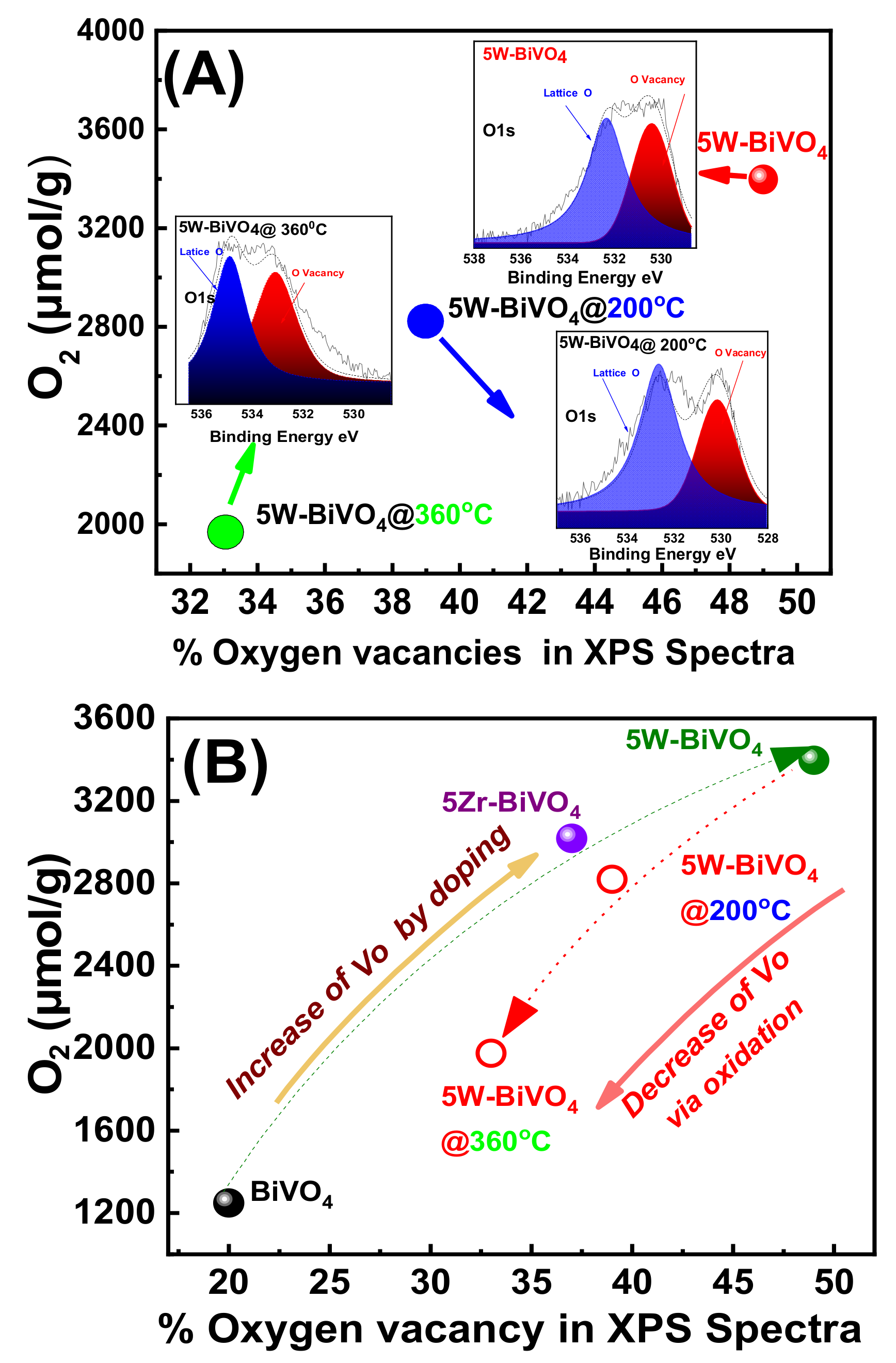
Doping with W or Zr, resulted in an enhancement of the concentration of oxygen vacancies (Vo), see Figure 3. Thus, the XPS data in Figure 3, show that the as-prepared FSP-made BiVO4 contains O-vacancies, whose concentration can be promoted by W- or Zr-doping. This phenomenon, i.e., the correlation of W with O-vacancies sites, has been recently discussed in theoretical DFT studies [19,20,24]. More specifically, theoretical calculations indicate that surface O-vacancies enhance the electron density near the bottom of the conduction band of BiVO4 [19] Additionally, there is theoretical evidence [20,24] that O-vacancies may play a role in the interfacial charge transfer. Importantly, the DFT data entail that the role of W-doping is interlinked with the O-vacancies in the enhancement of the photocatalytic electron-hole separation [24]. As we show hereafter, our data on photocatalytic O2-evolution, provide corroborating experimental evidence for the positive effect of W- and Zr-doping in conjunction with the Vo in BiVO4.
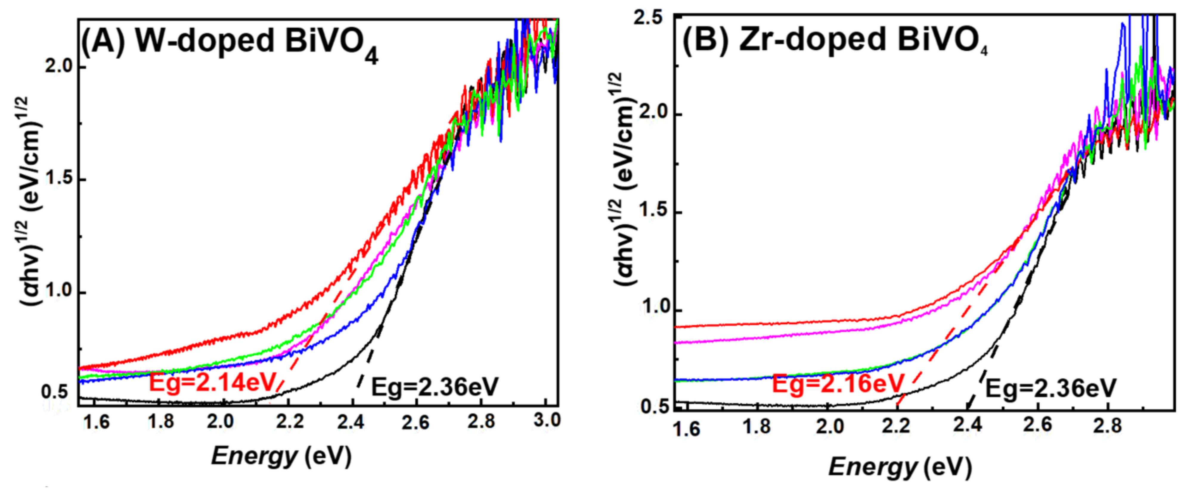
Diffuse Reflectance UV–Vis (DR-UV-Vis): Figure 4 presents DR-UV–Vis spectra and optical band gap energy determination using the Kubelka–Munk [25] formula (2) where α is the absorption coefficient, and the n-value is related to the type of photoexcited transition, i.e., direct or indirect [25].
αhv = A(hv − E)n/2
In our analysis, a value of n = 2 was set, i.e., since BiVO4 is a direct band gap semiconductor [25].
All our FSP-made materials showed adsorption edges with a tail extending towards low-energies, which was enhanced at increasing W- or Zr-doping, see Figure 4A and Figure 4B respectively. Optical band gap energies estimated from the DR-UV–Vis data, are marked by the tangent lines in Figure 4, while the full list of Eg values are presented in Table 1. A band-gap value of Eg = 2.36 eV is typical for pristine BiVO4 [24]. Notice that pristine BiVO4 showed also a low-energy tail, i.e., resolved as a weak hump at 1.6 eV in Figure 4A. This can be attributed to low-energy photons absorbed via intraband states created by the oxygen-vacancies [24] at the Fermi level of BiVO4.
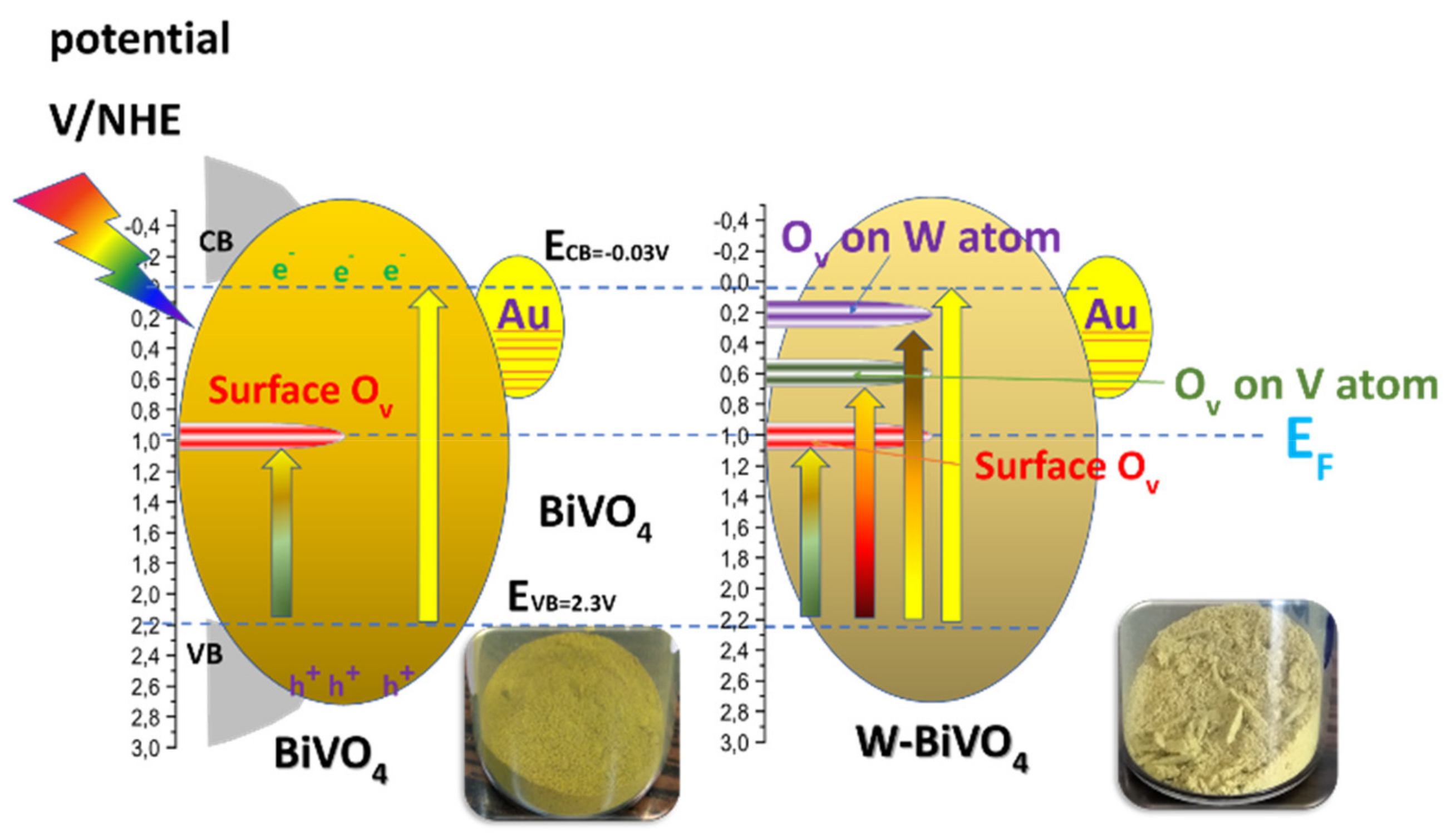
Figure 5 presents a schematic depiction of the energy profile of our BiVO4-based particles. Doping causes a progressive decrease of the Eg values, see Figure 4 and Table 1, i.e., the Eg was progressively decreased to 2.14 eV for 5W-BiVO4 and Eg = 2.16 eV for 5Zr-BiVO4. As analyzed previously in detail, for W-doped BiVO4 [19,20] these trends can be attributed to creation of intraband states, see Figure 5 (right). DFT calculations [5,19,20,24] indicate that the energy of the intraband states is sensitive to the exact location of the W-atom, i.e., whether an oxygen-vacancy occurs next to a W- or next to a V-atom [24], see Figure 5.
In our materials, in all cases, the Eg decrease was accompanied by enhanced light-absorbance at low-energies, verifying that such intraband states are indeed formed upon insertion of the W- or Zr-heteroatoms in to the BiVO4 lattice [24]. Notice that such low-energy light-absorbance is manifested in a more-opaque, less-sharp yellowish color of the W-BiVO4 and Zr-BiVO4 materials, see photos of the FSP-powders in Figure 5. Accordingly, we consider that in our FSP-made BiVO4, all the possible intraband states depicted in Figure 5 (right) might be formed, thus a quasi-continuum of low-energy photons are absorbed by the doped BiVO4 materials.
Overall, the present XPS, Raman and DRS-UV–Vis data provide converging evidence that: (i) our FSP-made BiVO4 contains oxygen vacancies. (ii) W-doping and Zr-doping increase the population of the oxygen vacancies. (iii) W-doping and Zr-doping generate intraband energy states, which enhance the photon-absorbance profile at increased wavelengths, i.e., lower energies down to 50% of the original Eg. These observations are of immediate relevance to the photocatalytic O2-evolution, as we show in the following.

3.2. Catalytic Results

3.2.1. Photocatalytic O2-Evolution from H2O

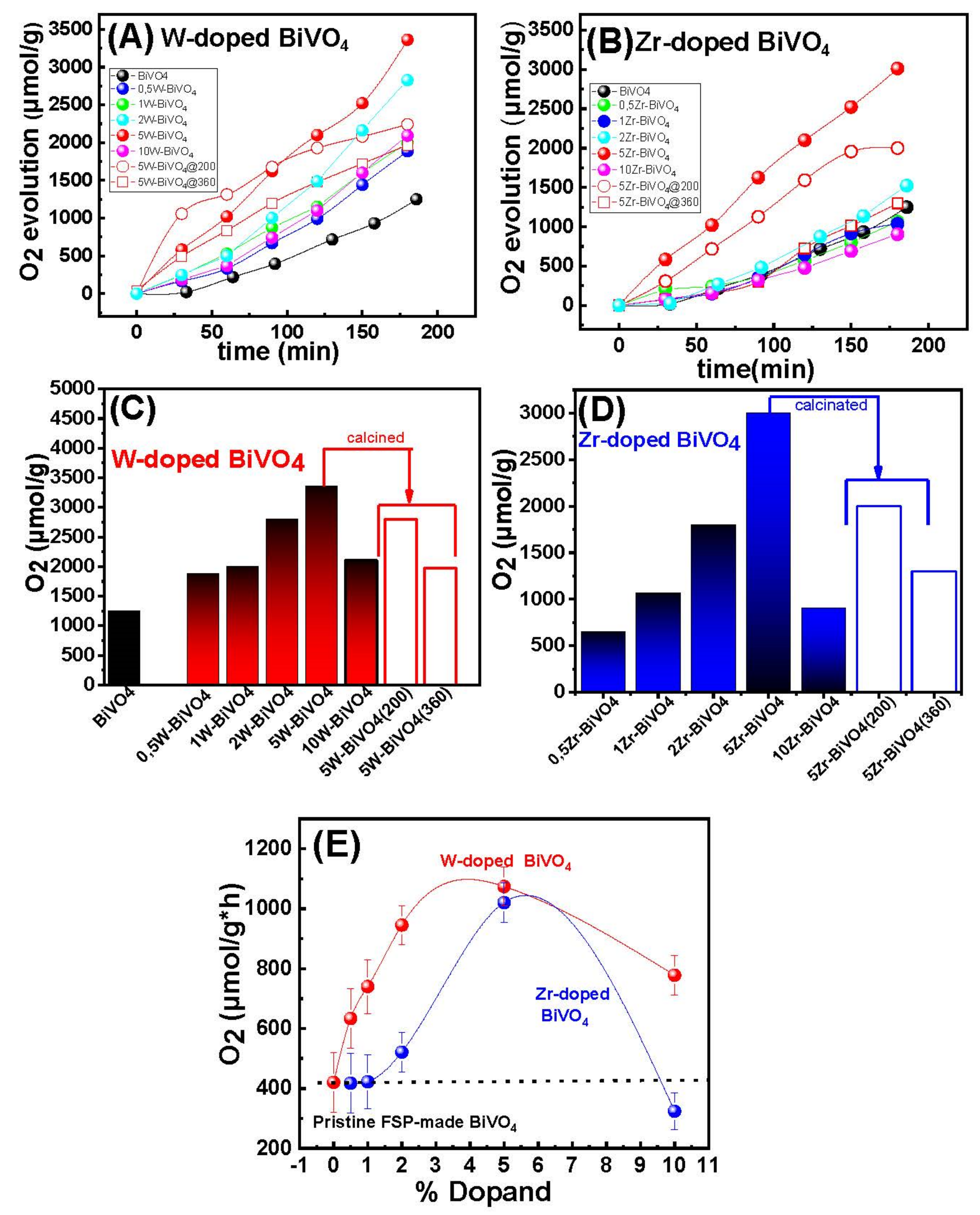
The photocatalytic water oxidation activity of our BiVO4-based catalysts was investigated, using Au as cocatalyst. Figure 6A,B presents data on the O2 evolution kinetics, for the W- and Zr-doped BiVO4-based catalysts.
The data in Figure 6 show that all photocatalysts produced O2 from H2O splitting. More precisely, after 180 min of irradiation, the O2 generated by the best-performing catalyst, 5W-BiVO4, was 2217 μmol per g of material, Figure 6A. The homologous material 5Zr-BiVO4 produced 3018 μmol/g within 180 min of reaction, Figure 6B. For comparison, Figure 6C,D presents the O2 evolution activities in (μmoles per gram of catalyst) at t = 180 min, based on the data of Figure 6A,B. The data are summarized in Table 2.
According to Figure 6E and Table 2, W-doping even at 0.5% resulted in a drastic improvement of the O2 evolution activity vs. pristine BiVO4. Among them, 5 W-BiVO4 exhibited a 270% higher activity than pristine BiVO4 under the same catalytic conditions. When we compared the W-doped vs. Zr-doped materials in Figure 6E, we noticed that significantly higher Zr-doping is required to achieve comparable O2 production rate, as for W-doping. Take as example the 0.5%-doped materials: O2 production rate was 633 μmol/(g × h) for 0.5% W doped vs. 235 μmol/(g × h) for the 0.5% Zr-doped BiVO4. Notice that the two materials had analogous specific-surface-area, i.e., 45 m2/gr vs. 47 m2/g, see Table 1. Thus, if we would normalize per SSA, the W-doped material far more active than the Zr doped BiVO4. In all cases, high dopant-loading, i.e., 10%W or 10%Zr, resulted to lower O2-evolution efficiency.

3.2.2. Comparison with Literature

In Table 3 we listed the O2 evolution rates (μmol/g × h) reported so far for pertinent BiVO4 based photocatalysts, synthesized with various methods or doped with different ions.
The data in Table 3 show that the present 5W-BiVO4 and 5Zr-BiVO4 ranked among the best-performing O2-evolving photocatalysts. Taking into account the differences in the photon-flux, i.e., low-light 125 W of our irradiation system vs. 400 W in reference [26], further confirms that the present FSP-made materials stand at high performance rank. Our data exemplified that a concomitant control of O-vacancies and W-doping is a potent route for engineering of efficient low-cost O2-evolving photocatalysts.

3.2.3. Reusability of the Nanocatalysts

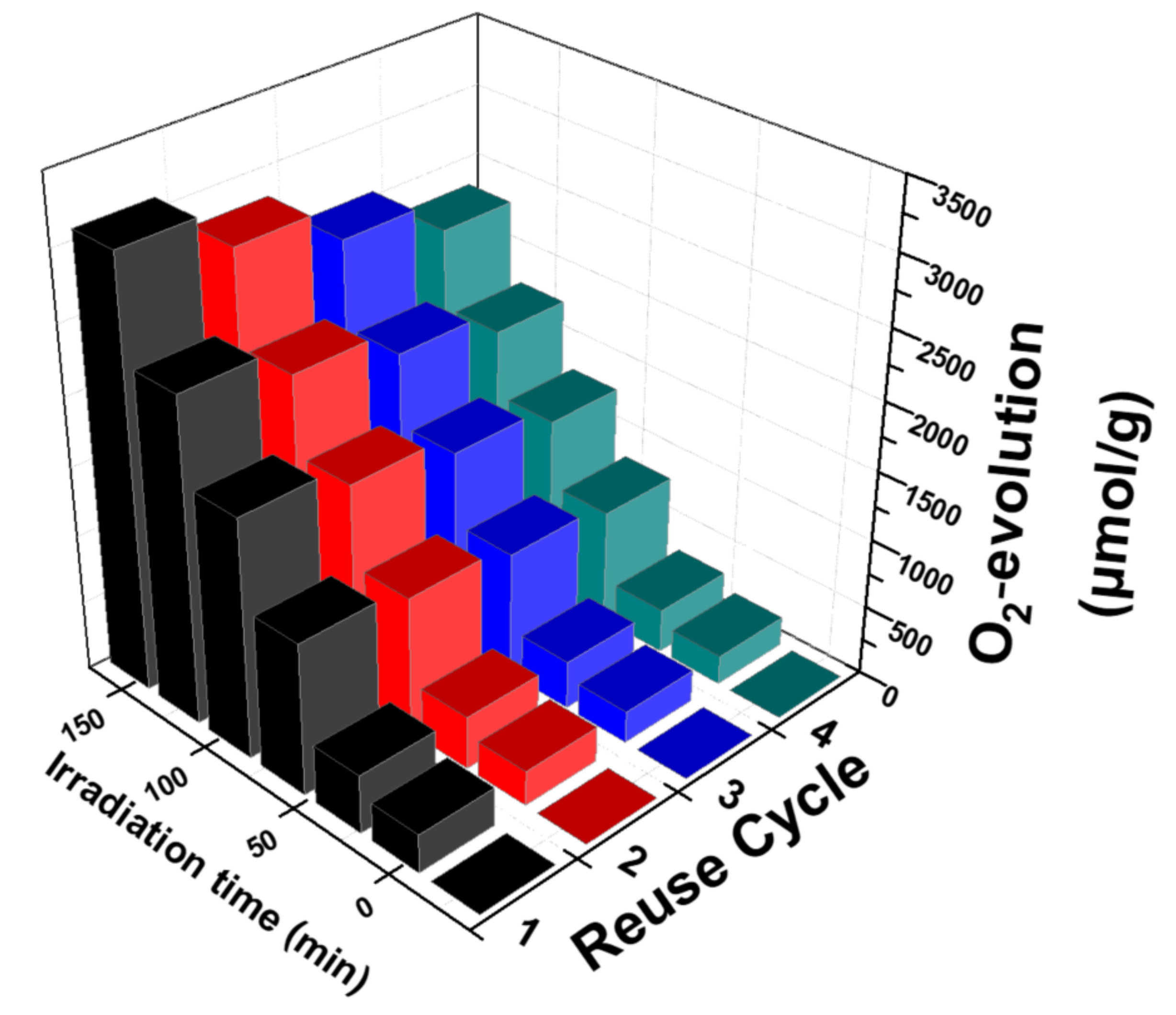
Among the main aims of the heterogeneous catalysis is the reusability of the catalyst. In the present work, the reusability of our best-performing photocatalyst, 5W-BiVO4 was evaluated. After each catalytic use, the solid catalyst was recovered by centrifugation (6000 rpm, 15 min). The liquid supernatant was discarded, and the solid was used for a new reaction under the same catalytic conditions—with no further addition of catalyst.
According to Figure 7, after four sequential catalytic cycles the 5W-BiVO4 catalyst retained 75% of its catalytic activity. The moderate loss of the catalytic activity is progressive and can be attributed to the loss of material mass at each centrifugation–recovery–reuse step, and aggregation phenomena of the particles as reported previously for other BiVO4 particles [26,28].

4. On the Mechanism of Photocatalytic Oxygen Evolution

Previous theoretical calculations suggest that surface oxygen-vacancies enhance the electron density near the bottom of the conduction band of BiVO4 [24]. Moreover, Vo and W-doping may play an important role in the interfacial charge transfer, and in this way W-doping is interlinked with the Vo [19,24] and the enhancement of the photocatalytic electron-hole separation [19,20,24]. To verify the role of the oxygen-vacancies, herein we oxidized, i.e., by mild calcination under O2, the best performing 5 W-BiVO4 and 5 Zr-BiVO4 particles.
The data, see Figure 6C (open bars), show a sharp drop in O2-production efficiency as a function of the calcination temperature. The XRD data, see Figure S2 in Supporting Information, show an increase of particle size, i.e., it becomes 37 nm upon calcination at 360 °C for 3 h. At the same time, the Raman spectra, Figure S3 in Supporting Information, show a restoration of the peak position of the V-O(s) mode at 830 cm−1 indicating the elimination of the O-vacancies from the lattice of W-BiVO4. Moreover, there is a strong correlation of calcination with the decline of the O-vacancies population as detected by XPS, see Figure 8. An analogous phenomenon has been reported by other researchers also [21,24,25,31]. Here, our data allow a better comprehension of the necessity of W-doping and O-vacancies occurrence for enhancing O2-evolution. W-doping alone, i.e., without O-vacancies does not suffice for enhanced O2-evolution. Notice that in Figure 6C, after calcination at 360 °C the efficiency of the best-performing 5W-BiVO4 dropped below the non-doped BiVO4.
In agreement with previous reports, [31], the present data show that the primary factor for enhanced O2-evolution is the optimal O-vacancy engineering. Herein the 5W-BiVO4 and 5 Zr-BiVO4 materials, when oxidized by calcination at mild temperatures 200 °C and 360 °C for 3 h under air their O2-production declined from 3326 μmol/g to 2804 μmol/g and 1973 μmol/g. Analogous results observed for the 5 Zr-BiVO4 (see Table 2, and Figure 6).

5. Conclusions

The present research bears two key-results (i) FSP-technology allows engineering of oxygen vacancies content in BiVO4 via W-doping or Zr-doping, and (ii) oxygen-vacancies are the primary factor to determine the photocatalytic O2-production via water oxidation. FSP allows engineering of BiVO4 nanoparticles, with controlled heteroatom-dopant content, and oxygen vacancy content. As a proof of concept, W- and Zr-doped BiVO4 photocatalysts prepared with different dopant loading were engineered. The correlation between the catalytic efficiency and the oxygen-vacancies content provides strong evidence that the presence of oxygen vacancies in W-BiVO4, Zr-BiVO4 improves drastically the O2 production efficiency. Our present data on doped BiVO4 corroborate the mechanistic suggestions, indicating that the beneficial role of oxygen vacancy is a general phenomenon, which should be operating in various photocatalysts.

Supplementary Materials

The following are available online at https://www.mdpi.com/2079-4991/11/2/501/s1, Figure S1: theoretical simulation of XRD patterns, Figure S2: XRD data for W doped BiVO4. Figure S3: Raman spectra of calcined W doped BiVO4.

Author Contributions

P.S. and M.S.: Data curation, Writing- Original draft preparation. Y.D.: Supervision, Writing- Reviewing and Editing. All authors have read and agreed to the published version of the manuscript.

Funding

The research work was supported by the Hellenic Foundation for Research and Innovation (H.F.R.I) under the “First Call for H.F.R.I Research Projects to support Faculty members and Researchers and the procurement of high-cost research equipment grant” (Project Number: HFRI-FM17-1888.

Data Availability Statement

Data is available upon the reasonable request from the corresponding author.

Conflicts of Interest

The authors declare no conflict of interest.

References

  1. Fujushima, A.; Honda, K. Electrochemical Photolysis of Water at a Semiconductor Electrode. Nature 1972, 238, 37–38. [Google Scholar] [CrossRef]
  2. Kudo, A.; Ueda, K.; Kato, H.; Mikami, I. Photocatalytic O2 evolution under visible light irradiation on BiVO4 in aqueous AgNO3 solution. Catal. Lett. 1998, 53, 229–230. [Google Scholar] [CrossRef]
  3. Moniz, S.J.A.; Shevlin, S.A.; Martin, D.J.; Guo, Z.X.; Tang, J. Visible-light driven heterojunction photocatalysts for water splitting-a critical review. Energy Environ. Sci. 2015, 8, 731–759. [Google Scholar] [CrossRef]
  4. Kato, H.; Hori, M.; Konta, R.; Shimodaira, Y.; Kudo, A. Construction of Z-scheme type heterogeneous photocatalysis systems for water splitting into H2 and O 2 under visible light irradiation. Chem. Lett. 2004, 33, 1348–1349. [Google Scholar] [CrossRef]
  5. Abdi, F.F.; Savenije, T.J.; May, M.M.; Dam, B.; Van De Krol, R. The Origin of Slow Carrier Transport in BiVO 4 Thin Film Photoanodes. J. Phys. Chem. Lett. 2013, 4, 2752–2757. [Google Scholar] [CrossRef]
  6. Zhang, B.; Zhang, H.; Wang, Z.; Zhang, X.; Qin, X.; Dai, Y.; Liu, Y.; Wang, P.; Li, Y.; Huang, B. Doping strategy to promote the charge separation in BiVO4 photoanodes. Appl. Catal. B Environ. 2017, 211, 258–265. [Google Scholar] [CrossRef]
  7. Ding, K.; Chen, B.; Fang, Z.; Zhang, Y.; Chen, Z. Why the photocatalytic activity of Mo-doped BiVO4 is enhanced: A comprehensive density functional study. Phys. Chem. Chem. Phys. 2014, 16, 13465–13476. [Google Scholar] [CrossRef] [PubMed]
  8. Shan, L.; Liu, H.; Wang, G. Preparation of tungsten-doped BiVO4 and enhanced photocatalytic activity. J. Nanopart. Res. 2015. [Google Scholar] [CrossRef]
  9. Yang, Y.; Zhao, Y.; Fan, W.; Shen, H.; Shi, W. A simple flame strategy for constructing W-doped BiVO4 photoanodes with enhanced photoelectrochemical water splitting. Int. J. Energy Res. 2020, 44, 10821–10831. [Google Scholar] [CrossRef]
  10. Ngoipala, A.; Ngamwongwan, L.; Fongkaew, I.; Jungthawan, S.; Hirunsit, P.; Limpijumnong, S.; Suthirakun, S. On the Enhanced Reducibility and Charge Transport Properties of Phosphorus-Doped BiVO4 as Photocatalysts: A Computational Study. J. Phys. Chem. C 2020, 124, 4352–4362. [Google Scholar] [CrossRef]
  11. Babu, P.; Mohanty, S.; Naik, B.; Parida, K. Serendipitous Assembly of Mixed Phase BiVO4 on B-Doped g-C3N4: An Appropriate p-n Heterojunction for Photocatalytic O2 evolution and Cr(VI) reduction. Inorg. Chem. 2019, 58, 12480–12491. [Google Scholar] [CrossRef]
  12. Jiang, Z.; Liu, Y.; Jing, T.; Huang, B.; Zhang, X.; Qin, X.; Dai, Y.; Whangbo, M.H. Enhancing the Photocatalytic Activity of BiVO4 for Oxygen Evolution by Ce Doping: Ce3+ Ions as Hole Traps. J. Phys. Chem. C 2016, 120, 2058–2063. [Google Scholar] [CrossRef]
  13. Ikeda, S.; Kawaguchi, T.; Higuchi, Y.; Kawasaki, N.; Harada, T. Effects of Zirconium Doping into a Monoclinic Scheelite BiVO 4 Crystal on Its Structural, Photocatalytic, and Photoelectrochemical Properties. Front. Chem. 2018, 6, 2–7. [Google Scholar] [CrossRef]
  14. Yin, W.; Wei, S.; Al-jassim, M.M.; Turner, J.; Yan, Y. Doping properties of monoclinic BiVO4 studied by first-principles density-functional theory. Phys. Rev. B 2011, 155102, 1–11. [Google Scholar]
  15. Parmar, K.P.S.; Kang, H.J.; Bist, A.; Dua, P.; Jang, J.S.; Lee, J.S. Photocatalytic and photoelectrochemical water oxidation over metal-doped monoclinic BiVO4 photoanodes. ChemSusChem 2012, 5, 1926–1934. [Google Scholar] [CrossRef]
  16. Camenzind, A.; Caseri, W.R.; Pratsinis, S.E. Flame-made nanoparticles for nanocomposites. Nano Today 2010, 5, 48–65. [Google Scholar] [CrossRef]
  17. Teoh, W.Y.; Amal, R.; Mädler, L. Flame spray pyrolysis: An enabling technology for nanoparticles design and fabrication. Nanoscale 2010, 2, 1324–1327. [Google Scholar] [CrossRef]
  18. Kho, Y.K.; Teoh, W.Y.; Iwase, A.; Mädler, L.; Kudo, A.; Amal, R. Flame preparation of visible-light-responsive BiVO 4 oxygen evolution photocatalysts with subsequent activation via aqueous route. ACS Appl. Mater. Interfaces 2011, 3, 1997–2004. [Google Scholar] [CrossRef] [PubMed]
  19. Wang, W.; Strohbeen, P.J.; Lee, D.; Zhou, C.; Kawasaki, J.K.; Choi, K.-S.; Liu, M.; Galli, G. The Role of Surface Oxygen Vacancies in BiVO 4. Chem. Mater. 2020, 32, 2899–2909. [Google Scholar] [CrossRef]
  20. Zhao, X.; Hu, J.; Yao, X.; Chen, S.; Chen, Z. Clarifying the Roles of Oxygen Vacancy in W-Doped BiVO4 for Solar Water Splitting. ACS Appl. Energy Mater. 2018, 1, 3410–3419. [Google Scholar] [CrossRef]
  21. Moularas, C.; Georgiou, Y.; Adamska, K.; Deligiannakis, Y. Thermoplasmonic Heat Generation Efficiency by Nonmonodisperse Core–Shell Ag 0@SiO 2 Nanoparticle Ensemble. J. Phys. Chem. C 2019, 123, 22499–22510. [Google Scholar] [CrossRef]
  22. Psathas, P.; Georgiou, Y.; Moularas, C.; Armatas, G.S.; Deligiannakis, Y. Controlled-Phase Synthesis of Bi2Fe4O9 & BiFeO3 by Flame Spray Pyrolysis and their evaluation as non-noble metal catalysts for efficient reduction of 4-nitrophenol. Powder Technol. 2020, 368, 268–277. [Google Scholar]
  23. Wu, J.M.; Chen, Y.; Pan, L.; Wang, P.; Cui, Y.; Kong, D.C.; Wang, L.; Zhang, X.; Zou, J.J. Multi-layer monoclinic BiVO4 with oxygen vacancies and V4+ species for highly efficient visible-light photoelectrochemical applications. Appl. Catal. B Environ. 2018, 221, 187–195. [Google Scholar] [CrossRef]
  24. Liu, G.; Li, F.; Zhu, Y.; Li, J.; Sun, L. Cobalt doped BiVO4 with rich oxygen vacancies for efficient photoelectrochemical water oxidation. RSC Adv. 2020, 10, 28523–28526. [Google Scholar] [CrossRef]
  25. Walsh, A.; Yan, Y.; Huda, M.N.; Al-Jassim, M.M.; Wei, S.H. Band edge electronic structure of BiVO 4: Elucidating the role of the Bi s and V d orbitals. Chem. Mater. 2009, 21, 547–551. [Google Scholar] [CrossRef]
  26. Chen, S.H.; Jiang, Y.S.; Lin, H.Y. Easy Synthesis of BiVO4 for Photocatalytic Overall Water Splitting. ACS Omega 2020, 5, 8927–8933. [Google Scholar] [CrossRef] [Green Version]
  27. Yoshino, S.; Iwase, A.; Ng, Y.H.; Amal, R.; Kudo, A. Z-Schematic Solar Water Splitting Using Fine Particles of H2-Evolving (CuGa)0.5ZnS2 Photocatalyst Prepared by a Flux Method with Chloride Salts. ACS Appl. Energy Mater. 2020, 3, 5684–5692. [Google Scholar] [CrossRef]
  28. Tan, H.L.; Amal, R.; Ng, Y.H. Exploring the Different Roles of Particle Size in Photoelectrochemical and Photocatalytic Water Oxidation on BiVO4. ACS Appl. Mater. Interfaces 2016, 8, 28607–28614. [Google Scholar] [CrossRef]
  29. Zhang, Y.; Shi, L.; Geng, Z.; Ren, T.; Yang, Z. The improvement of photocatalysis O2 production over BiVO4 with amorphous FeOOH shell modification. Sci. Rep. 2019, 9, 2–11. [Google Scholar] [CrossRef] [PubMed] [Green Version]
  30. Thalluri, S.M.; Hernández, S.; Bensaid, S.; Saracco, G.; Russo, N. Green-synthesized W- and Mo-doped BiVO4 oriented along the {040} facet with enhanced activity for the sun-driven water oxidation. Appl. Catal. B Environ. 2016, 180, 630–636. [Google Scholar] [CrossRef]
  31. Qiu, W.; Huang, Y.; Tang, S.; Ji, H.; Tong, Y. Thin-Layer Indium Oxide and Cobalt Oxyhydroxide Cobalt-Modified BiVO4 Photoanode for Solar-Assisted Water Electrolysis. J. Phys. Chem. C 2017, 121, 17150–17159. [Google Scholar] [CrossRef]
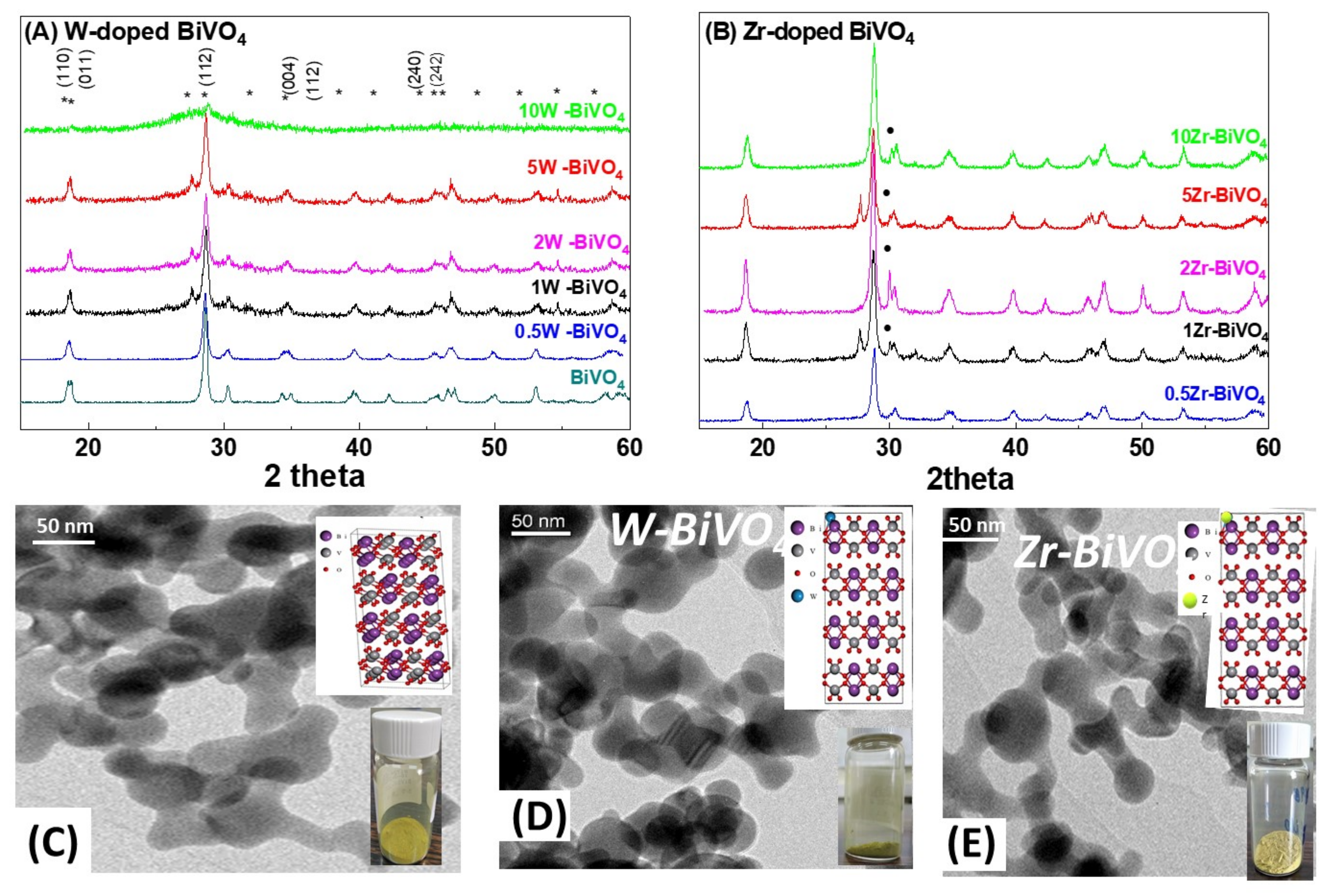
Figure 1. XRD patterns (upper row, W-doped (A) and Zr-doped (B)), and TEM images (lower row) for BiVO4 (C), 5W-BiVO4 (D) and 5 Zr-BiVO4 (E). Inserts in C-E: photos of the nanopowders and atomic structure of the materials. In figure also presented the miller index of BiVO4 structure. In (A), stars (*) mark the main Miller-planes of BiVO4. In (B) the dots (●) mark ZrO2 phase.
Nanomaterials 11 00501 g001
Figure 2. Raman spectra for the FSP-made BiVO4-based photocatalysts: (A) W-doped nanomaterials and (B) Zr-doped nanomaterials. Insert: shift of Raman V-O(s) peak at 830 cm−1.
Nanomaterials 11 00501 g002
Figure 3. XPS spectra of (A) pristine FSP-made BiVO4, (B) 5 W-BiVO4 and (C) 5 Zr-BiVO4 ((a) Bi4f, (b) V2p and (c) O1S).
Nanomaterials 11 00501 g003
Figure 4. Kubelka–Munk transformed diffuse reflectance UV–Vis spectra of FSP-made BiVO4-based nanocatalysts. (A) W-doped BiVO4 and (B) Zr-doped BiVO4.
Nanomaterials 11 00501 g004
Figure 5. Schematic depiction of the energy levels of FSP-made photocatalysts. Surface O-vacancies create Fermi-level states in BiVO4 (left). In addition, W-doping (right) generates additional intraband states whose position depends on the relative location of W, V atoms and O-vacancies [24].
Nanomaterials 11 00501 g005
Figure 6. (A) Kinetics of O2-evolution by the W-BiVO4 and (B) O2 evolution by Zr-BiVO4. Solid symbols are for as prepared materials. Open symbols for calcined at 200 °C (open cycles), or 360 °C (open squares). (C) Comparison of O2-evolution efficiencies of the present FSP-photocatalysts after 180 min of photocatalytic reaction (W-BiVO4), (D) (Zr-BiVO4) and (E) O2 evolution rate vs. the dopant loading. The lines in (E) are for guiding the eye.
Nanomaterials 11 00501 g006
Figure 7. Photocatalytic O2 evolution by sequential reuse of the same 5W-BiVO4-batch.
Nanomaterials 11 00501 g007
Figure 8. Photocatalytic O2-evolution evolution vs. the O-vacancies in the doped-BiVO4 catalysts. (A) Increase of O-vacancies is promoted by W- or Zr-doping in FSP-made BiVO4. Insert: O1s XPS spectra of the W-doped BiVO4. (B) Calcination under O2-rich atmosphere (oxidation) results in decrease of O-vacancies.
Nanomaterials 11 00501 g008
Table 1. Properties of the flame spray pyrolysis (FSP)-made BiVO4 nanocatalysts *.
MaterialNominal Dopant Content in the FSP Precursor (%w:w)Dopant Content Measured by XRFXRD Size (nm ± 0.5)SSA (m2g−1 ± 0.8)Band-Gap Eg (eV)
BiVO4--22.137.72.36
W-Doped nanocatalysts
0.5W-BiVO40.5Not detectable17.545.02.31
1W-BiVO410.619.846.82.25
2W-BiVO421.815.345.02.21
5W-BiVO454.417.147.22.14
10W-BiVO4107.8-49.02.13
Zr-Doped nanocatalysts
0.5Zr-BiVO40.5Not detectable17.847.12.27
1Zr-BiVO410.420.245.62.25
2Zr-BiVO421.719.845.72.20
5Zr-BiVO453.420.546.02.16
10Zr-BiVO4108.3-47.22.15
* Data based on average of three batches.
Table 2. O2-evolution by the present FSP-made BiVO4-based photocatalysts.
MaterialO2-Evolution (μmol/g) after 3 h ReactionO2-Evolution Rate (μmol/g × h)
BiVO41249 (±20)420 (±5)
W-Doped nanocatalysts
0.5W-BiVO41842 (±20)633 (±5)
1W-BiVO42002 (±20)667 (±5)
2W-BiVO42827 (±20)945 (±5)
5W-BiVO43326 (±20)1020 (±5)
10W-BiVO42100 (±20)778 (±5)
Zr-Doped nanocatalysts
0.5Zr-BiVO4600 (±20)235 (±5)
1Zr-BiVO41039 (±20)337 (±5)
2Zr-BiVO41512 (±20)521 (±5)
5Zr-BiVO43018 (±20)974 (±5)
10 Zr-BiVO4907 (±20)324 (±5)
Calcined nanocatalysts
5W-BiVO4@2002804 (±20)1001 (±5)
5W-BiVO4@3601973 (±20)720 (±5)
5Zr-BiVO4@2001988 (±20)710 (±5)
5Zr-BiVO4@3601299 (±20)448 (±5)
Table 3. Comparison of the O2 evolution rate from BiVO4-based photocatalysts.
PhotocatalystLight SourceSynthesis MethodXRD Size (nm)Electron AcceptorActivity (μmol/g*h)Ref.
BiVO4400 W medium-pressure halide lamp (Phillips HPA400, λmax = 360 nmHydrothermal method28.3AgNO32622[26]
BiVO4300 W Xe lamp with a 420 nm cut off filterFlame Synthesis of BiVO4 and Acid Modification72.0AgNO3333[27]
BiVO4300 W Xe lamp with a 420 nm cut off filterSolid-liquid state reaction in HNO391.0AgNO3800[28]
Ce-BiVO4300 W Xe lampHydrothermal methodNot referredAgNO3775[14]
A-FeOOH/BiVO4 (8 wt %)300 W Xe lamp with a 420 nm cut off filterPrecipitation methodNot referredNaIO41206[29]
Zr-BiVO4Perkin Elmer CERMAX LX-300BUV Xe lampPrecipitation methodNot referredAgNO3700[13]
W(0.9%w/w)-BiVO4Simulated plasma (Lumix model LIFI STA-40)Hydrothermal method92.0AgNO3942[30]
BiVO4125W Hg λmax = 440 nmFSP22.1AuHCl4420This work
5W-BiVO4125W Hg λmax = 440 nmFSP17.1AuHCl41074This work
5Zr-BiVO4125W Hg λmax = 440 nmFSP20.5AuHCl41020This work
Publisher’s Note: MDPI stays neutral with regard to jurisdictional claims in published maps and institutional affiliations.