Article

Large-Scale MoS2 Pixel Array for Imaging Sensor

1
State Key Laboratory of ASIC and System, School of Microelectronics, Zhangjiang Fudan International Innovation Center, Fudan University, Shanghai 200433, China
2
School of Electronic Information, Soochow University, Suzhou 215006, China
3
Shanghai Institute of Technical Physics, Chinese Academy of Sciences, Shanghai 200083, China
*
Authors to whom correspondence should be addressed.
These authors contributed equally to this work.
Nanomaterials 2022, 12(23), 4118; https://doi.org/10.3390/nano12234118
Submission received: 28 October 2022 / Revised: 15 November 2022 / Accepted: 18 November 2022 / Published: 22 November 2022
(This article belongs to the Special Issue Field-Effect Transistors Based on Two-Dimensional Materials)

Abstract

:
Two-dimensional molybdenum disulfide (MoS2) has been extensively investigated in the field of optoelectronic devices. However, most reported MoS2 phototransistors are fabricated using the mechanical exfoliation method to obtain micro-scale MoS2 flakes, which is laboratory- feasible but not practical for the future industrial fabrication of large-scale pixel arrays. Recently, wafer-scale MoS2 growth has been rapidly developed, but few results of uniform large-scale photoelectric devices were reported. Here, we designed a 12 × 12 pixels pixel array image sensor fabricated on a 2 cm × 2 cm monolayer MoS2 film grown by chemical vapor deposition (CVD). The photogating effect induced by the formation of trap states ensures a high photoresponsivity of 364 AW−1, which is considerably superior to traditional CMOS sensors (≈0.1 AW−1). Experimental results also show highly uniform photoelectric properties in this array. Finally, the concatenated image obtained by laser lighting stencil and photolithography mask demonstrates the promising potential of 2D MoS2 for future optoelectrical applications.

1. Introduction

Two-dimensional (2D) transition metal dichalcogenides (TMDs) have been developing rapidly and have received considerable research attention in the field of photodetection because of their superior electrical and optical properties [1,2,3,4]. Tremendous efforts have been dedicated to developing high-performance 2D TMDs-based photodetectors for potential applications in optical imaging, neural network vision sensor, and bioinspired in-sensor vision [5,6,7,8,9]. Molybdenum disulfide (MoS2), the most famous representative in the TMD family, has been extensively investigated for electronic and optoelectronic device applications owing to its unique properties, including the layer-dependent bandgap (1.8~1.2 eV from monolayer to bulk), relatively high electron mobility, and current on/off ratio (up to 109) [10,11,12,13]. Therefore, MoS2 has been considered a promising channel material for low-power logic devices [14,15,16] and photodetectors in the visible range [17]. Most previously reported results were based on isolated MoS2 flakes obtained via widely-used top-down approaches such as mechanical exfoliation [18,19]. Such exfoliated single crystalline flakes provide good performance for a single fabricated device, but their micro-scale flake sizes and randomly distributed thicknesses also result in low yield and reproducibility, which hinder practical device applications [18,20].
In recent years, the large-scale fabrication of MoS2 devices has become mainstream since various wafer-scale bottom-up growth methods have been developed, such as CVD, atomic layer deposition (ALD), and metal-organic CVD (MOCVD). For example, Peng et al. presented CVD-grown MoS2 phototransistors with a high photoresponsivity of 6650 AW−1 and detectivity of 1.23 × 10¹² [21]. Chu et al. produced a molybdenum-based phototransistor with an ultrasensitive detectivity of 9.8 × 1016 cm Hz1/2 W−1 [22]. Guo et al. reported the optoelectrical performances of stacked ML-MoS2 phototransistors [23]. In addition to the film fabrication, various device structures have been proposed to improve the performance of MoS2 photodetectors. Chen et al. presented a bilayer MoS2/graphene heterostructure array with the photoresponsivity of 32 mAW−1 [24]. Jeong et al. demonstrated a periodically arrayed nanopore structures for improving the efficiency of multilayered p-WSe2/n-MoS2 phototransistors with a photoresponsivity of 1.7 × 10⁴ AW−1 [25]. However, none of the above focused on the homogeneity of large-scale 2D TMD photodetector arrays, which is a key to realize a practical image sensor. Recently, several reported results tackled the fabrication of pixel array MoS2 image sensors. Park et al. reported a 4 × 4 multilayer MoS2 phototransistors array grown using a post-sulfurization process, which gives rise to a high uniformity but at the cost of a relatively low photoresponsivity of 3.7 AW−1 [26]. Hong et al. designed an 8 × 8 active pixel image sensor array based on a bilayer MoS2 film with a maximum photoresponsivity of 119.16 AW−1 [27]. In these works, how to maintain a uniform high performance while increasing the scale of the pixel array still remains an unsolved problem. In this study, we presented a 12 × 12 pixel array image sensor built on a 2 cm × 2 cm monolayer MoS2 film. Compared with previous work, all 144 individual pixels exhibit the desired optoelectrical properties (photoresponsivity of 364 AW−1, photo detectivity of 2.13 × 10¹⁰) with a high uniformity. Moreover, the MoS2 image sensor, placed under photolithography masks, was exposed to lights of visible-band wavelengths from a laser emitter. By illumination with a different wavelength and different stencils, three sets of photocurrent data were collected and converted to a visualization image, respectively. Thus, this work introduces a new platform for optoelectronic application of wafer-scale 2D-TMDs such as ultra-thin image sensors, transparent image sensors, artificial intelligence photo sensors, and selective light-detecting imagers.

2. Materials Synthesis and Characterizations

Our pixel array image sensor was formed using a 2 cm × 2 cm CVD-grown monolayer MoS2 film, as shown in Figure 1a. This film was synthesized directly on a cleaned silicon oxide (SiO2) substrate without any transfer processes [28]. Compared with the transferring method, the CVD growing method does not use complex processes, implying a higher productivity and lower cost. More details on the film growth are provided in the Supplementary Materials. Prior to the device fabrication, multiple material characteristics, Raman spectra, photoluminescence (PL) spectra, atomic transmission microscope (AFM), and SHG were tested, and the corresponding results were given in Figure 1. In Figure 1b, the Raman spectra under the irradiation of a 514 nm laser were obtained from five different locations on the MoS2 film. The difference between two dominant peaks, i.e., the in-plane (E12g) vibration mode at ~384.3 cm−1 and the out-of-plane (A1g) vibration mode at ~404.7 cm−1 was around 20 cm−1, which was consistent with previous work [18]. Moreover, the prominent consistency and nonexistence of splitting of all Raman spectra curves indicated the high uniformity of our CVD-grown MoS2 film. Figure 1c demonstrated the PL spectra from five suspended samples excited with a solid-state laser at a wavelength of 514 nm. A low laser power of 50 μW (on the sample) was used to avoid heating and PL saturation. The peak value at ≈1.84 eV in the PL spectra was a signal from the MoS2 with silicon substrate [28]. Furthermore, the near-identical peak positions of all curves validated the wafer-scale uniformity of the film as well. The height profile of MoS2 film was measured by AFM (Bruker Dimension Edge). The average height difference along the red line, as shown in Figure 1e, was around 0.78 nm, which corresponded exactly to the thickness of monolayer MoS2 film [11]. Since the information on domain size and boundaries was missing through AFM, a second harmonic generation (SHG) technique was applied to reveal more detailed morphology. As shown in Figure 1f, the domain size was about 10–20 µm in our monolayer, MoS2 and the grain boundaries could be clearly recognized from SHG images. Such capability of direct visualization of the grain information in MoS2 is attributed to the suppressed SHG signal at the grain boundaries. The difference in crystal orientations resulted in the destructive interference and annihilation of the nonlinear waves [29,30]. Above all, all the measurement results indicated the excellent uniformity of our CVD-grown film, which is crucial for further MoS2 phototransistor device fabrication.
The mature back-gate phototransistor structure was adopted here, and the detailed process is presented in Figure 1. The optical microscopic image of the pixel array photodetector with a large-area monolayer MoS2 film is demonstrated in Figure 1g. The device was composed of 12 × 12 MoS2 phototransistors with the same geometric size. A more detailed single device structure was illustrated in Figure 1h. Each phototransistor consists of a MoS2 channel with W/L of 30 μm/20 μm and Au electrodes for source and drain contacts. Figure 1i–k displays a schematic fabrication flow of our MoS2 pixelarray image sensor. As described before, the monolayer MoS2 film was grown on a SiO2/Si substrate, Au (35 nm), as the source/drain (S/D) electrodes were deposited using an electron-beam evaporator and photolithography via a lift-off technique, and then the channel isolation was realized by the CF4 reactive ion etching using a photoresist mask, and a MoS2 channel was etched to the designated dimensions. Finally, Aluminum oxide (40 nm) was deposited as the protective insulator via ALD. The details of the device fabrication are presented in the Supplementary Information.
Compared with the top-gate phototransistor structure, the back gate structured phototransistor can absorb light more efficiently. Moreover, MoS2 is directly grown on a SiO2/Si substrate without additional processing (e.g., transferring MoS2 onto a glass or flexible substrate [31]), which also contributes to a higher uniformity and lower fabrication cost.

3. Results and Discussion

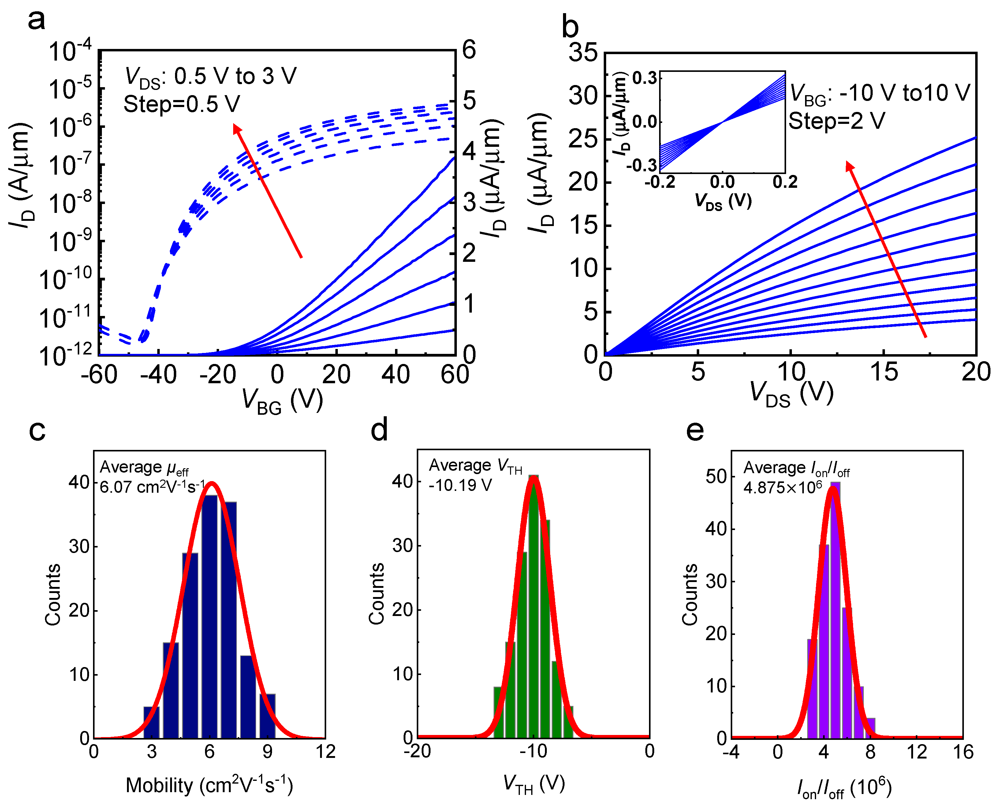
Then, we tested the electrical properties of as-fabricated MoS2 phototransistors. The typical transfer characteristics (ID-VBG) of a single MoS2 phototransistor was plotted in Figure 2a. The current on/off ratio (Ion/Ioff) of ≈106 and the threshold voltage (VTH) of −9.2 V at a drain voltage (VDS) of 1 V indicated the strong gate modulation of the designed MoS2 device. Figure 2b displayed the output characteristics of a typical MoS2 phototransistor in the pixel array image sensor. The drain current (ID) was saturated at a high drain bias (>20 V) because of pinch-off at the drain region. Due to a satisfied contact formation between the monolayer MoS2 film and the S/D electrodes (Au), the output characteristics, ID-VDS, in the insets of Figure 2b exhibited a linear behavior of ID at a low drain bias [32]. Field-effect carrier mobility μeff could also be extracted from the linear region of the transfer curve using the following equation μ eff = g m L c W c C ox V DS , where gm is the transconductance, Lc and Wc are the length and width of the channel, respectively, Cox is the capacitance of the gate insulator, and VDS is the drain voltage [33].
To further demonstrate the uniformity of our image sensor in terms of electrical properties, histogram and corresponding fitted Gaussian distribution were also calculated for mobility (μeff), threshold voltage (VTH), and on/off current ratio (Ion/Ioff) measured from all 12 × 12 phototransistors, as summarized in Figure 2c–e, respectively. The corresponding fitted Gaussian curves (the red solid lines) were also plotted for each parameter. All the phototransistors exhibited a satisfactory electrical performance with the following average values: a μeff of 6.07 cm2 V−1 s−1, a VTH of −10.2 V, and an Ion/Ioff of 4.87 × 10⁶. According the equation S T D D = i = 1 144 D i D ¯ 2 144 / D ¯ , where D could be replaced by μeff, VTH and Ion/Ioff. The standard deviations (STD) of carrier mobility μeff is 23%. The STD of threshold voltage VTH and current ratio Ion/Ioff are 2.8% and 25.2%, respectively. Compared with previous works [26,27], our image sensor exhibited better electrical performance and much smaller pixel-level variation with a much greater number of phototransistors.
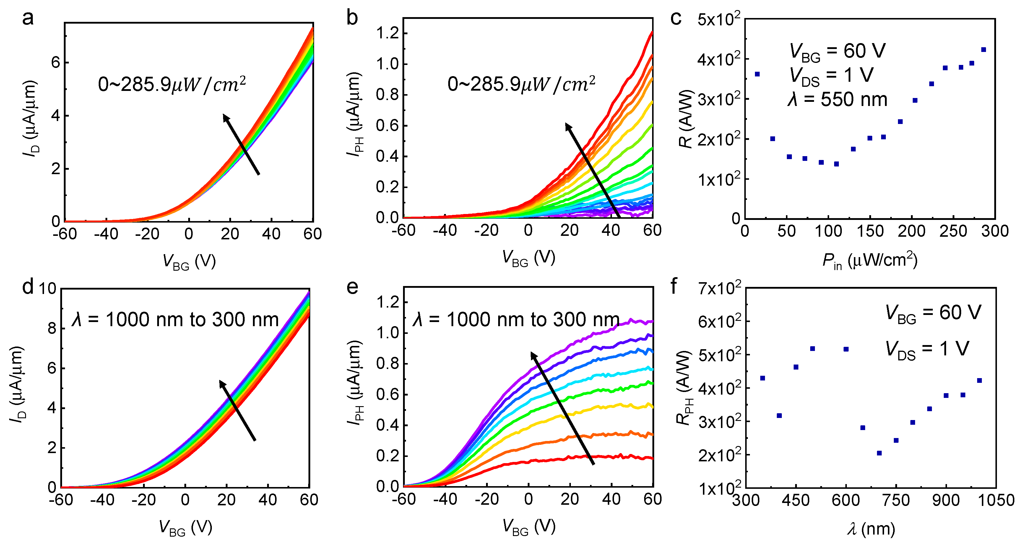
In addition to the electrical characteristics, the key opto-electric characteristics of a single MoS2 phototransistor were measured in Figure 3. Figure 3a exhibited the photoinduced transfer characteristics ID-VBG for a typical MoS2 photodetector 550 nm illumination wavelength at given incident power densities (Pin) ranging from 14.7 to 285.9 μW/cm2. The photocurrent curve of Figure 3b was further extracted from Figure 3a by the following equation: IPH = IilluminationIdark. The photocurrent of the monolayer MoS2 phototransistor gradually increased with increasing Pin according to the photocurrent value under light with different power densities. Moreover, the photoinduced transfer characteristics and photocurrent curve under light with changeable excitation wavelength are presented in Figure 3d,e. Monolayer MoS2 is more sensitive to visible wavelength band (420–680 nm) compared with near-infrared band (700–1200 nm). Figure 3c,f displayed the curve of the calculated photoresponsivity RPH as a function of incident power density and excitation wavelength, which were important figures of merit for phototransistors. The RPH was extracted from the transfer characteristics in Figure 3b,e using the equation of RPH = IPH/Pin (unit: AW−1), where IPH and Pin are the photocurrent and incident power density, respectively. The main mechanism for the high RPH of the monolayer MoS2 phototransistor is the photogating (PG) effect by the formation of trap states near the valance band due to the structural imperfection and defects of MoS2 [34,35,36,37].
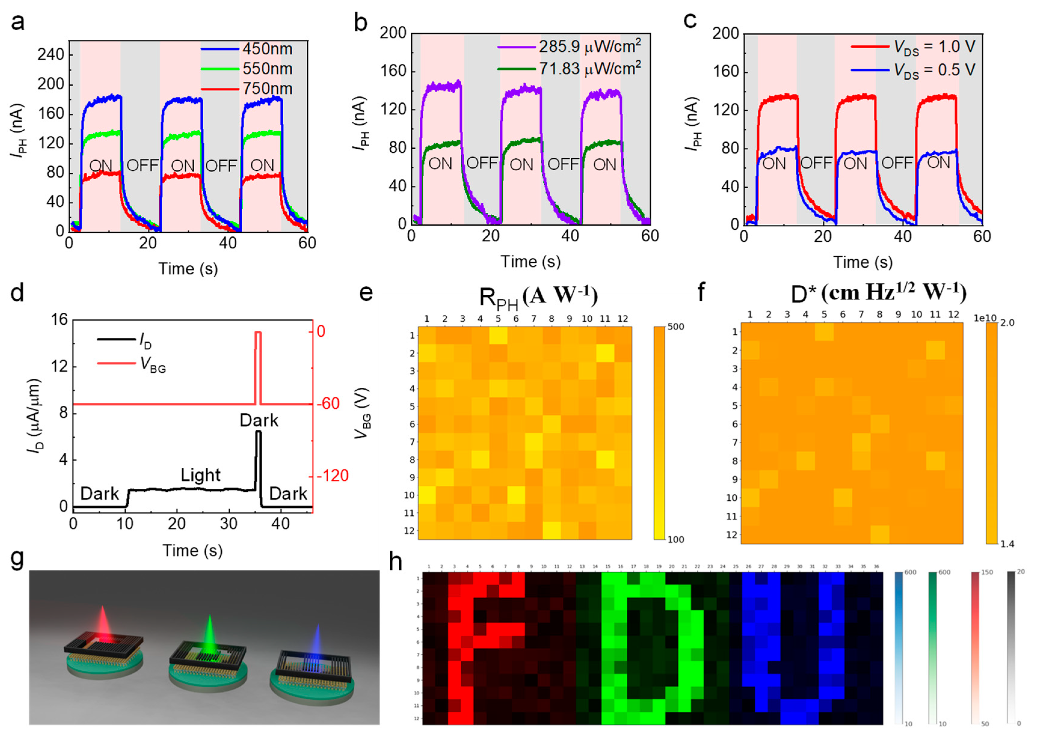
Finally, Figure 4 presents the photo-response speed and an overall uniformity of the 12 × 12 MoS2 array. Figure 4a showed its switching behaviors under pulsed RGB light illumination. As it can be noticed, the photoresponsivity under 450 nm laser was much larger but with a slower response than that of the 750 nm laser. The rise time and decay time increased as light wavelength decreases due to a more excited number of photogenerated charge carriers [36,38]. Figure 4b showed a light intensity dependence, in which the IPH and response time are all positively correlated to incident power, which is also attributed to the photogating effect [17]. Figure 4c presents the photo-switching behavior of IPH under different VDS values. The photoresponsivity increases at the expense of a longer response time. The switching characteristics under different back-gate voltages (VBG) are displayed in Figure S1. When VBG was over-high (>10 V) or over-low (<−10 V), the recovery time would be longer for photo-excited carriers to detrap from the subgap state [27], whereas the gate pulse method by changing back-gate voltage VBG would improve greatly the response speed of our image sensor array. As shown in Figure 4d, when the illumination switched from light to dark, the VBG was injected simultaneously with a short-time (≈1 s) pulse voltage from 8 V to 15 V to suppress carriers as soon as possible. As a result, the falling time decreased by 90% compared with Figure 4a–c. A more detailed gate pulse effect was displayed in Figure S2 and Table S1. A short duration gate voltage pulse could reduce the decay time significantly due to the detrapping of the trapped holes in subgap states, which enables the high-speed operation of the MoS2 image sensor [27,39].
To demonstrate the overall uniformity of our image sensor, a statistical distribution of the photoelectrical properties, i.e., responsivity and detectivity of 12 × 12 MoS2 phototransistors was confirmed in the mapping images that presented the current level of each phototransistor under illumination with Pin of 285.9 μW/cm2 (Figure 4e,f), respectively, where all currents were measured at a VDS of 1 V. The detectivity (D*) is obtained by the equation of D * = A 2 q I D R PH , where A is the channel area, q is unit electric charge, ID is dark current, and RPH is photoresponsivity. The average photoresponsivity was 364.00 AW−1 with a standard deviation of 99 AW−1. The detectivity was 2.16 × 1010 cm Hz1/2 W−1 with a standard deviation of 3.23 × 109 cm Hz1/2 W−1. The photocurrent maps under illumination with different wavelengths (red: 750 nm, green: 550 nm, blue: 450 nm) are demonstrated in Figure S3, which also proves the uniformity of our device.
Moreover, three sets of masks (each was 12 × 12 pixels) with different characters (“F”, “D” and “U”) were prepared and patterned using a laser cutting system, as shown in Figure 4g, to evaluate the image-sensing characteristics of the monolayer MoS2 image sensor array. These character masks were sequentially placed on the image sensor array during light projection (red: 750 nm, green: 550 nm, blue: 450 nm). The 2D photocurrent data, collected under light stencil, was firstly normalized from float type to then concatenated horizontally to form the final 36 × 12 image (total pixel: 576). Due to the uniform photo-related properties of all 12 × 12 phototransistors, the photosensitivity mapping result could display three characters clearly. Table 1 compared the fundamental properties with former works.

4. Conclusions

In this paper, a 12 × 12 phototransistor pixel array image sensor based on a wafer-scale monolayer MoS2 film was fabricated to present the potential of the next generation photodetector. The integrated pixel number is significantly increased without sacrificing photodetector performance. The fabricated MoS2 devices showed high uniformity in electrical properties, including carrier mobility (≈6.07 cm2 V−1 s−1), Ion/Ioff (≈4.875 × 10⁶), and threshold voltage VTH (≈−10.19 V). The measured photoresponsivity RPH (≈364 AW−1) and detectivity (≈2.13 × 10¹⁰) were superior to traditional CMOS image sensors [40,41], which were attributed to the predominant photogeneration mechanism of the PG effect induced by the formation of trap states near the valance band due to the structural imperfection and defects of MoS2. These results provide a blueprint for the future development of wafer-scale 2D TMD optoelectrical application and suggest further application scenarios requiring a high dynamic range, such as with artificial retinas.

Supplementary Materials

The following supporting information can be downloaded at: https://www.mdpi.com/article/10.3390/nano12234118/s1, Figure S1: Photoswitching characteristics of the MoS2 phototransistor under temporal light illumination with varying back gate voltage VBG from −30 V to 30 V with a step of 10 V. All switching curves were measured at VDS = 1 V with illumination frequency of 0.05 Hz; Figure S2: A Time resolved photoresponsive characteristics of the monolayer MoS2 phototransistor under temporal light illumination with λ = 550 nm without and with gate voltage pulse. The fall time is improved from 7.59 s to 6.24 s; Figure S3: Photocurrent mapping of 12 × 12 MoS2 phototransistors at VDS = 1 V, VBG = −10 V under RGB light illumination (λ = 750, 550, and 450 nm), indicating uniform photocurrent photoresponses; Table S1: The rise time and fall time with different case. Reference [42] was cited in supplementary materials.

Author Contributions

Conceptualization, data curation, software, K.L. and X.W.; formal analysis, K.L., X.W. and H.S.; resources, supervision, L.S., H.C. and W.B.; writing—review and editing, K.L., X.W., H.S., X.C., D.W. and J.G. All authors have read and agreed to the published version of the manuscript.

Funding

This research was funded by the National Key Research and Development Program (Grant No. 2021YFA1200500), in part by the Innovation Program of Shanghai Municipal Education Commission (Grant No. 2021-01-07-00-07-E00077), and Shanghai Municipal Science and Technology Commission (Grant No. 21DZ1100900). We also thank the support of the young scientist project from the MOE innovation platform.

Data Availability Statement

The data presented in this study are available on request from the corresponding author.

Conflicts of Interest

The authors declare no conflict of interest.

References

  1. Xu, X.; Pan, Y.; Liu, S.; Han, B. Seeded 2D epitaxy of large-area single-crystal films of the van der Waals semiconductor 2H MoTe2. Science 2021, 372, 195–200. [Google Scholar] [CrossRef] [PubMed]
  2. Michailow, W.; Spencer, P.; Almond, N.W.; Kindness, S.J.; Wallis, R.; Mitchell, T.A.; Degl’Innocenti, R.; Mikhailov, S.A.; Beere, H.E.; Ritchie, D.A. An in-plane photoelectric effect in two-dimensional electron systems for terahertz detection. Sci. Adv. 2022, 8, eabi8398. [Google Scholar] [CrossRef] [PubMed]
  3. Wang, Q.H.; Kalantar-Zadeh, K.; Kis, A.; Coleman, J.N.; Strano, M.S. Electronics and optoelectronics of two-dimensional transition metal dichalcogenides. Nat. Nanotechnol. 2012, 7, 699–712. [Google Scholar] [CrossRef] [PubMed]
  4. Xia, F.; Wang, H.; Xiao, D.; Dubey, M.; Ramasubramaniam, A. Two-dimensional material nanophotonics. Nat. Photonics 2014, 8, 899–907. [Google Scholar] [CrossRef] [Green Version]
  5. Koppens, F.H.; Mueller, T.; Avouris, P.; Ferrari, A.C.; Vitiello, M.S.; Polini, M. Photodetectors based on graphene, other two-dimensional materials and hybrid systems. Nat. Nanotechnol. 2014, 9, 780–793. [Google Scholar] [CrossRef]
  6. Mennel, L.; Symonowicz, J.; Wachter, S.; Polyushkin, D.K.; Molina-Mendoza, A.J.; Mueller, T. Ultrafast machine vision with 2D material neural network image sensors. Nature 2020, 579, 62–66. [Google Scholar] [CrossRef]
  7. Liao, F.; Zhou, Z.; Kim, B.; Chen, J.; Wang, J.; Wan, T.; Zhou, Y.; Hoang, A.; Wang, C.; Kang, J.; et al. Bioinspired in-sensor visual adaptation for accurate perception. Nat. Electron. 2022, 5, 84–91. [Google Scholar] [CrossRef]
  8. Yu, J.; Yang, X.; Gao, G.; Xiong, Y.; Wang, Y.; Han, J.; Chen, Y.; Zhang, H.; Sun, Q.; Wang, Z.L. Bioinspired mechano-photonic artificial synapse based on graphene/MoS2 heterostructure. Sci. Adv. 2021, 7, eabd9117. [Google Scholar] [CrossRef]
  9. Hu, Y.; Dai, M.; Feng, W.; Zhang, X.; Gao, F.; Zhang, S.; Tan, B.; Zhang, J.; Shuai, Y.; Fu, Y.; et al. Ultralow Power Optical Synapses Based on MoS2 Layers by Indium-Induced Surface Charge Doping for Biomimetic Eyes. Adv. Mater. 2021, 33, 2104960. [Google Scholar] [CrossRef]
  10. Liu, L.; Li, T.; Ma, L.; Li, W.; Gao, S.; Sun, W.; Dong, R.; Zou, X.; Fan, D.; Shao, L.; et al. Uniform nucleation and epitaxy of bilayer molybdenum disulfide on sapphire. Nature 2022, 605, 69–75. [Google Scholar] [CrossRef]
  11. Radisavljevic, B.; Radenovic, A.; Brivio, J.; Giacometti, V.; Kis, A. Single-layer MoS2 transistors. Nat. Nanotechnol. 2011, 6, 147–150. [Google Scholar] [CrossRef] [PubMed]
  12. Yoon, Y.; Ganapathi, K.; Salahuddin, S. How good can monolayer MoS2 transistors be? Nano Lett. 2011, 11, 3768–3773. [Google Scholar] [CrossRef] [PubMed]
  13. Mak, K.F.; Lee, C.; Hone, J.; Shan, J.; Heinz, T.F. Atomically thin MoS2: A new direct-gap semiconductor. Phys. Rev. Lett. 2010, 105, 136805. [Google Scholar] [CrossRef] [Green Version]
  14. Dodda, A.; Trainor, N.; Redwing, J.M.; Das, S. All-in-one, bio-inspired, and low-power crypto engines for near-sensor security based on two-dimensional memtransistors. Nat. Commun. 2022, 13, 3587. [Google Scholar] [CrossRef] [PubMed]
  15. Waltl, M.; Knobloch, T.; Tselios, K.; Filipovic, L.; Stampfer, B.; Hernandez, Y.; Waldhor, D.; Illarionov, Y.; Kaczer, B.; Grasser, T. Perspective of 2D Integrated Electronic Circuits: Scientific Pipe Dream or Disruptive Technology? Adv. Mater. 2022, 2201082. [Google Scholar] [CrossRef]
  16. Zhu, K.; Wen, C.; Aljarb, A.; Xue, F.; Xu, X.; Tung, V.; Zhang, X.; Alshareef, H.; Lanza, M. The development of integrated circuits based on two-dimensional materials. Nat. Electron. 2021, 4, 775–785. [Google Scholar] [CrossRef]
  17. Long, M.; Wang, P.; Fang, H.; Hu, W. Progress, challenges, and opportunities for 2D material based photodetectors. Adv. Funct. Mater. 2019, 29, 1803807. [Google Scholar] [CrossRef]
  18. Lee, C.; Yan, H.; Brus, L.; Heinz, T.; Hone, J.; Ryu, S. Anomalous lattice vibrations of single-and few-layer MoS2. ACS Nano 2010, 4, 2695–2700. [Google Scholar] [CrossRef] [Green Version]
  19. Ermolaev, G.A.; El-Sayed, M.A.; Yakubovsky, D.I.; Voronin, K.V.; Romanov, R.I.; Tatmyshevskiy, M.K.; Doroshina, N.V.; Nemtsov, A.B.; Voronov, A.A.; Novikov, S.M. Optical Constants and Structural Properties of Epitaxial MoS2 Monolayers. Nanomaterials 2021, 11, 1411. [Google Scholar] [CrossRef]
  20. Coleman, J.N.; Lotya, M.; O’Neill, A.; Bergin, S.D.; King, P.J.; Khan, U.; Young, K.; Gaucher, A.; De, S.; Smith, R.J. Two-dimensional nanosheets produced by liquid exfoliation of layered materials. Science 2011, 331, 568–571. [Google Scholar] [CrossRef]
  21. Peng, M.; Xie, R.; Wang, Z.; Wang, P.; Wang, F.; Ge, H.; Wang, Y.; Zhong, F.; Wu, P.; Ye, J.; et al. Blackbody-sensitive room-temperature infrared photodetectors based on low-dimensional tellurium grown by chemical vapor deposition. Sci. Adv. 2021, 7, eabf7358. [Google Scholar] [CrossRef] [PubMed]
  22. Feng, S.; Liu, C.; Zhu, Q.; Su, X.; Qian, W.; Sun, Y.; Wang, C.; Li, B.; Chen, M.; Chen, L. An ultrasensitive molybdenum-based double-heterojunction phototransistor. Nat. Commun. 2021, 12, 4094. [Google Scholar] [CrossRef] [PubMed]
  23. Guo, X.; Chen, H.; Bian, J.; Liao, F.; Ma, J.; Zhang, S.; Zhang, X.; Zhu, J.; Luo, C.; Zhang, Z.; et al. Stacking monolayers at will: A scalable device optimization strategy for two-dimensional semiconductors. Nano Res. 2022, 15, 6620–6627. [Google Scholar] [CrossRef]
  24. Chen, C.; Feng, Z.; Feng, Y.; Yue, Y.; Qin, C.; Zhang, D.; Feng, W. Large-scale synthesis of a uniform film of bilayer MoS2 on graphene for 2D heterostructure phototransistors. ACS Appl. Mater. Interfaces 2016, 8, 19004–19011. [Google Scholar] [CrossRef]
  25. Jeong, M.H.; Ra, H.S.; Lee, S.H.; Kwak, D.H.; Ahn, J.; Yun, W.S. Multilayer WSe2/MoS2 Heterojunction Phototransistors through Periodically Arrayed Nanopore Structures for Bandgap Engineering. Adv. Mater. 2022, 34, e2108412. [Google Scholar] [CrossRef]
  26. Park, H.; Liu, N.; Kim, B.H.; Kwon, S.H.; Baek, S.; Kim, S.; Lee, H.; Yoon, Y.J.; Kim, S. Exceptionally uniform and scalable multilayer MoS2 phototransistor array based on large-scale MoS2 grown by RF sputtering, electron beam irradiation, and sulfurization. ACS Appl. Mater. Interfaces 2020, 12, 20645–20652. [Google Scholar] [CrossRef] [PubMed]
  27. Hong, S.; Liu, N.; Kim, B.H.; Kwon, S.H.; Baek, S.; Kim, S.; Lee, H.K.; Yoon, Y.J.; Kim, S. Highly sensitive active pixel image sensor array driven by large-area bilayer MoS2 transistor circuitry. Nat. Commun. 2021, 12, 3559. [Google Scholar] [CrossRef]
  28. Xu, H.; Zhang, H.; Guo, Z.; Shan, Y.; Wu, S.; Wang, J. High-performance wafer-scale MoS2 transistors toward practical application. Small 2018, 14, 1803465. [Google Scholar] [CrossRef]
  29. Yin, X.; Ye, Z.; Chenet, D.A.; Ye, Y.; O’Brien, K.; Hone, J.C.; Zhang, X. Edge nonlinear optics on a MoS2 atomic monolayer. Science 2014, 344, 488–490. [Google Scholar] [CrossRef]
  30. Li, D.; Xiao, Z.; Mu, S.; Wang, F.; Liu, Y.; Song, J. A facile space-confined solid-phase sulfurization strategy for growth of high-quality ultrathin molybdenum disulfide single crystals. Nano Lett. 2018, 18, 2021–2032. [Google Scholar] [CrossRef]
  31. Choi, M.; Bae, S.R.; Hu, L.; Hoang, A.T.; Kim, S.Y.; Ahn, J.H. Full-color active-matrix organic light-emitting diode display on human skin based on a large-area MoS2 backplane. Sci. Adv. 2020, 6, eabb5898. [Google Scholar] [CrossRef] [PubMed]
  32. Das, S.; Chen, H.Y.; Penumatcha, A.V.; Appenzeller, J. High performance multilayer MoS2 transistors with scandium contacts. Nano Lett. 2013, 13, 100–105. [Google Scholar] [CrossRef] [PubMed]
  33. Zheng, X.; Calò, A.; Albisetti, E.; Liu, X.; Alharbi, A.; Arefe, G.; Liu, X.; Spieser, M.; Yoo, W.; Taniguchi, T.; et al. Patterning metal contacts on monolayer MoS2 with vanishing Schottky barriers using thermal nanolithography. Nat. Electron. 2019, 2, 17–25. [Google Scholar] [CrossRef] [Green Version]
  34. Buscema, M.; Island, J.O.; Groenendijk, D.J.; Blanter, S.I.; Steele, G.A.; van der Zant, H.S.; Castellanos-Gomez, A. Photocurrent generation with two-dimensional van der Waals semiconductors. Chem. Soc. Rev. 2015, 44, 3691–3718. [Google Scholar] [CrossRef] [PubMed] [Green Version]
  35. Butt, N.Z.; Sarker, B.K.; Chen, Y.P.; Alam, M. Substrate-induced photofield effect in graphene phototransistors. IEEE Trans. Electron Devices 2015, 62, 3734–3741. [Google Scholar] [CrossRef]
  36. Fang, H.; Hu, W. Photogating in low dimensional photodetectors. Adv. Sci. 2017, 4, 1700323. [Google Scholar] [CrossRef]
  37. Kim, S.; Maassen, J.; Lee, J.; Kim, S.M.; Han, G.; Kwon, J.; Hong, S.; Park, J.; Liu, N.; Park, Y.C.; et al. Interstitial Mo-assisted photovoltaic effect in multilayer MoSe2 phototransistors. Adv. Mater. 2018, 30, 1705542. [Google Scholar] [CrossRef]
  38. Lan, Z.; Lei, Y.; Chan WK, E.; Chen, S.; Luo, D.; Zhu, F. Near-infrared and visible light dual-mode organic photodetectors. Sci. Adv. 2020, 6, eaaw8065. [Google Scholar] [CrossRef] [Green Version]
  39. Lopez-Sanchez, O.; Lembke, D.; Kayci, M.; Radenovic, A.; Kis, A. Ultrasensitive photodetectors based on monolayer MoS2. Nat. Nanotechnol. 2013, 8, 497–501. [Google Scholar] [CrossRef]
  40. Gao, J.; Zhang, B.; Nie, K.; Xu, J. Research on a pulse-based high-line-rate TDI CMOS image sensor. Microelectron. J. 2021, 111, 105021. [Google Scholar] [CrossRef]
  41. Zhang, X.; Fan, W.; Xi, J.; He, L.; Hu, C. 14-Bit Fully Differential SAR ADC with PGA Used in Readout Circuit of CMOS Image Sensor. J. Sens. 2021, 2021, 6651642. [Google Scholar] [CrossRef] [PubMed]
  42. Yu, H.; Liao, M.; Zhao, W.; Liu, G.; Zhou, X.J.; Wei, Z.; Xu, X.; Liu, K.; Hu, Z.; Deng, K.; et al. Wafer-Scale Growth and Transfer of Highly-Oriented Monolayer MoS2 Continuous Films. ACS Nano 2017, 11, 12001–12007. [Google Scholar] [CrossRef] [PubMed]
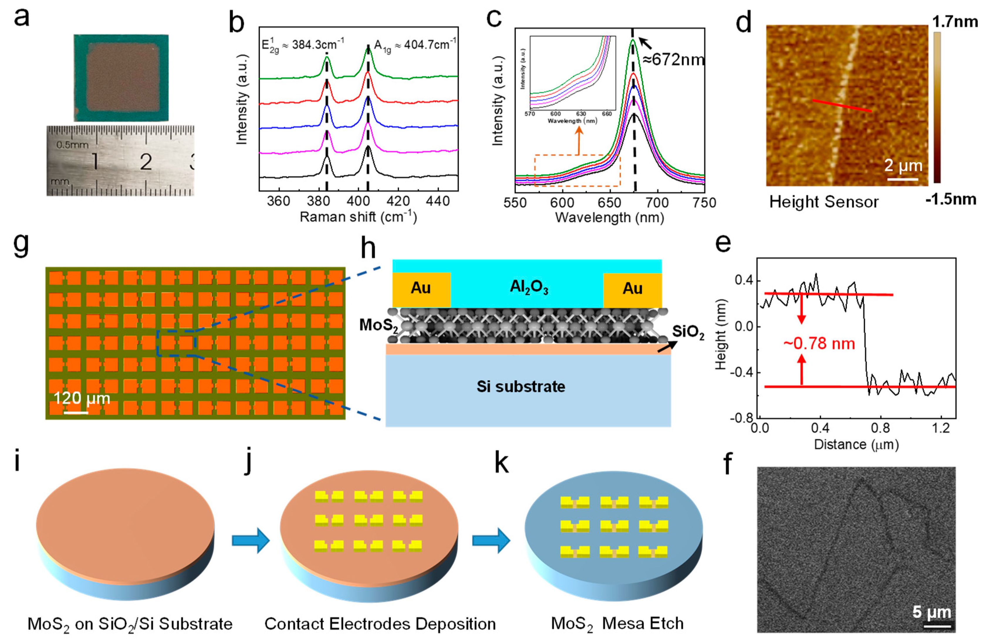
Figure 1. Spectroscopic analysis of the CVD-grown monolayer MoS2 film and process of the device structure. (a) Photograph of an as-fabricated centimeter scale MoS2 with FETs. (b) Raman spectra curves of five randomly selected points from the film, (c) PL spectra curves of five points with the same positions from the film, (e) thickness scan along the red line across the boundary of the film. (d) AFM image of the monolayer MoS2 film. (f) SHG mapping in an area of 30 μm × 30 μm of the film, scale bar: 5 μm. (g) Optical microscopy image of the as-fabricated 12 × 12 MoS2 phototransistors array. The scale bar is 120 μm. (h) 3D schematic image of a single MoS2-based phototransistor. (ik) diagram for the fabrication process of MoS2 film to phototransistors devices employing back metal gates device structure.
Nanomaterials 12 04118 g001
Figure 2. Electrical characteristics and statistical analysis of MoS2 phototransistors. (a) ID-VBG curves of a typical MoS2 phototransistor at VDS from 0.5 to 3 V with the step of 0.5 V. (b) ID-VDS curves of MoS2 at VBG from −10 to 10 V with the step of 2 V. Inset: ID-VDS curve acquired at a small range of VDS. The Histograms of (c) field-effect mobility (average μeff = 6.07 cm2 V−1 s−1), (d) threshold voltage (average VTH = −10.19 V), and (e) Ion/Ioff current ratio (average Ion/Ioff = 4.875 × 10⁶) with VDS = 1 V of all 12 × 12 MoS2 phototransistor pixels.
Nanomaterials 12 04118 g002
Figure 3. Photo-responsive characteristics of a monolayer MoS2 phototransistor in the image sensor array. (a) Transfer curves of ID for dark and illumination conditions and (b) Photocurrent as the function of VBG when VDS = 1 V with varying lighting intensity, ranging from 14.7 to 285.9 μW/cm2 with an average step of 17.87 μW/cm2. (c) Responsivity as a function of incident power when VBG = 60 V and VDS = 1 V. (d) Transfer curves for illumination conditions with varying light wavelengths. (e) Photocurrent as a function of VBG when VDS = 1 V with different wavelengths, ranging from 1000 nm to 300 nm with a step of −100 nm. (f) Responsivity as a function of the light wavelength.
Nanomaterials 12 04118 g003
Figure 4. (a) Time-trace of the photodetector under illumination with the same intensity (Pin = 200 μW/cm2) and VDS = 1 V but at different wavelengths (λ = 450, 550 and 750 nm). (b) Same measure for different light intensities (Pin = 285.9 μW/cm2 and 71.83 μW/cm2) with the wavelength of 550 nm and VDS = 1 V. (c) Same measurement for different VDS values (0.5 and 1.0 V) under the illumination of 285.9 μW/cm2 with 550 nm wavelength. (d) Photo-switching characteristics with gate pulse. (e,f) Photo-responsivity and detectivity mapping of 144 MoS2 phototransistors under the illumination of 285.9 μW/cm2 with 550 nm wavelength. (g) Measurement concept using the light stencil projection for image detection of the image sensor array. The 12 × 12 monolayer MoS2 image sensor array is placed behind character masks and measured photoelectricity under RGB light illumination (wavelength λ = 750, 550, and 450 nm). (h) Horizontal concatenated normalized image with a resolution of 36 × 12 pixels.
Nanomaterials 12 04118 g004
Table 1. Comparison of photo-related properties of fabricated pixel array image sensors.
IndicatorPark et al. [26]Hong et al. [27]Ours
Pixel size
(width × height)
4 × 48 × 812 × 12
Layer of MoS2 film2 LMultilayer1 L
Average responsivity
(Unit: A W−1)
0.503119.16364.00
Std responsivity
(Unit: percentage %)
15--27.2
Average detectivity
(Unit: cm Hz1/2 W−1)
1.4 × 1044.66 × 1062.13 × 1010
Std detectivity
(Unit: percentage %)
12--15
Publisher’s Note: MDPI stays neutral with regard to jurisdictional claims in published maps and institutional affiliations.