Review

The Role of Integrins for Mediating Nanodrugs to Improve Performance in Tumor Diagnosis and Treatment

1
College of Pharmaceutical and Biological Engineering, Shenyang University of Chemical Technology, Shenyang 110142, China
2
Institute of Materials Engineering, College of Engineering and Applied Sciences, Nanjing University, Nanjing 210093, China
3
Department of Biochemistry and Molecular Biology, Medical College, Guangxi University of Science and Technology, Liuzhou 545005, China
*
Authors to whom correspondence should be addressed.
These authors contributed equally to this work.
Nanomaterials 2023, 13(11), 1721; https://doi.org/10.3390/nano13111721
Submission received: 29 April 2023 / Revised: 18 May 2023 / Accepted: 23 May 2023 / Published: 24 May 2023
(This article belongs to the Section Biology and Medicines)

Abstract

:
Integrins are heterodimeric transmembrane proteins that mediate adhesive connections between cells and their surroundings, including surrounding cells and the extracellular matrix (ECM). They modulate tissue mechanics and regulate intracellular signaling, including cell generation, survival, proliferation, and differentiation, and the up-regulation of integrins in tumor cells has been confirmed to be associated with tumor development, invasion, angiogenesis, metastasis, and therapeutic resistance. Thus, integrins are expected to be an effective target to improve the efficacy of tumor therapy. A variety of integrin-targeting nanodrugs have been developed to improve the distribution and penetration of drugs in tumors, thereby, improving the efficiency of clinical tumor diagnosis and treatment. Herein, we focus on these innovative drug delivery systems and reveal the improved efficacy of integrin-targeting methods in tumor therapy, hoping to provide prospective guidance for the diagnosis and treatment of integrin-targeting tumors.

1. Introduction

Tumors, as one of the most high-incidence diseases that seriously threatens human health, have become a leading cause of death worldwide [1]. Frequent invasion and metastasis have become important biological characteristics of malignant tumors as well as the main factors affecting the clinical therapeutic effect and prognosis of tumors [2,3]. Recently, it has been discovered that several proteins, including adhesion receptor families, receptor tyrosine kinases, cytoskeleton proteins, and adaptor and signaling proteins, are linked to the invasion and metastatic properties of tumor cells [4,5,6]. Among them, integrins, as a member of adhesion receptor families, comprise a wide number of cellular receptors and are important proteins on the cell surface [7,8]. They are involved in practically every stage of tumor progression, from initial tumor formation to metastasis [9].
Integrins are transmembrane glycoproteins that attach the cells to the extracellular matrix (ECM) and serve as bidirectional hubs transmitting signals to mediate cell-cell and cell-ECM interactions [10,11,12]. They modulate tissue mechanics and regulate intracellular signaling, including cell faith, survival, proliferation, and differentiation, and the up-regulation of integrins in tumor cells has been confirmed to be associated with tumor development, invasion, angiogenesis, metastasis, and therapeutic resistance. Thus, reasonably regulating the activity of integrins in tumor cells can effectively improve the therapeutic effect of oncotherapy and inhibit tumor invasion and metastasis.
Recently, with the rapid development of nanotechnology, nanodrugs have attracted wide attention due to their excellent biocompatibility and modifiability. As an effective target for tumor therapy, integrins can not only be used to regulate tumor microenvironment but also can promote nanodrugs accumulation in tumor tissues. Herein, we will review the structural characteristics, biological functions, mechanisms of action of integrins in tumor cells, the research progress of the interaction between integrins and tumor microenvironment, as well as the application of integrin-targeting nanomedicine in tumor therapy.

2. The Structure and Function of Integrins

2.1. The Structure of Integrins

Integrins, as adhesion molecules and transmembrane receptors that consist of α and β subunits, heterodimers, are widely located on the surface of vertebrate cells and are involved in cell-cell and cell-matrix interactions [11]. Integrins feature an extracellular domain, a single-pass transmembrane domain and a short cytoplasmic domain (except β4) [13]. Of these, the extracellular domain can interact with ECM ligands to transmit signals [13,14]. Generally, there are 18α and 8β subunits that can combine to generate 24 distinct receptors with varying binding capabilities and tissue distribution [15,16]. Specifically, nine of these α subunits contain an αI domain at the N-terminus consisting of approximately 190–200 amino acid residues and can be strongly bound by ligands [17]. Moreover, metal ion binding sites at the top of the αI domain have been demonstrated to bind divalent metal ions, such as magnesium ions and manganese ions, thus, mediating the interaction of integrins and ligands [18,19]. For the α subunits that lack the I domain (α3, α4, α5, α6, α7, α8, α9, αv, and αIIb), the β-propeller of the α subunits and the I domain of the β subunit can be joined to form the binding site [20]. Moreover, many integrin ligands, as well as the integrins themselves, present structural alterations under specific conditions, which may compromise signaling pathways that are constitutively triggered under physiological conditions [21,22].
Of the 24 human integrin subtypes known to date, eight integrin dimers can recognize Arg-Gly-Asp (RGD) peptide in extracellular matrix proteins [23]. In addition, there are four types of integrins based on their connective ligand: leukocyte, collagen-binding, Arg-Gly-Asp (RGD)-binding, and laminin-binding integrins (Figure 1). The short cytoplasmic domain provides binding sites for these adaptors, signaling proteins, and cytoskeleton-associated proteins, all of which are required for bidirectional integrin signaling [12,24]. Moreover, the integrins also serve as receptors for certain toxins (such as snake venom), viruses, and other pathogens [25]. In addition, fibronectin is regarded as a potential integrin ligand and the extra deposition of fibronectin in tissue leads to inflamed diseases [26,27].

2.2. The Function of Integrins

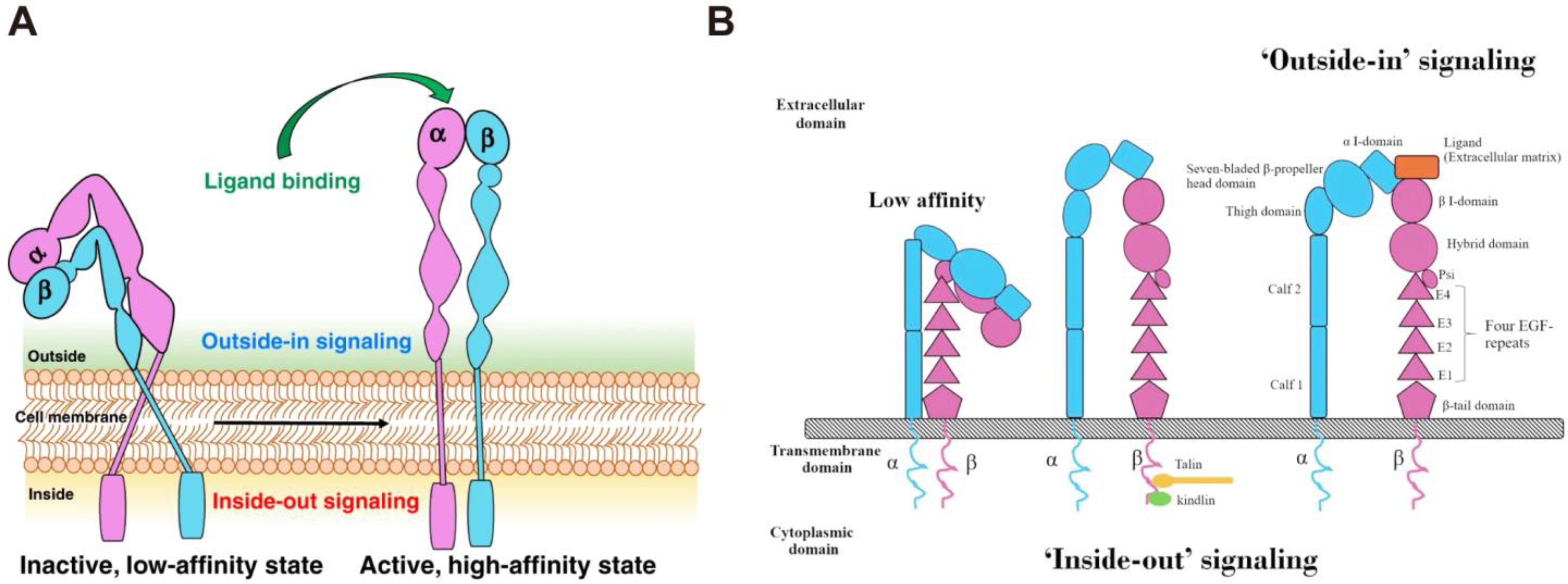
Integrins are essential for cell growth and survival. They tightly link cells to ECM and deliver signals both internally and outwardly, acting as a communication bridge between the internal and external worlds of cells [28]. Integrins are activated in the extracellular domain via conformational rearrangement from bent-closed to open-active states [29,30]. In this process, the ECM ligand binding would rapidly lead to the conversion of the integrins to the high-affinity extended-open form that permits coupling to the actin cytoskeleton. This bridging to actin supports cell adhesion and the recruitment of additional intracellular binding partners that mechanically reinforce the link and allow subsequent downstream signal transduction [31]. The bidirectional transmembrane signals are delivered through “inside-out” and “outside-in” pathways (Figure 2). Briefly, some intracellular signal molecules, such as talin and paxillin, can bind to the intracellular region of integrins, inducing conformational changes in the intracellular region and initiating a series of subsequent conformational changes in the extracellular domain, a process known as “inside-out” signaling [32]. In this process, the binding of ligands to integrins causes changes in the intracellular cytoskeleton and the recruitment of important intracellular signal molecules [33]. Furthermore, the intracellular domains of α and β subunits are separated and bind to intracellular signal molecules, activating relevant intracellular signaling pathways and mediating cell functions. This is known as “outside-in” signal transduction. The bidirectional signal transduction ability of integrins is critical for cell proliferation, invasion, angiogenesis, and apoptosis resistance, contributing to more aggressive diseases, such as tumors [8,34]. For example, Integrin α2 (ITGA2) is overexpressed in solid tumors, including pancreatic, gastric, liver, prostate, and breast tumor, and its expression level is highly correlated with tumor proliferation and invasion [35,36,37,38]. In addition, integrin β1, β3, β6, αv, and α5 are also found in many tumors and play an important role in tumor therapies [39,40,41]. Therefore, an in-depth understanding of the interaction between integrins and tumors is beneficial in improving tumor therapy.

2.3. The Effect of Integrins on Tumors

Integrins have been found to play multiple key functions in practically every stage of tumor progression, from basic tumor growth to metastasis, and hence integrin modulation has been widely used in tumor therapy [9,43,44]. For example, the pre-treatment of cell monolayers with different matrix components (laminin, fibronectin, and collagen), makes neoplastic cells more resistant to cytotoxic insults mediated by different classes of chemotherapeutic agents in an event dependent on integrin-ECM components interaction [45]. The overexpression of ECM components was proven to result in chemoresistance [46,47,48]. However, because tumor incidence and growth are exceedingly complicated, many concerns remain unsolved, particularly the impact on tumor metastasis and angiogenesis [49,50,51]. Further research into the mechanisms of the integrins family in tumor metastasis and tumor angiogenesis will help to expand the use of integrins in tumor therapy.
Compared with normal tissues or cells, the expression of integrins is maintained at a high level in tumors and plays an important role in tumor proliferation and invasion [52,53,54]. For example, integrin β1, a member of the integrins family, has been confirmed to be overexpressed in nasopharyngeal carcinoma tissues [37]. It promotes the migration and metastasis of nasopharyngeal carcinoma cells and reduces the sensitivity of cells to radiotherapy [55]. It has been discovered that the inhibition of integrin β1 expression can effectively improve the radiation sensitivity of nasopharyngeal carcinoma [56]. In addition, the high expression of integrin α5β1 in hepatocellular carcinoma is closely associated with the occurrence and metastasis of hepatocellular carcinoma, the antagonists of integrin α5β1 can significantly reduce the migration ability of hepatocellular carcinoma [57,58].

3. The Role of Integrin αvβ3 in Nanodrugs Antitumor Therapy

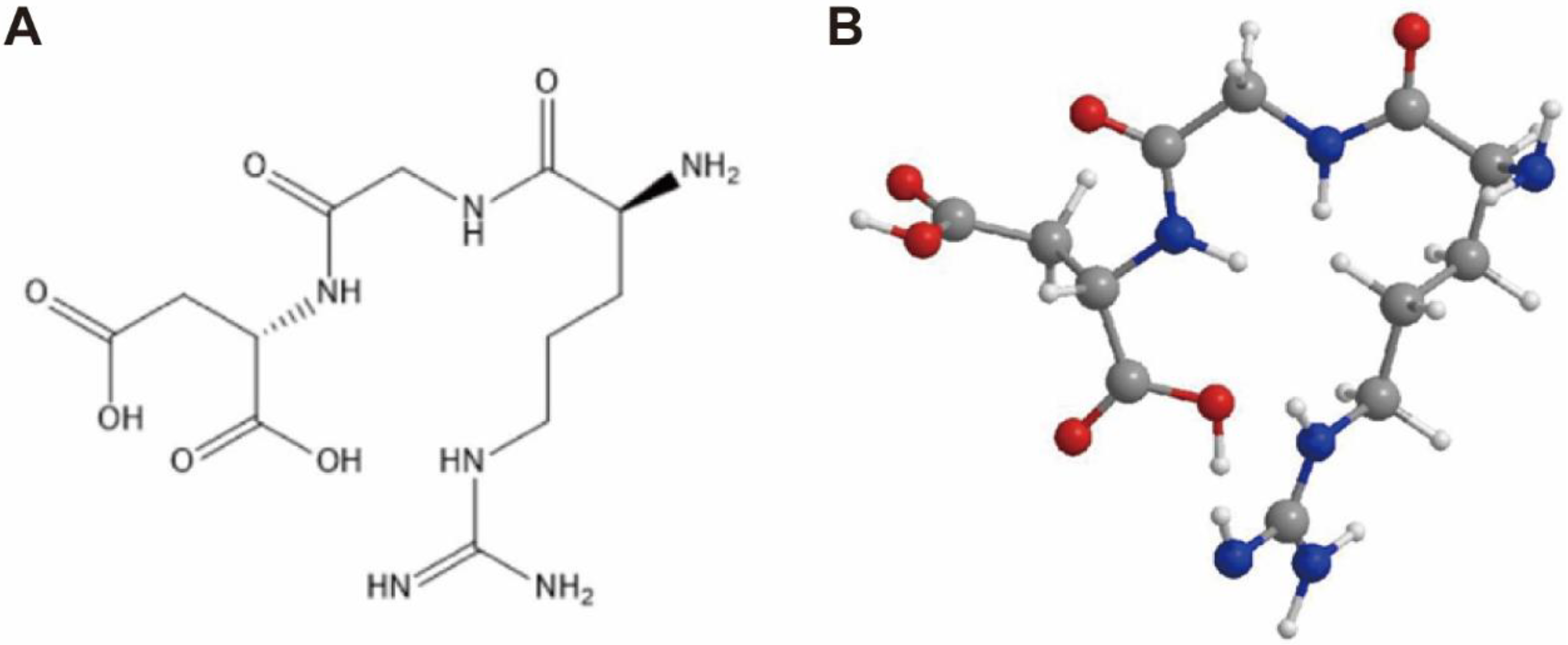
Nanotechnology has a wide range of applications in medicine, such as nanodrugs delivery, nano contrast agents and medical materials. Compared with small molecule drugs, nanodrugs have the characteristics of size effect, large specific surface area, and photothermal effect, which helps to prolong the biological half-life of drugs in vivo, and improve the antitumor effect and imaging ability [59,60,61]. Previous studies have shown that integrin αvβ3 expression on the surface of tumor cells is substantially higher than in normal tissues and that it is easily recognized and bound by RGD and other molecules [27,54,62] (Figure 3). Therefore, the design of nanodrugs targeting integrin αvβ3 is of great practical significance for tumor treatment and diagnosis by enhancing the targeting of nanodrugs in tumor tissues, achieving optimal antitumor potency with minimized toxicity.

3.1. Chemotherapy

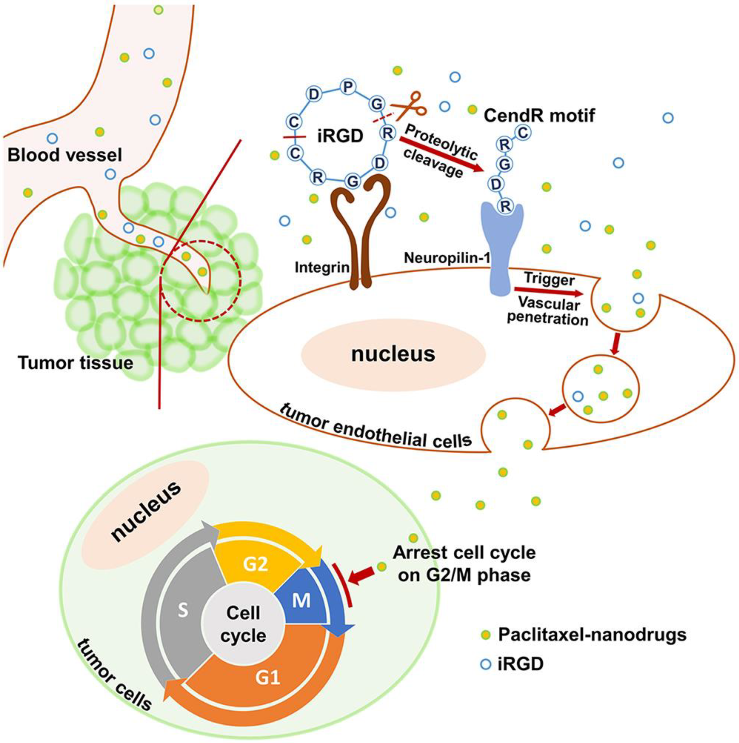
Chemotherapy is a common form of clinical antitumor therapy. Although chemotherapy has shown some efficacy in antitumor treatment, drug enrichment in tumors is limited due to the in vivo complex environment and physiological obstacles [63,64,65]. According to the report from Warren Chan et al., the amount of nanodrugs entering tumor tissue is even less than 1% of the injected amount [66]. Although this conclusion is controversial, it is not satisfactory to show that nanodrugs only relies on the enhanced permeability and retention (EPR) effect to enter tumor tissue and exert antitumor effects [67]. Therefore, some studies use antibodies and other means to analyze drug aggregation at the tumor site, hoping to improve the low-targeting efficiency [68,69,70]. Currently, chemotherapy based on targeting integrins has attracted widespread attention, mainly for better drug targeting, reducing the off-target effect, and improving the antitumor efficacy [71,72]. The iRGD is a bifocal ligand containing RGD and a C-end sequence (CendR), which can target integrin αvβ3 and neuropilin-1, respectively [73]. For instance, polylactic acid-glycolic acid copolymer (PLGA) was modified by iRGD to form iRGD-PLGA-PTX nanomicelles. The nanomicelles were then intravenously injected into colorectal tumor-loaded mice. The results showed that the inhibition rate of the iRGD-PLGA-PTX group was higher than the PLGA-PTX group, indicating that iRGD promoted endocytosis of PLGA-PTX nanomicelles by colon tumor cells and improved the antitumor efficiency (Figure 4) [74]. In another instance, PLGA combined with RGDfk to prepare nanodrugs for the treatment of breast tumor. PLGA is often used in drug and gene delivery due to its excellent biocompatibility, high stability, and degradability [75,76]. The RGDfk-modified nanodrugs can not only increase drug solubility but also improved the biological activity of the drug and prolonged the half-life of the drug in vivo [77]. In addition, the EGF-Like Repeats and Discoidin I-Like Domains Protein 3 (EDIL3) was highly expressed in paclitaxel-resistant breast tumor, but low in sensitive breast tumor. Ding, Y et al. revealed that blocking the interaction between integrin αvβ3 and EDIL3 by Cilengitide improved the sensitivity of tumor tissues to paclitaxel and reversed the epithelial-mesenchymal transition (EMT) of paclitaxel-resistant tumors, reducing the probability of tumor resistance [78].

3.2. Radiotherapy

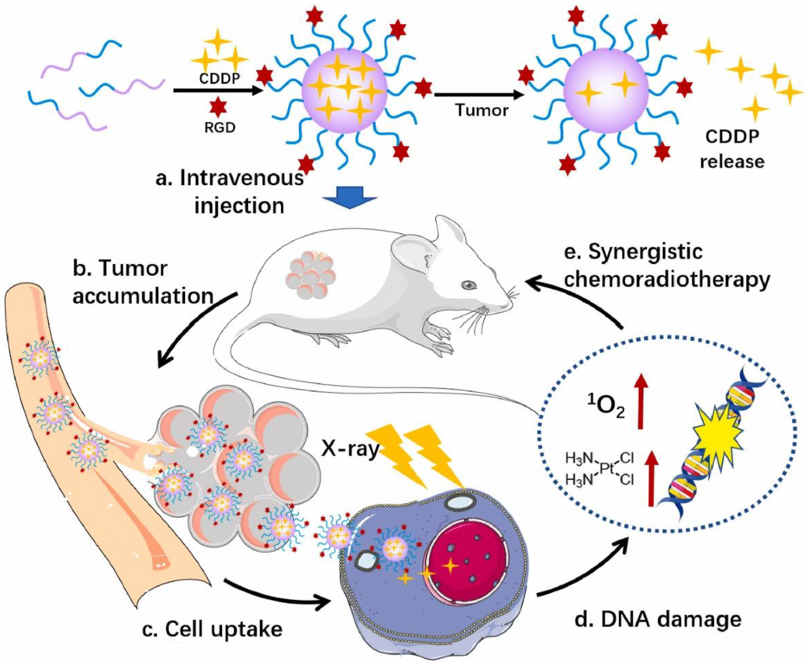
Radiotherapy can induce the instantaneous, dynamic, and local “bursting” of tumor microvessels, thus, increasing the permeability of blood vessels and facilitating the entry of nanoparticles from outside the blood vessels into the tumor, promoting drug enrichment in tumor site, enhancing the local drug concentration, and reducing drug resistance [79,80]. Gold nanoparticles are often regarded as radiosensitizers because of their unique physical and chemical properties and excellent biocompatibility [81]. Gold nanoparticles can kill tumor cells at low doses of radiation, reducing the damage of radiation therapy to normal tissues [82]. For in vivo CT imaging and radiosensitization evaluation, c(RGDyC)-AuNPs enhanced the sensitivity of radiotherapy and improved the effect of radiotherapy in 4T1 tumor-bearing mice with integrin αvβ3 modified nanodrugs [83]. Compared with gold, copper (64) is also a radiosensitizer. The 64Cu-Pyro-3PRGD2 probe was synthesized by 3PRGD2-modified Pyropheophorbiide-A (Pyro), which showed high-tumor specificity in both positron emission tomography (PET) and optical imaging [84]. 3PRGD2 has a higher affinity with integrin αvβ3, which can reduce drug dose and tumor resistance, and inhibit tumor metastasis [85]. Meantime, 64Cu-Pyro-3PRGD2 has good water solubility and has very low accumulation in normal organs, which can be quickly cleared through renal metabolism to avoid potential damage to normal tissues [84]. Platinum drugs are not only one of the chemotherapeutics but also used in clinical radiotherapy sensitizers [86]. By modifying platinum-based nanoparticles (PtNPs) with RGD to form RPNs, the uptake of cisplatin by tumor cells was enhanced through RGD targeting, and DNA fragmentation was promoted [76]. In vitro and in vivo experimental results demonstrated that the RPNs + X-ray group had a higher level of tumor apoptosis and lower survival rate of nasopharyngeal carcinoma cells than the RPNs alone treatment group, confirming that RPNs combined with X-ray enhanced the inhibitory effect on the nasopharyngeal carcinoma cells (Figure 5).
In addition, targeted radionuclide therapy (TRT) is also an important component of radiotherapy [87]. By being encapsulated into nanocarriers, radionuclides can naturally accumulate in tumors by positive targeting [88,89]. The TRT radiotherapy can be used for local radiotherapy and metastatic tumor. For example, the programmed death ligand 1 (PD-L1) combined with RGD-modified TRT (177Lu as a radionuclide) nanoparticles was used in colon tumor-bearing mice [90]. The CT imaging studies in small animals have demonstrated that anti-PD-L1 antibody combined with integrin αvβ3-targeted 177Lu-EB-RGD nanodrugs can improve antitumor efficacy with reduced tumor resistance, prolonging overall survival compared with other groups [90].

3.3. Immunotherapy

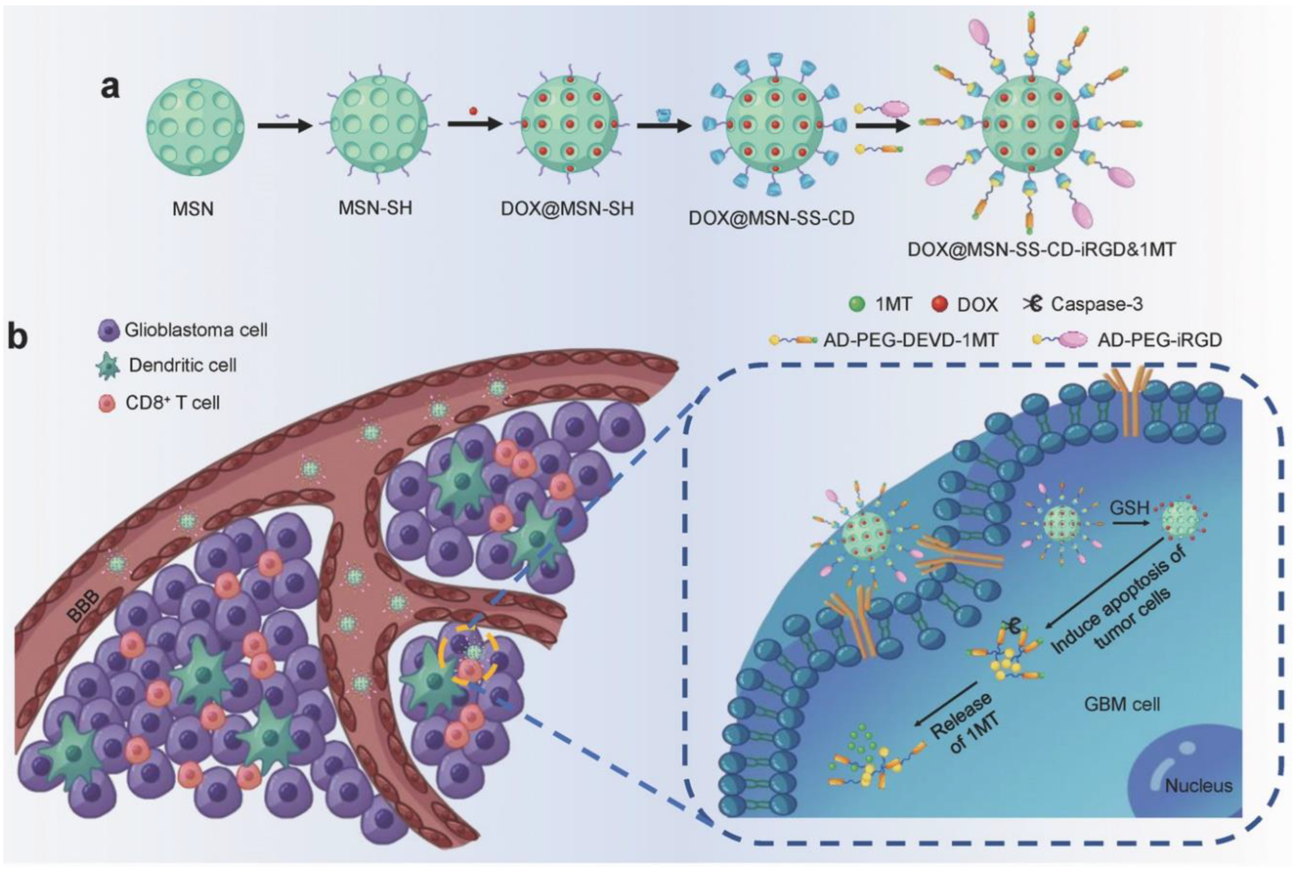
Immunotherapy is a rapidly developing antitumor strategy in recent years with high clinical application value [91]. The integrin-based targeting delivery system can transport the radiosensitizer GTe (L-glutathione-decorated triangle-shaped nanodrugs) to tumor cells, enhancing the X-ray sensitivity of tumor cells, inducing reactive oxygen species (ROS) to destroy mitochondria and promote cell apoptosis [92]. Compared with GTE-RGD + X-ray therapy, GTe-RGD + X-ray/anti-PD-1 has a more significant antitumor effect, suggesting that GTE-RGD + X-ray/anti-PD-1 can activate immune function and inhibit tumor growth. 1-methyl tryptophan (1MT) inhibits indoleamine 2, 3-dioxygenase (IDO) specifically [93]. Integrin-targeting DOX@MSN-SS-iRGD&1MT (iRGD-modified doxorubicin and 1MT co-loaded nanodrugs) were synthesized as previously reported [94]. In recent research, an integrin-facilitated lysosomal degradation molecular was designed by a cyclic RGD peptide connecting with PD-L1 [95]. This method allowed integrin-facilitated lysosomal degradation of PD-L1 by bifunctional compound to enhance antitumor immunotherapy. In other researches, the inhibition of integrin αvβ6 and αvβ3 also showed a remarkable antitumor effect in different tumors [96,97,98]. The results showed that iRGD-modified nanoparticles were enriched in tumor sites, inhibiting the tumor proliferation and also up-regulating the differentiation of cytotoxic CD8+ T cells, confirming that the integrin-targeting nanoparticles can activate the immune response against tumor (Figure 6).

3.4. Other Treatment Strategies

Photodynamic therapy (PDT) has attracted increasing attention due to its advantages of low damage to normal tissues, low invasion, economy, and little drug resistance [99,100]. However, the effect of PDT therapy is limited by the accumulation of photosensitizer (PS), poor tumor targeting, and hypoxic tumor microenvironment, so it is worth studying to improve PDT by improving photosensitizer accumulation in tumors [101,102]. Au-RGD-miR-320a nanoparticles targeting integrin αvβ3 were constructed for enhanced photosensitive therapy and gene-targeted therapy. This nanodrugs combined with laser irradiation can significantly inhibit the proliferation and metastasis of lung tumors and enhance the apoptosis of tumor cells, and can effectively prevent tumor metastasis. In addition to optimizing PS delivery, the combination of laser irradiation also improves drug release behavior of other antitumor drugs [103,104]. Laser irradiation can also be used for nanocarriers to improve oxygen content in tumor tissues, alleviating tumor hypoxia and slowing down tumor drug resistance. Avraham Dayan’s lab has developed an integrin-targeting nanophotodynamical drug [105]. By modifying TiO2 with the RGD group, a nanobiological complex is generated, and when combined with laser irradiation, can specifically bind to tumor cells, promoting sufficient drug release and cell death, thus, improving the therapeutic effect and reducing drug resistance (Figure 7). Combining photothermal therapy with integrin-targeting nanodrugs can not only reduce the damage to normal tissues but also improve the antitumor effect, making this a promising tumor treatment strategy [106].

4. The Nanoparticles Targeting Integrin αvβ3

Tumor-targeting therapy is the administration of antitumor drugs to tumor tissue to prevent tumor formation, migration, and invasion [107]. Compared with traditional chemotherapy and surgery, tumor-targeted therapy has a higher tumor suppressive effect with reduced side effects [108,109].

4.1. Protein-Based Nano-Delivery System

Albumin is currently used in clinical antitumor therapy due to its excellent physiological properties when combined with paclitaxel to form nanodrugs [110]. RGD polypeptide targeting integrin αvβ3 was modified with 131I-loaded bovine serum albumin (BSA) to construct RGD-BSA-polycaprolactone (131I-RGD-BSA-PCL) [111]. It was found that although both BSA-PCL with or without RGD could produce fluorescence after being co-cultured with cells, the tumor absorption rate of BSA-PCL with RGD NPs was higher than that of BSA-PCL without RGD NPs, indicating that integrin αvβ3 could be a molecular target in tumor diagnosis and treatment.

4.2. Polymers

Pegylated paclitaxel has been widely used in clinical antitumor therapy since it was approved by FDA [112]. This is mainly because polyethylene glycol (PEG) has low toxicity, high biocompatibility, and negligible immunogenicity [113,114]. Therefore, studies of various polymer micelles for nanodrugs delivery are still attracting a lot of attention. Amphiphilic nanopolymers, which contain both hydrophilic and hydrophobic chains, can self-assemble to form nanoparticles in an aqueous solution [115]. The hydrophilic part is located on the surface while the hydrophobic chain and antitumor drugs are wrapped in the inner side, allowing the antitumor drugs to reach the tumor tissue through the circulatory system [116,117]. To improve the target efficacy of the polymer, integrin αvβ3-based targeting nanomicelles were constructed for increased drug concentration and improved tumor death [118]. At the same dose of doxorubicin (DOX), the RGD-modified polymer micelles inhibited the growth of tumor cells more significantly than free doxorubicin [119]. More importantly, there was no obvious toxic side effect in the RGD-modified polymer micelles group in normal organs.

4.3. Inorganic Nanomaterials

In recent years, inorganic nanoparticles have attracted much attention in tumor imaging, photothermal therapy and radiosensitization therapy, including copper sulfide nanoparticles, magnetic nanoparticles, mesoporous silica nanoparticles, gold nanoparticles, and quantum dots [120,121,122]. Integrin-based strategies were implemented as an effective means of achieving active tumor targeting to further improve the antitumor effect of nanodrugs. Targeting molecules are usually modified with inorganic nanomaterials to enable nanodrugs to aggregate in tumor. For example, RGD-modified gold nanoparticles (125I-RGD@AuNPs) were constructed to promote the specific targeting of gold nanoparticles by integrin αvβ3 receptor [123]. In tumor tissues, γ rays were released from 125I-RGD@AuNPs to inhibit tumor cell growth. RGD-modified polyamide dendrimers coated with gold nanoparticles were also designed for in vivo imaging, improving the biocompatibility of gold nanoparticles [124]. The CT imaging effect of the nanomaterial is superior to that of the clinical contrast agent iodihydramol (Omnipaque). At low-dose X-ray, RGD-modified gold nanoparticles showed a higher signal-to-noise ratio and more obvious lung MR and CT imaging effects than iodihydramol. In addition, targeting nanoprobe cRGD-IONPs were constructed based on ultrafine magnetic iron oxide nanoparticles (IONPs). The 125I labeled cRGD-IONPs probe can be enriched in tumor sites by cRGD targeting with integrin αvβ3 receptor, showing excellent tumor imaging [125]. Quantum dots (QDs) are nanomaterials with unique fluorescence properties that have promising applications in tumor imaging and early detection [126,127]. The QDs-RGD probe coupled with RGD peptide showed an obvious tumor imaging effect in SW1990 pancreatic tumor model [128]. When QDs-RGD NPs were combined with gemcitabine, the tumor inhibition rate of the combined treatment group (QDs-RGD NPs+ laser irradiation + gemcitabine) was much higher than that of the gemcitabine group alone, suggesting that QDs-RGD NPs has a synergistic effect on inhibiting the proliferation of SW1990 pancreatic tumors.

4.4. Membrane

Recently, a new direction of research is decorating nanoparticles with cell membranes to prevent drugs from being captured and removed by the immune system [129]. Among them, red blood cells, stem cells, platelets, phagocytes, and tumor cells are effective natural carriers with excellent biocompatibility and a unique ability to evade immune system [130,131,132,133]. For example, nanoparticles modified by homologous hepatocellular carcinoma cell membranes or neutrophil membranes can improve nanodrugs retention in tumor sites and effectively inhibit tumor growth [134,135]. An RGD-decorated macrophage membrane was coated on the magnetic nanocluster to deliver siRNA. Such feature-packed magnetic nanodrugs enable us to gain success in high-performance siRNA delivery through superior stealth effect, magnetic resonance imaging, magnetic accumulation, RGD targeting, and favorable cytoplasm trafficking [136]. Similarly, RGD peptide-modified platelet membranes coated with melanin nanoparticles and doxorubicin (DOX) have a significant inhibition effect on the proliferation and metastasis of drug-resistant tumors [137].

5. Physical Barriers to Integrin-Targeting Nanodrugs

Integrins are overexpressed on the surface of tumor cells [52,54]. The aggressive growth of tumors requires a lot of nutrients to promote the formation of new blood vessels [138]. Endogenous αvβ3 integrin transmembrane glycoprotein, which is widely distributed in tumor neovascularization, is thought to be an angiogenesis marker [8,139]. Integrins and immunoglobulin superfamily cell adhesion molecules (IgCAM) and junction adhesion molecules (JAM) have a series of interactions, including leukocyte extravasation in the blood, immune monitoring in the gut, and hematopoietic stem cells homing and mobilization [140]. In addition, ECM ligands enhance interactions between pathogens and phagocytic immune cells, acting as phagocytic primers and inducers for neutrophil extracellular traps [141]. Integrins can also activate signal transduction pathways after binding to ECM, mediating cell signals in cell growth, survival, division, and migration [142]. Therefore, integrins were often used for specific ligands for targeting drug delivery (targeting tumor cells or ECM). Integrins can be also used to reduce the loss of nanodrugs and increase their half-life and bioavailability in vivo [143,144].

5.1. Opsonization

Opsonization is the molecular mechanism whereby molecules, microbes, or apoptotic cells are chemically modified to have stronger interactions with cell surface receptors on phagocytes and antibodies to identify invading particles. Integrin-targeting nanodrugs need to reduce opsonic as much as possible in vivo to avoid phagocytosis by macrophages [145]. Due to the surface properties, many serum proteins (also called opsonins) can be deposited on the surface of nanoparticles or can enter the nanoparticles to form dynamic protein crowns (the protein-rich layer formed around nanoparticles), which can be effectively recognized by the immune system and assimilated by macrophages [146,147]. Integrin modification can increase the targeting efficiency of nanodrugs and reduce the excessive adhesion of nanodrugs to cells, thus, enhancing the efficacy of nanodrugs [148].
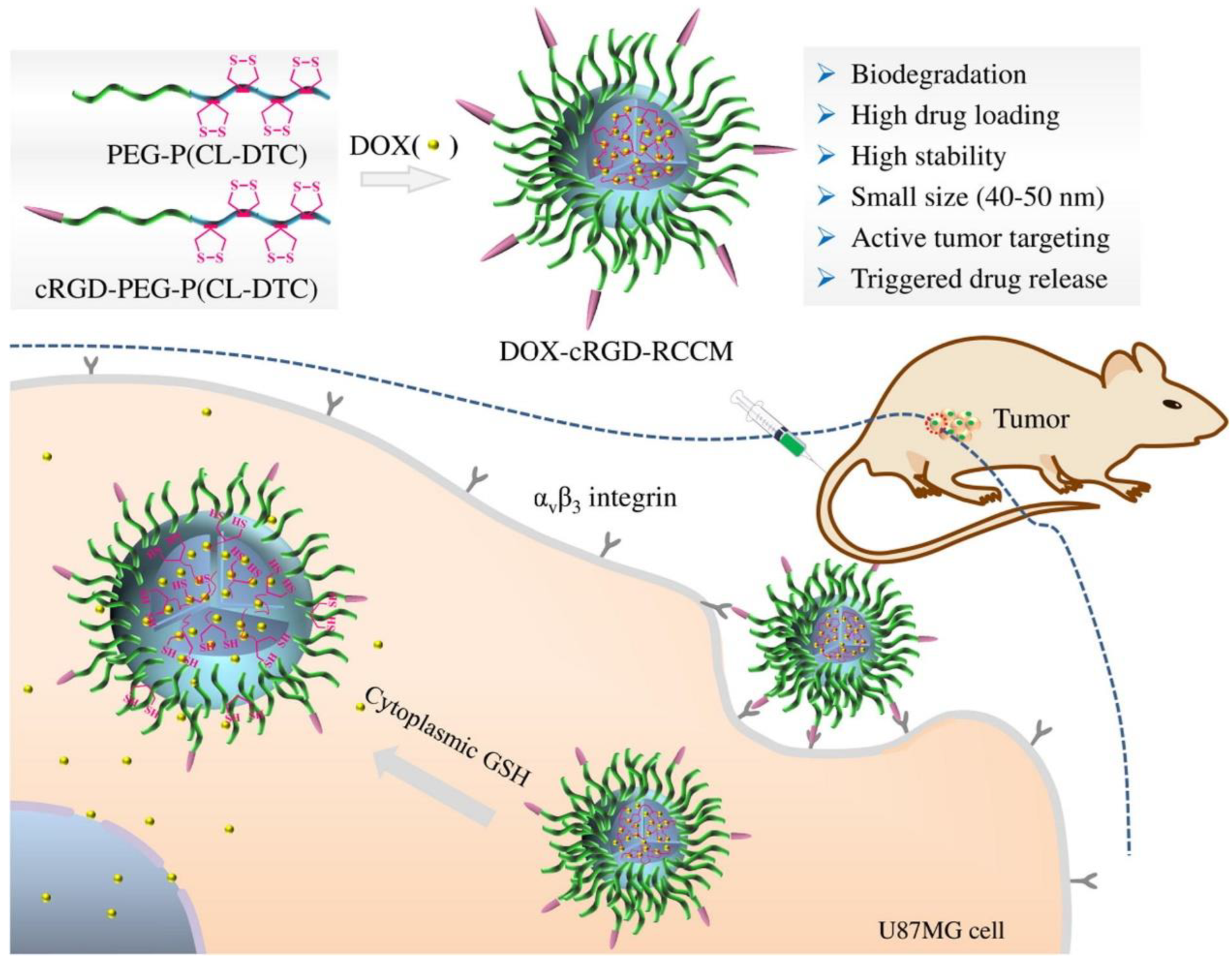
As is known to all, liposomes have higher drug delivery efficiency and wider drug delivery spectrum than small molecules and can achieve targeting drug delivery after being combined with targeting units [149,150,151]. The protein crown formed by liposomes and plasma proteins after entering blood circulation has a certain influence on the targeting function of liposomes [152,153]. Protein crowns of liposomes are rich in opsonins and coagulation proteins can activate immune cells and promote phagocytes to absorb nanoparticles through receptor-mediated phagocytosis [154,155] In Fang et al.’s study, PEG-PCL was cross-linked with RGD and coated with DOX to form DOX-cRGD-RCCMS nanoparticles, and the metabolic effects of the cross-linked nanodrugs showed an elimination half-life of 4.7 h, 11.7 times longer than the free DOX (0.4 h), and 3.9 times longer than the non-cross-linked DOX-cRGD-PEG-PCL (1.2 h), demonstrating the important role of RGD cross-linked nanomicelles in drug pharmacokinetics in vivo [156]. Therefore, RGD crosslinked nanodrugs can prolong their half-life in plasma and reduce their opsonance (Figure 8).

5.2. Penetration in Tumor

The combination of nanoparticles and targeted integrins increases the targeting efficiency of nanodrugs. However, the complex tumor microenvironment possesses unique structure and physiological functions, which not only hinders tumor penetration of chemotherapeutics but also becomes a physiological barrier to tumor penetration of integrin-targeting nanodrugs [157]. As a result, many antitumor drugs can only be enriched on the surface of tumor tissues rather than entering the deep tumor tissues and inhibiting tumor proliferation or metastasis [158]. iRGD is a tumor-targeting transmembrane peptide of circular RGD peptide linked by a disulfide bond, which can bind specifically to integrins and mediate the penetration of tumor cells [159]. The iRGD-heparin nanocarrier iRGD-Heparin (iHP) modified by iRGD has confirmed that iHP can target tumor cells for cisplatin and dye delivery, which can provide accurate diagnosis and treatment for gastric tumor and reduce the toxicity of drugs in normal tissues. To further demonstrate the effect of peptides on cell penetration, the uptake capacity of PEGylated PLGA nanoparticles modified by cRGD at different densities in cells was studied. Confocal microscopy showed that the penetration of nanoparticles into U87MG 3D multicellular spheres was enhanced with the increase incRGD density. The localization and penetration of these nanomaterials to lysosomes were also observed in 2D culture. In addition, the effects of the same incubation time and temperature on the penetrability of cRGD-modified polymer micelles (CCPM) preparations with different densities were compared. The degree of cell internalization increased with the increase in cRGD concentration. Under different culture conditions, CCPM uptake of 5% cRGD was observed to be significantly higher than 0% cRGD by immunofluorescence staining, which proved that integrins receptor-mediated CCPM uptake was enhanced with the increase in surface CRGD [160].

5.3. Endocytosis

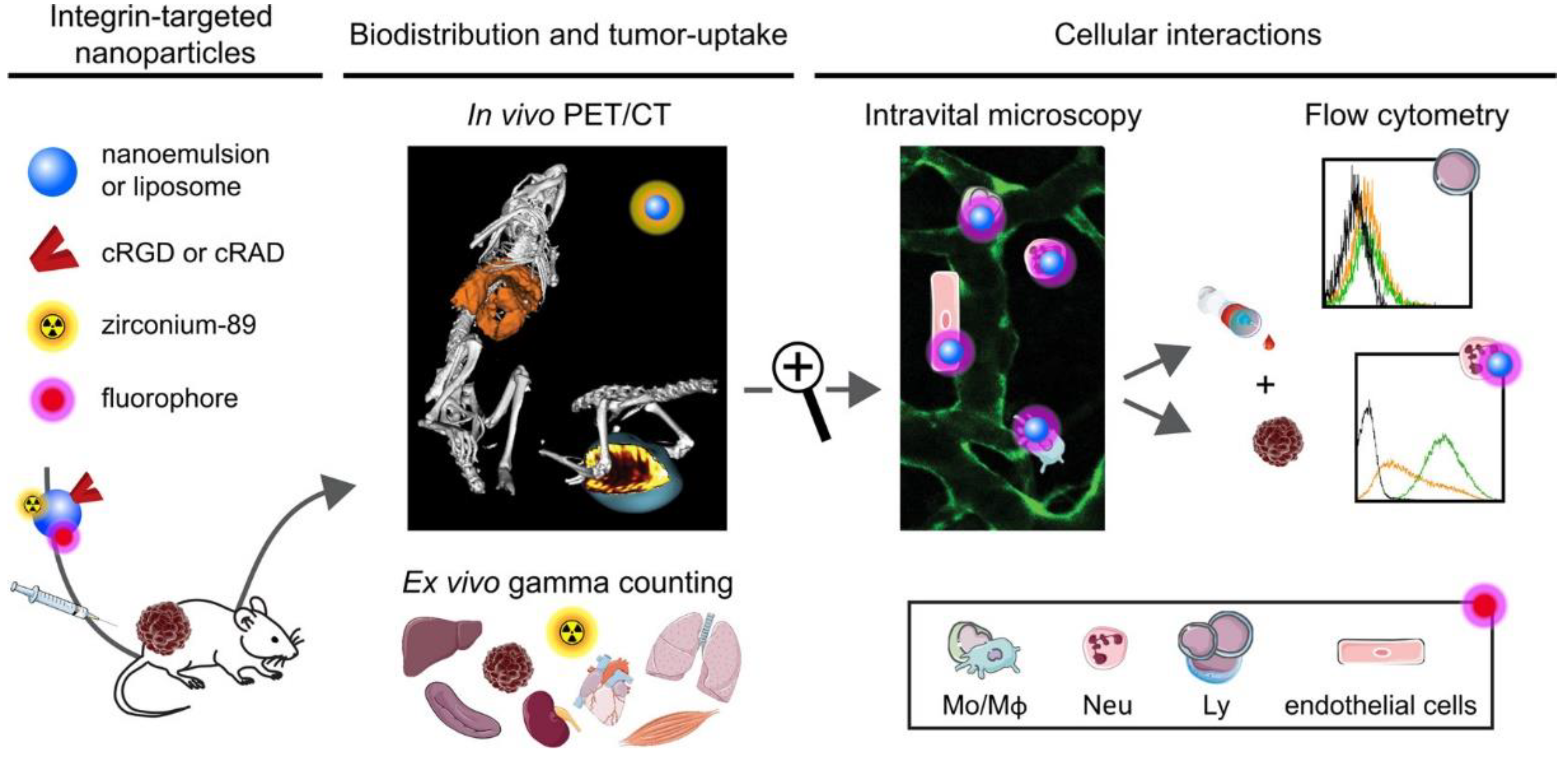
Various factors in vivo can interfere with nanodrugs, leading to poor drug targeting and difficulty in entering tumor tissues [158]. Thus, most of them enter normal tissues, resulting in the release of the tumor treatment drugs loaded in the nanomaterials into normal tissues, thereby increasing the toxic side effects on the body [59,161]. Nanoparticles wrapped in cell membranes can disguise as autologous cells to avoid elimination by the immune system [162]. For instance, natural neutrophil membranes wrapped on the surface of synthetic nanostructures constitute a potential biomimetic nanoparticle. cRGD and non-targeted control polypeptide cRAD were modified with nanoparticles to explore the mechanism of action of targeted drugs in vivo (Figure 9). The distribution and metabolic effects of cRAD-NPs and cRGD-NPs in mice were compared using living microscope and flow cytometry. The results confirmed that cRGD-NPs were transferred to the tumor using phagocyte hitchhiking, revealing that neutrophils are the main phagocytic contributor to this process. The results also revealed that neutrophils showed a clear preference for cRGD-NPs. Therefore, cRGD-NPs injected into the microbe were widely disseminated into tumor tissues by immune cells [163]. In another research, DOX was loaded in RGD-modified liposomes, the loaded liposomes with or without RGD modification and free DOX were tested for antitumor effect [164]. The results showed that DOX-loaded RGD-liposomes could effectively target and inhibit tumors and significantly improve the survival rate of tumor-bearing mice. At the same time, DOX-loaded RGD-liposomes can reduce the aggregation and release of drugs in normal tissues while abundantly accumulating in tumor site. Therefore, DOX-loaded RGD-liposomes can be used as a safe and effective antitumor drug delivery system to improve antitumor efficacy.

6. Conclusions and Perspectives

Integrin-mediated adhesion between cells and cells to the ECM is crucial for the physiological development and functioning of tissues, and targeting integrins has become an effective means to improve the efficacy of tumor therapy. In this review, the structure and function of integrins, the role of integrin αvβ3 in antitumor therapy, the nanoparticles targeting integrin αvβ3, as well as the physical barriers to integrin-targeting nanodrugs are well discussed. We systematically analyzed the advantages and enhancement strategies associated with integrin-targeting nanodrugs, hoping to lay the foundation for the potential future application of tumor diagnosis and treatment with nanodrugs therapies.
The investigation into the relationship between integrin and tumors remains ongoing, with increasing clarity regarding the mechanism of action of integrin in tumor therapy. With the discovery of new integrins or ligands, the role of integrins in tumor treatment will become even more important; other types of integrins, such as ανβ6, α4β1, α2β1, and α5β1 are also gaining interest. More importantly, the development of nanomedicine has led to the emergence of a variety of functional nanomedicines, and integrin is poised to become the preferred choice for targeted delivery due to its impact on tumors.
Tumor-targeting nanodrugs are an emerging area that has begun to show clinical success, and various nano-based diagnostic and therapeutic drugs are currently undergoing clinical trials (ClinicalTrials.gov). Despite the results to date, there are still many challenges and limitations that need to be resolved. Drug carriers should effectively deliver cargo directly to target cells rather than normal organs and tissues. In the meantime, the carriers should possess the ability of biocompatibility and biodegradability, avoiding extra toxicity. Moreover, these carriers must have long circulation to avoid premature drug release. Integrins possess the ability to specifically interact with target cells to enhance drug delivery efficiency and accuracy. However, integrins are also expressed in normal cells; thus, integrin-targeting methods have hidden dangers of off-target effect. This may cause severe side effects in clinical treatments. Overall, we believe that research efforts on integrin-targeting nanodrugs that are used for antitumor therapy will result in highly efficient, safe, and curative treatment modalities that meet the prerequisites of personalized medicine and achieve permanent cures without side effects.

Author Contributions

Writing—original draft preparation, C.Y.; Writing—review and editing, W.J.; Conceptualization, C.Y., D.L. and B.L.; Supervision, Y.H. and D.L.; Project administration, Y.H.; Funding acquisition, B.L. and W.J. All authors have read and agreed to the published version of the manuscript.

Funding

This work was supported in part by the Doctoral Research Start-up Fund of Guangxi University of Science and Technology (Xiaokebo21Z31), Guangxi Science and Technology Bases and Talents (AD21238014), the Liuzhou Science and Technology Plan Project (2022SB033) and the Natural Science Foundation of Jiangsu Province (Youth Foundation, BK20220784).

Data Availability Statement

In this review manuscript, no new data were created.

Conflicts of Interest

The authors declare no conflict of interest.

References

  1. Sung, H.; Ferlay, J.; Siegel, R.L.; Laversanne, M.; Soerjomataram, I.; Jemal, A.; Bray, F. Global Cancer Statistics 2020: GLOBOCAN Estimates of Incidence and Mortality Worldwide for 36 Cancers in 185 Countries. CA Cancer J. Clin. 2021, 71, 209–249. [Google Scholar] [CrossRef] [PubMed]
  2. Liu, M.; Yang, J.; Xu, B.; Zhang, X. Tumor Metastasis: Mechanistic Insights and Therapeutic Interventions. MedComm 2021, 2, 587–617. [Google Scholar] [CrossRef] [PubMed]
  3. Meirson, T.; Gil-Henn, H.; Samson, A.O. Invasion and Metastasis: The Elusive Hallmark of Cancer. Oncogene 2020, 39, 2024–2026. [Google Scholar] [CrossRef] [PubMed]
  4. Fares, J.; Fares, M.Y.; Khachfe, H.H.; Salhab, H.A.; Fares, Y. Molecular Principles of Metastasis: A Hallmark of Cancer Revisited. Sig. Transduct. Target. Ther. 2020, 5, 28. [Google Scholar] [CrossRef]
  5. Friedl, P.; Wolf, K. Tumour-Cell Invasion and Migration: Diversity and Escape Mechanisms. Nat. Rev. Cancer 2003, 3, 362–374. [Google Scholar] [CrossRef]
  6. Wittekind, C.; Neid, M. Cancer Invasion and Metastasis. Oncology 2005, 69 (Suppl. 1), 14–16. [Google Scholar] [CrossRef]
  7. Bachmann, M.; Kukkurainen, S.; Hytönen, V.P.; Wehrle-Haller, B. Cell Adhesion by Integrins. Physiol. Rev. 2019, 99, 1655–1699. [Google Scholar] [CrossRef]
  8. Mezu-Ndubuisi, O.J.; Maheshwari, A. The Role of Integrins in Inflammation and Angiogenesis. Pediatr. Res. 2021, 89, 1619–1626. [Google Scholar] [CrossRef]
  9. Hamidi, H.; Ivaska, J. Every Step of the Way: Integrins in Cancer Progression and Metastasis. Nat. Rev. Cancer 2018, 18, 533–548. [Google Scholar] [CrossRef]
  10. Danen, E.H.J. Integrins: An Overview of Structural and Functional Aspects; Landes Bioscience: Austin, TX, USA, 2013. [Google Scholar]
  11. Berman, A.E.; Kozlova, N.I. Integrins: Structure and Functions. Membr. Cell Biol. 2000, 13, 207–244. [Google Scholar]
  12. van der Flier, A.; Sonnenberg, A. Function and Interactions of Integrins. Cell Tissue Res. 2001, 305, 285–298. [Google Scholar] [CrossRef]
  13. Niu, G.; Chen, X. Why Integrin as a Primary Target for Imaging and Therapy. Theranostics 2011, 1, 30–47. [Google Scholar] [CrossRef]
  14. Takada, Y.; Ye, X.; Simon, S. The Integrins. Genome Biol. 2007, 8, 215. [Google Scholar] [CrossRef]
  15. Shi, J.; Wang, F.; Liu, S. Radiolabeled Cyclic RGD Peptides as Radiotracers for Tumor Imaging. Biophys. Rep. 2016, 2, 1–20. [Google Scholar] [CrossRef]
  16. Shimaoka, M.; Springer, T.A. Therapeutic Antagonists and Conformational Regulation of Integrin Function. Nat. Rev. Drug Discov. 2003, 2, 703–716. [Google Scholar] [CrossRef]
  17. Barczyk, M.; Carracedo, S.; Gullberg, D. Integrins. Cell Tissue Res. 2010, 339, 269–280. [Google Scholar] [CrossRef]
  18. Humphries, M.J.; Symonds, E.J.; Mould, A.P. Mapping Functional Residues onto Integrin Crystal Structures. Curr. Opin. Struct. Biol. 2003, 13, 236–243. [Google Scholar] [CrossRef]
  19. Arimori, T.; Miyazaki, N.; Mihara, E.; Takizawa, M.; Taniguchi, Y.; Cabañas, C.; Sekiguchi, K.; Takagi, J. Structural Mechanism of Laminin Recognition by Integrin. Nat. Commun. 2021, 12, 4012. [Google Scholar] [CrossRef]
  20. Pan, L.; Zhao, Y.; Yuan, Z.; Qin, G. Research Advances on Structure and Biological Functions of Integrins. SpringerPlus 2016, 5, 1094. [Google Scholar] [CrossRef]
  21. Kolasangiani, R.; Bidone, T.C.; Schwartz, M.A. Integrin Conformational Dynamics and Mechanotransduction. Cells 2022, 11, 3584. [Google Scholar] [CrossRef]
  22. Tong, D.; Soley, N.; Kolasangiani, R.; Schwartz, M.A.; Bidone, T.C. Integrin AIIbβ3 Intermediates: From Molecular Dynamics to Adhesion Assembly. Biophys. J. 2023, 122, 533–543. [Google Scholar] [CrossRef] [PubMed]
  23. Assa-Munt, N.; Jia, X.; Laakkonen, P.; Ruoslahti, E. Solution Structures and Integrin Binding Activities of an RGD Peptide with Two Isomers. Biochemistry 2001, 40, 2373–2378. [Google Scholar] [CrossRef] [PubMed]
  24. Hood, J.D.; Cheresh, D.A. Role of Integrins in Cell Invasion and Migration. Nat. Rev. Cancer 2002, 2, 91–100. [Google Scholar] [CrossRef] [PubMed]
  25. Vasconcelos, A.A.; Estrada, J.C.; David, V.; Wermelinger, L.S.; Almeida, F.C.L.; Zingali, R.B. Structure-Function Relationship of the Disintegrin Family: Sequence Signature and Integrin Interaction. Front. Mol. Biosci. 2021, 8, 1178. [Google Scholar] [CrossRef] [PubMed]
  26. Su, Y.; Iacob, R.E.; Li, J.; Engen, J.R.; Springer, T.A. Dynamics of Integrin α5β1, Fibronectin, and Their Complex Reveal Sites of Interaction and Conformational Change. J. Biol. Chem. 2022, 298, 102323. [Google Scholar] [CrossRef]
  27. Pang, X.; He, X.; Qiu, Z.; Zhang, H.; Xie, R.; Liu, Z.; Gu, Y.; Zhao, N.; Xiang, Q.; Cui, Y. Targeting Integrin Pathways: Mechanisms and Advances in Therapy. Sig. Transduct. Target. Ther. 2023, 8, 1. [Google Scholar] [CrossRef]
  28. Thomas Parsons, J.; Slack-Davis, J.K.; Tilghman, R.W.; Iwanicki, M.; Martin, K.H. Chapter 66—Integrin Signaling: Cell Migration, Proliferation, and Survival. In Handbook of Cell Signaling, 2nd ed.; Bradshaw, R.A., Dennis, E.A., Eds.; Academic Press: San Diego, CA, USA, 2010; pp. 491–499. ISBN 978-0-12-374145-5. [Google Scholar]
  29. Takagi, J.; Springer, T.A. Integrin Activation and Structural Rearrangement. Immunol. Rev. 2002, 186, 141–163. [Google Scholar] [CrossRef]
  30. Kim, M.; Carman, C.V.; Springer, T.A. Bidirectional Transmembrane Signaling by Cytoplasmic Domain Separation in Integrins. Science 2003, 301, 1720–1725. [Google Scholar] [CrossRef]
  31. Kanchanawong, P.; Calderwood, D.A. Organization, Dynamics and Mechanoregulation of Integrin-Mediated Cell-ECM Adhesions. Nat. Rev. Mol. Cell Biol. 2023, 24, 142–161. [Google Scholar] [CrossRef]
  32. Kulke, M.; Langel, W. Molecular Dynamics Simulations to the Bidirectional Adhesion Signaling Pathway of Integrin αVβ3. Proteins Struct. Funct. Bioinform. 2020, 88, 679–688. [Google Scholar] [CrossRef]
  33. Calderwood, D.A.; Campbell, I.D.; Critchley, D.R. Talins and Kindlins: Partners in Integrin-Mediated Adhesion. Nat. Rev. Mol. Cell Biol. 2013, 14, 503–517. [Google Scholar] [CrossRef]
  34. Bouti, P.; Webbers, S.D.S.; Fagerholm, S.C.; Alon, R.; Moser, M.; Matlung, H.L.; Kuijpers, T.W. β2 Integrin Signaling Cascade in Neutrophils: More Than a Single Function. Front. Immunol. 2021, 11, 619925. [Google Scholar] [CrossRef]
  35. Ren, D.; Zhao, J.; Sun, Y.; Li, D.; Meng, Z.; Wang, B.; Fan, P.; Liu, Z.; Jin, X.; Wu, H. Overexpressed ITGA2 Promotes Malignant Tumor Aggression by Up-Regulating PD-L1 Expression through the Activation of the STAT3 Signaling Pathway. J. Exp. Clin. Cancer Res. 2019, 38, 485. [Google Scholar] [CrossRef]
  36. Guo, P.; Moses-Gardner, A.; Huang, J.; Smith, E.R.; Moses, M.A. ITGA2 as a Potential Nanotherapeutic Target for Glioblastoma. Sci. Rep. 2019, 9, 6195. [Google Scholar] [CrossRef]
  37. Xiong, J.; Yan, L.; Zou, C.; Wang, K.; Chen, M.; Xu, B.; Zhou, Z.; Zhang, D. Integrins Regulate Stemness in Solid Tumor: An Emerging Therapeutic Target. J. Hematol. Oncol. 2021, 14, 177. [Google Scholar] [CrossRef]
  38. Rattanasinchai, C.; Navasumrit, P.; Ruchirawat, M. Elevated ITGA2 Expression Promotes Collagen Type I-Induced Clonogenic Growth of Intrahepatic Cholangiocarcinoma. Sci. Rep. 2022, 12, 22429. [Google Scholar] [CrossRef]
  39. Slack, R.J.; Macdonald, S.J.F.; Roper, J.A.; Jenkins, R.G.; Hatley, R.J.D. Emerging Therapeutic Opportunities for Integrin Inhibitors. Nat. Rev. Drug Discov. 2022, 21, 60–78. [Google Scholar] [CrossRef]
  40. Niu, J.; Li, Z. The Roles of Integrin Avβ6 in Cancer. Cancer Lett. 2017, 403, 128–137. [Google Scholar] [CrossRef]
  41. Wei, L.; Zhou, Q.; Tian, H.; Su, Y.; Fu, G.-H.; Sun, T. Integrin β3 Promotes Cardiomyocyte Proliferation and Attenuates Hypoxia-Induced Apoptosis via Regulating the PTEN/Akt/MTOR and ERK1/2 Pathways. Int. J. Biol. Sci. 2020, 16, 644–654. [Google Scholar] [CrossRef]
  42. Aksorn, N.; Chanvorachote, P. Integrin as a Molecular Target for Anti-Cancer Approaches in Lung Cancer. Anticancer Res. 2019, 39, 541–548. [Google Scholar] [CrossRef]
  43. Li, M.; Wang, Y.; Li, M.; Wu, X.; Setrerrahmane, S.; Xu, H. Integrins as Attractive Targets for Cancer Therapeutics. Acta. Pharm. Sin. B 2021, 11, 2726–2737. [Google Scholar] [CrossRef] [PubMed]
  44. Bergonzini, C.; Kroese, K.; Zweemer, A.J.M.; Danen, E.H.J. Targeting Integrins for Cancer Therapy—Disappointments and Opportunities. Front. Cell Dev. Biol. 2022, 10, 863850. [Google Scholar] [CrossRef] [PubMed]
  45. Henke, E.; Nandigama, R.; Ergün, S. Extracellular Matrix in the Tumor Microenvironment and Its Impact on Cancer Therapy. Front. Mol. Biosci. 2019, 6, 160. [Google Scholar] [CrossRef] [PubMed]
  46. Li, X.; Sun, X.; Kan, C.; Chen, B.; Qu, N.; Hou, N.; Liu, Y.; Han, F. COL1A1: A Novel Oncogenic Gene and Therapeutic Target in Malignancies. Pathol. Res. Pract. 2022, 236, 154013. [Google Scholar] [CrossRef] [PubMed]
  47. Baltes, F.; Pfeifer, V.; Silbermann, K.; Caspers, J.; Wantoch von Rekowski, K.; Schlesinger, M.; Bendas, G. β1-Integrin Binding to Collagen Type 1 Transmits Breast Cancer Cells into Chemoresistance by Activating ABC Efflux Transporters. Biochim. Biophys. Acta. Mol. Cell Res. 2020, 1867, 118663. [Google Scholar] [CrossRef]
  48. Reis, J.S.D.; Santos, M.A.R.d.C.; da Costa, K.M.; Freire-de-Lima, C.G.; Morrot, A.; Previato, J.O.; Previato, L.M.; da Fonseca, L.M.; Freire-de-Lima, L. Increased Expression of the Pathological O-Glycosylated Form of Oncofetal Fibronectin in the Multidrug Resistance Phenotype of Cancer Cells. Matrix Biol. 2023, 118, 47–68. [Google Scholar] [CrossRef]
  49. Majidpoor, J.; Mortezaee, K. Steps in Metastasis: An Updated Review. Med. Oncol. 2021, 38, 3. [Google Scholar] [CrossRef]
  50. Zhang, Y.; Qian, J.; Gu, C.; Yang, Y. Alternative Splicing and Cancer: A Systematic Review. Signal. Transduct. Target. Ther. 2021, 6, 78. [Google Scholar] [CrossRef]
  51. Hanahan, D.; Weinberg, R.A. Hallmarks of Cancer: The next Generation. Cell 2011, 144, 646–674. [Google Scholar] [CrossRef]
  52. Valdembri, D.; Serini, G. The Roles of Integrins in Cancer. Fac. Rev. 2021, 10, 45. [Google Scholar] [CrossRef]
  53. Moschos, S.J.; Drogowski, L.M.; Reppert, S.L.; Kirkwood, J.M. Integrins and Cancer. Oncology 2007, 21, 13–20. [Google Scholar]
  54. Desgrosellier, J.S.; Cheresh, D.A. Integrins in Cancer: Biological Implications and Therapeutic Opportunities. Nat. Rev. Cancer 2010, 10, 9–22. [Google Scholar] [CrossRef]
  55. Cordes, N.; Park, C.C. Beta1 Integrin as a Molecular Therapeutic Target. Int. J. Radiat. Biol. 2007, 83, 753–760. [Google Scholar] [CrossRef]
  56. Blandin, A.-F.; Renner, G.; Lehmann, M.; Lelong-Rebel, I.; Martin, S.; Dontenwill, M. β1 Integrins as Therapeutic Targets to Disrupt Hallmarks of Cancer. Front. Pharmacol. 2015, 6, 279. [Google Scholar] [CrossRef]
  57. Hou, J.; Yan, D.; Liu, Y.; Huang, P.; Cui, H. The Roles of Integrin α5β1 in Human Cancer. Onco Targets Ther. 2020, 13, 13329–13344. [Google Scholar] [CrossRef]
  58. Fang, Z.; Yao, W.; Fu, Y.; Wang, L.-Y.; Li, Z.; Yang, Y.; Shi, Y.; Qiu, S.; Fan, J.; Zha, X. Increased Integrin A5β1 Heterodimer Formation and Reduced C-Jun Expression Are Involved in Integrin β1 Overexpression-Mediated Cell Growth Arrest. J. Cell. Biochem. 2010, 109, 383–395. [Google Scholar] [CrossRef]
  59. Jeevanandam, J.; Chan, Y.S.; Danquah, M.K. Nano-Formulations of Drugs: Recent Developments, Impact and Challenges. Biochimie 2016, 128–129, 99–112. [Google Scholar] [CrossRef]
  60. Zhang, Y.; Chan, H.F.; Leong, K.W. Advanced Materials and Processing for Drug Delivery: The Past and the Future. Adv. Drug Deliv. Rev. 2013, 65, 104–120. [Google Scholar] [CrossRef]
  61. Tibbitt, M.W.; Dahlman, J.E.; Langer, R. Emerging Frontiers in Drug Delivery. J. Am. Chem. Soc. 2016, 138, 704–717. [Google Scholar] [CrossRef]
  62. Danhier, F.; Le Breton, A.; Preat, V. RGD-Based Strategies To Target Alpha(v) Beta(3) Integrin in Cancer Therapy and Diagnosis. Mol. Pharm. 2012, 9, 2961–2973. [Google Scholar] [CrossRef]
  63. Xiao, Y.; Yu, D. Tumor Microenvironment as a Therapeutic Target in Cancer. Pharmacol. Ther. 2021, 221, 107753. [Google Scholar] [CrossRef] [PubMed]
  64. Senthebane, D.A.; Rowe, A.; Thomford, N.E.; Shipanga, H.; Munro, D.; Al Mazeedi, M.A.M.; Almazyadi, H.A.M.; Kallmeyer, K.; Dandara, C.; Pepper, M.S.; et al. The Role of Tumor Microenvironment in Chemoresistance: To Survive, Keep Your Enemies Closer. Int. J. Mol. Sci. 2017, 18, 1586. [Google Scholar] [CrossRef] [PubMed]
  65. Hui, L.; Chen, Y. Tumor Microenvironment: Sanctuary of the Devil. Cancer Lett. 2015, 368, 7–13. [Google Scholar] [CrossRef] [PubMed]
  66. Wilhelm, S.; Tavares, A.J.; Dai, Q.; Ohta, S.; Audet, J.; Dvorak, H.F.; Chan, W.C.W. Analysis of Nanoparticle Delivery to Tumours. Nat. Rev. Mater. 2016, 1, 16014. [Google Scholar] [CrossRef]
  67. Sindhwani, S.; Syed, A.M.; Ngai, J.; Kingston, B.R.; Maiorino, L.; Rothschild, J.; MacMillan, P.; Zhang, Y.; Rajesh, N.U.; Hoang, T.; et al. The Entry of Nanoparticles into Solid Tumours. Nat. Mater. 2020, 19, 566–575. [Google Scholar] [CrossRef]
  68. Peng, J.Q.; Fumoto, S.; Suga, T.; Miyamoto, H.; Kuroda, N.; Kawakami, S.; Nishida, K. Targeted Co-Delivery of Protein and Drug to a Tumor in Vivo by Sophisticated RGD-Modified Lipid-Calcium Carbonate Nanoparticles. J. Control. Release 2019, 302, 42–53. [Google Scholar] [CrossRef]
  69. Filipczak, N.; Joshi, U.; Attia, S.A.; Berger Fridman, I.; Cohen, S.; Konry, T.; Torchilin, V. Hypoxia-Sensitive Drug Delivery to Tumors. J. Control. Release 2022, 341, 431–442. [Google Scholar] [CrossRef]
  70. Li, Y.; Hong, W.; Zhang, H.; Zhang, T.T.; Chen, Z.; Yuan, S.; Peng, P.; Xiao, M.; Xu, L. Photothermally Triggered Cytosolic Drug Delivery of Glucose Functionalized Polydopamine Nanoparticles in Response to Tumor Microenvironment for the GLUT1-Targeting Chemo-Phototherapy. J. Control. Release 2020, 317, 232–245. [Google Scholar] [CrossRef]
  71. Mansur, A.A.P.; Carvalho, S.M.; Lobato, Z.I.P.; Leite, M.D.F.; Cunha, A.d.S.; Mansur, H.S. Design and Development of Polysaccharide-Doxorubicin-Peptide Bioconjugates for Dual Synergistic Effects of Integrin-Targeted and Cell-Penetrating Peptides for Cancer Chemotherapy. Bioconjugate Chem. 2018, 29, 1973–2000. [Google Scholar] [CrossRef]
  72. Ahmad, K.; Lee, E.J.; Shaikh, S.; Kumar, A.; Rao, K.M.; Park, S.-Y.; Jin, J.O.; Han, S.S.; Choi, I. Targeting Integrins for Cancer Management Using Nanotherapeutic Approaches: Recent Advances and Challenges. Semin. Cancer Biol. 2021, 69, 325–336. [Google Scholar] [CrossRef]
  73. Saifi, M.A.; Sathish, G.; Bazaz, M.R.; Godugu, C. Exploration of Tumor Penetrating Peptide IRGD as a Potential Strategy to Enhance Tumor Penetration of Cancer Nanotherapeutics. Biochim. Et Biophys. Acta (BBA)-Rev. Cancer 2023, 1878, 188895. [Google Scholar] [CrossRef]
  74. Zhong, Y.; Su, T.; Shi, Q.; Feng, Y.; Tao, Z.; Huang, Q.; Li, L.; Hu, L.; Li, S.; Tan, H.; et al. Co-Administration of IRGD Enhances Tumor-Targeted Delivery and Anti-Tumor Effects of Paclitaxel-Loaded PLGA Nanoparticles for Colorectal Cancer Treatment. Int. J. Nanomed. 2019, 14, 8543–8560. [Google Scholar] [CrossRef]
  75. Liang, G.; Jin, X.; Zhang, S.; Xing, D. RGD Peptide-Modified Fluorescent Gold Nanoclusters as Highly Efficient Tumor-Targeted Radiotherapy Sensitizers. Biomaterials 2017, 144, 95–104. [Google Scholar] [CrossRef]
  76. Ding, Y.; Xiao, X.; Zeng, L.; Shang, Q.; Jiang, W.; Xiong, S.; Duan, X.; Shen, J.; Wang, R.; Guo, J.; et al. Platinum-Crosslinking Polymeric Nanoparticle for Synergetic Chemoradiotherapy of Nasopharyngeal Carcinoma. Bioact. Mater. 2021, 6, 4707–4716. [Google Scholar] [CrossRef]
  77. Liu, Y.; Li, L.; Liu, J.; Yang, M.; Wang, H.; Chu, X.; Zhou, J.; Huo, M.; Yin, T. Biomineralization-Inspired Dasatinib Nanodrug with Sequential Infiltration for Effective Solid Tumor Treatment. Biomaterials 2021, 267, 120481. [Google Scholar] [CrossRef]
  78. Izci, M.; Maksoudian, C.; Manshian, B.B.; Soenen, S.J. The Use of Alternative Strategies for Enhanced Nanoparticle Delivery to Solid Tumors. Chem. Rev. 2021, 121, 1746–1803. [Google Scholar] [CrossRef]
  79. De Ruysscher, D.; Niedermann, G.; Burnet, N.G.; Siva, S.; Lee, A.W.M.; Hegi-Johnson, F. Radiotherapy Toxicity. Nat. Rev. Dis. Primers 2019, 5, 13. [Google Scholar] [CrossRef]
  80. Hallahan, D.E.; Qu, S.; Geng, L.; Cmelak, A.; Chakravarthy, A.; Martin, W.; Scarfone, C.; Giorgio, T. Radiation-Mediated Control of Drug Delivery. Am. J. Clin. Oncol. 2001, 24, 473–480. [Google Scholar] [CrossRef]
  81. Chen, Y.; Yang, J.; Fu, S.; Wu, J. Gold Nanoparticles as Radiosensitizers in Cancer Radiotherapy. Int. J. Nanomed. 2020, 15, 9407–9430. [Google Scholar] [CrossRef]
  82. Cui, L.; Her, S.; Borst, G.R.; Bristow, R.G.; Jaffray, D.A.; Allen, C. Radiosensitization by Gold Nanoparticles: Will They Ever Make It to the Clinic? Radiother. Oncol. 2017, 124, 344–356. [Google Scholar] [CrossRef]
  83. Yang, Y.; Zhang, L.; Cai, J.; Li, X.; Cheng, D.; Su, H.; Zhang, J.; Liu, S.; Shi, H.; Zhang, Y.; et al. Tumor Angiogenesis Targeted Radiosensitization Therapy Using Gold Nanoprobes Guided by MRI/SPECT Imaging. ACS Appl. Mater. Interfaces 2016, 8, 1718–1732. [Google Scholar] [CrossRef] [PubMed]
  84. Fan, D.; Wang, K.; Gao, H.; Luo, Q.; Wang, X.; Li, X.; Tong, W.; Zhang, X.; Luo, C.; Yang, G.; et al. A 64 Cu-Porphyrin-Based Dual-Modal Molecular Probe with Integrin Av Β3 Targeting Function for Tumour Imaging. J. Labelled. Comp. Radiopharm. 2020, 63, 212–221. [Google Scholar] [CrossRef] [PubMed]
  85. Xiao, L.; Xin, J. Advances in Clinical Oncology Research on 99mTc-3PRGD2 SPECT Imaging. Front. Oncol. 2022, 12, 898764. [Google Scholar] [CrossRef] [PubMed]
  86. Zhang, C.; Xu, C.; Gao, X.; Yao, Q. Platinum-Based Drugs for Cancer Therapy and Anti-Tumor Strategies. Theranostics 2022, 12, 2115–2132. [Google Scholar] [CrossRef]
  87. Goldsmith, S.J. Targeted Radionuclide Therapy: A Historical and Personal Review. Semin. Nucl. Med. 2020, 50, 87–97. [Google Scholar] [CrossRef]
  88. Niu, G.; Chen, X. When Radionuclides Meet Nanoparticles. Nat. Nanotech 2018, 13, 359–360. [Google Scholar] [CrossRef]
  89. Dash, A.; Russ Knapp, F.F.; Pillai, M.R.A. Targeted Radionuclide Therapy—An Overview. Curr. Radiopharm. 2013, 6, 152–180. [Google Scholar] [CrossRef]
  90. Chen, H.; Zhao, L.; Fu, K.; Lin, Q.; Wen, X.; Jacobson, O.; Sun, L.; Wu, H.; Zhang, X.; Guo, Z.; et al. Integrin αvβ3-Targeted Radionuclide Therapy Combined with Immune Checkpoint Blockade Immunotherapy Synergistically Enhances Anti-Tumor Efficacy. Theranostics 2019, 9, 7948–7960. [Google Scholar] [CrossRef]
  91. Waldman, A.D.; Fritz, J.M.; Lenardo, M.J. A Guide to Cancer Immunotherapy: From T Cell Basic Science to Clinical Practice. Nat. Rev. Immunol. 2020, 20, 651–668. [Google Scholar] [CrossRef]
  92. Huang, W.; He, L.; Ouyang, J.; Chen, Q.; Liu, C.; Tao, W.; Chen, T. Triangle-Shaped Tellurium Nanostars Potentiate Radiotherapy by Boosting Checkpoint Blockade Immunotherapy. Matter 2020, 3, 1725–1753. [Google Scholar] [CrossRef]
  93. Yuasa, H.J.; Ball, H.J.; Austin, C.J.D.; Hunt, N.H. 1-L-Methyltryptophan Is a More Effective Inhibitor of Vertebrate IDO2 Enzymes than 1-D-Methyltryptophan. Comp. Biochem. Physiol. B Biochem. Mol. Biol. 2010, 157, 10–15. [Google Scholar] [CrossRef]
  94. Kuang, J.; Song, W.; Yin, J.; Zeng, X.; Han, S.; Zhao, Y.-P.; Tao, J.; Liu, C.-J.; He, X.-H.; Zhang, X.-Z. iRGD Modified Chemo-Immunotherapeutic Nanoparticles for Enhanced Immunotherapy against Glioblastoma. Adv. Funct. Mater. 2018, 28, 1800025. [Google Scholar] [CrossRef]
  95. Zheng, J.; He, W.; Li, J.; Feng, X.; Li, Y.; Cheng, B.; Zhou, Y.; Li, M.; Liu, K.; Shao, X.; et al. Bifunctional Compounds as Molecular Degraders for Integrin-Facilitated Targeted Protein Degradation. J. Am. Chem. Soc. 2022, 144, 21831–21836. [Google Scholar] [CrossRef]
  96. Busenhart, P.; Montalban-Arques, A.; Katkeviciute, E.; Morsy, Y.; Van Passen, C.; Hering, L.; Atrott, K.; Lang, S.; Garzon, J.F.G.; Naschberger, E.; et al. Inhibition of Integrin αvβ6 Sparks T-Cell Antitumor Response and Enhances Immune Checkpoint Blockade Therapy in Colorectal Cancer. J. Immunother. Cancer 2022, 10, e003465. [Google Scholar] [CrossRef]
  97. Zhou, Y.; Song, S.; Yuan, B.; Wu, Y.; Gao, Y.; Wan, G.; Li, G. A Novel CTLA-4 Affinity Peptide for Cancer Immunotherapy by Increasing the Integrin Avβ3 Targeting. Discov. Oncol. 2022, 13, 99. [Google Scholar] [CrossRef]
  98. Pan, X.; Yi, M.; Liu, C.; Jin, Y.; Liu, B.; Hu, G.; Yuan, X. Cilengitide, an αvβ3-Integrin Inhibitor, Enhances the Efficacy of Anti-Programmed Cell Death-1 Therapy in a Murine Melanoma Model. Bioengineered 2022, 13, 4557–4572. [Google Scholar] [CrossRef]
  99. Dolmans, D.E.; Fukumura, D.; Jain, R.K. Photodynamic Therapy for Cancer. Nat. Rev. Cancer 2003, 3, 380–387. [Google Scholar] [CrossRef]
  100. Correia, J.H.; Rodrigues, J.A.; Pimenta, S.; Dong, T.; Yang, Z. Photodynamic Therapy Review: Principles, Photosensitizers, Applications, and Future Directions. Pharmaceutics 2021, 13, 1332. [Google Scholar] [CrossRef]
  101. Yu, M.; Cao, R.; Ma, Z.; Zhu, M. Development of “Smart” Drug Delivery Systems for Chemo/PDT Synergistic Treatment. J. Mater. Chem. B 2023, 11, 1416–1433. [Google Scholar] [CrossRef]
  102. Calixto, G.M.F.; Bernegossi, J.; De Freitas, L.M.; Fontana, C.R.; Chorilli, M. Nanotechnology-Based Drug Delivery Systems for Photodynamic Therapy of Cancer: A Review. Molecules 2016, 21, 342. [Google Scholar] [CrossRef]
  103. Peng, J.; Wang, R.; Sun, W.; Huang, M.; Wang, R.; Li, Y.; Wang, P.; Sun, G.; Xie, S. Delivery of MiR-320a-3p by Gold Nanoparticles Combined with Photothermal Therapy for Directly Targeting Sp1 in Lung Cancer. Biomater. Sci. 2021, 9, 6528–6541. [Google Scholar] [CrossRef] [PubMed]
  104. Zou, J.; Zhu, J.; Yang, Z.; Li, L.; Fan, W.; He, L.; Tang, W.; Deng, L.; Mu, J.; Ma, Y.; et al. A Phototheranostic Strategy to Continuously Deliver Singlet Oxygen in the Dark and Hypoxic Tumor Microenvironment. Angew. Chem. Int. Ed. Engl. 2020, 59, 8833–8838. [Google Scholar] [CrossRef] [PubMed]
  105. Dayan, A.; Fleminger, G.; Ashur-Fabian, O. RGD-Modified Dihydrolipoamide Dehydrogenase Conjugated to Titanium Dioxide Nanoparticles—Switchable Integrin-Targeted Photodynamic Treatment of Melanoma Cells. RSC Adv. 2018, 8, 9112–9119. [Google Scholar] [CrossRef] [PubMed]
  106. Li, Y.; Hu, P.; Wang, X.; Hou, X.; Liu, F.; Jiang, X. Integrin αvβ3-Targeted Polydopamine-Coated Gold Nanostars for Photothermal Ablation Therapy of Hepatocellular Carcinoma. Regen. Biomater. 2021, 8, rbab046. [Google Scholar] [CrossRef]
  107. Vasir, J.K.; Labhasetwar, V. Targeted Drug Delivery in Cancer Therapy. Technol. Cancer Res. Treat. 2005, 4, 363–374. [Google Scholar] [CrossRef]
  108. Chen, B.; Dai, W.; He, B.; Zhang, H.; Wang, X.; Wang, Y.; Zhang, Q. Current Multistage Drug Delivery Systems Based on the Tumor Microenvironment. Theranostics 2017, 7, 538–558. [Google Scholar] [CrossRef]
  109. Dai, L.; Liu, J.; Luo, Z.; Li, M.; Cai, K. Tumor Therapy: Targeted Drug Delivery Systems. J. Mater. Chem. B 2016, 4, 6758–6772. [Google Scholar] [CrossRef]
  110. Elzoghby, A.O.; Samy, W.M.; Elgindy, N.A. Albumin-Based Nanoparticles as Potential Controlled Release Drug Delivery Systems. J. Control. Release 2012, 157, 168–182. [Google Scholar] [CrossRef]
  111. Ming, H.; Fang, L.; Gao, J.; Li, C.; Ji, Y.; Shen, Y.; Hu, Y.; Li, N.; Chang, J.; Li, W.; et al. Antitumor Effect of Nanoparticle 131I-Labeled Arginine-Glycine-Aspartate-Bovine Serum Albumin-Polycaprolactone in Lung Cancer. AJR Am. J. Roentgenol. 2017, 208, 1116–1126. [Google Scholar] [CrossRef]
  112. Mu, J.; Zhong, H.; Zou, H.; Liu, T.; Yu, N.; Zhang, X.; Xu, Z.; Chen, Z.; Guo, S. Acid-Sensitive PEGylated Paclitaxel Prodrug Nanoparticles for Cancer Therapy: Effect of PEG Length on Antitumor Efficacy. J. Control. Release 2020, 326, 265–275. [Google Scholar] [CrossRef]
  113. Knop, K.; Hoogenboom, R.; Fischer, D.; Schubert, U.S. Poly(Ethylene Glycol) in Drug Delivery: Pros and Cons as Well as Potential Alternatives. Angew. Chem. Int. Ed. Engl. 2010, 49, 6288–6308. [Google Scholar] [CrossRef]
  114. D’souza, A.A.; Shegokar, R. Polyethylene Glycol (PEG): A Versatile Polymer for Pharmaceutical Applications. Expert Opin. Drug Deliv. 2016, 13, 1257–1275. [Google Scholar] [CrossRef]
  115. Huo, M.; Wang, H.; Zhang, Y.; Cai, H.; Zhang, P.; Li, L.; Zhou, J.; Yin, T. Co-Delivery of Silybin and Paclitaxel by Dextran-Based Nanoparticles for Effective Anti-Tumor Treatment through Chemotherapy Sensitization and Microenvironment Modulation. J. Control. Release 2020, 321, 198–210. [Google Scholar] [CrossRef]
  116. Shi, Q.; Tong, Y.; Zheng, Y.; Liu, Y.; Yin, T. PDT-Sensitized ROS-Responsive Dextran Nanosystem for Maximizing Antitumor Potency of Multi-Target Drugs. Int. J. Pharm. 2023, 633, 122567. [Google Scholar] [CrossRef]
  117. Du, Q.; Lv, F.; Huang, J.; Tang, X.; Zhao, Z.; Chen, J. A Multiple Environment-Sensitive Prodrug Nanomicelle Strategy Based on Chitosan Graftomer for Enhanced Tumor Therapy of Gambogic Acid. Carbohydr. Polym. 2021, 267, 118229. [Google Scholar] [CrossRef]
  118. Raj, A.; Saraf, P.; Javali, N.M.; Li, X.; Jasti, B. Binding and Uptake of Novel RGD Micelles to the αvβ3 Integrin Receptor for Targeted Drug Delivery. J. Drug Target. 2014, 22, 518–527. [Google Scholar] [CrossRef]
  119. Xu, W.; Yan, X.; Liu, N.; Wu, G. P1c Peptide Decorated Liposome Targeting αvβ3-Expressing Tumor Cells In Vitro and In Vivo. RSC Adv. 2018, 8, 25575–25583. [Google Scholar] [CrossRef]
  120. Paul, W.; Sharma, C.P. 8—Inorganic Nanoparticles for Targeted Drug Delivery. In Biointegration of Medical Implant Materials; Sharma, C.P., Ed.; Woodhead Publishing Series in Biomaterials; Woodhead Publishing: Sawston, UK, 2010; pp. 204–235. ISBN 978-1-84569-509-5. [Google Scholar]
  121. Liu, Q.; Kim, Y.-J.; Im, G.-B.; Zhu, J.; Wu, Y.; Liu, Y.; Bhang, S.H. Inorganic Nanoparticles Applied as Functional Therapeutics. Adv. Funct. Mater. 2021, 31, 2008171. [Google Scholar] [CrossRef]
  122. Bayda, S.; Hadla, M.; Palazzolo, S.; Riello, P.; Corona, G.; Toffoli, G.; Rizzolio, F. Inorganic Nanoparticles for Cancer Therapy: A Transition from Lab to Clinic. Curr. Med. Chem. 2018, 25, 4269–4303. [Google Scholar] [CrossRef]
  123. Kim, Y.-H.; Jeon, J.; Hong, S.H.; Rhim, W.-K.; Lee, Y.-S.; Youn, H.; Chung, J.-K.; Lee, M.C.; Lee, D.S.; Kang, K.W.; et al. Tumor Targeting and Imaging Using Cyclic RGD-PEGylated Gold Nanoparticle Probes with Directly Conjugated Iodine-125. Small 2011, 7, 2052–2060. [Google Scholar] [CrossRef]
  124. Cheng, Y.; Ji, Y. RGD-Modified Polymer and Liposome Nanovehicles: Recent Research Progress for Drug Delivery in Cancer Therapeutics. Eur. J. Pharm. Sci. 2019, 128, 8–17. [Google Scholar] [CrossRef] [PubMed]
  125. Su, N.; Dang, Y.; Liang, G.; Liu, G. Iodine-125-Labeled CRGD-Gold Nanoparticles as Tumor-Targeted Radiosensitizer and Imaging Agent. Nanoscale Res. Lett. 2015, 10, 160. [Google Scholar] [CrossRef] [PubMed]
  126. Fang, M.; Chen, M.; Liu, L.; Li, Y. Applications of Quantum Dots in Cancer Detection and Diagnosis: A Review. J. Biomed. Nanotechnol. 2017, 13, 1–16. [Google Scholar] [CrossRef] [PubMed]
  127. Abbas, M.; Kurian, P. Quantum Probes in Cancer Research. Nat. Rev. Cancer 2022, 22, 378–379. [Google Scholar] [CrossRef]
  128. Li, S.; Yang, J.; Lei, X.; Zhang, J.; Yang, H.; Li, K.; Xu, C. Peptide-Conjugated Quantum Dots Act as the Target Marker for Human Pancreatic Carcinoma Cells. Cell. Physiol. Biochem. 2016, 38, 1121–1128. [Google Scholar] [CrossRef]
  129. Narain, A.; Asawa, S.; Chhabria, V.; Patil-Sen, Y. Cell Membrane Coated Nanoparticles: Next-Generation Therapeutics. Nanomedicine 2017, 12, 2677–2692. [Google Scholar] [CrossRef]
  130. Xiang, W.; Liu, X.; Zhang, L.; Liu, C.; Liu, G. Cell Membrane-Encapsulated Nanoparticles for Vaccines and Immunotherapy. Particuology 2022, 64, 35–42. [Google Scholar] [CrossRef]
  131. Zou, S.; Wang, B.; Wang, C.; Wang, Q.; Zhang, L. Cell Membrane-Coated Nanoparticles: Research Advances. Nanomedicine 2020, 15, 625–641. [Google Scholar] [CrossRef]
  132. Han, X.; Shen, S.; Fan, Q.; Chen, G.; Archibong, E.; Dotti, G.; Liu, Z.; Gu, Z.; Wang, C. Red Blood Cell–Derived Nanoerythrosome for Antigen Delivery with Enhanced Cancer Immunotherapy. Sci. Adv. 2019, 5, eaaw6870. [Google Scholar] [CrossRef]
  133. Li, Z.; Cai, H.; Li, Z.; Ren, L.; Ma, X.; Zhu, H.; Gong, Q.; Zhang, H.; Gu, Z.; Luo, K. A Tumor Cell Membrane-Coated Self-Amplified Nanosystem as a Nanovaccine to Boost the Therapeutic Effect of Anti-PD-L1 Antibody. Bioact. Mater. 2023, 21, 299–312. [Google Scholar] [CrossRef]
  134. Ji, B.; Cai, H.; Yang, Y.; Peng, F.; Song, M.; Sun, K.; Yan, F.; Liu, Y. Hybrid Membrane Camouflaged Copper Sulfide Nanoparticles for Photothermal-Chemotherapy of Hepatocellular Carcinoma. Acta Biomater. 2020, 111, 363–372. [Google Scholar] [CrossRef]
  135. Subhan, M.A.; Torchilin, V.P. Neutrophils as an Emerging Therapeutic Target and Tool for Cancer Therapy. Life Sci. 2021, 285, 119952. [Google Scholar] [CrossRef]
  136. Zhang, F.; Zhao, L.; Wang, S.; Yang, J.; Lu, G.; Luo, N.; Gao, X.; Ma, G.; Xie, H.-Y.; Wei, W. Construction of a Biomimetic Magnetosome and Its Application as a SiRNA Carrier for High-Performance Anticancer Therapy. Adv. Funct. Mater. 2018, 28, 1703326. [Google Scholar] [CrossRef]
  137. Jing, L.; Qu, H.; Wu, D.; Zhu, C.; Yang, Y.; Jin, X.; Zheng, J.; Shi, X.; Yan, X.; Wang, Y. Platelet-Camouflaged Nanococktail: Simultaneous Inhibition of Drug-Resistant Tumor Growth and Metastasis via a Cancer Cells and Tumor Vasculature Dual-Targeting Strategy. Theranostics 2018, 8, 2683–2695. [Google Scholar] [CrossRef]
  138. Lugano, R.; Ramachandran, M.; Dimberg, A. Tumor Angiogenesis: Causes, Consequences, Challenges and Opportunities. Cell. Mol. Life Sci. 2020, 77, 1745–1770. [Google Scholar] [CrossRef]
  139. Hodivala-Dilke, K. Avβ3 Integrin and Angiogenesis: A Moody Integrin in a Changing Environment. Curr. Opin. Cell Biol. 2008, 20, 514–519. [Google Scholar] [CrossRef]
  140. Harjunpää, H.; Llort Asens, M.; Guenther, C.; Fagerholm, S.C. Cell Adhesion Molecules and Their Roles and Regulation in the Immune and Tumor Microenvironment. Front. Immunol. 2019, 10, 1078. [Google Scholar] [CrossRef]
  141. LaFoya, B.; Munroe, J.A.; Miyamoto, A.; Detweiler, M.A.; Crow, J.J.; Gazdik, T.; Albig, A.R. Beyond the Matrix: The Many Non-ECM Ligands for Integrins. Int. J. Mol. Sci. 2018, 19, 449. [Google Scholar] [CrossRef]
  142. Kadry, Y.A.; Calderwood, D.A. Chapter 22: Structural and Signaling Functions of Integrins. Biochim. Et Biophys. Acta (BBA)-Biomembr. 2020, 1862, 183206. [Google Scholar] [CrossRef]
  143. Schnittert, J.; Bansal, R.; Storm, G.; Prakash, J. Integrins in Wound Healing, Fibrosis and Tumor Stroma: High Potential Targets for Therapeutics and Drug Delivery. Adv. Drug Deliv. Rev. 2018, 129, 37–53. [Google Scholar] [CrossRef]
  144. Arosio, D.; Casagrande, C. Advancement in Integrin Facilitated Drug Delivery. Adv. Drug Deliv. Rev. 2016, 97, 111–143. [Google Scholar] [CrossRef] [PubMed]
  145. Moghimi, S.M.; Szebeni, J. Stealth Liposomes and Long Circulating Nanoparticles: Critical Issues in Pharmacokinetics, Opsonization and Protein-Binding Properties. Prog. Lipid Res. 2003, 42, 463–478. [Google Scholar] [CrossRef] [PubMed]
  146. Li, Y.; Wang, G.; Griffin, L.; Banda, N.K.; Saba, L.M.; Groman, E.V.; Scheinman, R.; Moghimi, S.M.; Simberg, D. Complement Opsonization of Nanoparticles: Differences between Humans and Preclinical Species. J. Control. Release 2021, 338, 548–556. [Google Scholar] [CrossRef] [PubMed]
  147. Sosale, N.G.; Spinler, K.R.; Alvey, C.; Discher, D.E. Macrophage Engulfment of a Cell or Nanoparticle Is Regulated by Unavoidable Opsonization, a Species-Specific ‘Marker of Self’ CD47, and Target Physical Properties. Curr. Opin. Immunol. 2015, 35, 107–112. [Google Scholar] [CrossRef]
  148. Cox, N.; Kintzing, J.R.; Smith, M.; Grant, G.A.; Cochran, J.R. Integrin-Targeting Knottin Peptide-Drug Conjugates Are Potent Inhibitors of Tumor Cell Proliferation. Angew. Chem.-Int. Edit. 2016, 55, 9894–9897. [Google Scholar] [CrossRef]
  149. Wang, S.; Chen, Y.; Guo, J.; Huang, Q. Liposomes for Tumor Targeted Therapy: A Review. Int. J. Mol. Sci. 2023, 24, 2643. [Google Scholar] [CrossRef]
  150. Guimaraes, D.; Cavaco-Paulo, A.; Nogueira, E. Design of Liposomes as Drug Delivery System for Therapeutic Applications. Int. J. Pharm. 2021, 601, 120571. [Google Scholar] [CrossRef]
  151. Large, D.E.; Abdelmessih, R.G.; Fink, E.A.; Auguste, D.T. Liposome Composition in Drug Delivery Design, Synthesis, Characterization, and Clinical Application. Adv. Drug Deliv. Rev. 2021, 176, 113851. [Google Scholar] [CrossRef]
  152. Capriotti, A.L.; Cavaliere, C.; Piovesana, S. Liposome Protein Corona Characterization as a New Approach in Nanomedicine. Anal. Bioanal. Chem. 2019, 411, 4313–4326. [Google Scholar] [CrossRef]
  153. Onishchenko, N.; Tretiakova, D.; Vodovozova, E. Spotlight on the Protein Corona of Liposomes. Acta Biomater. 2021, 134, 57–78. [Google Scholar] [CrossRef]
  154. Foteini, P.; Pippa, N.; Naziris, N.; Demetzos, C. Physicochemical Study of the Protein-Liposome Interactions: Influence of Liposome Composition and Concentration on Protein Binding. J. Liposome Res. 2019, 29, 313–321. [Google Scholar] [CrossRef]
  155. Giulimondi, F.; Digiacomo, L.; Pozzi, D.; Palchetti, S.; Vulpis, E.; Capriotti, A.L.; Chiozzi, R.Z.; Laganà, A.; Amenitsch, H.; Masuelli, L.; et al. Interplay of Protein Corona and Immune Cells Controls Blood Residency of Liposomes. Nat. Commun. 2019, 10, 3686. [Google Scholar] [CrossRef]
  156. Fang, Y.; Jiang, Y.; Zou, Y.; Meng, F.; Zhang, J.; Deng, C.; Sun, H.; Zhong, Z. Targeted Glioma Chemotherapy by Cyclic RGD Peptide-Functionalized Reversibly Core-Crosslinked Multifunctional Poly(Ethylene Glycol)-b-Poly(ε-Caprolactone) Micelles. Acta Biomater. 2017, 50, 396–406. [Google Scholar] [CrossRef]
  157. Anderson, N.M.; Simon, M.C. The Tumor Microenvironment. Curr. Biol. 2020, 30, R921–R925. [Google Scholar] [CrossRef]
  158. Liu, Y.; Zhou, J.; Li, Q.; Li, L.; Jia, Y.; Geng, F.; Zhou, J.; Yin, T. Tumor Microenvironment Remodeling-Based Penetration Strategies to Amplify Nanodrug Accessibility to Tumor Parenchyma. Adv. Drug. Deliv. Rev. 2021, 172, 80–103. [Google Scholar] [CrossRef]
  159. Kang, S.; Lee, S.; Park, S. IRGD Peptide as a Tumor-Penetrating Enhancer for Tumor-Targeted Drug Delivery. Polymers 2020, 12, 1906. [Google Scholar] [CrossRef]
  160. Rios De La Rosa, J.M.; Spadea, A.; Donno, R.; Lallana, E.; Lu, Y.; Puri, S.; Caswell, P.; Lawrence, M.J.; Ashford, M.; Tirelli, N. Microfluidic-Assisted Preparation of RGD-Decorated Nanoparticles: Exploring Integrin-Facilitated Uptake in Cancer Cell Lines. Sci. Rep. 2020, 10, 14505. [Google Scholar] [CrossRef]
  161. Bao, J.; Zhang, Q.; Duan, T.; Hu, R.; Tang, J. The Fate of Nanoparticles In Vivo and the Strategy of Designing Stealth Nanoparticle for Drug Delivery. Curr. Drug. Targets 2021, 22, 922–946. [Google Scholar] [CrossRef]
  162. Glassman, P.M.; Hood, E.D.; Ferguson, L.T.; Zhao, Z.; Siegel, D.L.; Mitragotri, S.; Brenner, J.S.; Muzykantov, V.R. Red Blood Cells: The Metamorphosis of a Neglected Carrier into the Natural Mothership for Artificial Nanocarriers. Adv. Drug Deliv. Rev. 2021, 178, 113992. [Google Scholar] [CrossRef]
  163. Sofias, A.M.; Toner, Y.C.; Meerwaldt, A.E.; van Leent, M.M.T.; Soultanidis, G.; Elschot, M.; Gonai, H.; Grendstad, K.; Flobak, Å.; Neckmann, U.; et al. Tumor Targeting by Avβ3-Integrin-Specific Lipid Nanoparticles Occurs via Phagocyte Hitchhiking. ACS Nano 2020, 14, 7832–7846. [Google Scholar] [CrossRef]
  164. Ren, Y.; Yuan, B.; Hou, S.; Sui, Y.; Yang, T.; Lv, M.; Zhou, Y.; Yu, H.; Li, S.; Peng, H.; et al. Delivery of RGD-Modified Liposome as a Targeted Colorectal Carcinoma Therapy and Its Autophagy Mechanism. J. Drug Target. 2021, 29, 863–874. [Google Scholar] [CrossRef] [PubMed]
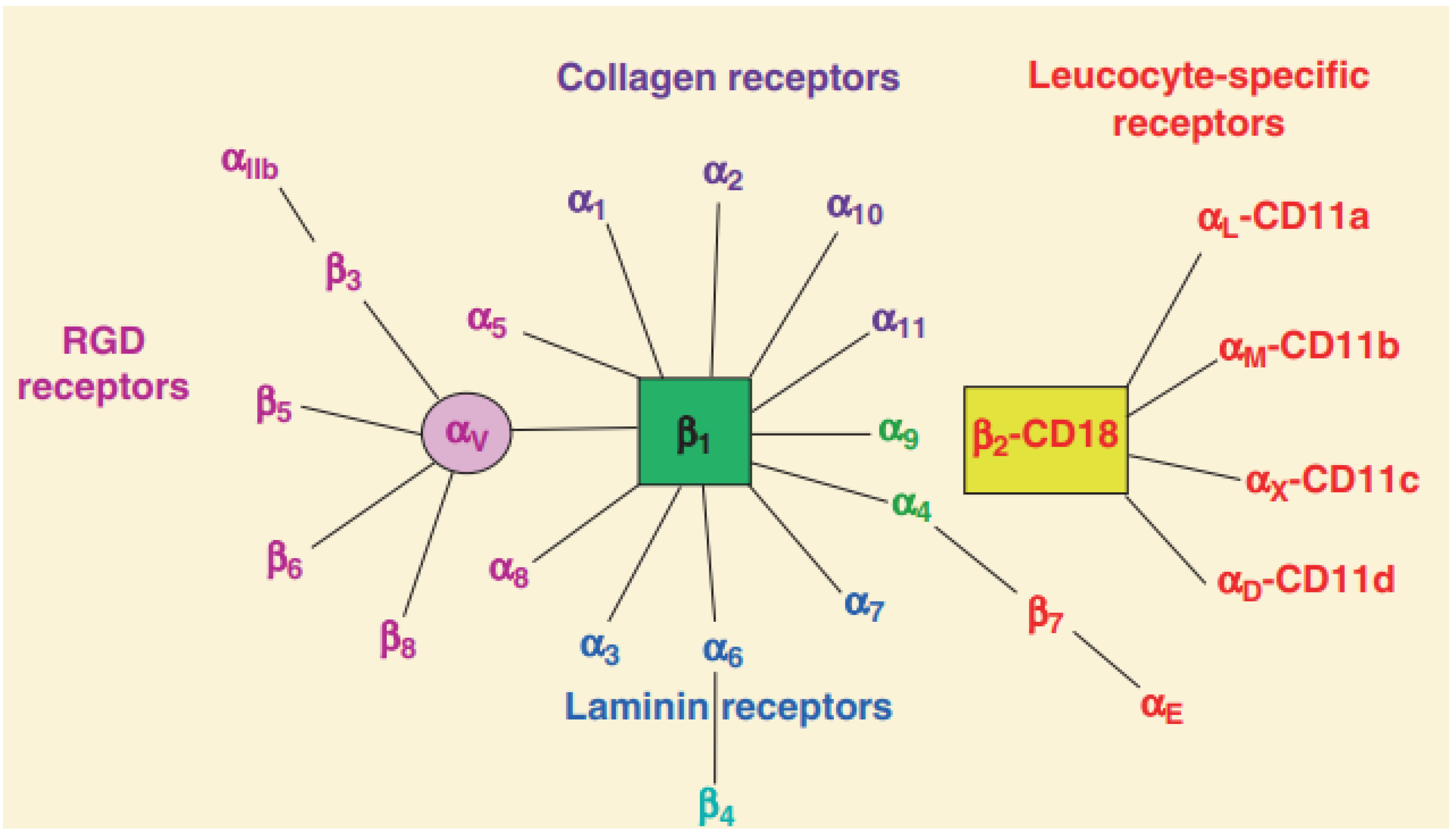
Figure 1. Classification of integrins family [8]. Integrin heterodimers were composed of different combinations of α and β subunits. In terms of ligand specificity, integrins can be divided into four groups: collagen-binding integrins (α1β1, α2β1, α10β1, and α11β1), RGD-recognizing integrins (α5β1, αVβ1, αVβ3, αVβ5, αVβ6, αVβ8, and αIIbβ3), laminin-binding integrins (α3β1, α6β1, α7β1, and α6β4), and leukocyte integrins (αLβ2, αMβ2, αXβ2, and αDβ2). The β2 integrin subunit (CD18) can pair with one of the four α subunits (αL-CD11a, αM-CD11b, αX-CD11c, and αD-CD11d), forming different leukocyte function-associated antigens.
Nanomaterials 13 01721 g001
Figure 2. (A) The structure and activation pathways of integrins [8]. Integrins in the bent resting conformation reveal low-affinity binding to their ligands. After being activated, integrins were able to have a high affinity with their ligands. (B) Schematic illustration of “Outside-in” and “Inside-out” integrins signaling pathways [42]. Integrins can be activated from two directions, from the inside by the regulated binding of proteins to the cytoplasmic tails, and from the outside by multivalent ligand binding. For “Outside-in” signaling, signals received by other receptors foster the binding of talin and kindlin to the cytoplasmic end of the integrin β subunit, at sites of actin polymerization. As for “Outside-in” signaling, ligand binding to the external domain causes conformational changes that increase ligand affinity, modify protein-interaction sites in the cytoplasmic domains and thence the resulting signals.
Nanomaterials 13 01721 g002
Figure 3. (A) Structure diagram of RGD sequence. (B) Ball-and-stick model of RGD sequence.
Nanomaterials 13 01721 g003
Figure 4. Schematic illustration of iRGD-PLGA-PTX nanomicelles used in antitumor research [74]. When the co-administered nanodrugs and iRGD peptide reach the tumor tissue, iRGD binds to the integrin receptor and is proteolytically cleaved. The exposed tissue-penetrating motif CendR interacts with the neuropilin-1 receptor and triggers nanodrugs penetration into tumor tissues. Ultimately, paclitaxel nanodrugs induce tumor cell cycle arrest and sequential apoptosis.
Nanomaterials 13 01721 g004
Figure 5. Platinum-based nanoparticles (PtNPs) were functionalized with RGD ligands to selectively target tumor cells and enhance DNA fragmentation [76]. Taking advantage of the targeting performance of RGD, the RPNs can effectively bind to the RGD receptor distributed in the tumor cell membrane to improve drug delivery efficiency and enhance synergistic chemoradiotherapy.
Nanomaterials 13 01721 g005
Figure 6. Integrin-targeting DOX@MSN-SS-iRGD&1MT nanoparticles were synthesized to upregulate cytotoxic CD8+ T cells for tumor immunotherapy therapy [94]. (a) Schematic synthesis of DOX@MSN-SS-iRGD&1MT. (b) Schematic illustration of DOX@MSN-SS-iRGD&1MT elicited antitumor immunity against glioblastoma. The introduction of iRGD significantly improves the accumulation efficiency of DOX@MSN-SS-iRGD&1MT into tumors and the cytotoxic CD8+ T cell recruitment in tumors.
Nanomaterials 13 01721 g006
Figure 7. TiO2 nanoparticles were modified with RGD groups to promote tumor cell uptake of photosensitizer [105]. Resultantly, the PDT antitumor effect was enhanced with reduced drug resistance.
Nanomaterials 13 01721 g007
Figure 8. RGD cross-linked nanomicelles showed a long circulation time and efficient glioma accumulation and retention [156].
Nanomaterials 13 01721 g008
Figure 9. cRGD and non-targeted control polypeptide cRAD were modified with nanoparticles to explore the mechanism of action of targeted drugs in vivo [163]. αvβ3-integrin-targeting nanodrugs were delivered to tumor site via phagocyte hitchhiking.
Nanomaterials 13 01721 g009
Disclaimer/Publisher’s Note: The statements, opinions and data contained in all publications are solely those of the individual author(s) and contributor(s) and not of MDPI and/or the editor(s). MDPI and/or the editor(s) disclaim responsibility for any injury to people or property resulting from any ideas, methods, instructions or products referred to in the content.