
Magnetic Iron Oxide Nanoparticles Coated by Coumarin-Bound Copolymer for Enhanced Magneto- and Photothermal Heating and Luminescent Thermometry
 , , , and
Abstract
:1. Introduction
2. Materials and Methods
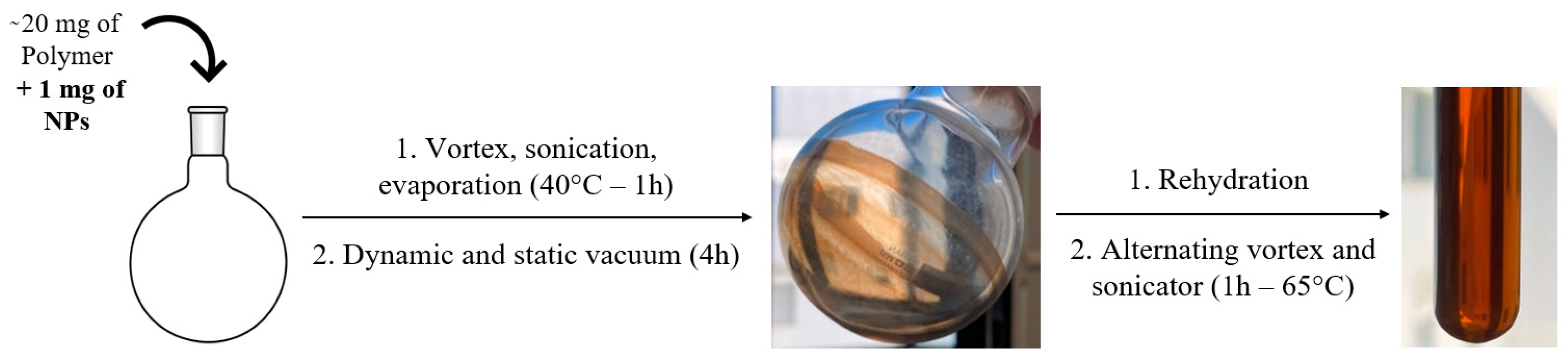
2.1. Syntheses
2.2. Physical Methods
2.3. Simulations and Fitting
3. Results and Discussion
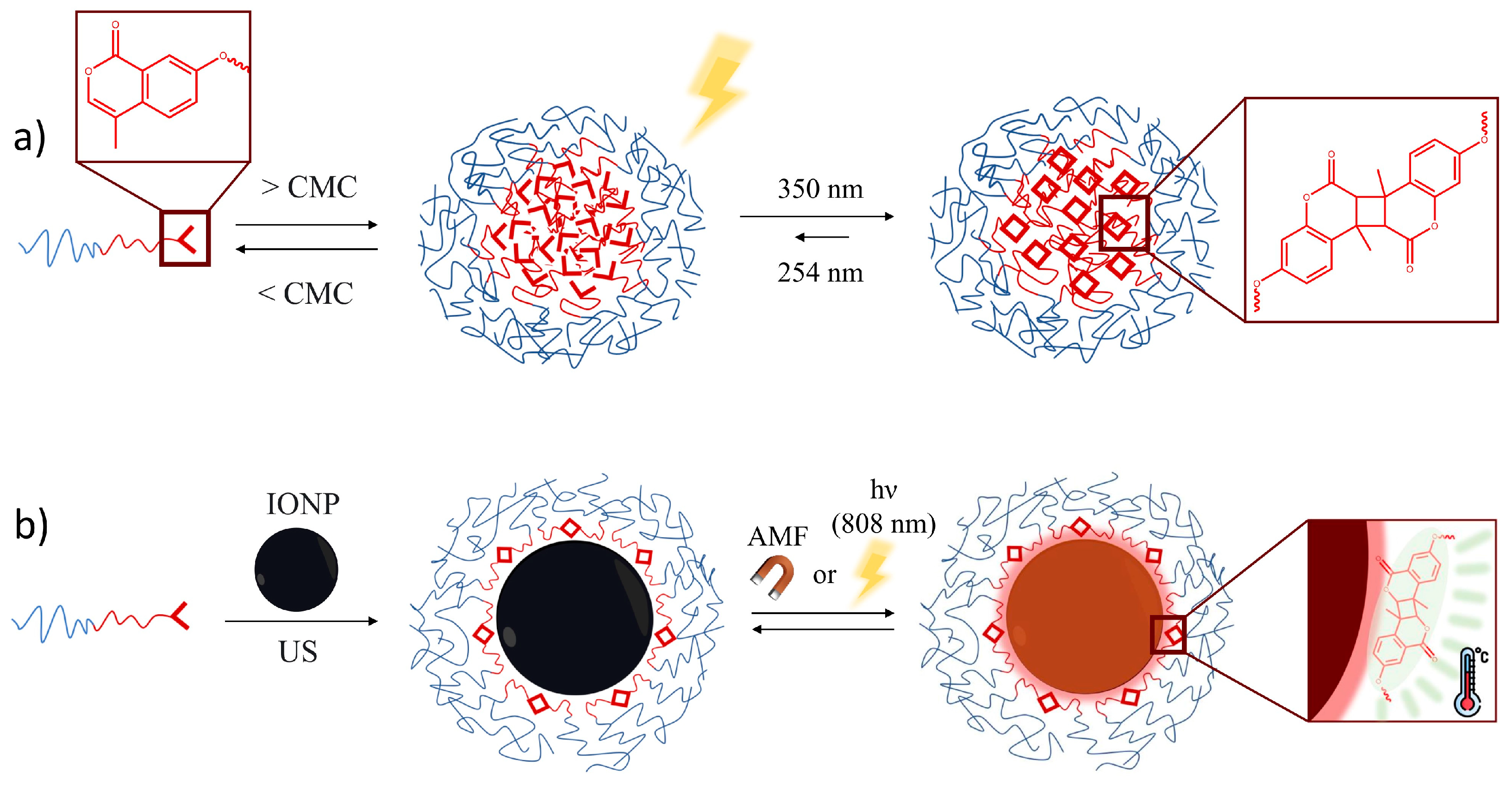
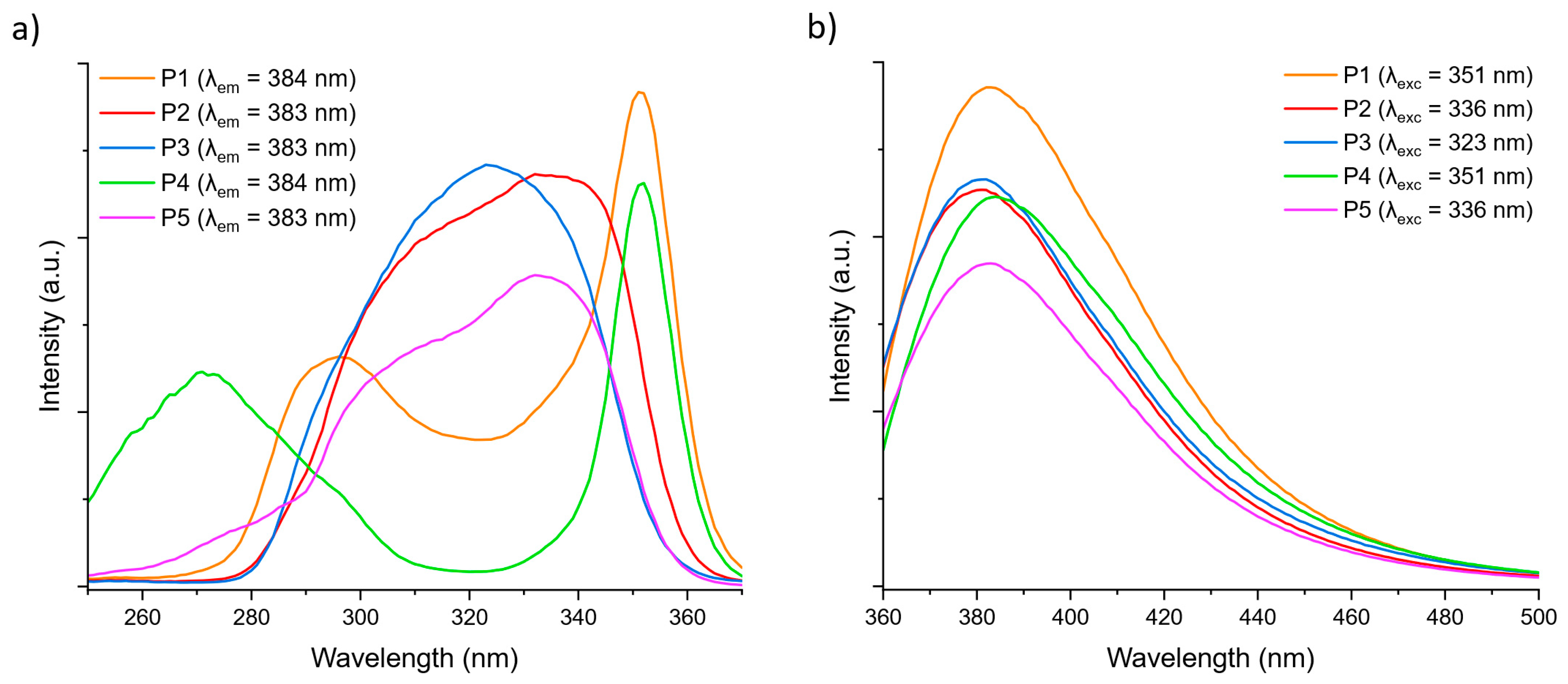
3.1. Amphiphilic Polymeric Micelles as Luminescent Thermometers
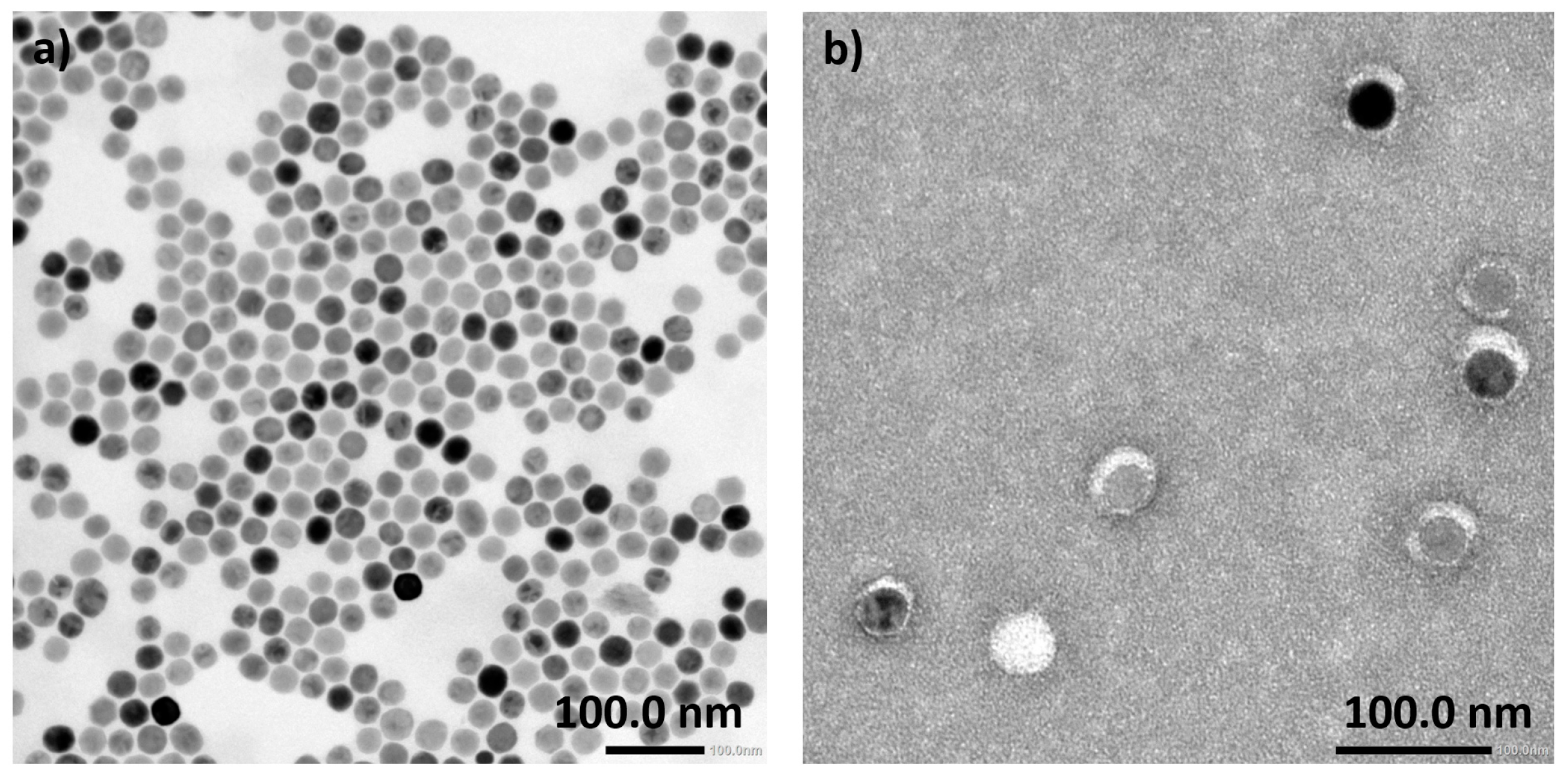
3.2. IONP Coating with Amphiphilic Bloc Copolymers
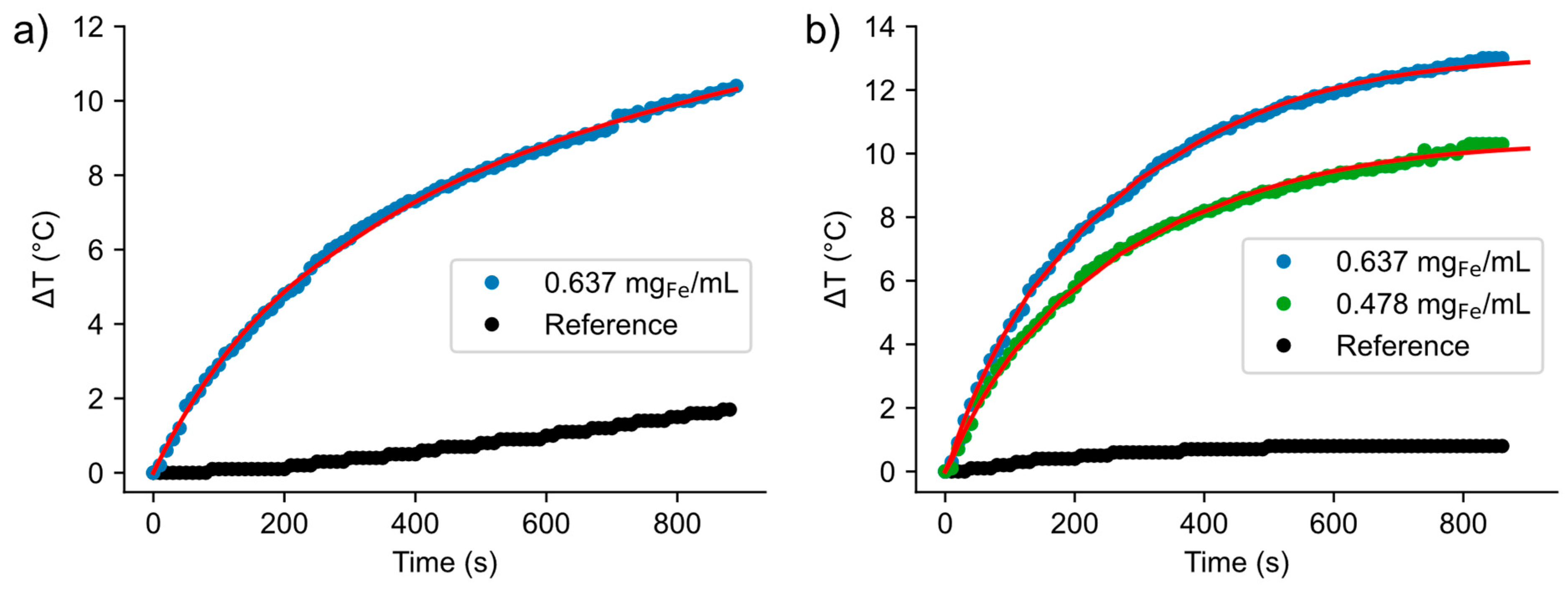
3.3. Magneto- and Photothermal Heating
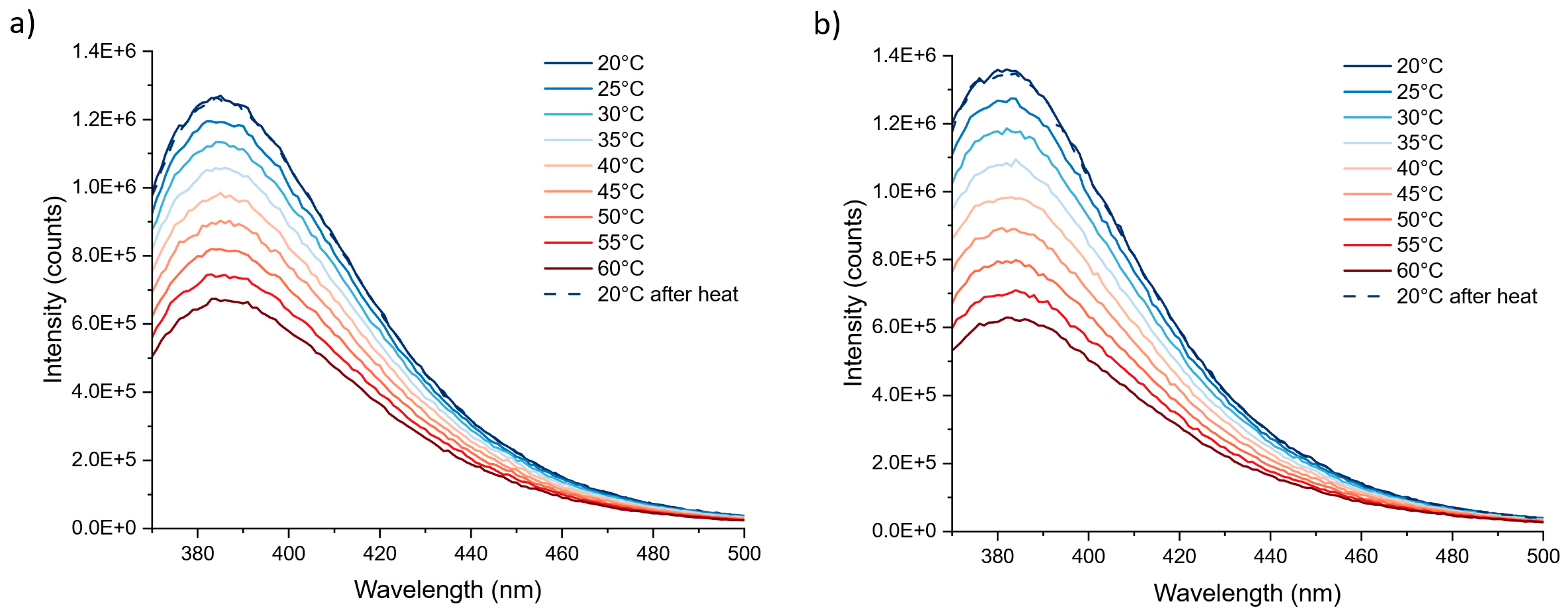
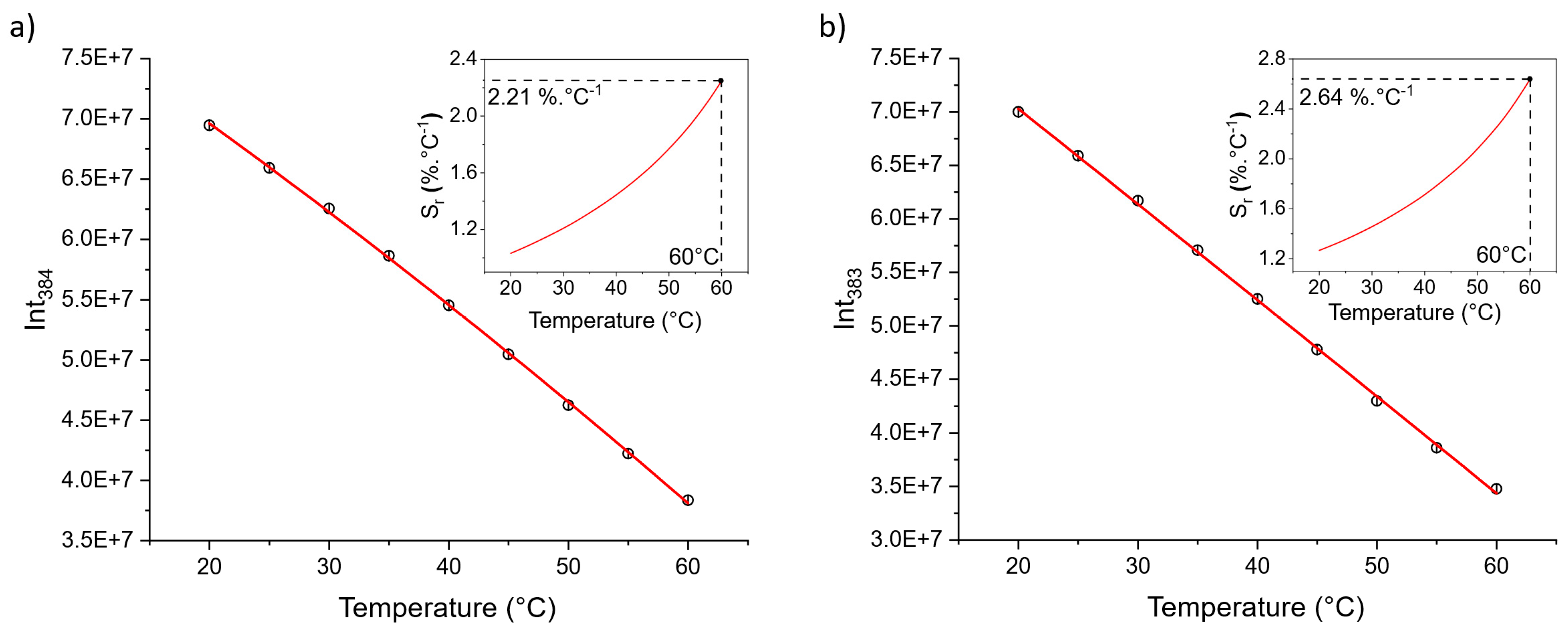
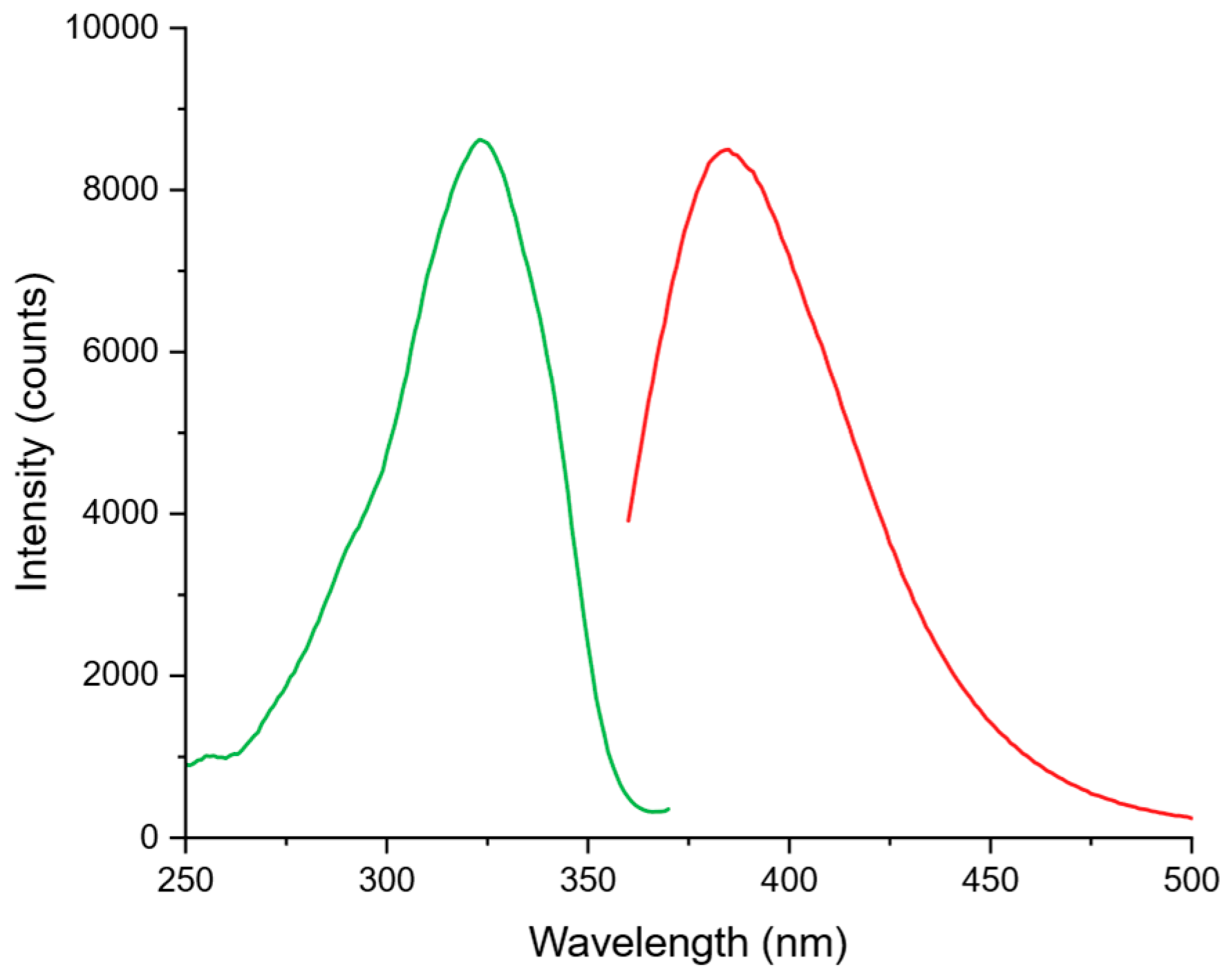
3.4. Luminesence and Luminescent Thermometry
4. Conclusions
Supplementary Materials
Author Contributions
Funding
Data Availability Statement
Acknowledgments
Conflicts of Interest
References
- Thiesen, B.; Jordan, A. Clinical Applications of Magnetic Nanoparticles for Hyperthermia. Int. J. Hyperth. 2008, 24, 467–474. [Google Scholar] [CrossRef] [PubMed]
- Reddy, L.H.; Arias, J.L.; Nicolas, J.; Couvreur, P. Magnetic Nanoparticles: Design and Characterization, Toxicity and Biocompatibility, Pharmaceutical and Biomedical Applications. Chem. Rev. 2012, 112, 5818–5878. [Google Scholar] [CrossRef] [PubMed]
- Zhu, Y.; Li, Q.; Wang, C.; Hao, Y.; Yang, N.; Chen, M.; Ji, J.; Feng, L.; Liu, Z. Rational Design of Biomaterials to Potentiate Cancer Thermal Therapy. Chem. Rev. 2023, 123, 7326–7378. [Google Scholar] [CrossRef] [PubMed]
- de Toledo, L.d.A.S.; Rosseto, H.C.; Bruschi, M.L. Iron Oxide Magnetic Nanoparticles as Antimicrobials for Therapeutics. Pharm. Dev. Technol. 2018, 23, 316–323. [Google Scholar] [CrossRef] [PubMed]
- Usov, N.A.; Nesmeyanov, M.S.; Tarasov, V.P. Magnetic Vortices as Efficient Nano Heaters in Magnetic Nanoparticle Hyperthermia. Sci. Rep. 2018, 8, 1224. [Google Scholar] [CrossRef] [PubMed]
- Ibelli, T.; Templeton, S.; Levi-Polyachenko, N. Progress on Utilizing Hyperthermia for Mitigating Bacterial Infections. Int. J. Hyperth. 2018, 34, 144–156. [Google Scholar] [CrossRef] [PubMed]
- Mahmoud, N.N.; Alkilany, A.M.; Khalil, E.A.; Al-Bakri, A.G. Nano-Photothermal Ablation Effect of Hydrophilic and Hydrophobic Functionalized Gold Nanorods on Staphylococcus aureus and Propionibacterium acnes. Sci. Rep. 2018, 8, 6881. [Google Scholar] [CrossRef] [PubMed]
- Rodrigues, D.; Bañobre-López, M.; Espiña, B.; Rivas, J.; Azeredo, J. Effect of Magnetic Hyperthermia on the Structure of Biofilm and Cellular Viability of a Food Spoilage Bacterium. Biofouling 2013, 29, 1225–1232. [Google Scholar] [CrossRef] [PubMed]
- Mura, S.; Nicolas, J.; Couvreur, P. Stimuli-Responsive Nanocarriers for Drug Delivery. Nat. Mater. 2013, 12, 991–1003. [Google Scholar] [CrossRef]
- Ovejero, J.G.; Armenia, I.; Serantes, D.; Veintemillas-Verdaguer, S.; Zeballos, N.; López-Gallego, F.; Grüttner, C.; de la Fuente, J.M.; del Puerto Morales, M.; Grazu, V. Selective Magnetic Nanoheating: Combining Iron Oxide Nanoparticles for Multi-Hot-Spot Induction and Sequential Regulation. Nano Lett. 2021, 21, 7213–7220. [Google Scholar] [CrossRef]
- Stanley, S.A.; Gagner, J.E.; Damanpour, S.; Yoshida, M.; Dordick, J.S.; Friedman, J.M. Radio-Wave Heating of Iron Oxide Nanoparticles Can Regulate Plasma Glucose in Mice. Science 2012, 336, 604–608. [Google Scholar] [CrossRef]
- Huang, H.; Delikanli, S.; Zeng, H.; Ferkey, D.M.; Pralle, A. Remote Control of Ion Channels and Neurons through Magnetic-Field Heating of Nanoparticles. Nat. Nanotechnol. 2010, 5, 602–606. [Google Scholar] [CrossRef]
- Marbaix, J.; Mille, N.; Lacroix, L.-M.; Asensio, J.M.; Fazzini, P.-F.; Soulantica, K.; Carrey, J.; Chaudret, B. Tuning the Composition of FeCo Nanoparticle Heating Agents for Magnetically Induced Catalysis. ACS Appl. Nano Mater. 2020, 3, 3767–3778. [Google Scholar] [CrossRef]
- Lim, Y.; Noh, S.; Shin, T.-H.; Lee, J.; Lungerich, D.; Lee, J.-H.; Cheon, J. Magnetothermally Activated Nanometer-Level Modular Functional Group Grafting of Nanoparticles. Nano Lett. 2021, 21, 3649–3656. [Google Scholar] [CrossRef]
- Cui, X.; Ruan, Q.; Zhuo, X.; Xia, X.; Hu, J.; Fu, R.; Li, Y.; Wang, J.; Xu, H. Photothermal Nanomaterials: A Powerful Light-to-Heat Converter. Chem. Rev. 2023, 123, 6891–6952. [Google Scholar] [CrossRef]
- Bian, W.; Wang, Y.; Pan, Z.; Chen, N.; Li, X.; Wong, W.-L.; Liu, X.; He, Y.; Zhang, K.; Lu, Y.-J. Review of Functionalized Nanomaterials for Photothermal Therapy of Cancers. ACS Appl. Nano Mater. 2021, 4, 11353–11385. [Google Scholar] [CrossRef]
- Ma, Z.; Mohapatra, J.; Wei, K.; Liu, J.P.; Sun, S. Magnetic Nanoparticles: Synthesis, Anisotropy, and Applications. Chem. Rev. 2023, 123, 3904–3943. [Google Scholar] [CrossRef]
- Gavilán, H.; Avugadda, S.K.; Fernández-Cabada, T.; Soni, N.; Cassani, M.; Mai, B.T.; Chantrell, R.; Pellegrino, T. Magnetic Nanoparticles and Clusters for Magnetic Hyperthermia: Optimizing Their Heat Performance and Developing Combinatorial Therapies to Tackle Cancer. Chem. Soc. Rev. 2021, 50, 11614–11667. [Google Scholar] [CrossRef]
- Guardia, P.; Di Corato, R.; Lartigue, L.; Wilhelm, C.; Espinosa, A.; Garcia-Hernandez, M.; Gazeau, F.; Manna, L.; Pellegrino, T. Water-Soluble Iron Oxide Nanocubes with High Values of Specific Absorption Rate for Cancer Cell Hyperthermia Treatment. ACS Nano 2012, 6, 3080–3091. [Google Scholar] [CrossRef]
- Martinez-Boubeta, C.; Simeonidis, K.; Makridis, A.; Angelakeris, M.; Iglesias, O.; Guardia, P.; Cabot, A.; Yedra, L.; Estradé, S.; Peiró, F.; et al. Learning from Nature to Improve the Heat Generation of Iron-Oxide Nanoparticles for Magnetic Hyperthermia Applications. Sci. Rep. 2013, 3, 1652. [Google Scholar] [CrossRef]
- de Walle, A.V.; Figuerola, A.; Espinosa, A.; Abou-Hassan, A.; Estrader, M.; Wilhelm, C. Emergence of Magnetic Nanoparticles in Photothermal and Ferroptotic Therapies. Mater. Horiz. 2023, 10, 4757–4775. [Google Scholar] [CrossRef]
- Espinosa, A.; Di Corato, R.; Kolosnjaj-Tabi, J.; Flaud, P.; Pellegrino, T.; Wilhelm, C. Duality of Iron Oxide Nanoparticles in Cancer Therapy: Amplification of Heating Efficiency by Magnetic Hyperthermia and Photothermal Bimodal Treatment. ACS Nano 2016, 10, 2436–2446. [Google Scholar] [CrossRef]
- Brites, C.D.S.; Balabhadra, S.; Carlos, L.D. Lanthanide-Based Thermometers: At the Cutting-Edge of Luminescence Thermometry. Adv. Opt. Mater. 2019, 7, 1801239. [Google Scholar] [CrossRef]
- Brites, C.D.S.; Marin, R.; Suta, M.; Carneiro Neto, A.N.; Ximendes, E.; Jaque, D.; Carlos, L.D. Spotlight on Luminescence Thermometry: Basics, Challenges, and Cutting-Edge Applications. Adv. Mater. 2023, 35, 2302749. [Google Scholar] [CrossRef]
- Bednarkiewicz, A.; Marciniak, L.; Carlos, L.D.; Jaque, D. Standardizing Luminescence Nanothermometry for Biomedical Applications. Nanoscale 2020, 12, 14405–14421. [Google Scholar] [CrossRef]
- Jaque, D.; Vetrone, F. Luminescence Nanothermometry. Nanoscale 2012, 4, 4301–4326. [Google Scholar] [CrossRef]
- Brites, C.D.S.; Lima, P.P.; Silva, N.J.O.; Millán, A.; Amaral, V.S.; Palacio, F.; Carlos, L.D. A Luminescent Molecular Thermometer for Long-Term Absolute Temperature Measurements at the Nanoscale. Adv. Mater. 2010, 22, 4499–4504. [Google Scholar] [CrossRef]
- Piñol, R.; Brites, C.D.S.; Bustamante, R.; Martínez, A.; Silva, N.J.O.; Murillo, J.L.; Cases, R.; Carrey, J.; Estepa, C.; Sosa, C.; et al. Joining Time-Resolved Thermometry and Magnetic-Induced Heating in a Single Nanoparticle Unveils Intriguing Thermal Properties. ACS Nano 2015, 9, 3134–3142. [Google Scholar] [CrossRef]
- Gu, Y.; Piñol, R.; Moreno-Loshuertos, R.; Brites, C.D.S.; Zeler, J.; Martínez, A.; Maurin-Pasturel, G.; Fernández-Silva, P.; Marco-Brualla, J.; Téllez, P.; et al. Local Temperature Increments and Induced Cell Death in Intracellular Magnetic Hyperthermia. ACS Nano 2023, 17, 6822–6832. [Google Scholar] [CrossRef]
- Dong, J.; Zink, J.I. Taking the Temperature of the Interiors of Magnetically Heated Nanoparticles. ACS Nano 2014, 8, 5199–5207. [Google Scholar] [CrossRef]
- Castellanos-Rubio, I.; Barón, A.; Luis-Lizarraga, O.; Rodrigo, I.; de Muro, I.G.; Orue, I.; Martínez-Martínez, V.; Castellanos-Rubio, A.; López-Arbeloa, F.; Insausti, M. Efficient Magneto-Luminescent Nanosystems Based on Rhodamine-Loaded Magnetite Nanoparticles with Optimized Heating Power and Ideal Thermosensitive Fluorescence. ACS Appl. Mater. Interfaces 2022, 14, 50033–50044. [Google Scholar] [CrossRef]
- Davis, H.C.; Kang, S.; Lee, J.-H.; Shin, T.-H.; Putterman, H.; Cheon, J.; Shapiro, M.G. Nanoscale Heat Transfer from Magnetic Nanoparticles and Ferritin in an Alternating Magnetic Field. Biophys. J. 2020, 118, 1502–1510. [Google Scholar] [CrossRef]
- Venturini, P.; Fleutot, S.; Cleymand, F.; Hauet, T.; Dupin, J.-C.; Ghanbaja, J.; Martinez, H.; Robin, J.-J.; Lapinte, V. Facile One-Step Synthesis of Polyoxazoline-Coated Iron Oxide Nanoparticles. ChemistrySelect 2018, 3, 11898–11901. [Google Scholar] [CrossRef]
- Chen, C.-H.; Niko, Y.; Konishi, G. Amphiphilic Gels of Solvatochromic Fluorescent Poly(2-Oxazoline)s Containing D–π–A Pyrenes. RSC Adv. 2016, 6, 42962–42970. [Google Scholar] [CrossRef]
- Guillerm, B.; Monge, S.; Lapinte, V.; Robin, J.-J. How to Modulate the Chemical Structure of Polyoxazolines by Appropriate Functionalization. Macromol. Rapid Commun. 2012, 33, 1600–1612. [Google Scholar] [CrossRef]
- Belkhir, K.; Cerlati, O.; Heaugwane, D.; Tosi, A.; Benkhaled, B.T.; Brient, P.-L.; Chatard, C.; Graillot, A.; Catrouillet, S.; Balor, S.; et al. Synthesis and Self-Assembly of UV-Cross-Linkable Amphiphilic Polyoxazoline Block Copolymers: Importance of Multitechnique Characterization. Langmuir 2022, 38, 16144–16155. [Google Scholar] [CrossRef]
- Mao, D.; Liu, X.; Qiao, Q.; Yin, W.; Zhao, M.; Cole, J.M.; Cui, J.; Xu, Z. Coumarin 545: An Emission Reference Dye with a Record-Low Temperature Coefficient for Ratiometric Fluorescence Based Temperature Measurements. Analyst 2015, 140, 1008–1013. [Google Scholar] [CrossRef]
- Hu, X.; Li, Y.; Liu, T.; Zhang, G.; Liu, S. Responsive Polymer-Based Multicolor Fluorescent Probes for Temperature and Zn2+ Ions in Aqueous Media. Sci. China Chem. 2014, 57, 615–623. [Google Scholar] [CrossRef]
- Wang, B.; Guan, X.; Hu, Y.; Su, Z. Preparation and Fluorescent Properties of Poly(Vinyl Alcohol) Bearing Coumarin. Polym. Adv. Technol. 2007, 18, 529–534. [Google Scholar] [CrossRef]
- Inal, S.; Kölsch, J.D.; Chiappisi, L.; Kraft, M.; Gutacker, A.; Janietz, D.; Scherf, U.; Gradzielski, M.; Laschewsky, A.; Neher, D. Temperature-Regulated Fluorescence Characteristics of Supramolecular Assemblies Formed By a Smart Polymer and a Conjugated Polyelectrolyte. Macromol. Chem. Phys. 2013, 214, 435–445. [Google Scholar] [CrossRef]
- Hu, X.; Li, Y.; Liu, T.; Zhang, G.; Liu, S. Intracellular Cascade FRET for Temperature Imaging of Living Cells with Polymeric Ratiometric Fluorescent Thermometers. ACS Appl. Mater. Interfaces 2015, 7, 15551–15560. [Google Scholar] [CrossRef]
- Yu, W.W.; Falkner, J.C.; Yavuz, C.T.; Colvin, V.L. Synthesis of Monodisperse Iron Oxide Nanocrystals by Thermal Decomposition of Iron Carboxylate Salts. Chem. Commun. 2004, 2306–2307. [Google Scholar] [CrossRef]
- Bouvet, B.; Sene, S.; Félix, G.; Havot, J.; Audran, G.; Marque, S.R.A.; Larionova, J.; Guari, Y. Cascade Strategy for Triggered Radical Release by Magnetic Nanoparticles Grafted with Thermosensitive Alkoxyamine. Nanoscale 2022, 15, 144–153. [Google Scholar] [CrossRef]
- COMSOL® Software Version 6.0 Release Highlights. Available online: https://www.comsol.com/release/6.0 (accessed on 18 April 2024).
- Wildeboer, R.R.; Southern, P.; Pankhurst, Q.A. On the Reliable Measurement of Specific Absorption Rates and Intrinsic Loss Parameters in Magnetic Hyperthermia Materials. J. Phys. D Appl. Phys. 2014, 47, 495003. [Google Scholar] [CrossRef]
- Lanier, O.L.; Korotych, O.I.; Monsalve, A.G.; Wable, D.; Savliwala, S.; Grooms, N.W.F.; Nacea, C.; Tuitt, O.R.; Dobson, J. Evaluation of Magnetic Nanoparticles for Magnetic Fluid Hyperthermia. Int. J. Hyperth. 2019, 36, 687–701. [Google Scholar] [CrossRef]
- Roca, A.G.; Lopez-Barbera, J.F.; Lafuente, A.; Özel, F.; Fantechi, E.; Muro-Cruces, J.; Hémadi, M.; Sepulveda, B.; Nogues, J. Iron Oxide Nanoparticles (Fe3O4, γ-Fe2O3 and FeO) as Photothermal Heat Mediators in the First, Second and Third Biological Windows. Phys. Rep. 2023, 1043, 1–35. [Google Scholar] [CrossRef]
- Qin, T.; Liu, B.; Zhu, K.; Luo, Z.; Huang, Y.; Pan, C.; Wang, L. Organic Fluorescent Thermometers: Highlights from 2013 to 2017. TrAC Trends Anal. Chem. 2018, 102, 259–271. [Google Scholar] [CrossRef]


| Name | Structure | Excitation and Emission Wavelengths |
|---|---|---|
| Coum-C11-PPhOx27-PMOx59 non-dimerized (P1) | 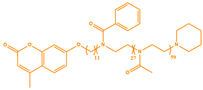 | λexc 1 = 297 nm λexc 2 = 351 nm λem = 384 nm |
| Coum-C11-PPhOx27-PMOx59 Dimerized (P2) | 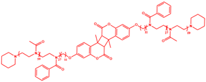 | λexc = 336 nm λem = 383 nm |
| PPhOx27-PMOx57 (P3) | 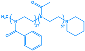 | λexc = 323 nm λem = 383 nm |
| Coum-C11-PButOx8-PMOx42 non-dimerized (P4) |  | λexc 1 = 270 nm λexc 2 = 351 nm λem = 384 nm |
| Coum-C11-PButOx8-PMOx42 Dimerized (P5) | 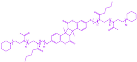 | λexc = 336 nm λem = 383 nm |
| Parameters | P1 | P2 | P3 | P4 | P5 | IONP@ Coum-C11-PPhOx27-PMOx59 |
|---|---|---|---|---|---|---|
| Relative maximal thermal sensitivity Srmax (%·°C−1) at 60 °C | 2.21 | 2.64 | 2.71 | 1.93 | 2.21 | 1.53 |
| Uncertainty δT(°C) at 60 °C | 0.28 | 0.40 | 0.87 | 0.14 | 0.29 | 0.82 |
Disclaimer/Publisher’s Note: The statements, opinions and data contained in all publications are solely those of the individual author(s) and contributor(s) and not of MDPI and/or the editor(s). MDPI and/or the editor(s) disclaim responsibility for any injury to people or property resulting from any ideas, methods, instructions or products referred to in the content. |
© 2024 by the authors. Licensee MDPI, Basel, Switzerland. This article is an open access article distributed under the terms and conditions of the Creative Commons Attribution (CC BY) license (https://creativecommons.org/licenses/by/4.0/).
Share and Cite
Féron, A.; Catrouillet, S.; Sene, S.; Félix, G.; Benkhaled, B.T.; Lapinte, V.; Guari, Y.; Larionova, J. Magnetic Iron Oxide Nanoparticles Coated by Coumarin-Bound Copolymer for Enhanced Magneto- and Photothermal Heating and Luminescent Thermometry. Nanomaterials 2024, 14, 906. https://doi.org/10.3390/nano14110906
Féron A, Catrouillet S, Sene S, Félix G, Benkhaled BT, Lapinte V, Guari Y, Larionova J. Magnetic Iron Oxide Nanoparticles Coated by Coumarin-Bound Copolymer for Enhanced Magneto- and Photothermal Heating and Luminescent Thermometry. Nanomaterials. 2024; 14(11):906. https://doi.org/10.3390/nano14110906
Chicago/Turabian StyleFéron, Alexiane, Sylvain Catrouillet, Saad Sene, Gautier Félix, Belkacem Tarek Benkhaled, Vincent Lapinte, Yannick Guari, and Joulia Larionova. 2024. "Magnetic Iron Oxide Nanoparticles Coated by Coumarin-Bound Copolymer for Enhanced Magneto- and Photothermal Heating and Luminescent Thermometry" Nanomaterials 14, no. 11: 906. https://doi.org/10.3390/nano14110906