Review

Transition Metal Oxide Nanomaterials: New Weapons to Boost Anti-Tumor Immunity Cycle

1
Department of Medical Oncology, Jinling Hospital, Nanjing University of Chinese Medicine, Nanjing 210000, China
2
Department of Medical Oncology, Nanjing Jinling Hospital, Affiliated Hospital of Medical School, Nanjing University, Nanjing 210000, China
3
Department of Gastroenterology, Nanjing Jinling Hospital, Affiliated Hospital of Medical School, Nanjing University, Nanjing 210023, China
*
Authors to whom correspondence should be addressed.
These authors contributed equally to this work.
Nanomaterials 2024, 14(13), 1064; https://doi.org/10.3390/nano14131064
Submission received: 30 April 2024 / Revised: 6 June 2024 / Accepted: 18 June 2024 / Published: 21 June 2024
(This article belongs to the Special Issue Advances in Wide-Bandgap Semiconductor Nanomaterials)

Abstract

:
Semiconductor nanomaterials have emerged as a significant factor in the advancement of tumor immunotherapy. This review discusses the potential of transition metal oxide (TMO) nanomaterials in the realm of anti-tumor immune modulation. These binary inorganic semiconductor compounds possess high electron mobility, extended ductility, and strong stability. Apart from being primary thermistor materials, they also serve as potent agents in enhancing the anti-tumor immunity cycle. The diverse metal oxidation states of TMOs result in a range of electronic properties, from metallicity to wide-bandgap insulating behavior. Notably, titanium oxide, manganese oxide, iron oxide, zinc oxide, and copper oxide have garnered interest due to their presence in tumor tissues and potential therapeutic implications. These nanoparticles (NPs) kickstart the tumor immunity cycle by inducing immunogenic cell death (ICD), prompting the release of ICD and tumor-associated antigens (TAAs) and working in conjunction with various therapies to trigger dendritic cell (DC) maturation, T cell response, and infiltration. Furthermore, they can alter the tumor microenvironment (TME) by reprogramming immunosuppressive tumor-associated macrophages into an inflammatory state, thereby impeding tumor growth. This review aims to bring attention to the research community regarding the diversity and significance of TMOs in the tumor immunity cycle, while also underscoring the potential and challenges associated with using TMOs in tumor immunotherapy.

1. Introduction

Immunotherapy has provided hope for numerous cancer patients; yet, it faces two main challenges. First, low immunogenicity hinders the effective initiation of immunogenic cell death (ICD) and diminishes antigen presentation efficiency [1]. Second, the suppressive tumor microenvironment (TME), comprising inhibitory physicochemical factors, such as low pH, hypoxia, high hydrogen peroxide (H2O2), high glutathione (GSH), and inhibitory immune cells, such as alternatively activated macrophages (M2), regulatory T cells (Tregs), and myeloid-derived suppressor cells (MDSCs), ultimately compromise immunotherapy efficacy [2,3]. To optimize tumor immunotherapy, transition metal oxide nanomaterials (TMOs), such as titanium oxide, manganese oxide, iron oxide, and zinc oxide, are extensively utilized to modulate tumor immunity cycle, namely, antigen presentation, priming, and activation, immune cell trafficking and infiltration, cancer cell recognition and elimination, and immune memory formation, ultimately capitalizing on their distinctive attributes to enhance the effectiveness of the treatment [4].
TMOs consist of oxygen atoms and transition metals, exhibiting high electron mobility, extended ductility, and strong stability. These materials are extensively utilized in various biomedical applications due to their favorable electrical, magnetic, and biocompatible properties, high specific surface area, good chemical and mechanical stability, unique structure, and biodegradability [5,6,7,8]. In addition to enhancing the generation of reactive oxygen species (ROS) independently, TMOs can synergize with photodynamic (PDT), photothermal (PTT), magnetic thermal (MHT), and sonodynamic (SDT) therapies to combine multiple treatments into a single platform. This significantly boosts ROS production efficiency, thereby enhancing ICD and dendritic cell (DC) maturation, further enhancing tumor immunogenicity and facilitating the initiation of the anti-tumor immunity cycle [9,10,11,12]. Moreover, TMOs possess unique properties such as improved electron transfer kinetics, strong adsorption capacity, high sensitivity, and excellent light absorption capabilities, making them ideal for doping into MXenes to enhance their optical properties for biomedical applications. This enhancement greatly improves the efficacy of PDT and PTT [5,8,13]. Notably, TMOs exhibit greater stability, require lower doses, offer enhanced efficacy, and have fewer side effects compared to metal ions [14]. TMO nanoparticles (NPs) offer advantages over free small molecule drugs as they can penetrate tumors via the activated transendothelial pathway. Leveraging the enhanced permeability and retention (EPR) effect (the altered permeability of tumor blood vessels and lymphatic drainage), TMO NPs can efficiently accumulate in tumor tissues [15]. The enhanced penetration and EPR effect facilitate the passive targeting of tumors by conventional TMOs. Nevertheless, when reaching distant tumor cells from blood vessels, the drug enrichment rate is limited, leading to off-target effects and suboptimal efficacy. Certain TMOs, with solubility at low pH levels, can function as appropriate pH-sensitive nanocarriers for delivering tumor-targeted drugs and releasing intracellular drugs. For example, in the acidic and hypoxic TME, TMOs like MnO2 and ZnO can decompose to generate oxygen, alleviating hypoxia and neutralizing H+, thereby reversing the inhibitory TME [16,17,18]. Furthermore, certain TMOs like Fe2O3 can stimulate the polarization of classical activated macrophages (M1) and enhance the anti-tumor response of T cells [19,20,21]. These properties make them suitable for designing pH-responsive nanocarriers to transport immune agents that are susceptible to enzymatic degradation in the body, ensuring their bioactive components remain intact until reaching the tumor site. This targeted delivery mechanism aids in activating CD4/CD8+ T cells, disrupting the inhibitory TME, and bolstering the anti-tumor immunity cycle [22,23,24]. Furthermore, actively targeting TMOs NPs can significantly improve therapeutic efficacy in cancer treatment by directly delivering cytotoxic drugs to tumor cells, reducing off-target effects, and enhancing the therapeutic index. For instance, the incorporation of internalizing RGD peptide (iRGD) into the outermost layer of MCMnO2 (carbon–manganese nanocomposite) can enhance the targeting and penetration capabilities of nanoparticles [25]. Moreover, coating gemcitabine zinc oxide, a first-line drug for pancreatic cancer treatment, with peptides that target the pancreatic TME enables active targeting for site-specific drug delivery, leading to high levels of cellular internalization mediated by the targeted peptides exposed on the nanostructure [26].
The remarkable functionalities of transition metal nanomaterials have sparked growing interest in tumor immunotherapy. While several reviews have comprehensively outlined the synthesis, properties, and therapeutic applications of individual TMO nanomaterials, there remains a dearth of systematic reviews on TMO tumor immunotherapy [27,28,29]. Hence, this review concentrates on the recent advancements of TMO nanoplatforms in tumor immunotherapy and deliberates on the challenges and future prospects of TMOs in tumor immunotherapy.

2. TMOs Based Cancer Immunotherapy

2.1. Shared Properties of TMOs

TMOs, ranging in size from 1 to 100 nm, are formed through chemical bonding between metals and oxygen elements [30]. Examples of these materials include manganese oxide, titanium oxide, iron oxide, zinc oxide, and copper oxide. The unique properties of these materials are attributed to the distinctive characteristics of oxygen ions present in their composition. The highly polarizable superoxide ion (O2•−) leads to non-linear, large, and uneven charge distributions within the crystal lattice of planar TMOs, resulting in electrostatic shielding on the nanoscale [10]. The fundamental properties of TMOs are heavily influenced by the metal cation species and their ability to change oxidation states [31]. In 2D TMOs, varying cation charge states and binding configurations enhance structural stability. Different oxidation states of the metallic components in TMOs give rise to a spectrum of electronic properties, from metallic behavior to wide-bandgap insulating characteristics. Local electronic states can also prompt significant changes in metal-insulator transitions under pressure and temperature variations, such as Mott and Verwey transitions. TMOs exhibit unique redox properties, often displaying reversible trends [32]. Additionally, TMOs showcase remarkable ferroelectric, ferromagnetic, photocatalytic, photoelectric, and magnetoelastic properties. Some TMOs can interact directly with biomolecules after surface modification, making them promising candidates for biomedical applications like targeted drug delivery, cancer treatment, tissue engineering, and biosensing. The simplicity, cost-effectiveness, ease of synthesis, and high theoretical specific capacity of TMOs have positioned them as a focal point in recent research [33].

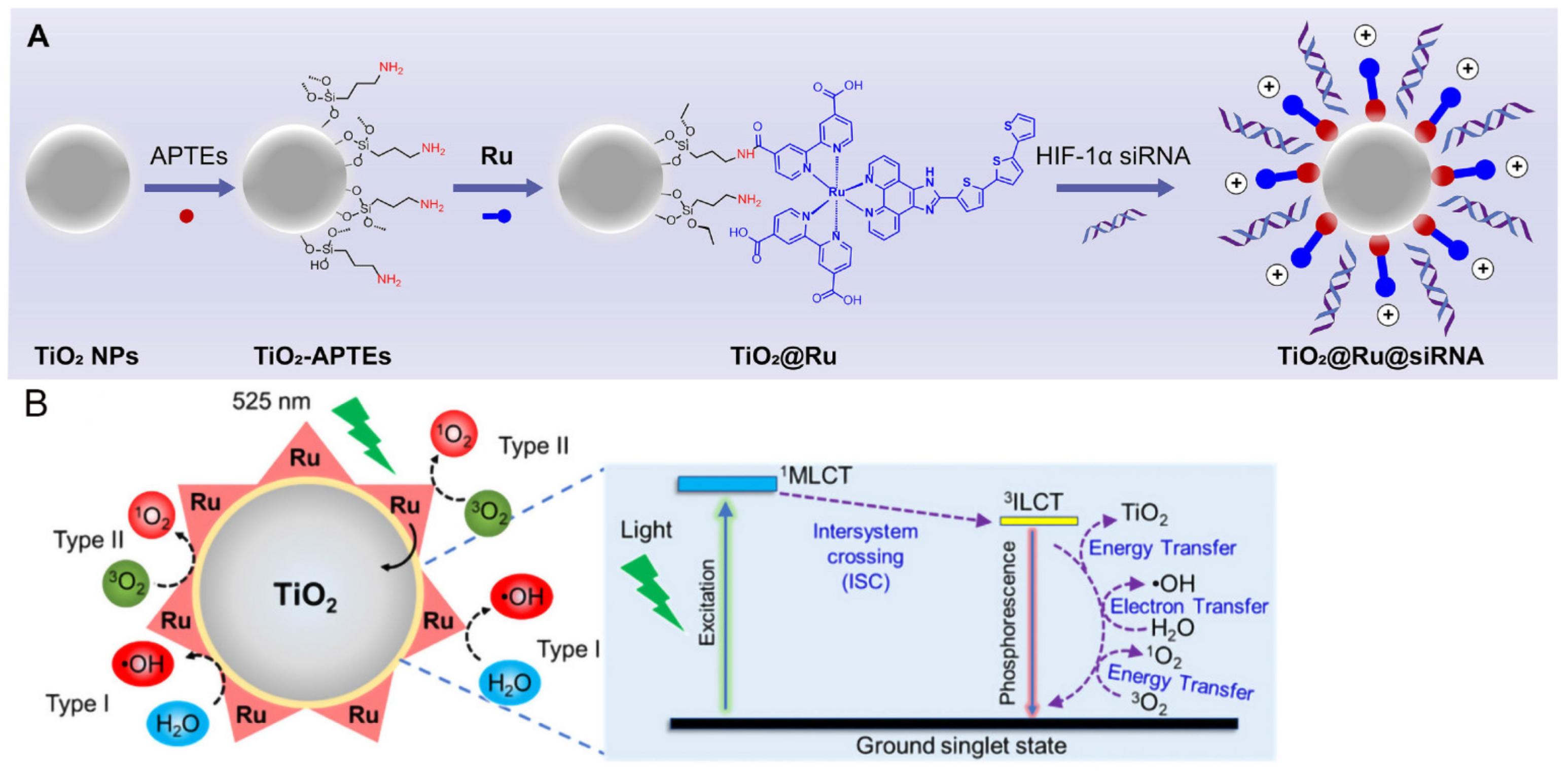
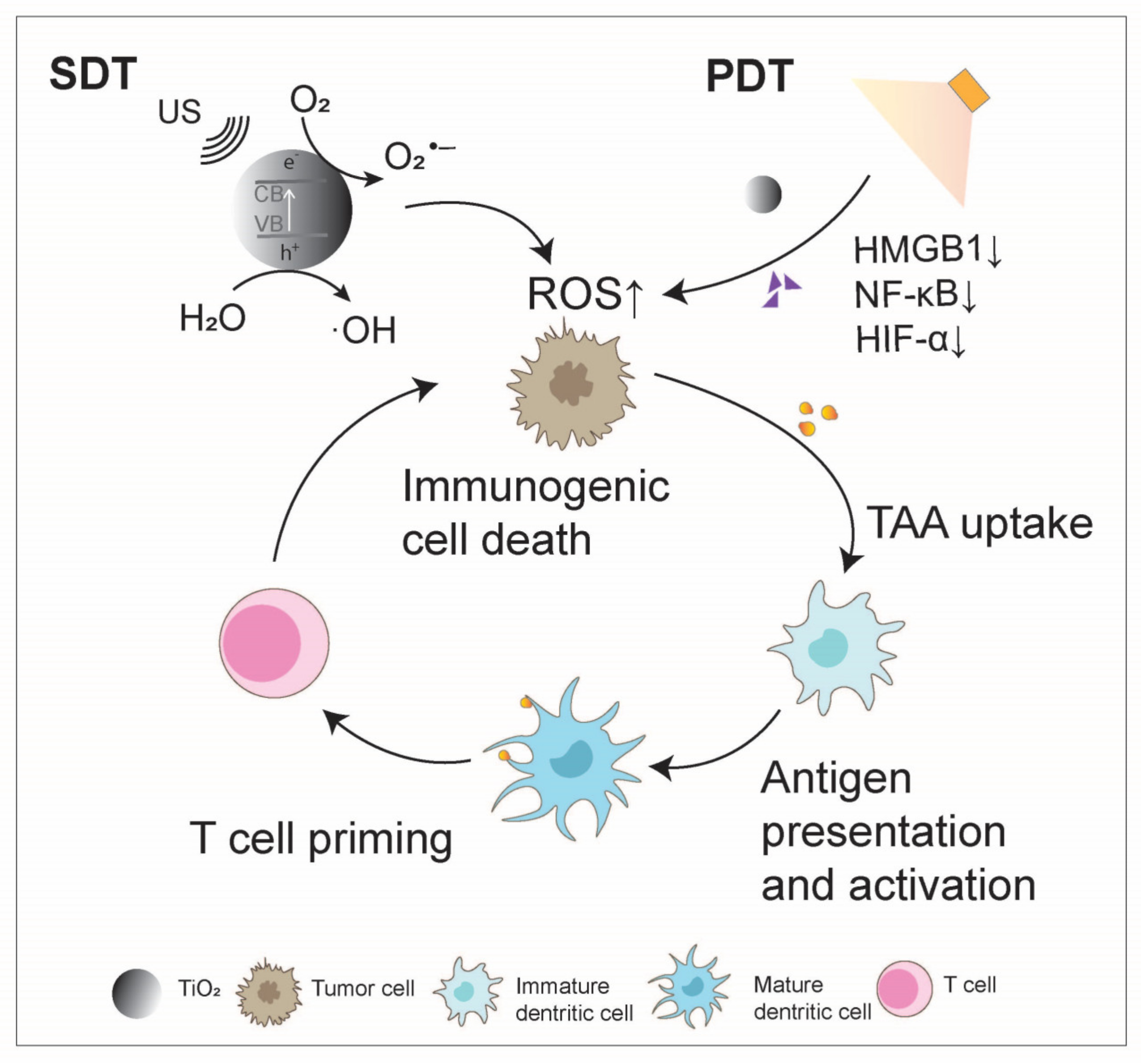
2.2. Titanium Oxide-Based Cancer Immunotherapy

Research on titanium dioxide (TiO2), a type of TMO, is continuously expanding. Due to its chemical inertness, long-term stability under physiological conditions, and nontoxicity to living cells, TiO2 NPs have received approval from the U.S. Food and Drug Administration (FDA) for various applications. The accumulation of TiO2 may lead to effects on gene expression, DNA damage, inflammatory responses, metabolic processes, apoptosis, cell cycle regulation, and more, potentially through genotoxic mechanisms, making it a candidate for use as an anticancer agent [34,35].
TiO2 NPs are considered advantageous as inorganic sonosensitizers due to their ability to generate ROS, their chemical inertness in biological fluids, and their low toxicity, which can induce ICD and enhance tumor immunogenicity. Under low-intensity ultrasound irradiation, TiO2 can be activated to induce a series of cell-killing effects, primarily the production of highly toxic ROS, along with high temperature and pressure, leading to SDT. By fine-tuning the frequency and intensity of ultrasound, it is feasible to target the tumor site accurately, thus reducing harm to surrounding healthy tissue [36]. Research has shown that black TiO2 could enhance the absorption of H2O and O2, separate electrons and holes, and generate ROS, thereby reducing hypoxia and improving the effectiveness of SDT [37].
As a semiconductor, TiO2 exhibits strong light absorption in the ultraviolet region and has the ability to generate singlet oxygen (1O2), enabling it to function as a photosensitizer for PDT [38]. However, the photoresponsive range of TiO2 is limited to the UV region, with poor penetration, prompting the use of different modifications to extend its absorption wavelength into the near-infrared (NIR) range. Efficient electron-hole separation can be achieved by transferring photogenerated electrons from the modifier (such as antennas or nanocomposite materials) to the conduction band (CB) of TiO2, facilitating the generation of 1O2 through O2•− reactions and hydroxyl radicals (·OH) and other ROS through hole-water reactions [39,40]. These characteristics make modified TiO2 NPs highly biocompatible and capable of exerting photodynamic effects in both oxygen-dependent and -independent manners. Shang et al. prepared rGO–TiO2 composites through a straightforward hydrothermal reduction method. These composites exhibit an absorption spectrum that extends from the ultraviolet to the visible light region, enhancing photodynamic processing performance compared to unmodified TiO2. Thus, rGO–TiO2 composite materials offer a novel approach for advancing the photocatalytic technology [41]. Zhou et al. utilized ruthenium-based (Ru) photosensitizers to modify TiO2 NPs as carriers, loading small interfering RNA targeting hypoxia-inducible factor-1α (HIF-1α) to create a novel nanocomposite material, namely, TiO2@Ru@siRNA (Figure 1). Upon visible light exposure, TiO2@Ru@siRNA induces type I and type II photodynamic effects, causing lysosome damage, silencing the HIF-1α gene, leading to the downregulation of high-mobility group box 1 protein (HMGB1) and NF-κB, alleviating hypoxia, promoting ICD, reducing key immune inhibitory factors, increasing immune cell factors, and activating CD4+ and CD8+ T cells to reshape the immune microenvironment [42] (Figure 2).
Due to its physiological stability and insolubility, TiO2 can also be used as a dopant to control the dissolution kinetics of copper ions, thereby enhancing tumor immunogenicity. Hesemans et al. conducted a study where they prepared TiO2 NPs doped with 33% copper to regulate the release kinetics and extent of Cu2+ ions from the residual TiO2 nanocrystals. These NPs efficiently activated DCs in vitro and, when transplanted into tumor-bearing mice, showed superior therapeutic outcomes compared to classical DC stimulation [43].

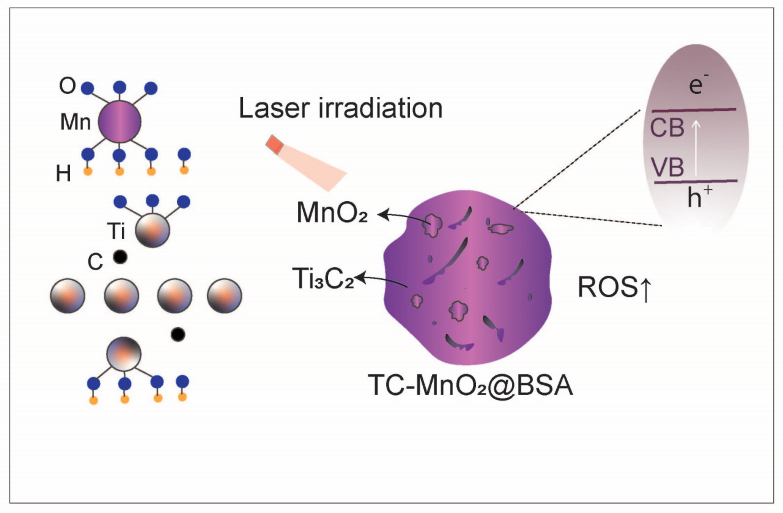
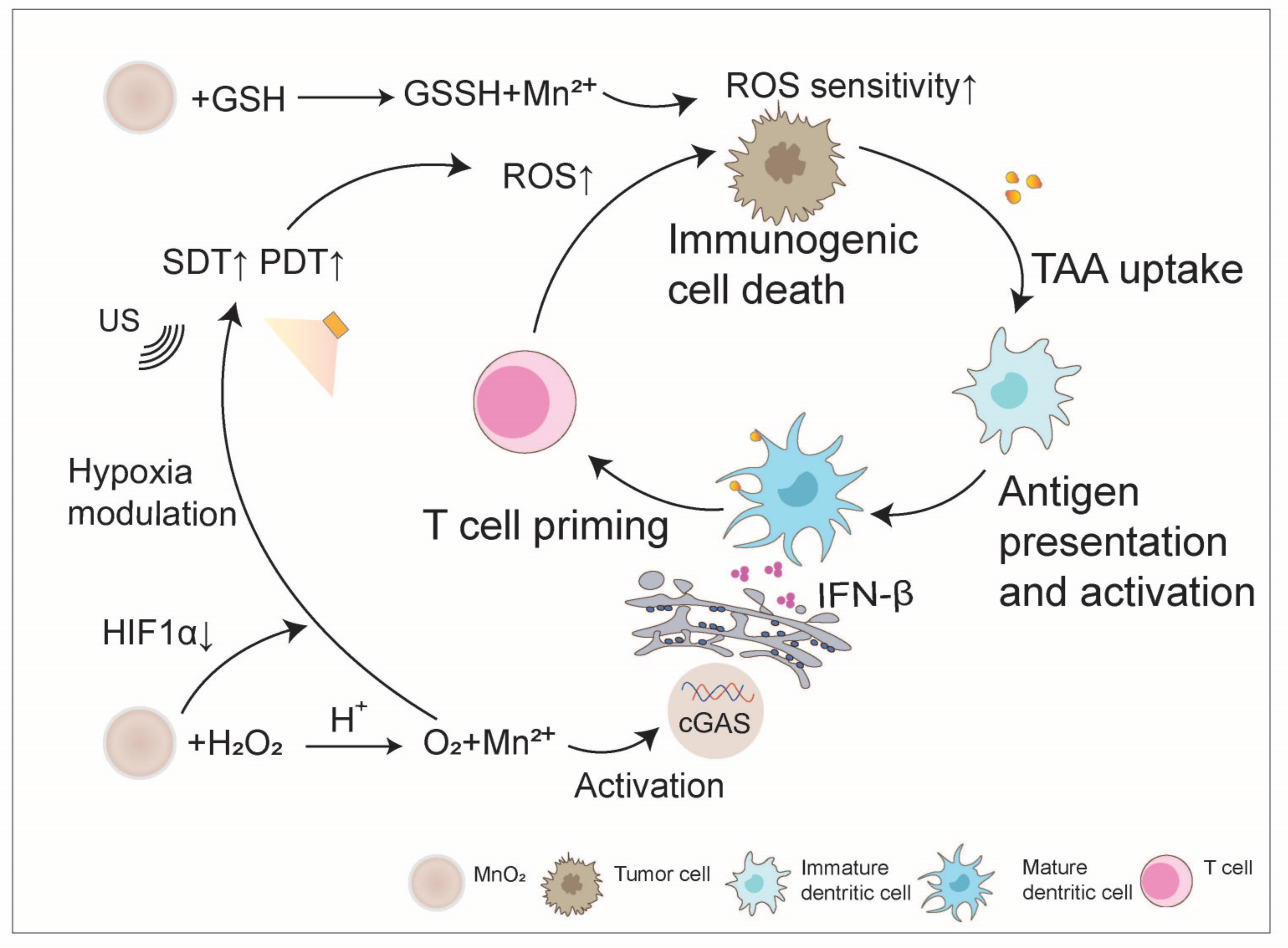
2.3. Manganese Oxide-Based Cancer Immunotherapy

Manganese dioxide (MnO2) nanomaterials, as a biodegradable material, have emerged as promising candidates in the field of tumor immunotherapy due to their stable structure, excellent physicochemical properties, and biocompatibility [16,44,45]. MnO2 demonstrates direct redox activity and possesses the ability to oxidize various reducing agents, such as GSH [46,47]. By utilizing this oxidative property, MnO2 has been engineered as an enhancer for PDT/PTT.
High levels of GSH and hypoxia in tumor tissues are two major factors hindering the efficacy of PDT, PTT, and SDT. MnO2 NPs can react with overexpressed GSH in tumor cells through redox reactions to generate Mn2+ and glutathione disulfide (GSSG), significantly reducing the content of antioxidant GSH in tumor cells, increasing the sensitivity of tumor cells to ROS, promoting immunogenic cell death of tumor cells, and releasing tumor-associated antigens (TAAs) to initiate adaptive immunity. Additionally, MnO2 can generate oxygen by decomposing endogenous H2O2 in tumors, and it can also promote the expression of HIF-1α, thereby alleviating tumor hypoxia, further increasing the production of ROS in oxygen-dependent PDT and SDT, thus enhancing the efficacy of PDT and SDT [23,48,49]. For example, Jian et al. utilized lipid NPs co-encapsulated with MnO2 NPs and zoledronic acid labeled with tumor-homing peptide LYP-1. This nanoplatform generates oxygen bubbles in the TME through MnO2 nanoparticles, providing sufficient oxygen for PDT [50]. Zhu et al. synthesized novel hollow organic silica nanospheres by embedding manganese oxide to serve as a sonosensitizer for nanoenzyme-enhanced SDT. The manganese oxide component exhibits catalase-like (CAT-like) activity, decomposing H2O2 to generate O2 to reverse hypoxia and promote ROS production in SDT [51].
However, in some solid tumors, the problem of a low ROS generation rate persists [52,53]. In order to further alleviate hypoxia and increase ROS production to enhance the efficacy of SDT and PDT/PTT, manganese oxide can be designed to form heterojunctions. For example, Zhu et al. formed heterojunctions by loading manganese oxide with multiple enzyme-like activities onto the surface of piezoelectric bismuth oxychloride nanosheets [51]. Under ultrasound (US) irradiation, the piezoelectric effect significantly promotes the separation and transfer of ultrasound-induced free charges, further enhancing ROS production in SDT. Meanwhile, the enzyme-like activity of manganese oxide not only downregulates GSH levels but also decomposes endogenous H2O2 to generate O2 and ·OH. This ultimately greatly promotes ROS production and reverses tumor hypoxia [51]. In another study, Song and her group anchored ultra-small γ-MnO2 nanodots onto bovine serum albumin-modified intrinsic metal Ti3C2(OH)2, forming a Schottky heterojunction (labeled as TC-MnO2@BSA) [54] (Figure 3). The Schottky heterojunction endows TC-MnO2@BSA with better photothermal conversion efficiency and ROS generation capacity. Through abundant ROS and a high temperature, tumor ICD is induced. The ICD and manganese ion generated from MnO2 decomposition synergistically amplify the cyclic GMP-AMP synthase (cGAS)-stimulator of interferon genes (STING) pathway, leading to an increase in type I interferon production, further promoting DC maturation and antigen presentation, thus enhancing immunogenicity [44].
Furthermore, by leveraging the characteristic decomposition of MnO2 in acidic environments and its material encapsulation, it can be designed as a pH-responsive nanocarrier, loading immunomodulators that are easily degraded by enzymes in the body. This prevents the hydrolysis of their bioactive components, maintains their activity, and delivers them to tumor sites, further promoting the anti-tumor immunity cycle. For instance, Wen et al. coated MnO2 on a chitosan-reversed protein core loaded with metformin, constructing a TME-responsive drug delivery system. The MnO2 coating protects metformin from premature release, but once it reaches the tumor site, the drug is rapidly released due to the degradation of MnO2 in the acidic TME. As an inhibitor of the programmed cell death protein 1 (PD-1)/programmed cell death ligand 1 (PD-L1) pathway, the released metformin disrupts the membrane localization of PD-L1, reducing its stability, thereby improving tumor immunotherapy [55]. Deng et al. [22] utilized a bio-mineralization method to conveniently synthesize αPDL1-coated MnO2 (αPDL1@MnO2) nanoparticles. Under acidic tumor pH conditions, the Mn2+ released by αPDL1@MnO2 can activate the cGAS-STING pathway of interferon genes, promoting DC maturation. Meanwhile, the αPDL1 released by αPDL1@MnO2 NPs further promotes the infiltration of CD8+ T cells in tumors and triggers a systemic anti-tumor response, thereby generating a potent in vitro effect, effectively inhibiting tumor metastasis (Figure 4).
The aforementioned TME modulation function of manganese oxide further enhances the efficacy of tumor immunotherapy. Its magnetic resonance imaging (MRI) functionality can be used to monitor the tumor treatment process, detect in vivo biodistribution, and evaluate the efficacy of tumor treatment. Therefore, manganese oxide-based nanomaterials can also be designed and constructed as traceable multifunctional nanoplatforms for effective tumor therapy [56,57].

2.4. Zinc Oxide-Based Cancer Immunotherapy

The anticancer drugs that generate ROS can exert their effects through two different mechanisms: (I) increasing the production of endogenous ROS in the mitochondria or (II) generating/transmitting exogenous ROS within the cells [58,59]. MnO2, as discussed above, mainly operates via the second mechanism to produce ROS, while zinc oxide NPs, by combining both endogenous and exogenous ROS, achieve enhanced oxidative damage to cancer cells. Lin et al. proposed a polymer-modified nanoscale zinc oxide ROS generator, which, upon internalization into tumor cells, undergoes decomposition under weak acidic pH conditions, leading to the controlled release of H2O2 and zinc ions. Zinc ions can synergistically exert anticancer effects with externally released H2O2 by inhibiting the electron transfer chain to increase the production of mitochondrial O2•− and H2O2, thereby promoting ICD in tumor cells and enhancing immunogenicity [60] (Figure 5).
Similar to MnO2, zinc oxide can also serve as a pH-responsive nanocarrier to reverse the TME and promote anti-tumor immunity when loaded with chemotherapy drugs. Zinc oxide NPs are stable at pH = 7.4 but can rapidly dissolve in acidic environments (pH < 5.5) [61,62]. Additionally, zinc oxide is easy to prepare, cost-effective, biodegradable, and exhibits sufficient responsiveness to acidity at pH < 5.5, with preferential cytotoxicity towards cancer cells, making it an ideal pH-responsive carrier [62]. Zhang et al. demonstrated a simple and versatile zinc oxide-terminated and doxorubicin (DOX)-loaded multifunctional nanocomposite material as an effective drug carrier, ensuring high DOX loading capacity and effective release in vitro at pH 5.0. Nanoscale zinc oxide not only exhibits selectivity against melanoma cells but also induces ICD, which promotes DC maturation, further stimulating the infiltration of CD4+ and CD8+ T cells into the tumor site, thereby preventing tumor growth and distant lung metastasis [63].
Zinc oxide can act as a carrier for photosensitizers like sodium porphyrin (SPS) to create therapeutic nanoplatforms that combine ROS generators and photosensitizers. These dual-action nanoplatforms not only reduce GSH levels but also enhance tumor targeting by depleting GSH levels due to improved permeability and retention effects, consequently lowering the dosage of photosensitizers and achieving effective tumor diagnosis and treatment [64,65]. SPS is attached to NPs through a liquid-phase reaction, and in the acidic TME, SPS@ZnO2 NPs break down into endogenous Zn2+ and H2O2, generating toxic 1O2 under 630 nm laser irradiation. This process is further enhanced by increased H2O2 levels that deplete intracellular GSH, enhancing the effects of PDT in a synergistic manner and ultimately reversing the inhibitory TME while boosting the anti-tumor activity effects of Molecular Dynamic Therapy (MDT) and PDT [17].
Among various wide-bandgap metal oxides, ZnO NPs are favored for their rigid structure, strong stability, versatility, and capacity to induce immunomodulatory and inflammatory responses in DCs and macrophages [66,67,68]. These particles have been extensively employed as vaccine adjuvants to harness their intrinsic adjuvant-like characteristics and immunomodulatory functions [69]. Studies have reported that zinc oxide NPs have the ability to produce immunomodulatory cytokines and act as immunostimulatory adjuvants in glioblastoma models [70]. Furthermore, the ductility of zinc oxide materials allows for the design of various shapes with unique functions to enhance their immune adjuvant function. Sharma et al. synthesized radially grown ZnO nanowires on poly-L-lactic acid microfibers with unique 3-dimensional structures. Based on the function in immune-stimulating adjuvant of ZnO, this inorganic–organic hybrid nanocomposite could reduce immune-suppressive Tregs and enhance the infiltration of CD4+ and CD8+ T cells, effectively inducing tumor antigen-specific immunity and significantly inhibiting tumor growth [24]. Zinc oxides can also be combined with other metals to create multi-faceted immunogenic nanocomposites. ZnO can not only guard pH-responsive targeted drug delivery and privileged phagocytic ability to melanoma cells but also play an immune-stimulating adjuvant role as they produce pro-inflammatory cytokines such as IFN-γ or TNF-α and induce an ICD effect, promoting DC maturation and the infiltration of effector CD4+ and CD8+ T cells into tumor sites, ultimately inhibiting tumor growth and distant lung metastases [63]. Moreover, research has shown that ZnO can be constructed as mesoporous ZnO nanocapsules, antigen-absorbed ZnO tetrapod nanoparticles, or Zn-doped mesoporous silica NPs, which can all boost the tumor immunity cycle by strengthening cytotoxic T lymphocyte (CTL) activity and enhancing levels of IFN-γ-producing CD4+ and CD8+ T cells [71,72,73]. The findings demonstrate the versatility of zinc oxide-based immunostimulatory adjuvants. Their cost-effective preparation, diverse structures, and simple surface modification offer promising clinical applications in the treatment of established cancers.

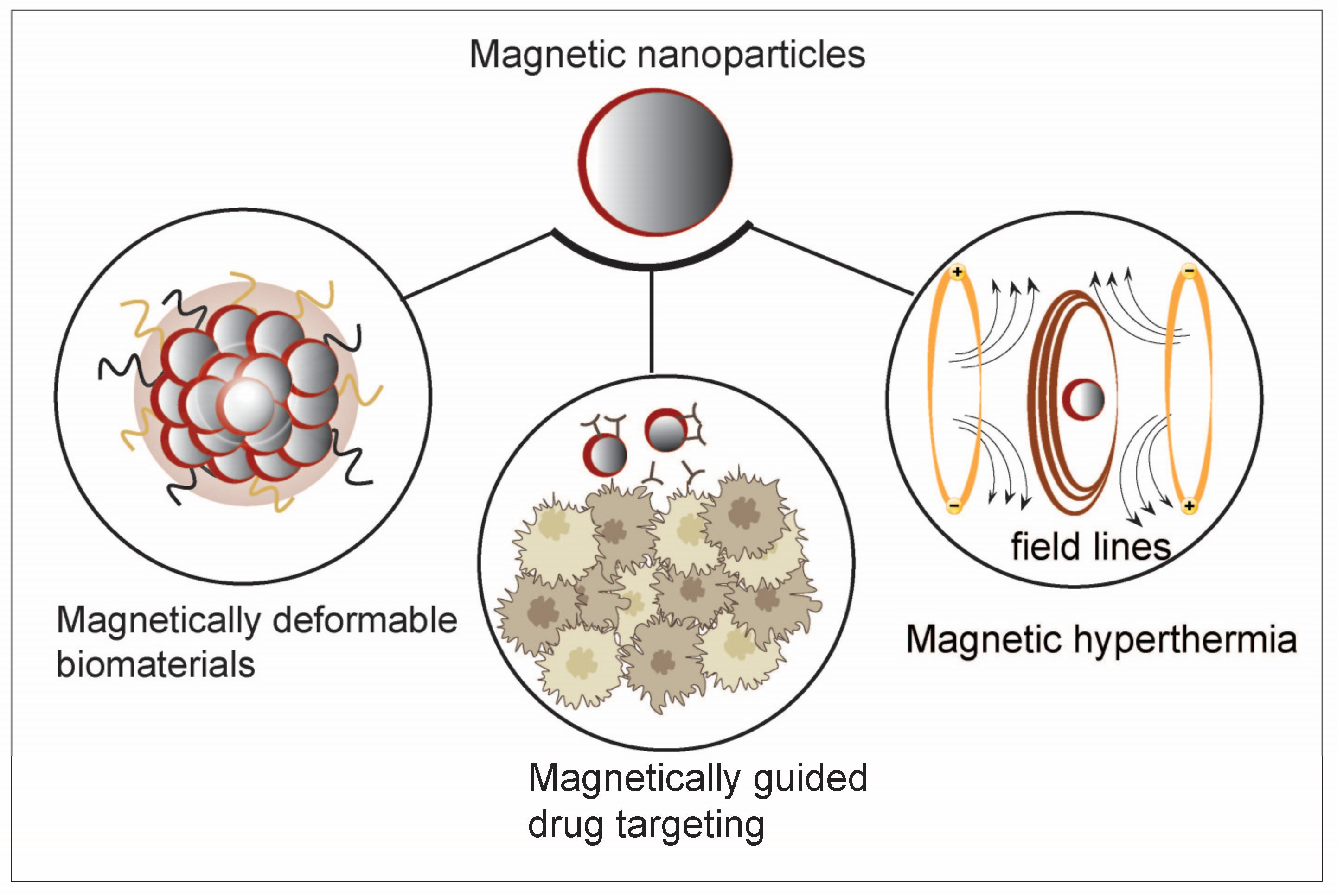
2.5. Iron Oxide-Based Cancer Immunotherapy

Iron oxide NPs (IONPs) in the acidic TME induce ferroptosis in tumor cells by releasing ions to initiate the Fenton reaction, resulting in the generation of ·OH. This leads to the exposure of TAAs in tumor cells, enhancing the immunogenicity of the microenvironment and providing potential clues for anticancer therapy [74,75,76]. Additionally, Fe3O4 NPs can form nanozymes when combined with ultra-small CaO2. In the weakly acidic TME, CaO2 triggers a cascade reaction to produce a significant amount of H2O2. The generated H2O2 then catalyzes a Fenton-like reaction mediated by Fe3O4 NPs, leading to the production of ·OH. This process promotes lipid peroxidation, inducing ferroptosis and facilitating the release of TAAs, ultimately creating an immunogenic TME. Moreover, this mechanism plays a crucial role in promoting DC maturation and enhancing immune modulation responses [77].
Iron oxide-based nanomaterials have been useful materials for tumor treatment due to their high relaxation, strong magnetic properties, and good biocompatibility, and they were among the first NPs approved for clinical use [78]. Unlike other metal-semiconductor nanomaterials, iron oxide is biodegradable and has no long-term toxicity [79,80]. As a typical class of iron-based materials, superparamagnetic IONPs can be used in magnetic particle imaging and MRI. IONPs have been incorporated into cancer immunotherapy, with applications including improving the efficiency of therapeutic and regulatory molecules on immune cells, increasing the presence of immune cells at the tumor site via magnetically guided cell delivery, and inducing local hyperthermia with immunostimulants as part of combination therapy and delivery [81] (Figure 6). In certain solid tumors, there is a problem of insufficient H2O2 content and imprecise antigen release. To further enhance anticancer immunity, the magnetic and superparamagnetic properties of iron oxide nanomaterials can be utilized to precisely control antigen release under external magnetic fields. Additionally, immunomodulators can be loaded onto these platforms, integrating ferroptosis and immunotherapy into one platform [77,82]. For instance, Zhang et al. constructed a biomimetic magnetic nanocomplex to promote the synergistic effect of ferroptosis and immunomodulation in cancer. This nanocomplex consists of Fe3O4 magnetic nanoparticle clusters as the core, pre-coated with leukocyte membrane for camouflage, loaded internally with a transforming growth factor-β inhibitor (Ti), and surface-anchored with a PD-1 antibody (pA). Upon intravenous injection, the membrane camouflage leads to prolonged circulation, and the magnetized and superparamagnetic Fe3O4 core enables MRI-guided magnetic targeting. Once inside the tumor, pA and Ti collaborate to create an immunogenic TME, increasing the H2O2 content in polarized M1 macrophages, which, in turn, promotes the Fenton reaction with iron ions released by Fe3O4. The generated ·OH subsequently induces ferroptosis in tumor cells, and the exposed tumor antigens, in turn, improve the immunogenicity of the microenvironment. Therefore, the synergistic effect of immunomodulation and ferroptosis in this cyclic manner results in a potent therapeutic effect [82].
In addition to addressing the issue of low immunogenicity in cold TME through the combination of ferroptosis and immunotherapy, magnetic IONPs can also enhance efficacy by synergizing with immunotherapy through MHT [83]. MHT is a novel tumor treatment method with good tissue penetration. Under an alternating magnetic field (AMF), IONPs have been applied to selectively kill tumor cells by generating heat (~41 °C) [84]. Under mild magnetic field heating, tumor cells can release damage-associated molecular patterns (DAMPs), promoting DC maturation and activating other components of the immune system, such as T lymphocytes and natural killer cells (NK cells) [85,86,87]. Additionally, research has shown that IONPs can induce M1 polarization to enhance tumor suppression [21]. The application of anti-PD-L1 antibodies after mild magnetic hyperthermia significantly promotes CTL infiltration, thereby inhibiting tumor metastasis [87]. PD-1 and its ligand PD-L1 are important immune checkpoint proteins that are overexpressed on tumor cells, leading to tumor immune evasion. Blocking the binding of PD-1 to PD-L1 with PD-1/PD-L1 inhibitors effectively enhances T cell immune response to tumors. Qiao et al. developed magnetic heat-triggered iron oxide nanocomposites by loading the immunomodulator JQ1 (an immune modulator) onto monodisperse IONPs. JQ1 directly downregulates PD-L1 levels via c-MYC, promoting anti-tumor immunity [88]. In the presence of AMF, iron oxide nanocomposites can rapidly heat up to 45 °C, triggering a strong immune response, including the activation of CTL cells and NK cells, Treg suppression, and M1 macrophage polarization. This approach completely eradicated primary tumors in 4T1 tumor-bearing mice and inhibited the growth of distant tumors in bilateral tumor models [89]. Zhang et al. developed a magneto-immunotherapy for solid tumors by combining a magnetic iron oxide nanocluster (IONC)-based heat and free radical generation with immune checkpoint blockade (ICB) therapy. The tumor cell death caused by the nanotherapeutic approach is extremely immunogenic, as demonstrated by the cell surface translocation of calreticulin (CRT) and Heat Shock Protein 70 (Hsp70) and the release of adenosine triphosphate (ATP), which has been shown to promote DC maturation. Under AMF, the combination of the IONC with anti-PD-1 ICB theatrically induced tumor-specific T cell response and enhanced tumor-infiltrating CD8+ T cells. Further, this magneto-immunotherapy also inspired a robust long-term immune memory effect against tumor metastasis and recurrence [90].
Tumor-associated macrophages (TAMs) play a crucial role in the tumor immunity cycle. Within the suppressive TME, TAMs are influenced by anti-inflammatory cytokines to adopt the M2 state, which in turn supports tumor cell proliferation [91,92]. However, by shifting TAMs towards the M1 state, the inhibitory TME can be partially reversed, leading to tumor cell apoptosis, enhanced CTL function, and tumor inhibition. The metabolic breakdown of IONPs within macrophages results in increased iron levels, directly stimulating M1 polarization [93]. Fe3O4 NP-loaded hyaluronic acid (HA)-modified doxorubicin (DOX) (Fe3O4-DOX-HA) has been constructed to mediate the specific delivery of Fe3O4 NPs to CD44-positive 4T1 tumor cells and TAMs. Combined with the targeted depletion of TAMs and M1 polarization effect of Fe3O4 NPs, Fe3O4-DOX-HA demonstrated strengthened anti-tumor and anti-metastasis effects in 4T1 orthotopic and metastatic mice models, representing a therapeutic candidate for anti-metastasis therapy against breast cancer [21]. Li and colleagues synthesized porous hollow IONPs to load a P13Kγ small molecule inhibitor, which was further modified by mannose to target TAMs. These IONPs could not merely polarize macrophages to reconstruct the immunosuppressive TME by increasing CD4+ and CD8+ T cells but also lessened the release of immunosuppressive cytokines by diminishing the proportion of Tregs and finally led to the effective inhibition of tumors [94] (Figure 7).

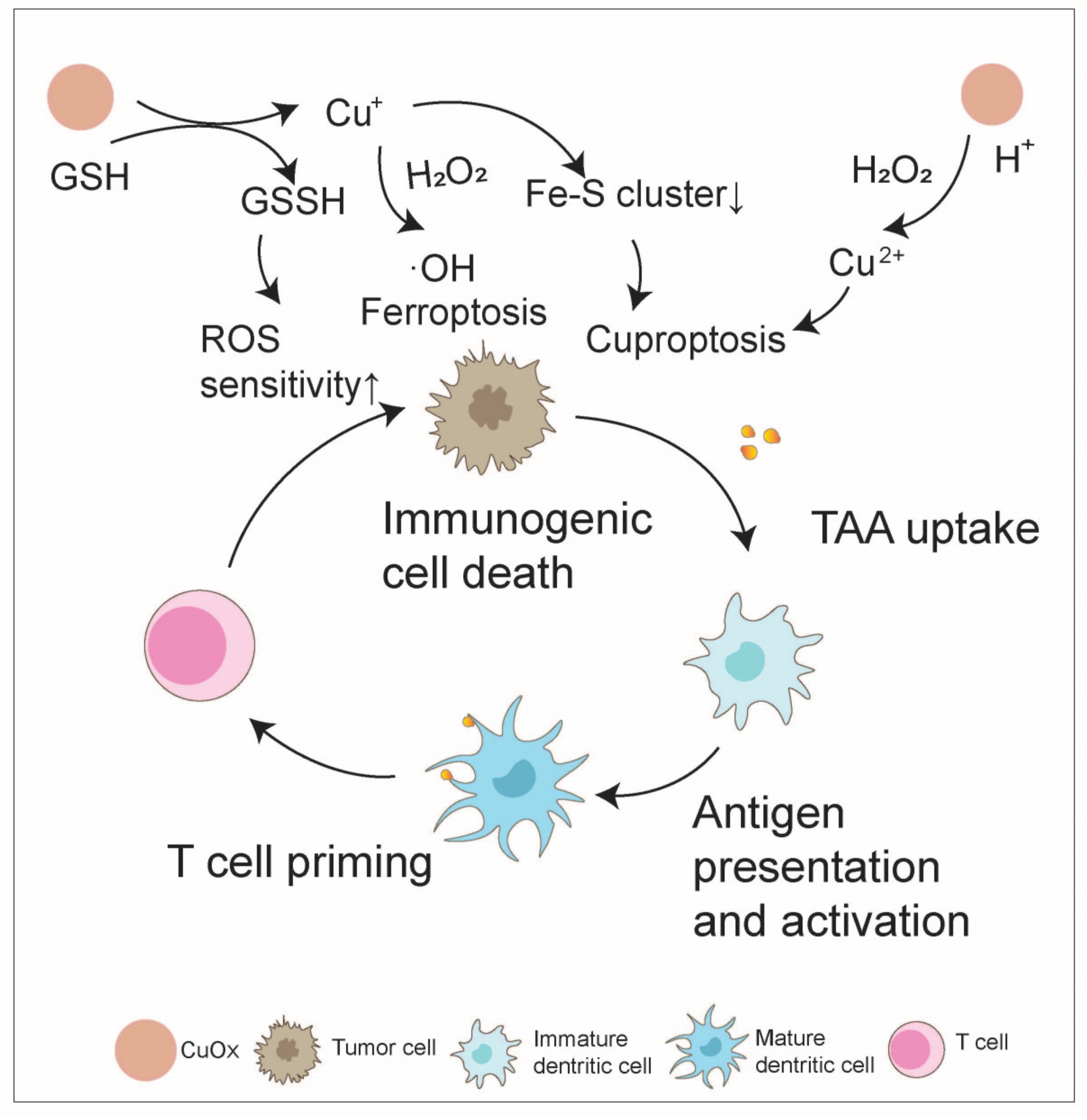
2.6. Copper Oxide-Based Cancer Immunotherapy

Due to the localized surface plasmon resonance (LSPR) characteristics of copper, nanomaterials exhibit excellent NIR absorption and remarkable photothermal properties [95,96]. In the TME, Cu2+ binds with H2O2 to generate O2, which is then reduced to Cu1+ by GSH, initiating a Fenton-like reaction. This reduces tumor hypoxia while generating ROS to eliminate tumor cells in conjunction with PTT and PDT, promoting ICD, and enhancing tumor immunogenicity [97]. To further address the issue of inadequate PDT efficiency, copper oxide can be designed into heterogeneous structures and loaded with immunoadjuvants to further amplify its efficacy. Jiang et al. employed a two-step hydrothermal method to combine semiconductor CuO with flower-like MoS2, designing a MoS2-CuO heterojunction structure loaded with bovine serum albumin (BSA) and the immunoadjuvant imiquimod (R837). Semiconductor CuO exhibits peroxidase-like activity similar to that of catalase, which can react with the overexpressed H2O2 in the TME via Haber–Weiss and Fenton reactions to generate ·OH. The multifunctional MoS2-CuO-BSA-R837 nanoplatform disrupts tumor cells under NIR irradiation, producing TAAs. The addition of R837 as an adjuvant triggers potent ICD, promoting the infiltration of CD4+ and CD8+ T cells and leading to a robust anti-tumor immune response, effectively eliminating primary and metastatic tumors [98].
In addition to enhancing immunogenicity, copper oxide NPs can also reverse tumor immune suppression through ferroptosis and cuproptosis mechanisms [97]. For instance, Ning and colleagues physically extruded a PTC system with AIE photosensitizer (TBP-2), Cu2O, and a platelet vesicle (PV). In an acidic TME, PTC was speedily degraded to release copper ions and H2O2, and the copper ions efflux could be inhibited considering that TBP-2 quickly entered the cell membrane and engendered ·OH to consume GSH under light irradiation. Ultimately, accumulated copper was able to cause lipoylated protein aggregation and iron–sulfur protein loss and lead to proteotoxic stress and, ultimately, cuproptosis, which increased the number of central memory CD4+ and CD8+ T cells collaborating with PDT, significantly inhibiting the lung metastasis of breast cancer and tumor recurrence [99]. In another case, engineered microbe–nanoparticle hybrid materials based on Escherichia coli (E. coli) and Cu2O NPs could accumulate at tumor sites after intravenous injection. Cu2O NPs consume endogenous hydrogen sulfide to convert to CuxS, displaying strong photothermal conversion in the NIR II biological window. Moreover, E. coli@Cu2O induces cell death via both ferroptosis and cuproptosis mechanisms through the deactivation of glutathione peroxidase (GPX4) and the aggregation of S-acyl transferase. Photothermally enhanced iron death/copper death mediated by E. coli@Cu2O reverses immune suppression in colorectal tumors by triggering DC maturation (by approximately 30%) and T cell activation (approximately 50% of CD8+ T cells). Combined with ICB, engineered microbe–NP hybrids can inhibit the growth of distant tumors under NIR irradiation [100] (Figure 8).

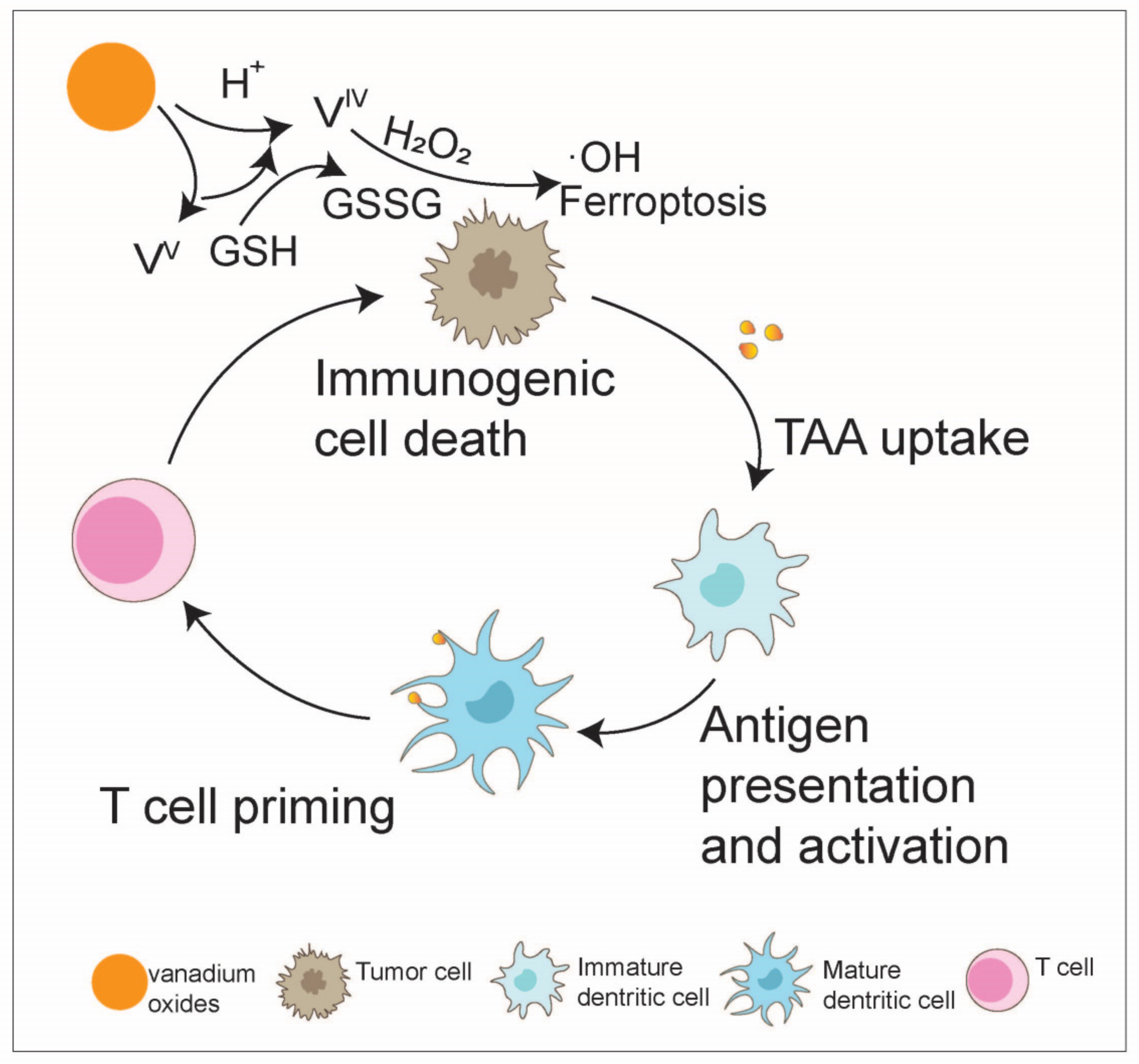
2.7. Other TMO-Based Cancer Immunotherapies

Cobalt oxide NPs (Co3O4 NPs) have received attention in recent years for their potential use as autophagy inhibitors, chemical sensitizers, and photosensitizers for synergistic anti-tumor treatment. Co3O4 NPs can not only induce autolysosomal accumulation and lysosomal function impairment by inhibiting the proteolytic activity of lysosomes and reducing intracellular ATP levels but can also be combined with the proteasome inhibitor Carfilzomib (CFZ) to strengthen the aggregation of autophagic substrates and endoplasmic reticulum stress, thereby inhibiting cancer progression. Co3O4 NPs can also be used as photothermal sensitizers to synergistically enhance the anticancer effect of CFZ owing to its photothermal conversion efficiency [101]. Moreover, cobalt oxides (Co3O4) play a role as a promising catalase-like nanozyme and can react with H2O2 in the TME to generate O2 to alleviate tumor hypoxia [102,103,104]. For example, the CuCo(O)/GOx@PCNs hybrid nanozyme was synthesized based on the Co3O4 nanozyme and achieved the function of oxygen supply, glucose consumption, and photothermal ablation, ultimately inhibiting both the growth of the primary tumor and distal tumor, together with boosting tumor immunity by promoting DC maturation, activating and proliferating CD8+ and CD4+ T cells, and enhancing the secretion of IL-12 and IFN-γ to interfere with tumor angiogenesis [105] (Figure 9).
The melting point of metallic vanadium is 1910 °C, and it can form an oxide with diverse oxidation states (from +II to +V) and different crystal structures [106]. The polyvalent state of the transition metal vanadium forwards its Fenton-like catalytic capacity, and many nanoplatforms based on vanadium have been reported [107,108,109,110,111]. A two-dimensional nanosheet containing VV has been synthesized, in which VV could produce ·OH and trigger apoptosis in human breast cancer cells [111]. In a study on a vanadium-based nanoplatform that synergizes ferroptosis-like therapy, it was found that under acidic conditions in tumor cells, vanadium oxides disassemble and release VV, VIV, and LND. The mixed valence of vanadium (VIV and VV) triggers ferroptosis by cyclically altering the valence of V, leading to the generation of ·OH for lipid peroxide accumulation (VIV → VV) and the depletion of GSH for GPX4 deactivation (VV → VIV). This process ultimately initiates ICD in tumor cells, activates CTLs, and inhibits B16F10 tumor growth and metastasis while enhancing immune memory [112] (Figure 10).

3. Conclusions and Future Perspectives

This paper provides an overview of the roles of TMOs in enhancing tumor immunogenicity and reversing the immune-suppressive TME, ultimately leading to the promotion of anti-tumor immune responses and improvement of immunotherapy efficacy. However, this field still faces many opportunities and challenges. To further promote the development and clinical translation of TMO-based tumor immunotherapy, efforts can be made in various aspects such as material preparation and modification, mechanism exploration, and the evaluation of biological properties.
First, the development of simple, practical, and reproducible synthetic methods is crucial for promoting the biological application of TMO nanomaterials. The biocompatibility of nanomaterials is crucial for their clinical translation [22,57]. Recently developed biomimetic mineralization strategies have been able to obtain TMO nanomaterials with good biocompatibility, making them the preferred choice for preparing manganese-based nanomaterials. Additionally, the surface modification of nanomaterials has a significant impact on their behavior in vivo. Therefore, the development of new strategies to finely modify the surface properties of TMO nanomaterials can significantly increase their accumulation at lesion sites and improve their therapeutic effects. For example, Schottky heterostructures can enhance the production of ROS by TMO nanomaterials, greatly promoting the efficacy of PDT and SDT [113,114].
Studies have shown that even in ectopic tumor models with high EPR effects, passive targeting of NPs only reaches 0.7% [115]. To address this limitation and improve the delivery of nanomedicines, the active targeting of TMOs has been proposed. This allows NPs to selectively reach tumor areas, minimizing toxicity. The development of tumor-targeting TMOs holds significant clinical translation value. For instance, the binding of iRGD facilitates the transport of TMOs to deep tumor tissues beyond blood vessels. By interacting with highly expressed integrin and neuronectin-1 receptors, iRGD effectively targets tumor cells and penetrates tumor tissue. Previous studies have introduced iRGD into MnO2 to enhance the targeting and penetration abilities of NPs for improved active targeting of TMOs [25]. In glioma, the presence of iRGD in nanosheets has shown a high capacity to cross the blood–brain barrier and penetrate tumor tissue. In addition to iRGD, TMOs can be combined with other targeting peptides for cancer immunotherapy [116]. For instance, liposome NPs labeled with LYP-1 peptide and embedded with MnO2 and photosensitizers preferentially target the tumor-homing peptide LYP-1. This approach effectively targets tumor cells and induces ROS production under NIR laser irradiation, leading to tumor cell death and modest immune activation. Gemcitabine and zinc oxide are coated and adsorbed by polypeptides that target the pancreatic TME, facilitating site-specific drug delivery. This system also includes a pancreatic tumor-targeting peptide (CKAAKN) that specifically targets pancreatic cancer cells and the TME. The combination of environmental vasculogenesis through the Wnt-2 pathway and the presence of the targeting peptide led to a significant increase of approximately 20% in positive outcomes in both cell lines. Furthermore, MnO2 nanosheets were combined with folic acid (FA), known for its high affinity towards folate receptors that are commonly overexpressed in human cancer cells. This combination was used to deliver the photosensitizer zinc phthalocyanine (ZnPc) for both PDT and bioimaging. The study revealed that FA-MnO2/ZnPc nanocarriers were efficiently internalized into cells through receptor-mediated endocytosis. Notably, after a 10-min irradiation with a 660 nm laser, significant inhibition of tumor growth was observed in mice treated with FA-MnO2/ZnPc along with irradiation [117].
In addition, TMOs can be modified to reduce potential off-target toxicity, such as sensitizing TiO2 with luciferase to enable activation by ATP. This activation leads to increased ATP levels, resulting in the localization of complex activity and the formation of O2•− within the tumor area. This, in turn, induces apoptosis in HCT116 colon cancer cells through a cascade reaction. Furthermore, by conjugating the C225 antibody to this luciferase–TiO2 nanocomposite (TiDoL), which can specifically recognize and bind to receptors expressed on the surface of HCT116 cells, the generation of ROS is limited to the surface of these cancer cells, enhancing the efficiency of O2•− interaction with neighboring cancer cells [118]. Substituting C225 with anti-IL13 as the anchoring antibody still resulted in effective therapeutic outcomes. This dual-locking approach not only minimizes the risk of the off-target release of radical species but also improves the effectiveness of delivering active species in close proximity to biological targets through in situ nanoparticle delivery. Moving forward, it is essential to further investigate and clarify the specific cellular targeting pathways and intratumoral barriers that may impede active targeting in order to enhance the effectiveness of actively targeting NPs in clinical applications [26].
Furthermore, the systematic investigation of the anti-tumor mechanisms of TMO nanomaterials is needed to guide the design and construction of multifunctional nanoplatforms. Although the immune mechanisms that are currently widely accepted include manganese ion and zinc ion activation of the cGAS-STING pathway and iron ions and copper ions activating the iron death pathway, these nanomaterials may still contain many other hidden mechanisms in practical applications.
Due to the complex immunomodulatory mechanism of TMOs, it is important to consider factors that may lead to immunosuppressive effects in order to maximize their immunostimulatory effects. For instance, research has shown that ZnO NPs of varying sizes and charges can trigger inflammation, potentially causing immunotoxicity [119]. Therefore, understanding the timing and mechanisms of inflammatory responses mediated by TMO NPs of different sizes is essential for the development of safe NPs with immunostimulatory properties. Additionally, surface coating, such as polyetherimide (PEI)-coating of IONPs, can promote immune activation by activating TLR4-mediated signaling and ROS production in macrophage cell lines [120]. It is crucial to control the time of exposure to TMOs as excessive ROS generated by TMO NPs can be harmful to immune cells. For example, prolonged exposure to TiO2 NPs in mouse models has been linked to a decrease in the number of NK cells. Therefore, careful consideration of these factors is necessary to avoid potential immunosuppressive effects and enhance the immunostimulatory effects of TMOs [121].
Lastly, a systematic evaluation of the biological properties and biosafety of TMO-based nanoplatforms is necessary, including cytotoxicity, blood compatibility, tissue compatibility, pharmacokinetics, and immunogenicity, although previous studies have shown that compared to metal ions, TMOs are more stable, have lower doses, and are more efficient with fewer side effects [122]. For instance, MnO2 nanomaterials, containing manganese, an essential trace element for the human body, exhibit a certain level of biocompatibility as the body can regulate its metabolism. Additionally, while the rapid renal clearance behavior of Mn2+ due to the degradation of the MnO2 nanostructure allows for easy excretion, resulting in negligible short-term toxicity, potential toxic effects of released Mn2+ ions must be considered as they can pose safety risks to cells and tissues, particularly at higher concentrations [123]. Previous studies have indicated that Mn2+ induces toxicity in hippocampal neurons by disrupting mitochondrial functions [124]. Similarly, rats that received intracerebral injections of Mn2+ (200 nL, i.e., 100 mM) exhibited neuronal toxicity and notable astrogliosis [125]. Zinc oxide NPs have shown promise in targeting cancer cells, enhancing cytotoxicity, and promoting cell death, thereby supporting the development of novel anti-tumor immunotherapies. Studies have indicated no hepatotoxic or nephrotoxic effects when using zinc oxide nanoparticles as anticancer agents. However, at elevated concentrations, such as 5 μg cm−2, zinc oxide has been observed to significantly reduce cell survival rates, alter cell morphology, and induce apoptosis in human colon cancer cells through the generation of ROS and the release of IL-8. Moreover, higher concentrations (10, 20, and 40 μg cm−2) can induce approximately 98% cytotoxicity, resulting in a cell survival rate of less than 5% after 24 h [13]. Previous studies have demonstrated that the dosage and duration of exposure to nano-TiO2 (21 Nm) significantly impact liver function enzymes, oxidative stress markers, and liver histological patterns. Genetic abnormalities were observed at high doses (500 mg kg−1 body weight) over extended periods (45 days). Furthermore, light exposure was found to intensify the genotoxic effects of oxidized NPs by increasing oxidative stress. The inhalation of iron oxide NPs was shown to induce oxidative stress by elevating ROS levels in the liver, spleen, lungs, and brain, resulting in inflammation, decreased cell viability, cell lysis, and disruptions in the coagulation system. Uncoated IONPs were non-cytotoxic and non-genotoxic, whereas oleic acid-coated iron oxide nanoparticles exhibited dose-dependent cytotoxicity and the potential to cause DNA damage, suggesting genotoxic properties [126]. Therefore, before clinical trials, the biological safety and effectiveness of manganese-based nanoplatforms should continue to be evaluated from small animal models (such as mice and rats) to large animal models (such as monkeys). Extensive toxicological studies on TMOs are needed to avoid metal dissolution catalysis or secondary pollution caused by NPs themselves. Based on their biosafety evaluation, we look forward to the promising application of TMO-based immunotherapy in nanomedicine.

Author Contributions

Conceptualization, Z.L. and X.C.; writing—original draft preparation, W.L., X.S. and Q.J.; writing—review and editing, X.S. and Q.J.; visualization, W.G. and J.L.; and supervision, Z.L., X.S. and X.C. All authors have read and agreed to the published version of the manuscript.

Funding

This work was supported by the China Postdoctoral Science Foundation (2020M670090ZX to Zengjie Lei), the National Natural Science Foundation of China (82072725 to Xiaoyuan Chu), and the Jiangsu Funding Program for Excellent Postdoctoral Talent (2023ZB381 to Xueru Song).

Conflicts of Interest

The authors declare no conflicts of interest.

References

  1. Binnewies, M.; Roberts, E.W.; Kersten, K.; Chan, V.; Fearon, D.F.; Merad, M.; Coussens, L.M.; Gabrilovich, D.I.; Ostrand-Rosenberg, S.; Hedrick, C.C.; et al. Understanding the tumor immune microenvironment (TIME) for effective therapy. Nat. Med. 2018, 24, 541–550. [Google Scholar] [CrossRef] [PubMed]
  2. Zhang, Y.; Zhang, Z. The history and advances in cancer immunotherapy: Understanding the characteristics of tumor-infiltrating immune cells and their therapeutic implications. Cell. Mol. Immunol. 2020, 17, 807–821. [Google Scholar] [CrossRef] [PubMed]
  3. Sahin, U.; Tureci, O. Personalized vaccines for cancer immunotherapy. Science 2018, 359, 1355–1360. [Google Scholar] [CrossRef] [PubMed]
  4. Yang, J.; Zhang, C. Regulation of cancer-immunity cycle and tumor microenvironment by nanobiomaterials to enhance tumor immunotherapy. WIREs Nanomed. Nanobiotechnol. 2020, 12, e1612. [Google Scholar] [CrossRef] [PubMed]
  5. Sundaram, A.; Francis, B.M.; Dhanabalan, S.C.; Ponraj, J.S. Transition metal carbide—MXene. In Handbook of Carbon-Based Nanomaterials; Elsevier: Amsterdam, The Netherlands, 2021; pp. 671–709. [Google Scholar]
  6. Wu, M.; Hou, P.; Dong, L.; Cai, L.; Chen, Z.; Zhao, M.; Li, J. Manganese dioxide nanosheets: From preparation to biomedical applications. Int. J. Nanomed. 2019, 14, 4781–4800. [Google Scholar] [CrossRef] [PubMed]
  7. Grigore, M.E.; Biscu, E.R.; Holban, A.M.; Gestal, M.C.; Grumezescu, A.M. Methods of Synthesis, Properties and Biomedical Applications of CuO Nanoparticles. Pharmarceuticals 2016, 9, 75. [Google Scholar] [CrossRef] [PubMed]
  8. Enoch, K.; Sundaram, A.; Ponraj, S.S.; Palaniyappan, S.; George, S.D.B.; Manavalan, R.K. Enhancement of MXene optical properties towards medical applications via metal oxide incorporation. Nanoscale 2023, 15, 16874–16889. [Google Scholar] [CrossRef] [PubMed]
  9. Hu, T.; Mei, X.; Wang, Y.; Weng, X.; Liang, R.; Wei, M. Two-dimensional nanomaterials: Fascinating materials in biomedical field. Sci. Bull. 2019, 64, 1707–1727. [Google Scholar] [CrossRef] [PubMed]
  10. Kalantar-zadeh, K.; Ou, J.Z.; Daeneke, T.; Mitchell, A.; Sasaki, T.; Fuhrer, M.S. Two dimensional and layered transition metal oxides. Appl. Mater. Today 2016, 5, 73–89. [Google Scholar] [CrossRef]
  11. Curcio, A.; Silva, A.K.A.; Cabana, S.; Espinosa, A.; Baptiste, B.; Menguy, N.; Wilhelm, C.; Abou-Hassan, A. Iron Oxide Nanoflowers @ CuS Hybrids for Cancer Tri-Therapy: Interplay of Photothermal Therapy, Magnetic Hyperthermia and Photodynamic Therapy. Theranostics 2019, 9, 1288–1302. [Google Scholar] [CrossRef]
  12. Yang, M.; Li, J.; Gu, P.; Fan, X. The application of nanoparticles in cancer immunotherapy: Targeting tumor microenvironment. Bioact. Mater. 2021, 6, 1973–1987. [Google Scholar] [CrossRef] [PubMed]
  13. Sengul, A.B.; Asmatulu, E. Toxicity of metal and metal oxide nanoparticles: A review. Environ. Chem. Lett. 2020, 18, 1659–1683. [Google Scholar] [CrossRef]
  14. Shen, F.; Fang, Y.; Wu, Y.; Zhou, M.; Shen, J.; Fan, X. Metal ions and nanometallic materials in antitumor immunity: Function, application, and perspective. J. Nanobiotechnol. 2023, 21, 20. [Google Scholar] [CrossRef] [PubMed]
  15. Sindhwani, S.; Syed, A.M.; Ngai, J.; Kingston, B.R.; Maiorino, L.; Rothschild, J.; MacMillan, P.; Zhang, Y.; Rajesh, N.U.; Hoang, T.; et al. The entry of nanoparticles into solid tumours. Nat. Mater. 2020, 19, 566–575. [Google Scholar] [CrossRef] [PubMed]
  16. Yang, G.; Ji, J.; Liu, Z. Multifunctional MnO2 nanoparticles for tumor microenvironment modulation and cancer therapy. Wiley Interdiscip. Rev. Nanomed. Nanobiotechnol. 2021, 13, e1720. [Google Scholar] [CrossRef] [PubMed]
  17. Zhang, D.-Y.; Huang, F.; Ma, Y.; Liang, G.; Peng, Z.; Guan, S.; Zhai, J. Tumor Microenvironment-Responsive Theranostic Nanoplatform for Guided Molecular Dynamic/Photodynamic Synergistic Therapy. ACS Appl. Mater. Interfaces 2021, 13, 17392–17403. [Google Scholar] [CrossRef] [PubMed]
  18. Liu, Y.; Wang, Y.; Song, S.; Zhang, H. Cascade-responsive nanobomb with domino effect for anti-tumor synergistic therapies. Natl. Sci. Rev. 2022, 9, nwab139. [Google Scholar] [CrossRef] [PubMed]
  19. Ding, H.; Zhang, Y.; Mao, Y.; Li, Y.; Shen, Y.; Sheng, J.; Gu, N. Modulation of macrophage polarization by iron-based nanoparticles. Med. Rev. 2023, 3, 105–122. [Google Scholar] [CrossRef]
  20. Guo, W.; Wu, X.; Wei, W.; Wang, Y.; Dai, H. Mesoporous hollow Fe3O4 nanoparticles regulate the behavior of neuro-associated cells through induction of macrophage polarization in an alternating magnetic field. J. Mater. Chem. B 2022, 10, 5633–5643. [Google Scholar] [CrossRef]
  21. Gong, T.; Dong, Z.; Fu, Y.; Gong, T.; Deng, L.; Zhang, Z. Hyaluronic acid modified doxorubicin loaded Fe3O4 nanoparticles effectively inhibit breast cancer metastasis. J. Mater. Chem. B 2019, 7, 5861–5872. [Google Scholar] [CrossRef]
  22. Deng, Z.; Xi, M.; Zhang, C.; Wu, X.; Li, Q.; Wang, C.; Fang, H.; Sun, G.; Zhang, Y.; Yang, G.; et al. Biomineralized MnO2 Nanoplatforms Mediated Delivery of Immune Checkpoint Inhibitors with STING Pathway Activation to Potentiate Cancer Radio-Immunotherapy. ACS Nano 2023, 17, 4495–4506. [Google Scholar] [CrossRef] [PubMed]
  23. Xie, L.; Wang, G.; Sang, W.; Li, J.; Zhang, Z.; Li, W.; Yan, J.; Zhao, Q.; Dai, Y. Phenolic immunogenic cell death nanoinducer for sensitizing tumor to PD-1 checkpoint blockade immunotherapy. Biomaterials 2021, 269, 120638. [Google Scholar] [CrossRef] [PubMed]
  24. Sharma, P.; Shin, J.B.; Park, B.C.; Lee, J.W.; Byun, S.W.; Jang, N.Y.; Kim, Y.J.; Kim, Y.; Kim, Y.K.; Cho, N.H. Application of radially grown ZnO nanowires on poly-l-lactide microfibers complexed with a tumor antigen for cancer immunotherapy. Nanoscale 2019, 11, 4591–4600. [Google Scholar] [CrossRef] [PubMed]
  25. Li, X.; Feng, X.; Sun, C.; Liu, Y.; Zhao, Q.; Wang, S. Mesoporous carbon-manganese nanocomposite for multiple imaging guided oxygen-elevated synergetic therapy. J. Control. Release 2020, 319, 104–118. [Google Scholar] [CrossRef] [PubMed]
  26. Barui, S.; Percivalle, N.M.; Conte, M.; Dumontel, B.; Racca, L.; Carofiglio, M.; Cauda, V. Development of doped ZnO-based biomimicking and tumor-targeted nanotheranostics to improve pancreatic cancer treatment. Cancer Nanotechnol. 2022, 13, 37. [Google Scholar] [CrossRef]
  27. Liu, X.; Rong, P. Recent Advances of Manganese-Based Hybrid Nanomaterials for Cancer Precision Medicine. Front. Oncol. 2021, 11, 707618. [Google Scholar] [CrossRef] [PubMed]
  28. Xu, W.; Qing, X.; Liu, S.; Yang, D.; Dong, X.; Zhang, Y. Hollow Mesoporous Manganese Oxides: Application in Cancer Diagnosis and Therapy. Small 2022, 18, e2106511. [Google Scholar] [CrossRef] [PubMed]
  29. Ding, B.; Zheng, P.; Ma, P.; Lin, J. Manganese Oxide Nanomaterials: Synthesis, Properties, and Theranostic Applications. Adv. Mater. 2020, 32, e1905823. [Google Scholar] [CrossRef]
  30. Ahmed, S.; Annu; Chaudhry, S.A.; Ikram, S. A review on biogenic synthesis of ZnO nanoparticles using plant extracts and microbes: A prospect towards green chemistry. J. Photochem. Photobiol. B 2017, 166, 272–284. [Google Scholar] [CrossRef]
  31. Chimene, D.; Alge, D.L.; Gaharwar, A.K. Two-Dimensional Nanomaterials for Biomedical Applications: Emerging Trends and Future Prospects. Adv. Mater. 2015, 27, 7261–7284. [Google Scholar] [CrossRef]
  32. Wang, H.; Zhuo, S.; Liang, Y.; Han, X.; Zhang, B. General Self-Template Synthesis of Transition-Metal Oxide and Chalcogenide Mesoporous Nanotubes with Enhanced Electrochemical Performances. Angew. Chem. Int. Ed. Engl. 2016, 55, 9055–9059. [Google Scholar] [CrossRef] [PubMed]
  33. Yao, S.; Wang, S.; Liu, Y.; Hou, Z.; Wang, J.; Gao, X.; Sun, Y.; Fu, W.; Nie, K.; Xie, J.; et al. High Flux and Stability of Cationic Intercalation in Transition-Metal Oxides: Unleashing the Potential of Mn t2g Orbital via Enhanced pi-Donation. J. Am. Chem. Soc. 2023, 145, 26699–26710. [Google Scholar] [CrossRef] [PubMed]
  34. Ziental, D.; Czarczynska-Goslinska, B.; Mlynarczyk, D.T.; Glowacka-Sobotta, A.; Stanisz, B.; Goslinski, T.; Sobotta, L. Titanium Dioxide Nanoparticles: Prospects and Applications in Medicine. Nanomaterials 2020, 10, 387. [Google Scholar] [CrossRef] [PubMed]
  35. Rehman, F.U.; Zhao, C.; Jiang, H.; Wang, X. Biomedical applications of nano-titania in theranostics and photodynamic therapy. Biomater. Sci. 2016, 4, 40–54. [Google Scholar] [CrossRef] [PubMed]
  36. Son, S.; Kim, J.H.; Wang, X.; Zhang, C.; Yoon, S.A.; Shin, J.; Sharma, A.; Lee, M.H.; Cheng, L.; Wu, J.; et al. Multifunctional sonosensitizers in sonodynamic cancer therapy. Chem. Soc. Rev. 2020, 49, 3244–3261. [Google Scholar] [CrossRef] [PubMed]
  37. Tao, N.; Li, H.; Deng, L.; Zhao, S.; Ouyang, J.; Wen, M.; Chen, W.; Zeng, K.; Wei, C.; Liu, Y.N. A Cascade Nanozyme with Amplified Sonodynamic Therapeutic Effects through Comodulation of Hypoxia and Immunosuppression against Cancer. ACS Nano 2022, 16, 485–501. [Google Scholar] [CrossRef] [PubMed]
  38. Zhang, R.; Yan, F.; Chen, Y. Exogenous Physical Irradiation on Titania Semiconductors: Materials Chemistry and Tumor-Specific Nanomedicine. Adv. Sci. 2018, 5, 1801175. [Google Scholar] [CrossRef]
  39. Lucky, S.S.; Idris, N.M.; Huang, K.; Kim, J.; Li, Z.; Thong, P.S.; Xu, R.; Soo, K.C.; Zhang, Y. In vivo Biocompatibility, Biodistribution and Therapeutic Efficiency of Titania Coated Upconversion Nanoparticles for Photodynamic Therapy of Solid Oral Cancers. Theranostics 2016, 6, 1844–1865. [Google Scholar] [CrossRef]
  40. Gilson, R.C.; Black, K.C.L.; Lane, D.D.; Achilefu, S. Hybrid TiO2–Ruthenium Nano-photosensitizer Synergistically Produces Reactive Oxygen Species in both Hypoxic and Normoxic Conditions. Angew. Chem. Int. Ed. Engl. 2017, 56, 10717–10720. [Google Scholar] [CrossRef]
  41. Shang, H.; Han, D.; Ma, M.; Li, S.; Xue, W.; Zhang, A. Enhancement of the photokilling effect of TiO2 in photodynamic therapy by conjugating with reduced graphene oxide and its mechanism exploration. J. Photochem. Photobiol. B 2017, 177, 112–123. [Google Scholar] [CrossRef]
  42. Zhou, J.-Y.; Wang, W.-J.; Zhang, C.-Y.; Ling, Y.-Y.; Hong, X.-J.; Su, Q.; Li, W.-G.; Mao, Z.-W.; Cheng, B.; Tan, C.-P.; et al. Ru(II)-modified TiO2 nanoparticles for hypoxia-adaptive photo-immunotherapy of oral squamous cell carcinoma. Biomaterials 2022, 289, 121757. [Google Scholar] [CrossRef]
  43. Hesemans, E.; Saffarzadeh, N.; Maksoudian, C.; Izci, M.; Chu, T.; Rios Luci, C.; Wang, Y.; Naatz, H.; Thieme, S.; Richter, C.; et al. Cu-doped TiO2 nanoparticles improve local antitumor immune activation and optimize dendritic cell vaccine strategies. J. Nanobiotechnol. 2023, 21, 87. [Google Scholar] [CrossRef]
  44. Zhang, K.; Qi, C.; Cai, K. Manganese-Based Tumor Immunotherapy. Adv. Mater. 2023, 35, e2205409. [Google Scholar] [CrossRef] [PubMed]
  45. Zhang, R.; Wang, C.; Guan, Y.; Wei, X.; Sha, M.; Yi, M.; Jing, M.; Lv, M.; Guo, W.; Xu, J.; et al. Manganese salts function as potent adjuvants. Cell. Mol. Immunol. 2021, 18, 1222–1234. [Google Scholar] [CrossRef]
  46. Fan, D.; Shang, C.; Gu, W.; Wang, E.; Dong, S. Introducing Ratiometric Fluorescence to MnO2 Nanosheet-Based Biosensing: A Simple, Label-Free Ratiometric Fluorescent Sensor Programmed by Cascade Logic Circuit for Ultrasensitive GSH Detection. ACS Appl. Mater. Interfaces 2017, 9, 25870–25877. [Google Scholar] [CrossRef] [PubMed]
  47. Yan, X.; Song, Y.; Zhu, C.; Song, J.; Du, D.; Su, X.; Lin, Y. Graphene Quantum Dot-MnO2 Nanosheet Based Optical Sensing Platform: A Sensitive Fluorescence “Turn Off-On” Nanosensor for Glutathione Detection and Intracellular Imaging. ACS Appl. Mater. Interfaces 2016, 8, 21990–21996. [Google Scholar] [CrossRef] [PubMed]
  48. Yang, G.; Xu, L.; Chao, Y.; Xu, J.; Sun, X.; Wu, Y.; Peng, R.; Liu, Z. Hollow MnO2 as a tumor-microenvironment-responsive biodegradable nano-platform for combination therapy favoring antitumor immune responses. Nat. Commun. 2017, 8, 902. [Google Scholar] [CrossRef]
  49. He, L.; Wang, J.; Zhu, P.; Chen, J.; Zhao, S.; Liu, X.; Li, Y.; Guo, X.; Yan, Z.; Shen, X.; et al. Intelligent manganese dioxide nanocomposites induce tumor immunogenic cell death and remould tumor microenvironment. Chem. Eng. J. 2023, 461, 141369. [Google Scholar] [CrossRef]
  50. Jian, H.; Wang, X.; Song, P.; Wu, X.; Zheng, R.; Wang, Y.; Zhang, H. Tumor microcalcification-mediated relay drug delivery for photodynamic immunotherapy of breast cancer. Acta Biomater. 2022, 140, 518–529. [Google Scholar] [CrossRef]
  51. Zhu, P.; Chen, Y.; Shi, J. Nanoenzyme-Augmented Cancer Sonodynamic Therapy by Catalytic Tumor Oxygenation. ACS Nano 2018, 12, 3780–3795. [Google Scholar] [CrossRef]
  52. Cheung, E.C.; Vousden, K.H. The role of ROS in tumour development and progression. Nat. Rev. Cancer 2022, 22, 280–297. [Google Scholar] [CrossRef] [PubMed]
  53. An, J.; Hu, Y.G.; Cheng, K.; Li, C.; Hou, X.L.; Wang, G.L.; Zhang, X.S.; Liu, B.; Zhao, Y.D.; Zhang, M.Z. ROS-augmented and tumor-microenvironment responsive biodegradable nanoplatform for enhancing chemo-sonodynamic therapy. Biomaterials 2020, 234, 119761. [Google Scholar] [CrossRef] [PubMed]
  54. Song, X.; Zhou, X.; Pan, Y.; Liang, K.; Luo, Y.; Xie, W.; Lv, Z.; Yang, D.; Wang, Y.; Wu, X.S.; et al. Schottky Heterojunction Realizes In Situ Vaccine-like Antitumor Efficacy and Microenvironment Remodeling upon Near-Infrared Laser Response in Cold Tumors. Adv. Funct. Mater. 2023, 33, 2306734. [Google Scholar] [CrossRef]
  55. Wen, Y.; Liu, Y.; Chen, C.; Chi, J.; Zhong, L.; Zhao, Y.; Zhao, Y. Metformin loaded porous particles with bio-microenvironment responsiveness for promoting tumor immunotherapy. Biomater. Sci. 2021, 9, 2082–2089. [Google Scholar] [CrossRef] [PubMed]
  56. Mi, P.; Kokuryo, D.; Cabral, H.; Wu, H.; Terada, Y.; Saga, T.; Aoki, I.; Nishiyama, N.; Kataoka, K. A pH-activatable nanoparticle with signal-amplification capabilities for non-invasive imaging of tumour malignancy. Nat. Nanotechnol. 2016, 11, 724–730. [Google Scholar] [CrossRef] [PubMed]
  57. Qi, C.; He, J.; Fu, L.H.; He, T.; Blum, N.T.; Yao, X.; Lin, J.; Huang, P. Tumor-Specific Activatable Nanocarriers with Gas-Generation and Signal Amplification Capabilities for Tumor Theranostics. ACS Nano 2021, 15, 1627–1639. [Google Scholar] [CrossRef] [PubMed]
  58. Harris, I.S.; DeNicola, G.M. The Complex Interplay between Antioxidants and ROS in Cancer. Trends Cell Biol. 2020, 30, 440–451. [Google Scholar] [CrossRef] [PubMed]
  59. Liang, J.L.; Jin, X.K.; Zhang, S.M.; Huang, Q.X.; Ji, P.; Deng, X.C.; Cheng, S.X.; Chen, W.H.; Zhang, X.Z. Specific activation of cGAS-STING pathway by nanotherapeutics-mediated ferroptosis evoked endogenous signaling for boosting systemic tumor immunotherapy. Sci. Bull. 2023, 68, 622–636. [Google Scholar] [CrossRef] [PubMed]
  60. Lin, L.-S.; Wang, J.-F.; Song, J.; Liu, Y.; Zhu, G.; Dai, Y.; Shen, Z.; Tian, R.; Song, J.; Wang, Z.; et al. Cooperation of endogenous and exogenous reactive oxygen species induced by zinc peroxide nanoparticles to enhance oxidative stress-based cancer therapy. Theranostics 2019, 9, 7200–7209. [Google Scholar] [CrossRef]
  61. Ye, D.X.; Ma, Y.Y.; Zhao, W.; Cao, H.M.; Kong, J.L.; Xiong, H.M.; Mohwald, H. ZnO-Based Nanoplatforms for Labeling and Treatment of Mouse Tumors without Detectable Toxic Side Effects. ACS Nano 2016, 10, 4294–4300. [Google Scholar] [CrossRef]
  62. Martinez-Carmona, M.; Gun’ko, Y.; Vallet-Regi, M. ZnO Nanostructures for Drug Delivery and Theranostic Applications. Nanomaterials 2018, 8, 268. [Google Scholar] [CrossRef] [PubMed]
  63. Zhang, Y.; Guo, C.; Liu, L.; Xu, J.; Jiang, H.; Li, D.; Lan, J.; Li, J.; Yang, J.; Tu, Q.; et al. ZnO-based multifunctional nanocomposites to inhibit progression and metastasis of melanoma by eliciting antitumor immunity via immunogenic cell death. Theranostics 2020, 10, 11197–11214. [Google Scholar] [CrossRef] [PubMed]
  64. Dong, S.; Xu, J.; Jia, T.; Xu, M.; Zhong, C.; Yang, G.; Li, J.; Yang, D.; He, F.; Gai, S.; et al. Upconversion-mediated ZnFe2O4 nanoplatform for NIR-enhanced chemodynamic and photodynamic therapy. Chem. Sci. 2019, 10, 4259–4271. [Google Scholar] [CrossRef] [PubMed]
  65. Prosser, B.L.; Ward, C.W.; Lederer, W.J. X-ROS signaling: Rapid mechano-chemo transduction in heart. Science 2011, 333, 1440–1445. [Google Scholar] [CrossRef] [PubMed]
  66. Ha, N.Y.; Shin, H.M.; Sharma, P.; Cho, H.A.; Min, C.K.; Kim, H.I.; Yen, N.T.; Kang, J.S.; Kim, I.S.; Choi, M.S.; et al. Generation of protective immunity against Orientia tsutsugamushi infection by immunization with a zinc oxide nanoparticle combined with ScaA antigen. J. Nanobiotechnol. 2016, 14, 76. [Google Scholar] [CrossRef] [PubMed]
  67. Roy, R.; Das, M.; Dwivedi, P.D. Toxicological mode of action of ZnO nanoparticles: Impact on immune cells. Mol. Immunol. 2015, 63, 184–192. [Google Scholar] [CrossRef] [PubMed]
  68. Roy, R.; Singh, S.K.; Das, M.; Tripathi, A.; Dwivedi, P.D. Toll-like receptor 6 mediated inflammatory and functional responses of zinc oxide nanoparticles primed macrophages. Immunology 2014, 142, 453–464. [Google Scholar] [CrossRef]
  69. Marques Neto, L.M.; Kipnis, A.; Junqueira-Kipnis, A.P. Role of Metallic Nanoparticles in Vaccinology: Implications for Infectious Disease Vaccine Development. Front. Immunol. 2017, 8, 239. [Google Scholar] [CrossRef]
  70. Shen, Q.; Yang, J.; Liu, R.Y.; Liu, L.Y.; Zhang, J.C.; Shen, S.G.; Zhang, X. Hybrid ‘clusterbombs’ as multifunctional nanoplatforms potentiate brain tumor immunotherapy. Mater. Horiz. 2019, 6, 810–816. [Google Scholar] [CrossRef]
  71. Afroz, S.; Medhi, H.; Maity, S.; Minhas, G.; Battu, S.; Giddaluru, J.; Kumar, K.; Paik, P.; Khan, N. Mesoporous ZnO nanocapsules for the induction of enhanced antigen-specific immunological responses. Nanoscale 2017, 9, 14641–14653. [Google Scholar] [CrossRef]
  72. Antoine, T.E.; Hadigal, S.R.; Yakoub, A.M.; Mishra, Y.K.; Bhattacharya, P.; Haddad, C.; Valyi-Nagy, T.; Adelung, R.; Prabhakar, B.S.; Shukla, D. Intravaginal Zinc Oxide Tetrapod Nanoparticles as Novel Immunoprotective Agents against Genital Herpes. J. Immunol. 2016, 196, 4566–4575. [Google Scholar] [CrossRef] [PubMed]
  73. Wang, X.; Li, X.; Ito, A.; Sogo, Y.; Watanabe, Y.; Tsuji, N.M.; Ohno, T. Biodegradable Metal Ion-Doped Mesoporous Silica Nanospheres Stimulate Anticancer Th1 Immune Response in Vivo. ACS Appl. Mater. Interfaces 2017, 9, 43538–43544. [Google Scholar] [CrossRef] [PubMed]
  74. Kim, K.-S.; Choi, B.; Choi, H.; Ko, M.J.; Kim, D.-H.; Kim, D.-H. Enhanced natural killer cell anti-tumor activity with nanoparticles mediated ferroptosis and potential therapeutic application in prostate cancer. J. Nanobiotechnol. 2022, 20, 428. [Google Scholar] [CrossRef]
  75. Chin, Y.-C.; Yang, L.-X.; Hsu, F.-T.; Hsu, C.-W.; Chang, T.-W.; Chen, H.-Y.; Chen, L.Y.-C.; Chia, Z.C.; Hung, C.-H.; Su, W.-C.; et al. Iron oxide@chlorophyll clustered nanoparticles eliminate bladder cancer by photodynamic immunotherapy-initiated ferroptosis and immunostimulation. J. Nanobiotechnol. 2022, 20, 373. [Google Scholar] [CrossRef]
  76. Chen, X.; Kang, R.; Kroemer, G.; Tang, D. Broadening horizons: The role of ferroptosis in cancer. Nat. Rev. Clin. Oncol. 2021, 18, 280–296. [Google Scholar] [CrossRef] [PubMed]
  77. Li, Z.; Rong, L. Cascade reaction-mediated efficient ferroptosis synergizes with immunomodulation for high-performance cancer therapy. Biomater. Sci. 2020, 8, 6272–6285. [Google Scholar] [CrossRef]
  78. Peng, Q.; Zhang, S.; Yang, H.; Sheng, B.; Xu, R.; Wang, Q.; Yu, Y. Boosting Potassium Storage Performance of the Cu2S Anode via Morphology Engineering and Electrolyte Chemistry. ACS Nano 2020, 14, 6024–6033. [Google Scholar] [CrossRef]
  79. Akbarzadeh, A.; Rezaei-Sadabady, R.; Davaran, S.; Joo, S.W.; Zarghami, N.; Hanifehpour, Y.; Samiei, M.; Kouhi, M.; Nejati-Koshki, K. Liposome: Classification, preparation, and applications. Nanoscale Res. Lett. 2013, 8, 102. [Google Scholar] [CrossRef]
  80. Han, J.; Zhao, D.; Li, D.; Wang, X.; Jin, Z.; Zhao, K. Polymer-Based Nanomaterials and Applications for Vaccines and Drugs. Polymers 2018, 10, 31. [Google Scholar] [CrossRef]
  81. Chung, S.; Revia, R.A.; Zhang, M. Iron oxide nanoparticles for immune cell labeling and cancer immunotherapy. Nanoscale Horiz. 2021, 6, 696–717. [Google Scholar] [CrossRef]
  82. Zhang, F.; Li, F.; Lu, G.-H.; Nie, W.; Zhang, L.; Lv, Y.; Bao, W.; Gao, X.; Wei, W.; Pu, K.; et al. Engineering Magnetosomes for Ferroptosis/Immunomodulation Synergism in Cancer. ACS Nano 2019, 13, 5662–5673. [Google Scholar] [CrossRef] [PubMed]
  83. Xue, F.; Zhu, S.; Tian, Q.; Qin, R.; Wang, Z.; Huang, G.; Yang, S. Macrophage-mediated delivery of magnetic nanoparticles for enhanced magnetic resonance imaging and magnetothermal therapy of solid tumors. J. Colloid Interface Sci. 2023, 629, 554–562. [Google Scholar] [CrossRef] [PubMed]
  84. Gavilan, H.; Avugadda, S.K.; Fernandez-Cabada, T.; Soni, N.; Cassani, M.; Mai, B.T.; Chantrell, R.; Pellegrino, T. Magnetic nanoparticles and clusters for magnetic hyperthermia: Optimizing their heat performance and developing combinatorial therapies to tackle cancer. Chem. Soc. Rev. 2021, 50, 11614–11667. [Google Scholar] [CrossRef] [PubMed]
  85. An, J.; Zhang, K.; Wang, B.; Wu, S.; Wang, Y.; Zhang, H.; Zhang, Z.; Liu, J.; Shi, J. Nanoenabled Disruption of Multiple Barriers in Antigen Cross-Presentation of Dendritic Cells via Calcium Interference for Enhanced Chemo-Immunotherapy. ACS Nano 2020, 14, 7639–7650. [Google Scholar] [CrossRef] [PubMed]
  86. Pan, J.; Xu, Y.; Wu, Q.; Hu, P.; Shi, J. Mild Magnetic Hyperthermia-Activated Innate Immunity for Liver Cancer Therapy. J. Am. Chem. Soc. 2021, 143, 8116–8128. [Google Scholar] [CrossRef] [PubMed]
  87. Liu, X.; Zheng, J.; Sun, W.; Zhao, X.; Li, Y.; Gong, N.; Wang, Y.; Ma, X.; Zhang, T.; Zhao, L.Y.; et al. Ferrimagnetic Vortex Nanoring-Mediated Mild Magnetic Hyperthermia Imparts Potent Immunological Effect for Treating Cancer Metastasis. ACS Nano 2019, 13, 8811–8825. [Google Scholar] [CrossRef] [PubMed]
  88. Qiao, H.; Chen, X.; Wang, Q.; Zhang, J.; Huang, D.; Chen, E.; Qian, H.; Zhong, Y.; Tang, Q.; Chen, W. Tumor localization of oncolytic adenovirus assisted by pH-degradable microgels with JQ1-mediated boosting replication and PD-L1 suppression for enhanced cancer therapy. Biomater. Sci. 2020, 8, 2472–2480. [Google Scholar] [CrossRef]
  89. Gao, M.; Feng, K.; Zhang, X.; Ruan, Y.; Zhao, G.; Liu, H.; Sun, X. Nanoassembly with self-regulated magnetic thermal therapy and controlled immuno-modulating agent release for improved immune response. J. Control. Release 2023, 357, 40–51. [Google Scholar] [CrossRef]
  90. Zhang, L.; Zhang, Q.; Hinojosa, D.T.; Jiang, K.; Pham, Q.K.; Xiao, Z.; Colvin, V.L.; Bao, G. Multifunctional Magnetic Nanoclusters Can Induce Immunogenic Cell Death and Suppress Tumor Recurrence and Metastasis. ACS Nano 2022, 16, 18538–18554. [Google Scholar] [CrossRef]
  91. Zhao, J.; Zhang, Z.; Xue, Y.; Wang, G.; Cheng, Y.; Pan, Y.; Zhao, S.; Hou, Y. Anti-tumor macrophages activated by ferumoxytol combined or surface-functionalized with the TLR3 agonist poly (I:C) promote melanoma regression. Theranostics 2018, 8, 6307–6321. [Google Scholar] [CrossRef]
  92. Orecchioni, M.; Ghosheh, Y.; Pramod, A.B.; Ley, K. Macrophage Polarization: Different Gene Signatures in M1(LPS+) vs. Classically and M2(LPS−) vs. Alternatively Activated Macrophages. Front. Immunol. 2019, 10, 1084. [Google Scholar] [CrossRef] [PubMed]
  93. Zanganeh, S.; Hutter, G.; Spitler, R.; Lenkov, O.; Mahmoudi, M.; Shaw, A.; Pajarinen, J.S.; Nejadnik, H.; Goodman, S.; Moseley, M.; et al. Iron oxide nanoparticles inhibit tumour growth by inducing pro-inflammatory macrophage polarization in tumour tissues. Nat. Nanotechnol. 2016, 11, 986–994. [Google Scholar] [CrossRef] [PubMed]
  94. Li, K.; Lu, L.; Xue, C.; Liu, J.; He, Y.; Zhou, J.; Xia, Z.; Dai, L.; Luo, Z.; Mao, Y.; et al. Polarization of tumor-associated macrophage phenotype via porous hollow iron nanoparticles for tumor immunotherapy in vivo. Nanoscale 2020, 12, 130–144. [Google Scholar] [CrossRef] [PubMed]
  95. Lee, H.E.; Ahn, H.Y.; Mun, J.; Lee, Y.Y.; Kim, M.; Cho, N.H.; Chang, K.; Kim, W.S.; Rho, J.; Nam, K.T. Amino-acid- and peptide-directed synthesis of chiral plasmonic gold nanoparticles. Nature 2018, 556, 360–365. [Google Scholar] [CrossRef] [PubMed]
  96. Gezgin, S.Y.; Kepceoglu, A.; Gundogdu, Y.; Zongo, S.; Zawadzka, A.; Kilic, H.S.; Sahraoui, B. Effect of Ar Gas Pressure on LSPR Property of Au Nanoparticles: Comparison of Experimental and Theoretical Studies. Nanomaterials 2020, 10, 71. [Google Scholar] [CrossRef] [PubMed]
  97. Zhuo, X.; Liu, Z.; Aishajiang, R.; Wang, T.; Yu, D. Recent Progress of Copper-Based Nanomaterials in Tumor-Targeted Photothermal Therapy/Photodynamic Therapy. Pharmaceutics 2023, 15, 2293. [Google Scholar] [CrossRef] [PubMed]
  98. Jiang, F.; Ding, B.; Liang, S.; Zhao, Y.; Cheng, Z.; Xing, B.; Ma, P.; Lin, J. Intelligent MoS2-CuO heterostructures with multiplexed imaging and remarkably enhanced antitumor efficacy via synergetic photothermal therapy/ chemodynamic therapy/ immunotherapy. Biomaterials 2021, 268, 120545. [Google Scholar] [CrossRef] [PubMed]
  99. Ning, S.; Lyu, M.; Zhu, D.; Lam, J.W.Y.; Huang, Q.; Zhang, T.; Tang, B.Z. Type-I AIE Photosensitizer Loaded Biomimetic System Boosting Cuproptosis to Inhibit Breast Cancer Metastasis and Rechallenge. ACS Nano 2023, 17, 10206–10217. [Google Scholar] [CrossRef] [PubMed]
  100. Ruan, Y.; Zhuang, H.; Zeng, X.; Lin, L.; Wang, X.; Xue, P.; Xu, S.; Chen, Q.; Yan, S.; Huang, W. Engineered Microbial Nanohybrids for Tumor-Mediated NIR II Photothermal Enhanced Ferroptosis/Cuproptosis and Immunotherapy. Adv. Healthc. Mater. 2023, 13, e2302537. [Google Scholar] [CrossRef]
  101. Huang, X.; Cai, H.; Zhou, H.; Li, T.; Jin, H.; Evans, C.E.; Cai, J.; Pi, J. Cobalt oxide nanoparticle-synergized protein degradation and phototherapy for enhanced anticancer therapeutics. Acta Biomater. 2021, 121, 605–620. [Google Scholar] [CrossRef]
  102. Dong, J.; Song, L.; Yin, J.J.; He, W.; Wu, Y.; Gu, N.; Zhang, Y. Co3O4 nanoparticles with multi-enzyme activities and their application in immunohistochemical assay. ACS Appl. Mater. Interfaces 2014, 6, 1959–1970. [Google Scholar] [CrossRef] [PubMed]
  103. Mu, J.; Zhang, L.; Zhao, M.; Wang, Y. Catalase mimic property of Co3O4 nanomaterials with different morphology and its application as a calcium sensor. ACS Appl. Mater. Interfaces 2014, 6, 7090–7098. [Google Scholar] [CrossRef] [PubMed]
  104. Guo, Y.; Jia, H.R.; Zhang, X.; Zhang, X.; Sun, Q.; Wang, S.Z.; Zhao, J.; Wu, F.G. A Glucose/Oxygen-Exhausting Nanoreactor for Starvation- and Hypoxia-Activated Sustainable and Cascade Chemo-Chemodynamic Therapy. Small 2020, 16, e2000897. [Google Scholar] [CrossRef] [PubMed]
  105. Wang, Q.; Niu, D.; Shi, J.; Wang, L. A Three-in-one ZIFs-Derived CuCo(O)/GOx@PCNs Hybrid Cascade Nanozyme for Immunotherapy/Enhanced Starvation/Photothermal Therapy. ACS Appl. Mater. Interfaces 2021, 13, 11683–11695. [Google Scholar] [CrossRef] [PubMed]
  106. Liu, M.; Su, B.; Tang, Y.; Jiang, X.; Yu, A. Recent Advances in Nanostructured Vanadium Oxides and Composites for Energy Conversion. Adv. Energy Mater. 2017, 7, 1700885. [Google Scholar] [CrossRef]
  107. Singh, N.; NaveenKumar, S.K.; Geethika, M.; Mugesh, G. A Cerium Vanadate Nanozyme with Specific Superoxide Dismutase Activity Regulates Mitochondrial Function and ATP Synthesis in Neuronal Cells. Angew. Chem. Int. Ed. Engl. 2021, 60, 3121–3130. [Google Scholar] [CrossRef]
  108. Chen, T.; Huang, R.; Liang, J.; Zhou, B.; Guo, X.L.; Shen, X.C.; Jiang, B.P. Natural Polyphenol-Vanadium Oxide Nanozymes for Synergistic Chemodynamic/Photothermal Therapy. Chemistry 2020, 26, 15159–15169. [Google Scholar] [CrossRef] [PubMed]
  109. Hu, D.; Xu, H.; Zhang, W.; Xu, X.; Xiao, B.; Shi, X.; Zhou, Z.; Slater, N.K.H.; Shen, Y.; Tang, J. Vanadyl nanocomplexes enhance photothermia-induced cancer immunotherapy to inhibit tumor metastasis and recurrence. Biomaterials 2021, 277, 121130. [Google Scholar] [CrossRef] [PubMed]
  110. Liang, S.; Liu, B.; Xiao, X.; Yuan, M.; Yang, L.; Ma, P.; Cheng, Z.; Lin, J. A Robust Narrow Bandgap Vanadium Tetrasulfide Sonosensitizer Optimized by Charge Separation Engineering for Enhanced Sonodynamic Cancer Therapy. Adv. Mater. 2021, 33, e2101467. [Google Scholar] [CrossRef]
  111. Nie, Y.; Zhang, W.; Xiao, W.; Zeng, W.; Chen, T.; Huang, W.; Wu, X.; Kang, Y.; Dong, J.; Luo, W.; et al. Novel biodegradable two-dimensional vanadene augmented photoelectro-fenton process for cancer catalytic therapy. Biomaterials 2022, 289, 121791. [Google Scholar] [CrossRef]
  112. Zhang, Y.; Du, X.; He, Z.; Gao, S.; Ye, L.; Ji, J.; Yang, X.; Zhai, G. A Vanadium-Based Nanoplatform Synergizing Ferroptotic-like Therapy with Glucose Metabolism Intervention for Enhanced Cancer Cell Death and Antitumor Immunity. ACS Nano 2023, 17, 11537–11556. [Google Scholar] [CrossRef] [PubMed]
  113. Zhang, T.; Zheng, Q.; Fu, Y.; Xie, C.; Fan, G.; Wang, Y.; Wu, Y.; Cai, X.; Han, G.; Li, X. alpha-Fe2O3@Pt heterostructure particles to enable sonodynamic therapy with self-supplied O2 and imaging-guidance. J. Nanobiotechnol. 2021, 19, 358. [Google Scholar] [CrossRef] [PubMed]
  114. Meng, N.; Xu, P.; Wen, C.; Liu, H.; Gao, C.; Shen, X.C.; Liang, H. Near-infrared-II-activatable sulfur-deficient plasmonic Bi2S3−x-Au heterostructures for photoacoustic imaging-guided ultrasound enhanced high performance phototherapy. J. Colloid Interface Sci. 2023, 644, 437–453. [Google Scholar] [CrossRef] [PubMed]
  115. Situ, J.; Yang, Y.; Zhang, L.; Yan, H.; Cheng, Y. Integration of O2-economised tumour-targeted photosensitive magnetic nanomaterials in the diagnosis and therapy of gastric cancer. RSC Adv. 2024, 14, 9920–9932. [Google Scholar] [CrossRef] [PubMed]
  116. Tan, J.; Duan, X.; Zhang, F.; Ban, X.; Mao, J.; Cao, M.; Han, S.; Shuai, X.; Shen, J. Theranostic Nanomedicine for Synergistic Chemodynamic Therapy and Chemotherapy of Orthotopic Glioma. Adv. Sci. 2020, 7, 2003036. [Google Scholar] [CrossRef] [PubMed]
  117. Kim, S.; Ahn, S.M.; Lee, J.S.; Kim, T.S.; Min, D.H. Functional manganese dioxide nanosheet for targeted photodynamic therapy and bioimaging in vitro and in vivo. 2D Mater. 2017, 4, 025069. [Google Scholar] [CrossRef]
  118. Rajh, T.; Koritarov, T.; Blaiszik, B.; Rizvi, S.F.Z.; Konda, V.; Bissonnette, M. Triggering cell death in cancers using self-illuminating nanocomposites. Front. Chem. 2022, 10, 962161. [Google Scholar] [CrossRef] [PubMed]
  119. Kim, C.S.; Nguyen, H.D.; Ignacio, R.M.; Kim, J.H.; Cho, H.C.; Maeng, E.H.; Kim, Y.R.; Kim, M.K.; Park, B.K.; Kim, S.K. Immunotoxicity of zinc oxide nanoparticles with different size and electrostatic charge. Int. J. Nanomed. 2014, 9 (Suppl. S2), 195–205. [Google Scholar] [CrossRef] [PubMed]
  120. Mulens-Arias, V.; Rojas, J.M.; Perez-Yague, S.; Morales, M.P.; Barber, D.F. Polyethylenimine-coated SPIONs trigger macrophage activation through TLR-4 signaling and ROS production and modulate podosome dynamics. Biomaterials 2015, 52, 494–506. [Google Scholar] [CrossRef]
  121. Sang, X.; Zheng, L.; Sun, Q.; Li, N.; Cui, Y.; Hu, R.; Gao, G.; Cheng, Z.; Cheng, J.; Gui, S.; et al. The chronic spleen injury of mice following long-term exposure to titanium dioxide nanoparticles. J. Biomed. Mater. Res. A 2012, 100, 894–902. [Google Scholar] [CrossRef]
  122. Tang, Y.; Qin, Z.; Yin, S.; Sun, H. Transition metal oxide and chalcogenide-based nanomaterials for antibacterial activities: An overview. Nanoscale 2021, 13, 6373–6388. [Google Scholar] [CrossRef] [PubMed]
  123. Zhu, D.; Zhu, X.H.; Ren, S.Z.; Lu, Y.D.; Zhu, H.L. Manganese dioxide (MnO2) based nanomaterials for cancer therapies and theranostics. J. Drug Target. 2021, 29, 911–924. [Google Scholar] [CrossRef] [PubMed]
  124. Adhikari, A.; Das, M.; Mondal, S.; Darbar, S.; Das, A.K.; Bhattacharya, S.S.; Pal, D.; Pal, S.K. Manganese neurotoxicity: Nano-oxide compensates for ion-damage in mammals. Biomater. Sci. 2019, 7, 4491–4502. [Google Scholar] [CrossRef] [PubMed]
  125. Daoust, A.; Saoudi, Y.; Brocard, J.; Collomb, N.; Batandier, C.; Bisbal, M.; Salome, M.; Andrieux, A.; Bohic, S.; Barbier, E.L. Impact of manganese on primary hippocampal neurons from rodents. Hippocampus 2014, 24, 598–610. [Google Scholar] [CrossRef]
  126. Feng, Q.; Liu, Y.; Huang, J.; Chen, K.; Huang, J.; Xiao, K. Uptake, distribution, clearance, and toxicity of iron oxide nanoparticles with different sizes and coatings. Sci. Rep. 2018, 8, 2082. [Google Scholar] [CrossRef]
Figure 1. (A) Construction of the nanocomposite TiO2@Ru@siRNA. (B) Mechanism of TiO2@Ru nanoparticles producing ROS through Type I and Type II pathways [42].
Nanomaterials 14 01064 g001
Figure 2. Titanium oxide boosts the anti-tumor immunity cycle. During the initiation and activation phase of the tumor immunity cycle, TiO2 can serve as a sonosensitizer and photosensitizer, inducing oxidative stress in tumor cells to generate reactive oxygen species (ROS). This process promotes the immunogenic cell death of tumor cells, leading to the release of tumor-associated antigens (TAAs) that enhance antigen presentation and activate dendritic cells (DCs). Ultimately, the activation of DCs promotes T cell function, enabling the recognition and elimination of tumor cells.
Nanomaterials 14 01064 g002
Figure 3. Schottky heterojunction endows TC-MnO2@BSA with better photothermal conversion efficiency and ROS generation capacity. Ultra-small γ-MnO2 nanodots are anchored on bovine serum albumin-modified intrinsic metal Ti3C2(OH)2 to form a Schottky heterojunction (labeled TC-MnO2@BSA), which enhances the reactive oxygen species (ROS) generation capabilities of TC-MnO2@BSA.
Nanomaterials 14 01064 g003
Figure 4. Manganese oxide boosts the anti-tumor immunity cycle. MnO2 plays an important role in tumor therapy. First, it generates oxygen by breaking down endogenous H2O2 in tumors, leading to the expression of hypoxia-inducible factor-α (HIF-α). This process helps alleviate tumor hypoxia, boosts the effectiveness of photodynamic (PDT) and sonodynamic therapies (SDT), and enhances the production of reactive oxygen species (ROS). Second, MnO2 nanoparticles interact with overexpressed glutathione (GSH) in tumor cells to produce Mn2+ and glutathione disulfide bonds (GSSGs). This action reduces the levels of the antioxidant GSH in tumor cells, making them more responsive to ROS. This increased sensitivity promotes immunogenic cell death and the release of tumor-associated antigens (TAAs), triggering an adaptive immune response. Moreover, Mn2+ released by MnO2 in acidic tumor pH conditions activates the cyclic GMP-AMP synthase (cGAS)-stimulator of interferon gene (STING) pathway of interferon genes, facilitates DC maturation, and collectively enhances the anti-tumor immune response.
Nanomaterials 14 01064 g004
Figure 5. Zinc oxide boosts the anti-tumor immunity cycle. Zinc oxide nanoparticles have the ability to increase oxidative damage to cancer cells by generating both endogenous and exogenous reactive oxygen species (ROS). In an acidic tumor microenvironment (TME), zinc oxide releases zinc ions, which disrupt the electron transfer chain, leading to an upsurge in endogenous ROS production within the mitochondria. This, combined with the externally generated ROS, synergistically contributes to the anticancer effects of zinc oxide nanoparticles and consequently facilitates tumor immunogenic cell death and enhances the anti-tumor immunity cycle.
Nanomaterials 14 01064 g005
Figure 6. The versatility of magnetic nanoparticles and their various types of biomedical applications. The schematic diagram illustrates that magnetic nanoparticles can be incorporated into different biomaterials and depicts their applications in magnetic targeted drug delivery and magnetothermal therapy.
Nanomaterials 14 01064 g006
Figure 7. Iron oxide boosts the anti-tumor immunity cycle. The schematic diagram illustrates that magnetic nanoparticles can be incorporated into different biomaterials and depicts their applications in magnetic targeted drug delivery and magnetothermal therapy. Under an alternating magnetic field (AMF), iron oxide magnetic nanoparticles have the ability to selectively eradicate tumor cells by producing heat. Furthermore, they can release iron ions in an acidic tumor microenvironment (TME), initiating the Fenton reaction to induce ferroptosis in tumor cells, unveil tumor-associated antigens (TAAs), and boost the immunogenicity of the microenvironment. Additionally, iron oxide nanoparticles can stimulate pro-inflammatory macrophage (M1-TAM) polarization, leading to enhanced T cell activity and ultimately boosting the anti-tumor immunity cycle to bolster tumor suppression.
Nanomaterials 14 01064 g007
Figure 8. Copper oxide boosts the anti-tumor immunity cycle. Copper oxide nanoparticles can interact with glutathione (GSH) and H2O2 in the tumor microenvironment (TME), leading to increased reactive oxygen species (ROS) production and sensitization to ROS via ferroptosis and cuproptosis pathways, and ultimately facilitates the immunogenic cell death of tumor cells, initiating the anti-tumor immunity cycle.
Nanomaterials 14 01064 g008
Figure 9. Cobalt oxide boosts the anti-tumor immunity cycle. Co3O4 nanoparticles can induce autolysosome accumulation and functional impairment by inhibiting the proteolytic activity of lysosomes, thereby inhibiting cancer progression. Additionally, they can serve as a photothermal agent to enhance reactive oxygen species (ROS) production and induce the death of tumor immunogenic cells, thereby facilitating the anti-tumor immunity cycle.
Nanomaterials 14 01064 g009
Figure 10. Vanadium oxide boosts the anti-tumor immunity cycle. The transition metal vanadium, with its multivalent states, exhibits high Fenton-like catalytic activity. The mixed valency of vanadium (VIV and VV) plays a key role in triggering ferroptosis by cyclically altering the valency of V, leading to the generation of hydroxyl radicals (·OH) for the accumulation of lipid peroxidation, ultimately initiating immunogenic cell death in tumor cells.
Nanomaterials 14 01064 g010
Disclaimer/Publisher’s Note: The statements, opinions and data contained in all publications are solely those of the individual author(s) and contributor(s) and not of MDPI and/or the editor(s). MDPI and/or the editor(s) disclaim responsibility for any injury to people or property resulting from any ideas, methods, instructions or products referred to in the content.

