A 28-Day Repeated Oral Administration Study of Mechanically Fibrillated Cellulose Nanofibers According to OECD TG407
Abstract
:1. Introduction
2. Experiments
2.1. Fib-CNF
2.2. Rats
2.3. Administration Method
2.4. Health Observations
2.5. Body Weight
2.6. Food Intake
2.7. Urinalysis
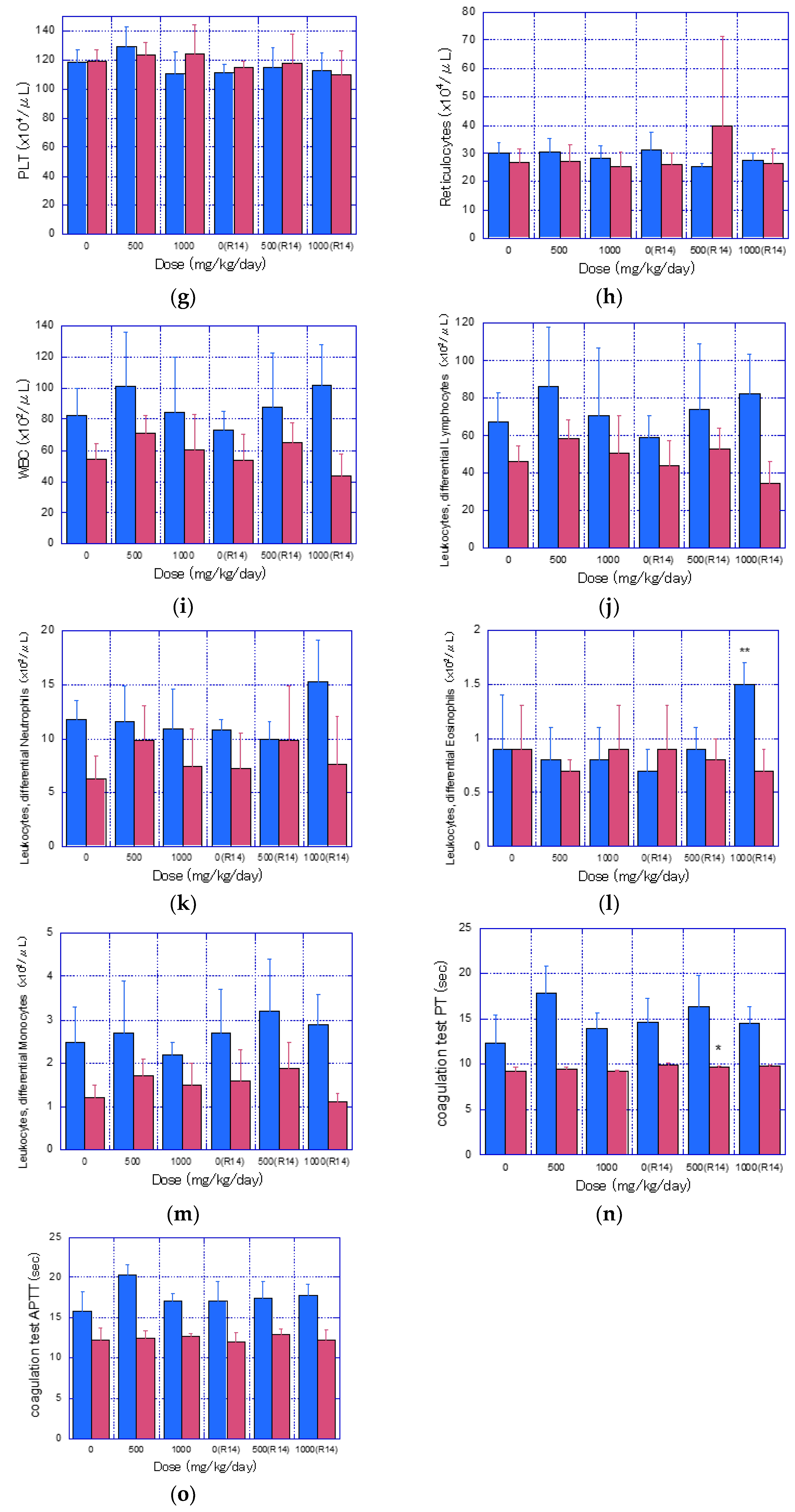
2.8. Hematological Examinations
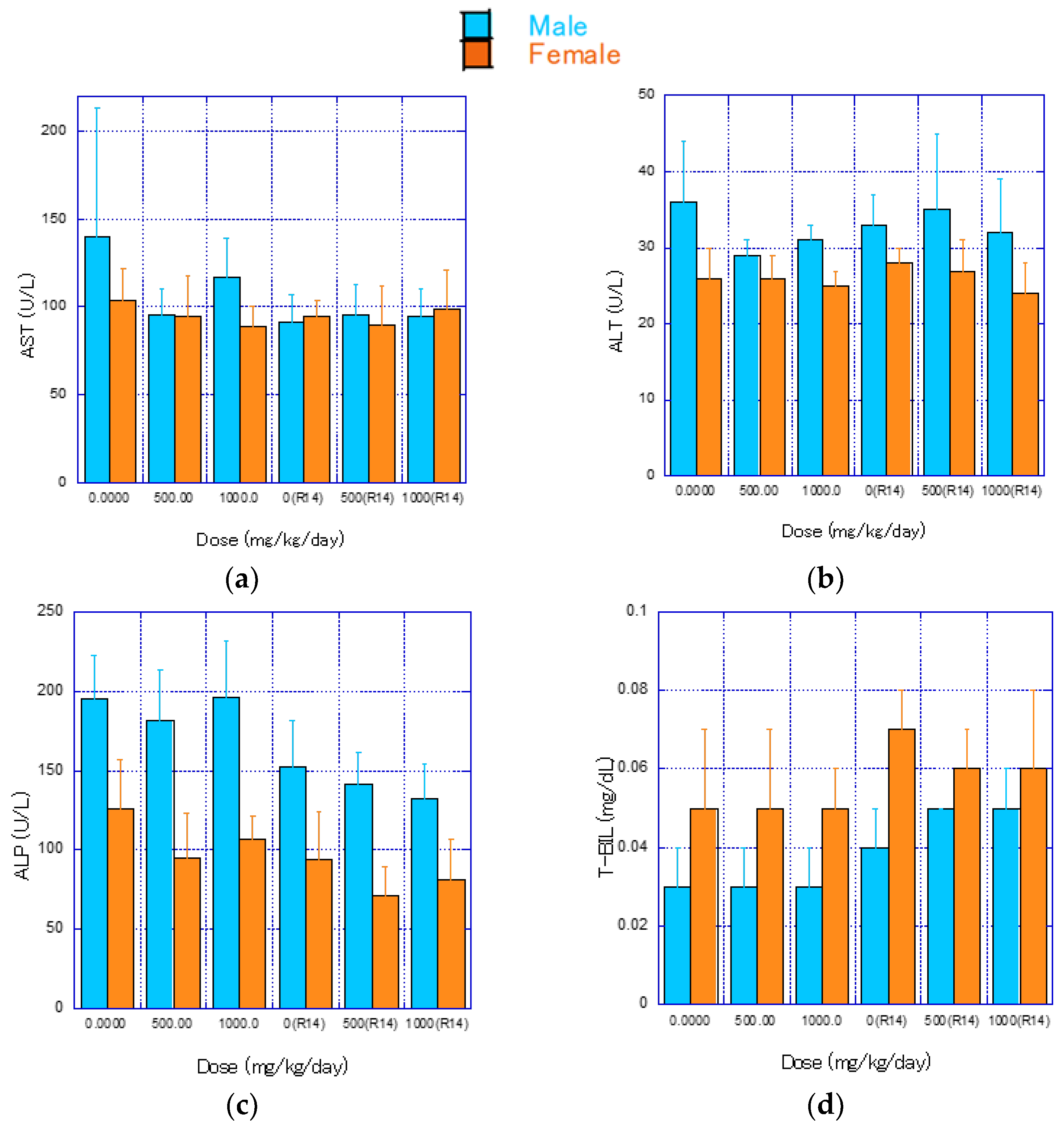
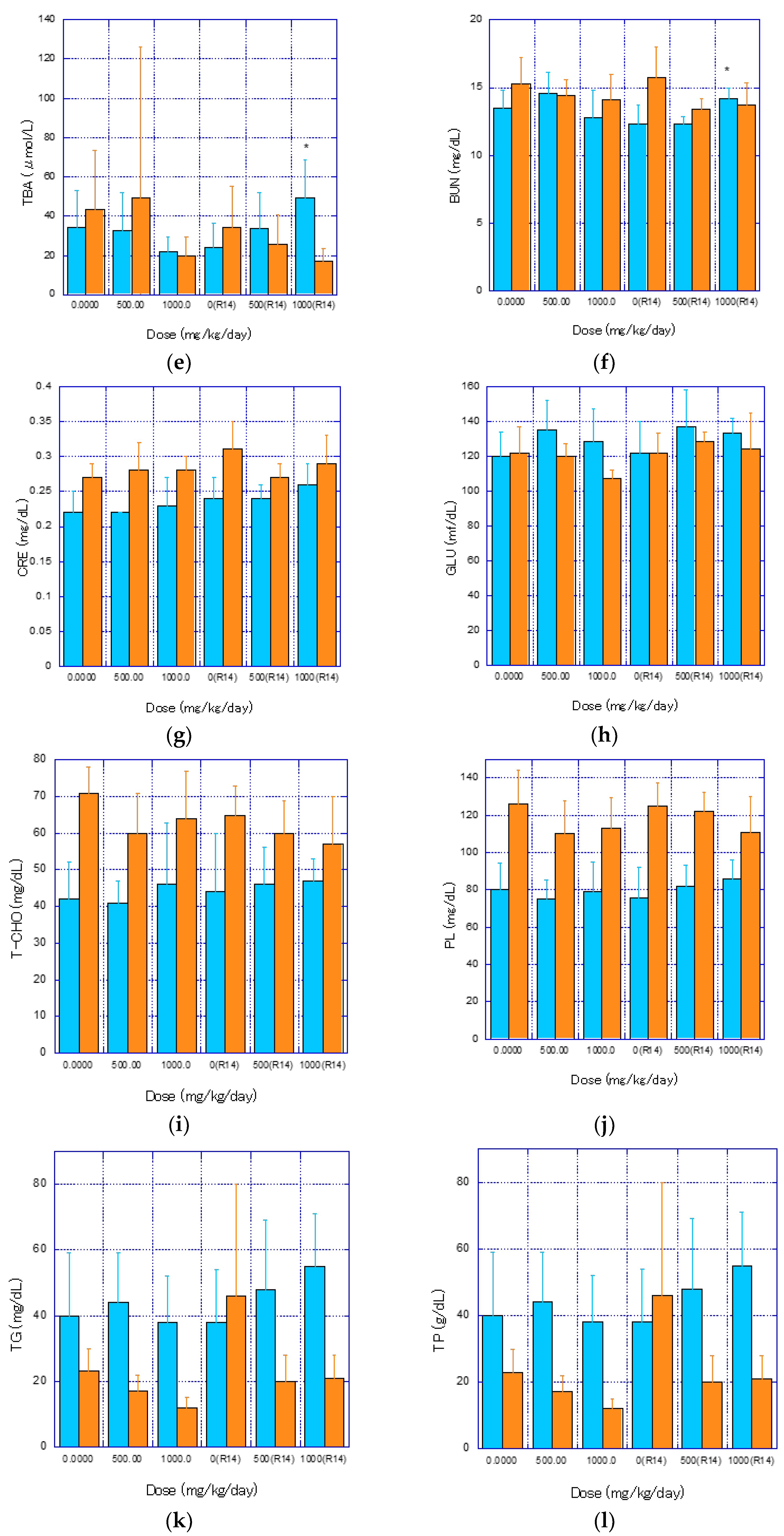
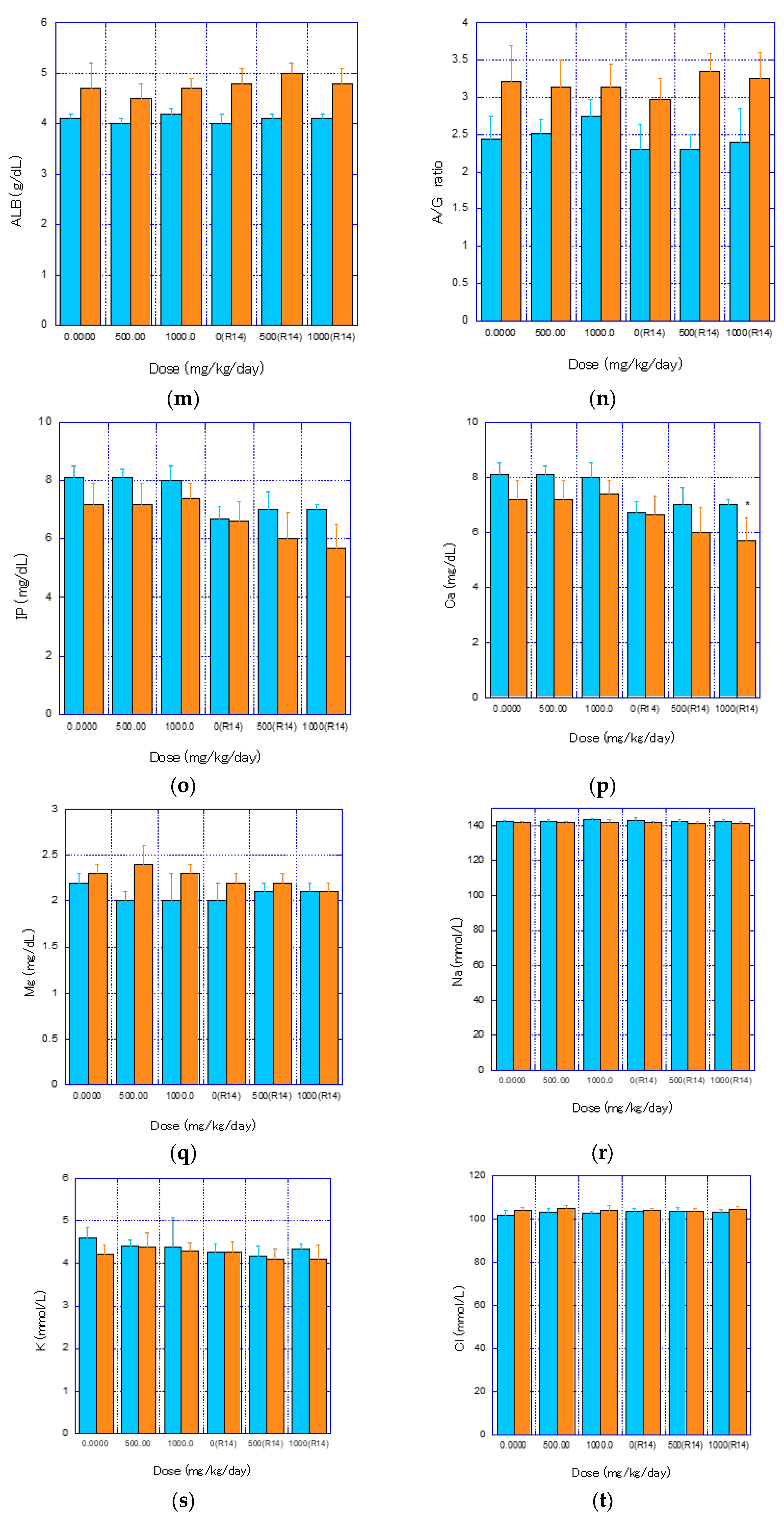
2.9. Blood Biochemical Examinations
2.10. Pathological Examination
2.11. Necropsy
2.12. Histopathological Examination
2.13. Statistical Treatment
3. Results
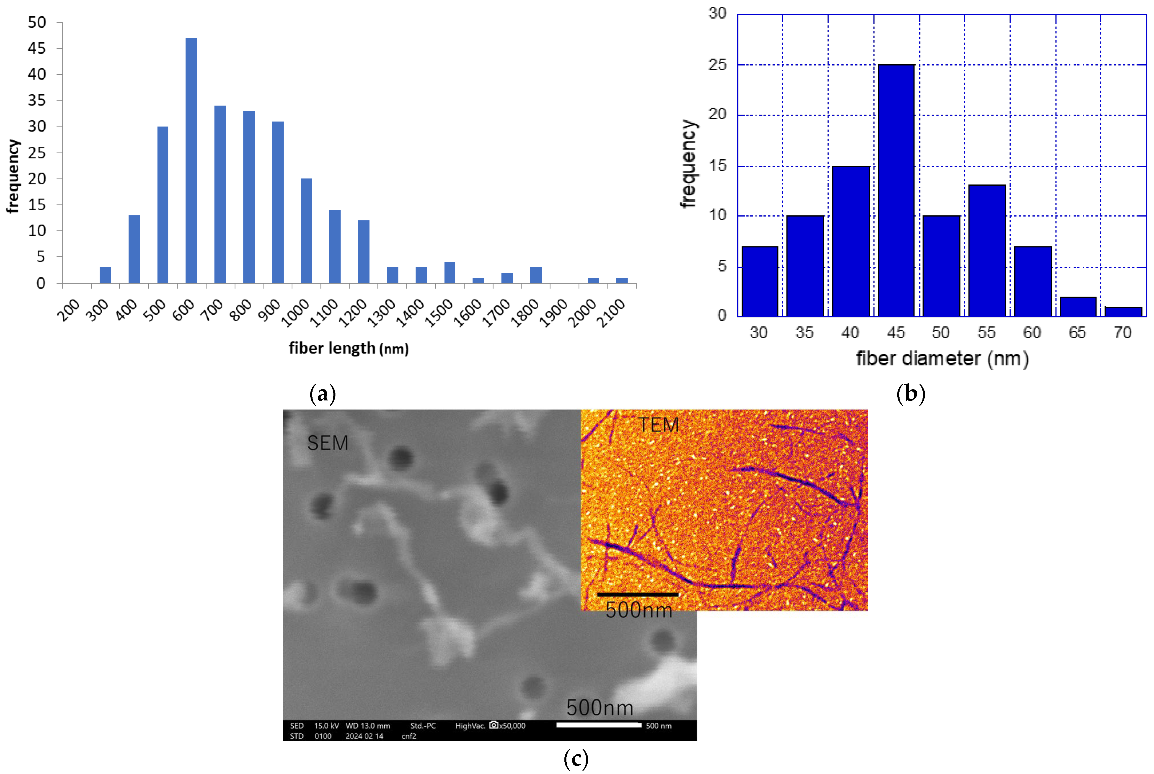
3.1. Fiber Length and Fiber Diameter of Fib-CNF
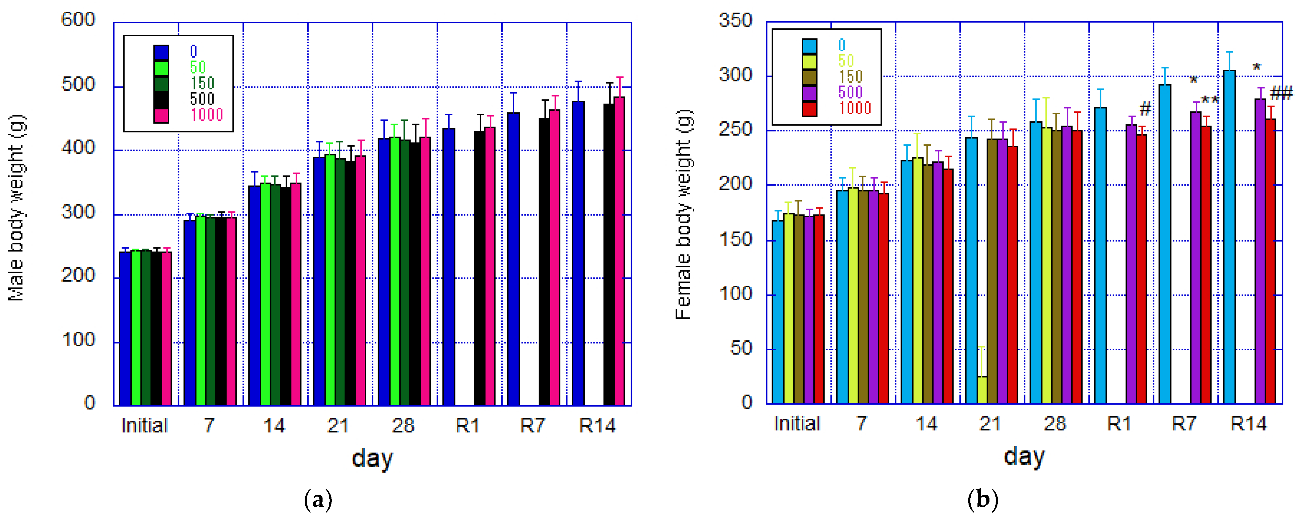
3.2. Weight
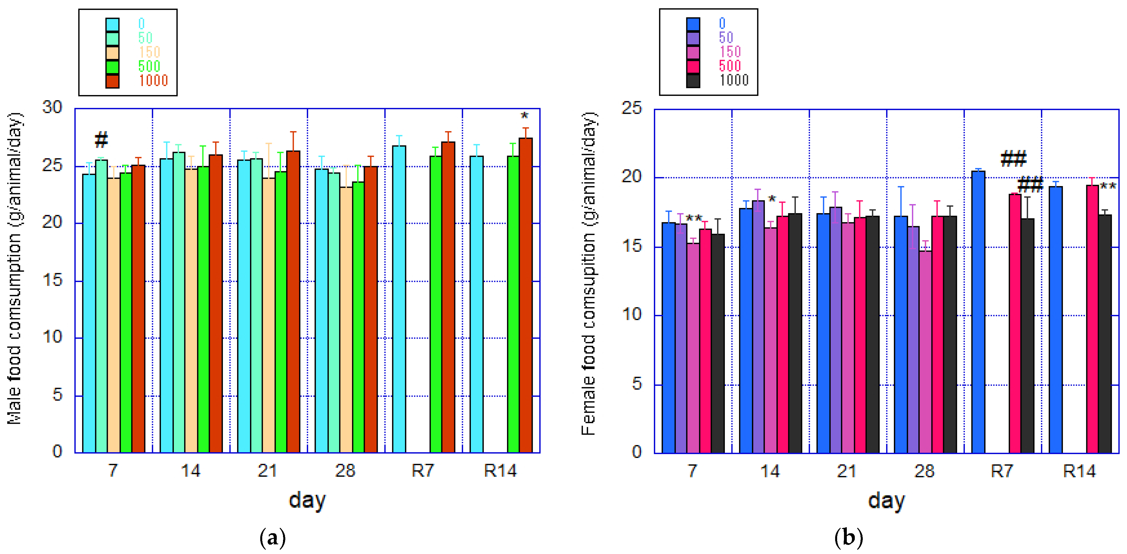
3.3. Food Intake
3.4. Urinalysis
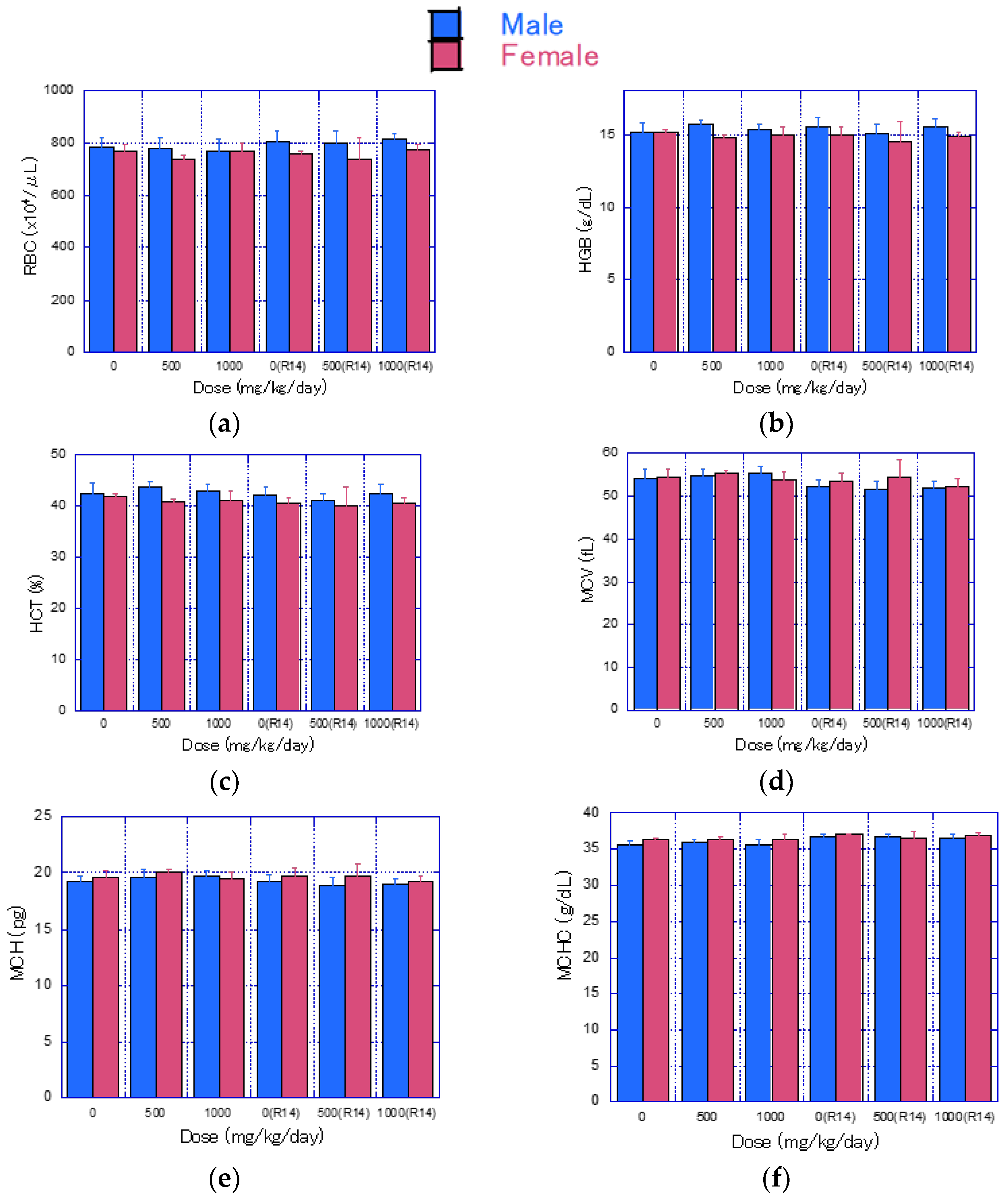
3.5. Hematological Examination
3.6. Blood Biochemical Tests
3.7. Pathological Examination
3.7.1. Necropsy
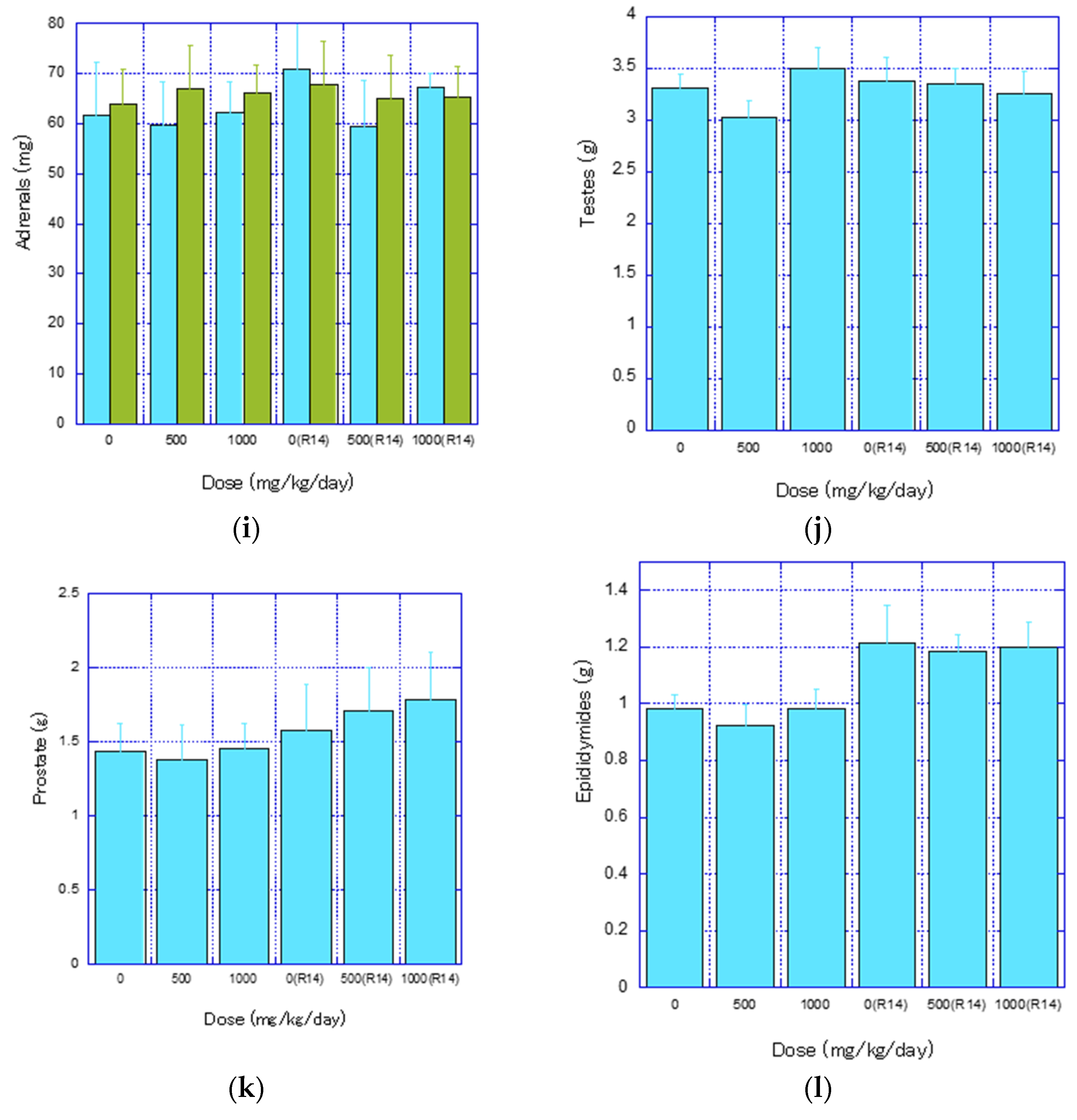
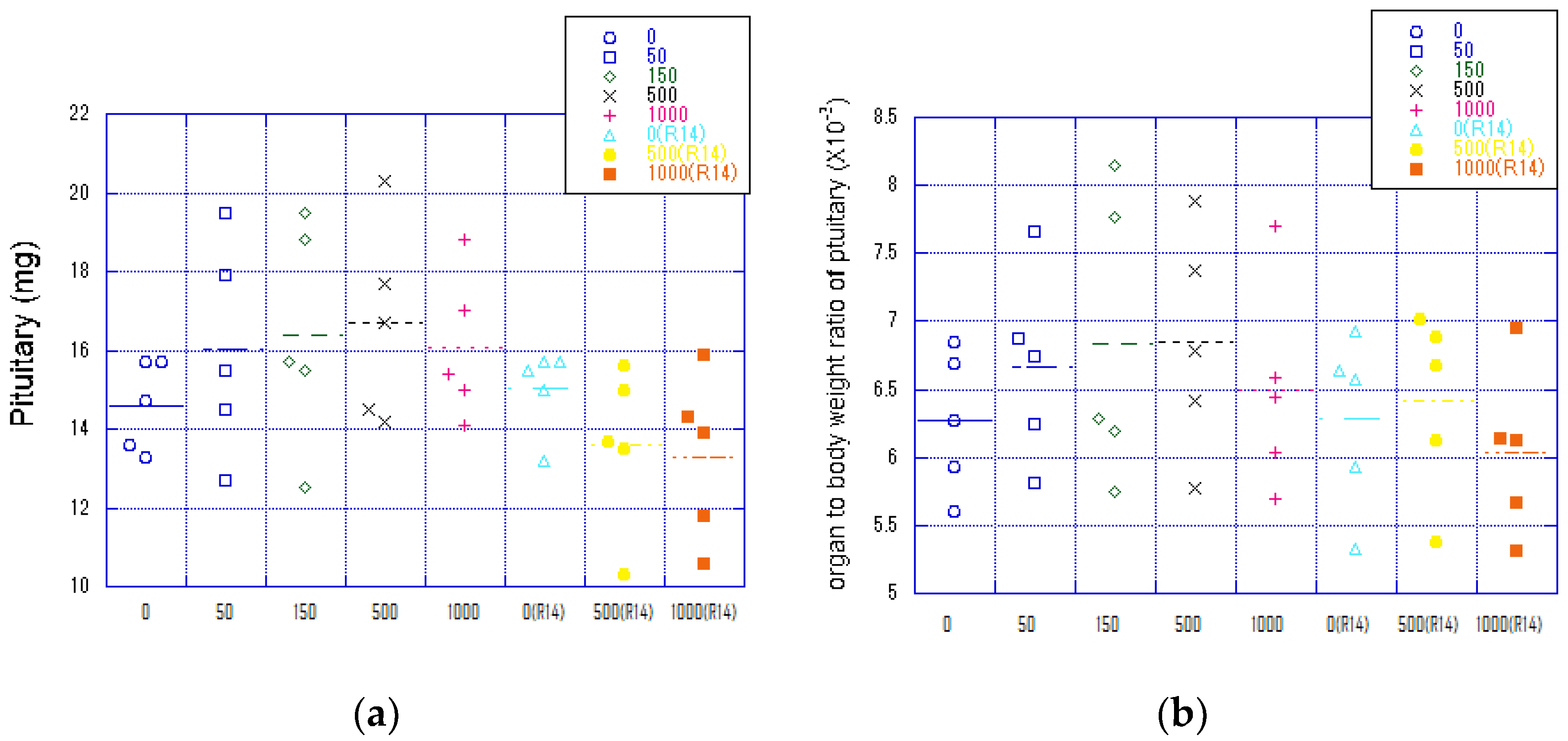
3.7.2. Organ Weights
3.8. Histopathological Examination
4. Discussion
5. Conclusions
Supplementary Materials
Author Contributions
Funding
Institutional Review Board Statement
Data Availability Statement
Conflicts of Interest
References
- Takemura, M.; Suzuki, T. Approach to Environmental, Health and Safety Issues of Nanotechnology in Japan. J. Disaster Res. 2011, 6, 506–513. [Google Scholar] [CrossRef]
- Liu, S.; Qamar, S.A.; Qamar, M.; Basharatd, K.; Bilal, M. Engineered nanocellulose-based hydrogels for smart drug delivery applications. Int. J. Biol. Macromol. 2021, 181, 275–290. [Google Scholar] [CrossRef] [PubMed]
- Canas-Gutierrez, A.; Gomez, H.C.; Velasquez-Cock, J.; Ganan, P.; Triana, O.; Cogollo-Florez, J.; Romero-Saez, M.; Romero-Saez, M.; Correa-Hincapie, N.; Zuluaga, R. Health and toxicological effects of nanocellulose when used as a food ingredient: A review. Carbohydr. Polym. 2024, 323, 121382. [Google Scholar] [CrossRef]
- Perumal, A.B.; Nambiar, R.B.; Moses, J.A.; Anandharamakrishnan, C. Nanocellulose: Recent trends and applications in the food industry. Food Hydrocoll. 2022, 127, 107484. [Google Scholar] [CrossRef]
- Gao, H.; Duan, B.; Lu, A.; Deng, H.; Du, Y.; Shi, X.; Zhang, L. Fabrication of cellulose nanofibers from waste brown algae and their potential application as milk thickeners. Food Hydrocoll. 2018, 79, 473–481. [Google Scholar] [CrossRef]
- Andrade, D.R.M.; Mendonca, M.H.; Helm, C.V.; Magalhaes, W.L.E.; Muniz, G.I.B.; Kestur, S.G. Assessment of Nano Cellulose from Peach Palm Residue as Potential Food Additive: Part II: Preliminary Studies. J. Food Sci. Technol. 2015, 52, 5641–5650. [Google Scholar] [CrossRef] [PubMed]
- Chen, Y.; Lin, Y.J.; Nagy, T.; Kong, F.; Guo, T.L. Subchronic exposure to cellulose nanofibrils induces nutritional risk by non-specifically reducing the intestinal absorption. Carbohydr. Polym. 2020, 229, 115536. [Google Scholar] [CrossRef] [PubMed]
- Fan, C.; Hou, L.; Che, G.; Shi, Y.; Liu, X.; Sun, L.; Jia, W.; Zhu, F.; Zhao, Z.; Xu, M.; et al. Dose- and time-dependent systemic adverse reactions of sodium carboxymethyl cellulose after intraperitoneal application in rats. J. Toxicol. Sci. 2021, 46, 223–234. [Google Scholar] [CrossRef] [PubMed]
- Zhang, K.; Zhang, H.; Wang, W. Toxicological studies and some functional properties of carboxymethylated cellulose nanofibrils as potential food ingredient. Int. J. Biol. Macromol. 2021, 190, 887–893. [Google Scholar] [CrossRef]
- Xu, H.S.; Chen, Y.; Patel, A.; Wang, Z.; McDonough, C.; Guo, T.L. Chronic exposure to nanocellulose altered depression-related behaviors in mice on a western diet: The role of immune modulation and the gut microbiome. Life Sci. 2023, 335, 122259. [Google Scholar] [CrossRef]
- Azuma, K.; Nagae, T.; Nagai, T.; Izawa, H.; Morimoto, M.; Murahata, Y.; Osaki, T.; Tsuka, T.; Imagawa, T.; Ito, N.; et al. Effects of Surface-Deacetylated Chitin Nanofibers in an Experimental Model of Hypercholesterolemia. Int. J. Mol. Sci. 2015, 16, 17445–17455. [Google Scholar] [CrossRef] [PubMed]
- Simokawa, T.; Magara, K.; Ikeda, T.; Hayashi, N.; Ogawa, M.; Takao, T.; Nakayama, E. Safety Assessment of Cellulose Nanofibers Obtained from Soda Cooking Bamboo Pulp: Bacterial Reverse Mutation Test, Mouse Lymphoma Thymidine Kinase Assay, Micronucleus Test, and Repeated dose 90-day Oral Toxicity Study in Rats. Jpn. TAPPI J. 2021, 75, 1018–1050. [Google Scholar] [CrossRef]
- Anraku, M.; Tabuchi, R.; Ifuku, S.; Nagae, T.; Iohara, D.; Tomida, H.; Uekama, K.; Maruyama, T.; Miyamura, S.; Hirayama, F.; et al. An oral absorbent, surface-deacetylated chitin nanofiber ameliorates renal injury and oxidative stress in 5/6 nephrectomized rats. Carbohydr. Polym. 2017, 161, 21–25. [Google Scholar] [CrossRef] [PubMed]
- Xu, Y.; Park, S.H.; Yoon, K.N.; Park, S.J.; Gye, M.C. Effects of citrate ester plasticizers and bis(2-ethylhexyl) phthalate in the OECD 28-day repeated-dose toxicity test (OECD TG 407). Environ. Res. 2019, 172, 675–683. [Google Scholar] [CrossRef] [PubMed]
- Mirza, A.C.; Panchal, S.S. Safety evaluation of syringic acid: Subacute oral toxicity studies in Wistar rats. Heliyon 2019, 5, e02129. [Google Scholar] [CrossRef] [PubMed]
- de Lima, P.C.A.; Fregonezi, N.F.; Carvalho, A.J.F.; Trovatti, E.; Resende, F.A. TEMPO-Oxidized Cellulose Nanofibers In Vitro Cyto-genotoxicity Studies. BioNanoScience 2020, 10, 766–772. [Google Scholar] [CrossRef]
- Iman, M.; Barati, A.; Safari, S. Characterization, in vitro antibacterial activity, and toxicity for rat of tetracycline in a nanocomposite hydrogel based on PEG and cellulose. Cellulose 2020, 27, 347–356. [Google Scholar] [CrossRef]
- Marcuello, C.; Foulon, L.; Chabbert, B.; Aguié-Béghin, V.; Michael Molinari, M. Atomic force microscopy reveals how relative humidity impacts the Young’s modulus of lignocellulosic polymers and their adhesion with cellulose nanocrystals at the nanoscale. Int. J. Biol. Macromol. 2020, 147, 1064–1075. [Google Scholar] [CrossRef] [PubMed]
- Nadia, V.; Celia, V.; Michel, K.; Joao, S.M.; Henriqueta, L. Toxicological Assessment of Cellulose Nanomaterials: Oral Exposure. Nanomaterials 2022, 12, 3375. [Google Scholar] [CrossRef]
- Moriyama, A.; Ogura, I.; Fujita, K. Potential issues specific to cytotoxicity tests of cellulose nanofibrils. J. Appl. Toxicol. 2023, 43, 195–207. [Google Scholar] [CrossRef]
- Menas, A.L.; Yanamala, N.; Farcas, M.T.; Russo, M.; Friend, S.; Fournier, P.M.; Star, A.; Iavicoli, I.; Shurin, G.V.; Vogel, U.B.; et al. Fibrillar vs crystalline nanocellulose pulmonary epithelial cell responses: Cytotoxicity or inflammation? Chemosphere 2017, 171, 671–680. [Google Scholar] [CrossRef] [PubMed]
- Nordli, H.R.; Chinga-Carrasco, G.; Rokstad, A.M.; Pukstad, B. Producing ultra pure wood cellulose nanofibrils and evaluating the cytotoxicity using human skin cells. Carbohydr. Polym. 2016, 150, 65–73. [Google Scholar] [CrossRef] [PubMed]
- Bhattacharya, K.; Kiliç, G.; Costa, P.M.; Fadeel, B. Cytotoxicity screening and cytokine profiling of nineteen nanomaterials enables hazard ranking and grouping based on inflammogenic potential. Nanotoxicology 2017, 11, 809–826. [Google Scholar] [PubMed]
- He, X.; Sun, C.; Zhao, J.; Zhang, Y.; Zhang, X.; Fang, Y. High Viscosity Slows the Utilization of Rapidly Fermentable Dietary Fiber by Human Gut Microbiota. J. Agric. Food Chem. 2023, 71, 19078–19087. [Google Scholar] [CrossRef] [PubMed]
- Baky, M.H.; Salah, M.; Ezzelarab, N.; Shao, P.; Elshahed, M.S.; Farag, M.A. Insoluble dietary fibers: Structure, metabolism, interactions with human microbiome, and role in gut homeostasis. Crit. Rev. Food Sci. Nutr. 2024, 64, 1954–1968. [Google Scholar] [CrossRef] [PubMed]
- Garschagen, L.S.; Franke, T.; Deppenmeier, U. An alternative pentose phosphate pathway in human gut bacteria for the degradation of C5 sugars in dietary fibers. FEBS J. 2021, 288, 1839–1858. [Google Scholar] [CrossRef]
- Moraïs, S.; Winkler, S.; Zorea, A.; Levin, L.; Mizrahi, I. Cryptic diversity of cellulose-degrading gut bacteria in industrialized humans. Science 2024, 383, eadj9223. [Google Scholar] [CrossRef] [PubMed]
- Lopes, V.R.; Stromme, M.; Ferraz, N. In Vitro Biological Impact of Nanocellulose Fibers on Human Gut Bacteria and Gastrointestinal Cells. Nanomaterials 2020, 10, 1159. [Google Scholar] [CrossRef] [PubMed]
- Zhou, L.; Wang, L.; Ma, N.; Wan, Y.; Qian, W. Real-time monitoring of interactions between dietary fibers and lipid layer and their impact on the lipolysis process. Food Hydrocoll. 2022, 125, 107445. [Google Scholar] [CrossRef]
- Ge, Q.; Li, H.Q.; Zheng, Z.Y.; Yang, K.; Li, P.; Xiao, Z.Q.; Xiao, G.M.; Mao, J.W. In vitro fecal fermentation characteristics of bamboo insoluble dietary fiber and its impacts on human gut microbiota. Food Res. Int. 2022, 156, 111173. [Google Scholar] [CrossRef]
- Li, M.; Wang, Y.; Guo, C.; Wang, S.; Zheng, L.; Bu, Y.; Ding, K. The claim of primacy of human gut Bacteroides ovatus in dietary cellobiose degradation. Gut Microbes 2023, 15, 2227434. [Google Scholar] [CrossRef] [PubMed]
- Bueno, D.; Freitas, C.; Brienzo, M. Enzymatic Cocktail Formulation for Xylan Hydrolysis into Xylose and Xylooligosaccharides. Molecules 2023, 28, 624. [Google Scholar] [CrossRef] [PubMed]
- Phuyal, M.; Budhathoki, U.; Bista, D.; Shakya, S.; Shrestha, R.; Shrestha, A.K. Xylanase-Producing Microbes and Their Real-World Application. Int. J. Chem. Eng. 2023, 2023, 3593035. [Google Scholar] [CrossRef]
- Tahiri, M.; Johnsrud, C.; Steffensen, I.L. Evidence and hypotheses on adverse effects of the food additives carrageenan (E 407)/processed Eucheuma seaweed (E 407a) and carboxymethylcellulose (E 466) on the intestines: A scoping review. Crit. Rev. Toxicol. 2023, 53, 521–571. [Google Scholar] [CrossRef] [PubMed]
- Nagano, T.; Yano, H. Dietary cellulose nanofiber modulates obesity and gut microbiota in high-fat-fed mice. Bioact. Carbohydr. Diet. Fibre 2020, 22, 100214. [Google Scholar] [CrossRef]
- Zhai, X.; Lin, D.; Zhao, Y.; Yang, X. Bacterial Cellulose Relieves Diphenoxylate-Induced Constipation in Rats. J. Agric. Food Chem. 2018, 66, 4106–4117. [Google Scholar] [CrossRef] [PubMed]
- Blevins, J.E.; Baskin, D.G. Translational and therapeutic potential of oxytocin as an anti-obesity strategy: Insights from rodents, nonhuman primates and humans. Physiol. Behav. 2015, 152, 438–449. [Google Scholar] [CrossRef] [PubMed]
- Matsuura, I.; Saitoh, T.; Tani, E.; Wako, Y.; Iwata, H.; Toyota, N.; Ishizuka, Y.; Namiki, M.; Hoshino, N.; Tsuchitani, M.; et al. Evaluation of a two-generation reproduction toxicity study adding endpoints to detect endocrine disrupting activity using lindane. J. Toxicol. Sci. 2006, 30, S135–S161. [Google Scholar] [CrossRef]
- Helmy, H.; Hamid, S.N.A.; Badawy, L.; Sayed, H. Mechanistic insights into the protective role of eugenol against stress-induced reproductive dysfunction in female rat model. Chem. Biol. Interact. 2022, 367, 110181. [Google Scholar] [CrossRef]
- Li, S.; Kim, Y.; Chen, J.D.Z.; Madhoun, M.F. Intestinal Electrical Stimulation Alters Hypothalamic Expression of Oxytocin and Orexin and Ameliorates Diet-Induced Obesity in Rats. Obes. Surg. 2021, 31, 1664–1672. [Google Scholar] [CrossRef]
- Ide, T.; Horii, M.; Yamamoto, T.; Kawashima, K. Contrasting effects of water-soluble and water-insoluble dietary fibers on bile acid conjugation and taurine metabolism in the rat. Lipids 1990, 25, 335–340. [Google Scholar] [CrossRef] [PubMed]
- Nagano, T.; Yano, H. Effect of dietary cellulose nanofiber and exercise on obesity and gut microbiota in mice fed a high-fat-diet. Biosci. Biotechnol. Biochem. 2020, 84, 613–620. [Google Scholar] [CrossRef] [PubMed]



| Sex | Group | Dose mg/kg/day | Dosing Volume mL/kg | Dose Concentration wt% | Number of Animals |
|---|---|---|---|---|---|
| Male | Control | 0 a | 10 mL, two divided doses | 0 | 5 |
| Control recovery | 0 a | 0 | |||
| Low dose | 50 | 0.25, two doses | |||
| Middle dose | 150 | 0.75, two doses | |||
| High dose | 500 | 2.5, two doses | |||
| High dose recovery | 500 | 2.5, two doses | |||
| Dosage limit | 1000 | 5.0, two doses | |||
| Dosage limit recovery | 1000 | 5.0, two doses | |||
| Female | Control | 0 a | 10 mL, two divided doses | 0 | 5 |
| Control recovery | 0 a | 0 | |||
| Low dose | 50 | 0.25, two doses | |||
| Middle dose | 150 | 0.75, two doses | |||
| High dose | 500 | 2.5, two doses | |||
| High dose recovery | 500 | 2.5, two doses | |||
| Dosage limit | 1000 | 5.0, two doses | |||
| Dosage limit recovery | 1000 | 5.0, two doses |
| Test Parameters | Measuring Methods and Equipment |
|---|---|
| (1) pH | CLINITEK Advantus Urine Chemistry Analyzer Multistix® 10 SG (Siemens Healthineers, Erlangen, Germany) Siemens Medical Solutions |
| (2) Blood | |
| (3) Ketone | |
| (4) Glucose | |
| (5) Protein | |
| (6) Urobilinogen | |
| (7) Bilirubin | |
| (8) Urine sediment | New Sternheimer stain microscope examination (Olympus BX46, Tokyo, Japan) |
| (9) Color tone | Observation with the naked eye |
| (10) Amount of urine | Weighing method Electronic balance LA4200 (Sartorius Co., Ltd., Göttingen, Germany) |
| (11) Specific gravity | ATAGO Serum Protein Flexure Meter N (ATAGO Co., Ltd., Tokyo, Japan) |
| Test Parameters | Measurement Method |
|---|---|
| (1) Red Blood Cell Count (RBC) | Sheath flow DC detection method |
| (2) Hematocrit value (HCT) | |
| (3) Platelet count (PLT) | |
| (4) Hemoglobin (HGB) | SLS hemoglobin method |
| (5) Mean corpuscular volume (MCV) | Sheath flow DC detection and calculation method |
| (6) Mean corpuscular hemoglobin (MCH) | |
| (7) Mean corpuscular hemoglobin concentration (MCHC) | |
| (8) Reticulocyte count | Flow cytometry method and calculation method |
| (9) White Blood Cell Count (WBC) | Flow cytometry method |
| (10) White Blood Cell Count by Type Lymphocyte Count Neutrophil Count Eosinophil Count Basophil Count Monocyte Count | |
| (11) Prothrombin time (PT) | Solidification Time Method: Light Scattering Inspection Method |
| (12) Activated partial thromboplastin time (APTT) |
| Test Parameters | Measurement Method |
|---|---|
| (1) Aspartate aminotransferase (AST) | JSCC recommendation method |
| (2) Alanine aminotransferase (ALT) | JSCC recommendation method |
| (3) Alkaline phosphatase (ALP) | IFCC Reference Measurement Operation Method |
| (4) Total pililpine (T-BIL) | Vanadate oxidation |
| (5) Bile acid (TBA) | Enzymatic cycling |
| (6) Urea nitrogen (BUN) | UV method (urease/GLDH method) |
| (7) Creatinine (CRE) | Enzymatic process |
| (8) Glucose (GLU) | HK-G-6-PDH method |
| (9) Total cholesterol (T-CHO) | Cholesterol oxidase method |
| (10) Phospholipids (PL) | Choline oxidase/DAOS method |
| (11) Triglycerides (TG) | FG scavenging enzyme method |
| (12) Total protein (TP) | Pioulet method |
| (13) Albumin (ALB) | BCG method |
| (14) A/G ratio (albumin/globulin ratio) | Computation method |
| (15) Inorganic phosphorus (IP) | Enzymatic process |
| (16) Calcium (Ca) | Enzymatic process |
| (17) Magnesium (Mg) | Xylidiliriproof method |
| (18) Sodium (Na) | Ion selective electrode method |
| (19) Potassium (K) | Ion selective electrode method |
| (20) Chlorine (Cl) | Ion selective electrode method |
Disclaimer/Publisher’s Note: The statements, opinions and data contained in all publications are solely those of the individual author(s) and contributor(s) and not of MDPI and/or the editor(s). MDPI and/or the editor(s) disclaim responsibility for any injury to people or property resulting from any ideas, methods, instructions or products referred to in the content. |
© 2024 by the authors. Licensee MDPI, Basel, Switzerland. This article is an open access article distributed under the terms and conditions of the Creative Commons Attribution (CC BY) license (https://creativecommons.org/licenses/by/4.0/).
Share and Cite
Yamashita, Y.; Tokunaga, A.; Aoki, K.; Ishizuka, T.; Fujita, S.; Tanoue, S. A 28-Day Repeated Oral Administration Study of Mechanically Fibrillated Cellulose Nanofibers According to OECD TG407. Nanomaterials 2024, 14, 1082. https://doi.org/10.3390/nano14131082
Yamashita Y, Tokunaga A, Aoki K, Ishizuka T, Fujita S, Tanoue S. A 28-Day Repeated Oral Administration Study of Mechanically Fibrillated Cellulose Nanofibers According to OECD TG407. Nanomaterials. 2024; 14(13):1082. https://doi.org/10.3390/nano14131082
Chicago/Turabian StyleYamashita, Yoshihiro, Akinori Tokunaga, Koji Aoki, Tamotsu Ishizuka, Satoshi Fujita, and Shuichi Tanoue. 2024. "A 28-Day Repeated Oral Administration Study of Mechanically Fibrillated Cellulose Nanofibers According to OECD TG407" Nanomaterials 14, no. 13: 1082. https://doi.org/10.3390/nano14131082
APA StyleYamashita, Y., Tokunaga, A., Aoki, K., Ishizuka, T., Fujita, S., & Tanoue, S. (2024). A 28-Day Repeated Oral Administration Study of Mechanically Fibrillated Cellulose Nanofibers According to OECD TG407. Nanomaterials, 14(13), 1082. https://doi.org/10.3390/nano14131082


