Carbon Nanodot–Microbe–Plant Nexus in Agroecosystem and Antimicrobial Applications
Abstract
:1. Introduction
2. Microbes and Plants: Amazing World
- Producing small peptides by microbes and/or plants and their role in plant–microbe interactions in the rhizosphere along with the change in the rhizosphere microbiomes. This nexus may be also useful in holobiont engineering, and the potential of exploring transgenic microbes to synthesize small peptides on a large scale [37].
- The role of plant–microbe interactions in regulating sterols including phytosterol biosynthesis, recognition, communication, transduction, and/or exchanges between partners through the expression of genes [38].
- The plant–microbe interactions and their impacts on polluted soil with microplastics through the microbial degradation microbial-mediated MPs bioremediation [39].
- Genome of the plant–microbe interactions under different ecological, physiological, and evolutionary implications of both plants and microbes and their impacts on stress tolerance, growth, and nutrient acquisition [40].
- The plant–microbe interactions and their role in controlling crop productivity through impacting the microbial communities, and activities, influencing endogenous and external growth factors, and potential targeted applications in agricultural production [41].
- The role of plant–microbe interactions in transferring immune signals between plant cells and plant pathogens including different bioactive molecules (as extracellular vesicles) like metabolites, proteins, lipids, and small RNAs and facilitating the exchange of such active substances between various species [40].
- Soil microbes and their play in nutrient cycling, soil health, and ecological restoration for food security and nutrient quality, which are controlled by both plants and microbes [42].
- The specific role of plant–metabolite–microbe or pathogen interactions and their complexity for the identification of specialized metabolite pathways using ecological, mechanistic, and evolutionary models [43].
- The role of agricultural practices (mainly plant grafting) in increasing or suppressing crop productivity, alleviating abiotic stress, controlling pathogens, and modulating the root microbiome [44].
- The interaction among nanoparticles, plants, and microbes and their roles in crop production and food security through the use of nano-devices/-products for agro-applications [45].
3. Relation between Microbes and Plants under Different Soil Conditions
4. Nanomaterial–Microbe–Plant Nexus
4.1. Carbon Nanodots as Inhibitors of Phytopathogens
| Microbe Species | Applied CNDs Dose | Types of CNDs and Their Precursors | Refs. |
|---|---|---|---|
| Gram-positive pathogenic organisms | |||
| Staphylococcus aureus | 100 μg mL−1 for 48 h | Vitamin C-derived CNDs | [62] |
| 200 μg/mL for >48 h | CNDs from tamarind plant (Tamarindus indica L.) | [63] | |
| 250 μg/mL for 60 min for spermidine-based CQDs | CNDs from three biogenic polyamines (i.e., spermidine, putrescine, and spermine) | [69] | |
| 100 μg/mL for 24 h | CNDs from henna plant (Lawsonia inermis L.) | [70] | |
| 75 μL mL−1 for 12 h | CNDs from 2,20-(ethylenedioxy)-bis(ethylamine) and malic acid | [71] | |
| 100 μg mL−1 for 12 h | CNDs from ciprofloxacin hydrochloride | [64] | |
| 256 μg mL−1 for 18 h | CNDs produced from D(1)-glucose monohydrate and diethylenetriamine | [66] | |
| (7.8–62.5) µg/L for 24 h | CNDs from olive solid wastes in a hybrid form of ZnO–CNDs | [72] | |
| Bacillus subtilis | 100 μg mL−1 for 48 h | Vitamin C-derived CNDs | [62] |
| Streptococcus mutans | 300 μg mL−1 for 24 h | CNDs from metronidazole under hydrothermal process | [65] |
| Gram-negative pathogenic organisms | |||
| Escherichia coli | 100 μg mL−1 for 48 h | Vitamin C-derived CNDs | [62] |
| 200 μg/mL for >48 h | CNDs from tamarind plant (Tamarindus indica L.) | [63] | |
| 250 μg/mL for 60 min for spermidine-based CQDs | CNDs from three biogenic polyamines (i.e., spermidine, putrescine, and spermine) | [69] | |
| 100 μg/mL for 24 h | CNDs from henna plant (Lawsonia inermis L.) | [70] | |
| 75 μL mL−1 for 12 h | CNDs from 2,20-(ethylenedioxy)-bis(ethylamine) and malic acid | [71] | |
| 100 μg mL−1 for 12 h | CNDs from ciprofloxacin hydrochloride | [64] | |
| 256 μg mL−1 for 18 h | CNDs produced from D(1)-glucose monohydrate and diethylenetriamine | [66] | |
| Pseudomonas aeruginosa | 200 μg/mL for >48 h | CNDs from tamarind plant (Tamarindus indica L.) | [63] |
| 250 μg/mL for 60 min for spermidine-based CQDs | CNDs from three biogenic polyamines (i.e., spermidine, putrescine, and spermine) | [69] | |
| 200 μg/mL for >48 h | CNDs from aminoguanidine and citric acid | [73] | |
| Salmonella enteritidis | 250 μg/mL for 60 min for spermidine-based CQDs | CNDs from three biogenic polyamines (i.e., spermidine, putrescine, and spermine) | [69] |
| Porphyromonas gingivalis | 300 μg mL−1 for 24 h | CNDs from metronidazole under hydrothermal process | [65] |
4.1.1. Microbial Interactions for Synthesis of CNDs
4.1.2. CNDs for Promoting Plant Growth and Stress Tolerance
4.1.3. Phytotoxicity Concerns of CNDs
4.1.4. The Transportation of CNDs in Plant Organs
4.2. Nano-Tellurium: A New Frontier in Nano-Microbe Nexus
4.2.1. Why Nano-Tellurium?
4.2.2. Antimicrobial Mechanisms of Nano-Tellurium
4.2.3. Nano-Tellurium: Microbial Synthesis
5. Transport of Nanomaterials to Different Organs
5.1. Soil Application and Uptake of NPs
5.2. Foliar Application and Uptake of NPs
5.3. Translocation of NPs
6. Nanotoxicity on Soil Microbes and Plants
6.1. Toxicity Concerns about Soil Microbes
6.2. Toxicity Concerns in Plants
6.2.1. Morpho-Physiological Concerns
6.2.2. Cellular and Biochemical Concerns
6.2.3. Molecular Concerns
7. Regulations of Nanotoxicity
7.1. Current Challenges of Nanotoxicity
7.1.1. Risk and Challenges in Ecological Level
7.1.2. Human Health-Related Risks and Challenges
7.2. Regulations for Risk Assessment and Management Related to Nanotoxicity
7.2.1. Scientific-Level Regulation
7.2.2. Organizational Level Regulations
8. Conclusions, Challenges, and Prospects
Author Contributions
Funding
Acknowledgments
Conflicts of Interest
References
- Agriculture and Food. Available online: https://www.worldbank.org/ (accessed on 20 June 2024).
- Li, Y.; Tang, Z.; Pan, Z.; Wang, R.; Wang, X.; Zhao, P.; Liu, M.; Zhu, Y.; Liu, C.; Wang, W.; et al. Calcium-Mobilizing Properties of Salvia miltiorrhiza-Derived Carbon Dots Confer Enhanced Environmental Adaptability in Plants. ACS Nano 2022, 16, 4357–4370. [Google Scholar] [CrossRef] [PubMed]
- Xiong, C.; Lu, Y. Microbiomes in Agroecosystem: Diversity, Function and Assembly Mechanisms. Environ. Microbiol. Rep. 2022, 14, 833–849. [Google Scholar] [CrossRef] [PubMed]
- Verma, K.K.; Song, X.-P.; Xu, L.; Huang, H.-R.; Liang, Q.; Seth, C.S.; Li, Y.-R. Nano-Microplastic and Agro-Ecosystems: A Mini-Review. Front. Plant Sci. 2023, 14, 1283852. [Google Scholar] [CrossRef] [PubMed]
- Okeke, E.S.; Chukwudozie, K.I.; Addey, C.I.; Okoro, J.O.; Chidike Ezeorba, T.P.; Atakpa, E.O.; Okoye, C.O.; Nwuche, C.O. Micro and Nanoplastics Ravaging Our Agroecosystem: A Review of Occurrence, Fate, Ecological Impacts, Detection, Remediation, and Prospects. Heliyon 2023, 9, e13296. [Google Scholar] [CrossRef] [PubMed]
- Hatt, S.; Döring, T.F. Designing Pest Suppressive Agroecosystems: Principles for an Integrative Diversification Science. J. Clean. Prod. 2023, 432, 139701. [Google Scholar] [CrossRef]
- Aguiar, M.; Conway, A.J.; Bell, J.K.; Stewart, K.J. Agroecosystem Edge Effects on Vegetation, Soil Properties, and the Soil Microbial Community in the Canadian Prairie. PLoS ONE 2023, 18, e0283832. [Google Scholar] [CrossRef] [PubMed]
- Beeckman, F.; Annetta, L.; Corrochano-Monsalve, M.; Beeckman, T.; Motte, H. Enhancing Agroecosystem Nitrogen Management: Microbial Insights for Improved Nitrification Inhibition. Trends Microbiol. 2024, 32, 590–601. [Google Scholar] [CrossRef] [PubMed]
- Wang, C.; Kuzyakov, Y. Rhizosphere Engineering for Soil Carbon Sequestration. Trends Plant Sci. 2024, 29, 447–468. [Google Scholar] [CrossRef] [PubMed]
- Mouratiadou, I.; Wezel, A.; Kamilia, K.; Marchetti, A.; Paracchini, M.L.; Bàrberi, P. The Socio-Economic Performance of Agroecology. A Review. Agron. Sustain. Dev. 2024, 44, 19. [Google Scholar] [CrossRef]
- Chowardhara, B.; Saha, B.; Awasthi, J.P.; Deori, B.B.; Nath, R.; Roy, S.; Sarkar, S.; Santra, S.C.; Hossain, A.; Moulick, D. An Assessment of Nanotechnology-Based Interventions for Cleaning up Toxic Heavy Metal/Metalloid-Contaminated Agroecosystems: Potentials and Issues. Chemosphere 2024, 359, 142178. [Google Scholar] [CrossRef]
- Umair, M.; Huma Zafar, S.; Cheema, M.; Usman, M. New Insights into the Environmental Application of Hybrid Nanoparticles in Metal Contaminated Agroecosystem: A Review. J. Environ. Manag. 2024, 349, 119553. [Google Scholar] [CrossRef] [PubMed]
- Kalpana, P.; Falkenberg, T.; Yasobant, S.; Saxena, D.; Schreiber, C. Agroecosystem Exploration for Antimicrobial Resistance in Ahmedabad, India: A Study Protocol [Version 2; Peer Review: 2 Approved]. F1000Research 2024, 12, 316. [Google Scholar] [CrossRef] [PubMed]
- Singh, A.; Rajput, V.D.; Varshney, A.; Ghazaryan, K.; Minkina, T. Small Tech, Big Impact: Agri-Nanotechnology Journey to Optimize Crop Protection and Production for Sustainable Agriculture. Plant Stress 2023, 10, 100253. [Google Scholar] [CrossRef]
- Yadav, N.; Garg, V.K.; Chhillar, A.K.; Rana, J.S. Recent Advances in Nanotechnology for the Improvement of Conventional Agricultural Systems: A Review. Plant Nano Biol. 2023, 4, 100032. [Google Scholar] [CrossRef]
- Shah, M.A.; Shahnaz, T.; Zehab-ud-Din; Masoodi, J.H.; Nazir, S.; Qurashi, A.; Ahmed, G.H. Application of Nanotechnology in the Agricultural and Food Processing Industries: A Review. Sustain. Mater. Technol. 2024, 39, e00809. [Google Scholar] [CrossRef]
- Preethi, B.; Karmegam, N.; Manikandan, S.; Vickram, S.; Subbaiya, R.; Rajeshkumar, S.; Gomadurai, C.; Govarthanan, M. Nanotechnology-Powered Innovations for Agricultural and Food Waste Valorization: A Critical Appraisal in the Context of Circular Economy Implementation in Developing Nations. Process Saf. Environ. Prot. 2024, 184, 477–491. [Google Scholar] [CrossRef]
- Vaidya, S.; Deng, C.; Wang, Y.; Zuverza-Mena, N.; Dimkpa, C.; White, J.C. Nanotechnology in Agriculture: A Solution to Global Food Insecurity in a Changing Climate? NanoImpact 2024, 34, 100502. [Google Scholar] [CrossRef] [PubMed]
- Ridhi, R.; Saini, G.S.S.; Tripathi, S.K. Nanotechnology as a Sustainable Solution for Proliferating Agriculture Sector. Mater. Sci. Eng. B 2024, 304, 117383. [Google Scholar] [CrossRef]
- Balusamy, S.R.; Joshi, A.S.; Perumalsamy, H.; Mijakovic, I.; Singh, P. Advancing Sustainable Agriculture: A Critical Review of Smart and Eco-Friendly Nanomaterial Applications. J. Nanobiotechnol. 2023, 21, 372. [Google Scholar] [CrossRef]
- Wahab, A.; Muhammad, M.; Ullah, S.; Abdi, G.; Shah, G.M.; Zaman, W.; Ayaz, A. Agriculture and Environmental Management through Nanotechnology: Eco-Friendly Nanomaterial Synthesis for Soil-Plant Systems, Food Safety, and Sustainability. Sci. Total Environ. 2024, 926, 171862. [Google Scholar] [CrossRef]
- Upadhayay, V.K.; Chitara, M.K.; Mishra, D.; Jha, M.N.; Jaiswal, A.; Kumari, G.; Ghosh, S.; Patel, V.K.; Naitam, M.G.; Singh, A.K.; et al. Synergistic Impact of Nanomaterials and Plant Probiotics in Agriculture: A Tale of Two-Way Strategy for Long-Term Sustainability. Front. Microbiol. 2023, 14, 1133968. [Google Scholar] [CrossRef] [PubMed]
- Lv, W.; Geng, H.; Zhou, B.; Chen, H.; Yuan, R.; Ma, C.; Liu, R.; Xing, B.; Wang, F. The Behavior, Transport, and Positive Regulation Mechanism of ZnO Nanoparticles in a Plant-Soil-Microbe Environment. Environ. Pollut. 2022, 315, 120368. [Google Scholar] [CrossRef]
- Zheng, T.; Zhou, Q.; Tao, Z.; Ouyang, S. Magnetic Iron-Based Nanoparticles Biogeochemical Behavior in Soil-Plant System: A Critical Review. Sci. Total Environ. 2023, 904, 166643. [Google Scholar] [CrossRef]
- Tran, T.-K.; Nguyen, M.-K.; Lin, C.; Hoang, T.-D.; Nguyen, T.-C.; Lone, A.M.; Khedulkar, A.P.; Gaballah, M.S.; Singh, J.; Chung, W.J.; et al. Review on Fate, Transport, Toxicity and Health Risk of Nanoparticles in Natural Ecosystems: Emerging Challenges in the Modern Age and Solutions toward a Sustainable Environment. Sci. Total Environ. 2024, 912, 169331. [Google Scholar] [CrossRef]
- Moulick, D.; Majumdar, A.; Choudhury, A.; Das, A.; Chowardhara, B.; Pattnaik, B.K.; Dash, G.K.; Murmu, K.; Bhutia, K.L.; Upadhyay, M.K.; et al. Emerging Concern of Nano-Pollution in Agro-Ecosystem: Flip Side of Nanotechnology. Plant Physiol. Biochem. 2024, 211, 108704. [Google Scholar] [CrossRef]
- Dolatabadian, A. Plant–Microbe Interaction. Biology 2021, 10, 15. [Google Scholar] [CrossRef]
- Cheng, Y.T.; Zhang, L.; He, S.Y. Plant-Microbe Interactions Facing Environmental Challenge. Cell Host Microbe 2019, 26, 183–192. [Google Scholar] [CrossRef] [PubMed]
- Nadarajah, K.; Abdul Rahman, N.S.N. Plant–Microbe Interaction: Aboveground to Belowground, from the Good to the Bad. Int. J. Mol. Sci. 2021, 22, 10388. [Google Scholar] [CrossRef] [PubMed]
- Diwan, D.; Rashid, M.M.; Vaishnav, A. Current Understanding of Plant-Microbe Interaction through the Lenses of Multi-Omics Approaches and Their Benefits in Sustainable Agriculture. Microbiol. Res. 2022, 265, 127180. [Google Scholar] [CrossRef]
- Zhu, Y.; Wang, Y.; He, X.; Li, B.; Du, S. Plant Growth-Promoting Rhizobacteria: A Good Companion for Heavy Metal Phytoremediation. Chemosphere 2023, 338, 139475. [Google Scholar] [CrossRef]
- Ali, M.H.; Muzaffar, A.; Khan, M.I.; Farooq, Q.; Tanvir, M.A.; Dawood, M.; Hussain, M.I. Microbes-Assisted Phytoremediation of Lead and Petroleum Hydrocarbons Contaminated Water by Water Hyacinth. Int. J. Phytoremediat. 2024, 26, 405–415. [Google Scholar] [CrossRef] [PubMed]
- James, A.; Rene, E.R.; Bilyaminu, A.M.; Chellam, P.V. Advances in Amelioration of Air Pollution Using Plants and Associated Microbes: An Outlook on Phytoremediation and Other Plant-Based Technologies. Chemosphere 2024, 358, 142182. [Google Scholar] [CrossRef] [PubMed]
- Singh, D.; Thapa, S.; Singh, J.P.; Mahawar, H.; Saxena, A.K.; Singh, S.K.; Mahla, H.R.; Choudhary, M.; Parihar, M.; Choudhary, K.B.; et al. Prospecting the Potential of Plant Growth-Promoting Microorganisms for Mitigating Drought Stress in Crop Plants. Curr. Microbiol. 2024, 81, 84. [Google Scholar] [CrossRef] [PubMed]
- Iqbal, A.; Maqsood Ur Rehman, M.; Sajjad, W.; Degen, A.A.; Rafiq, M.; Jiahuan, N.; Khan, S.; Shang, Z. Patterns of Bacterial Communities in the Rhizosphere and Rhizoplane of Alpine Wet Meadows. Environ. Res. 2024, 241, 117672. [Google Scholar] [CrossRef] [PubMed]
- Ameen, M.; Mahmood, A.; Sahkoor, A.; Zia, M.A.; Ullah, M.S. The Role of Endophytes to Combat Abiotic Stress in Plants. Plant Stress 2024, 12, 100435. [Google Scholar] [CrossRef]
- Tan, W.; Nian, H.; Tran, L.-S.P.; Jin, J.; Lian, T. Small Peptides: Novel Targets for Modulating Plant–Rhizosphere Microbe Interactions. Trends Microbiol. 2024; in press. [Google Scholar] [CrossRef] [PubMed]
- Der, C.; Courty, P.-E.; Recorbet, G.; Wipf, D.; Simon-Plas, F.; Gerbeau-Pissot, P. Sterols, Pleiotropic Players in Plant–Microbe Interactions. Trends Plant Sci. 2024, 29, 524–534. [Google Scholar] [CrossRef] [PubMed]
- Zhai, Y.; Bai, J.; Chang, P.; Liu, Z.; Wang, Y.; Liu, G.; Cui, B.; Peijnenburg, W.; Vijver, M.G. Microplastics in Terrestrial Ecosystem: Exploring the Menace to the Soil-Plant-Microbe Interactions. TrAC Trends Anal. Chem. 2024, 174, 117667. [Google Scholar] [CrossRef]
- Zhang, H.; Liu, H.; Han, X. Traits-Based Approach: Leveraging Genome Size in Plant–Microbe Interactions. Trends Microbiol. 2024, 32, 333–341. [Google Scholar] [CrossRef]
- Shi, X.; Zhao, Y.; Xu, M.; Ma, L.; Adams, J.M.; Shi, Y. Insights into Plant–Microbe Interactions in the Rhizosphere to Promote Sustainable Agriculture in the New Crops Era. New Crops 2024, 1, 100004. [Google Scholar] [CrossRef]
- Jagadesh, M.; Dash, M.; Kumari, A.; Singh, S.K.; Verma, K.K.; Kumar, P.; Bhatt, R.; Sharma, S.K. Revealing the Hidden World of Soil Microbes: Metagenomic Insights into Plant, Bacteria, and Fungi Interactions for Sustainable Agriculture and Ecosystem Restoration. Microbiol. Res. 2024, 285, 127764. [Google Scholar] [CrossRef]
- Kliebenstein, D.J. Specificity and Breadth of Plant Specialized Metabolite–Microbe Interactions. Curr. Opin. Plant Biol. 2024, 77, 102459. [Google Scholar] [CrossRef]
- Morais, M.C.; Torres, L.F.; Kuramae, E.E.; de Andrade, S.A.L.; Mazzafera, P. Plant Grafting: Maximizing Beneficial Microbe-Plant Interactions. Rhizosphere 2024, 29, 100825. [Google Scholar] [CrossRef]
- Handa, M.; Kalia, A. Nanoparticle-Plant-Microbe Interactions Have a Role in Crop Productivity and Food Security. Rhizosphere 2024, 30, 100884. [Google Scholar] [CrossRef]
- Omotayo, A.O.; Omotayo, O.P. Potentials of Microbe-Plant Assisted Bioremediation in Reclaiming Heavy Metal Polluted Soil Environments for Sustainable Agriculture. Environ. Sustain. Indic. 2024, 22, 100396. [Google Scholar] [CrossRef]
- Zhu, Q.; Yang, Z.; Zhang, Y.; Wang, Y.; Fei, J.; Rong, X.; Peng, J.; Wei, X.; Luo, G. Intercropping Regulates Plant- and Microbe-Derived Carbon Accumulation by Influencing Soil Physicochemical and Microbial Physiological Properties. Agric. Ecosyst. Environ. 2024, 364, 108880. [Google Scholar] [CrossRef]
- Bian, Q.; Zhao, L.; Cheng, K.; Jiang, Y.; Li, D.; Xie, Z.; Sun, B.; Wang, X. Divergent Accumulation of Microbe- and Plant-Derived Carbon in Different Soil Organic Matter Fractions in Paddy Soils under Long-Term Organic Amendments. Agric. Ecosyst. Environ. 2024, 366, 108934. [Google Scholar] [CrossRef]
- Kanjana, N.; Li, Y.; Shen, Z.; Mao, J.; Zhang, L. Effect of Phenolics on Soil Microbe Distribution, Plant Growth, and Gall Formation. Sci. Total Environ. 2024, 924, 171329. [Google Scholar] [CrossRef]
- Wang, L.; Lu, P.; Feng, S.; Hamel, C.; Sun, D.; Siddique, K.H.M.; Gan, G.Y. Strategies to Improve Soil Health by Optimizing the Plant–Soil–Microbe–Anthropogenic Activity Nexus. Agric. Ecosyst. Environ. 2024, 359, 108750. [Google Scholar] [CrossRef]
- Zhao, Z.; Liu, L.; Sun, Y.; Xie, L.; Liu, S.; Li, M.; Yu, Q. Combined Microbe-Plant Remediation of Cadmium in Saline-Alkali Soil Assisted by Fungal Mycelium-Derived Biochar. Environ. Res. 2024, 240, 117424. [Google Scholar] [CrossRef]
- D’Acunto, L.; Iglesias, M.A.; Poggio, S.L.; Semmartin, M. Land Cover, Plant Residue and Soil Microbes as Drivers of Soil Functioning in Temperate Agricultural Lands. A Microcosm Study. Appl. Soil Ecol. 2024, 193, 105133. [Google Scholar] [CrossRef]
- Shao, M.; Wang, C.; Zhou, L.; Peng, F.; Zhang, G.; Gao, J.; Chen, S.; Zhao, Q. Rhizosphere Soil Properties of Waxy Sorghum under Different Row Ratio Configurations in Waxy Sorghum-Soybean Intercropping Systems. PLoS ONE 2023, 18, e0288076. [Google Scholar] [CrossRef]
- Wang, H.; Fan, H.; Zheng, N.; Yao, H. Elevated CO2 Enhanced the Incorporation of 13C-Residue into Plant but Depressed It in the Microbe in the Tomato (Solanum lycopersicum L.) Rhizosphere Soils. Appl. Soil Ecol. 2024, 198, 105388. [Google Scholar] [CrossRef]
- Palansooriya, K.N.; Zhou, Y.; An, Z.; Cai, Y.; Chang, S.X. Microplastics Affect the Ecological Stoichiometry of Plant, Soil and Microbes in a Greenhouse Vegetable System. Sci. Total Environ. 2024, 924, 171602. [Google Scholar] [CrossRef]
- Xu, J.-M.; Zhang, Y.; Wang, K.; Zhang, G.; Liu, Y.; Xu, H.-R.; Zi, H.-Y.; Wang, A.-J.; Lv, Y.; Xu, K.; et al. Tissue-Specific Responses and Interactive Characteristics of Crop-Microbe “One Health” System to Soil Chromium and Ofloxacin Pollution. Process Saf. Environ. Prot. 2024, 186, 798–807. [Google Scholar] [CrossRef]
- Chen, S.; Sun, Y.; Wang, Y.; Luo, G.; Ran, J.; Zeng, T.; Zhang, P. Grazing Weakens the Linkages between Plants and Soil Biotic Communities in the Alpine Grassland. Sci. Total Environ. 2024, 913, 169417. [Google Scholar] [CrossRef]
- Li, M.; He, C.; Gong, F.; Zhou, X.; Wang, K.; Yang, X.; He, X. Seasonal and Soil Compartmental Responses of Soil Microbes of Gymnocarpos Przewalskii in a Hyperarid Desert. Appl. Soil Ecol. 2024, 200, 105447. [Google Scholar] [CrossRef]
- Lu, C.; Xiao, Z.; Li, H.; Han, R.; Sun, A.; Xiang, Q.; Zhu, Z.; Li, G.; Yang, X.; Zhu, Y.-G.; et al. Aboveground Plants Determine the Exchange of Pathogens within Air-Phyllosphere-Soil Continuum in Urban Greenspaces. J. Hazard. Mater. 2024, 465, 133149. [Google Scholar] [CrossRef]
- Chatzimitakos, T.; Stalikas, C. Chapter 14—Antimicrobial Properties of Carbon Quantum Dots. In Nanotoxicity; Rajendran, S., Mukherjee, A., Nguyen, T.A., Godugu, C., Shukla, R.K., Eds.; Elsevier: Amsterdam, The Netherlands, 2020; pp. 301–315. ISBN 978-0-12-819943-5. [Google Scholar]
- Kostov, K.; Andonova-Lilova, B.; Smagghe, G. Inhibitory activity of carbon quantum dots against Phytophthora infestans and fungal plant pathogens and their effect on dsRNA-induced gene silencing. Biotechnol. Biotechnol. Equip. 2022, 36, 949–959. [Google Scholar] [CrossRef]
- Li, H.; Huang, J.; Song, Y.; Zhang, M.; Wang, H.; Lu, F.; Huang, H.; Liu, Y.; Dai, X.; Gu, Z.; et al. Degradable Carbon Dots with Broad-Spectrum Antibacterial Activity. ACS Appl. Mater. Interfaces 2018, 10, 26936–26946. [Google Scholar] [CrossRef]
- Jhonsi, M.A.; Ananth, D.A.; Nambirajan, G.; Sivasudha, T.; Yamini, R.; Bera, S.; Kathiravan, A. Antimicrobial Activity, Cytotoxicity and DNA Binding Studies of Carbon Dots. Spectrochim. Acta Part A Mol. Biomol. Spectrosc. 2018, 196, 295–302. [Google Scholar] [CrossRef] [PubMed]
- Hou, P.; Yang, T.; Liu, H.; Li, Y.F.; Huang, C.Z. An Active Structure Preservation Method for Developing Functional Graphitic Carbon Dots as an Effective Antibacterial Agent and a Sensitive pH and Al(III) Nanosensor. Nanoscale 2017, 9, 17334–17341. [Google Scholar] [CrossRef]
- Liu, J.; Lu, S.; Tang, Q.; Zhang, K.; Yu, W.; Sun, H.; Yang, B. One-Step Hydrothermal Synthesis of Photoluminescent Carbon Nanodots with Selective Antibacterial Activity against Porphyromonas Gingivalis. Nanoscale 2017, 9, 7135–7142. [Google Scholar] [CrossRef] [PubMed]
- Zhao, C.; Wang, X.; Wu, L.; Wu, W.; Zheng, Y.; Lin, L.; Weng, S.; Lin, X. Nitrogen-Doped Carbon Quantum Dots as an Antimicrobial Agent against Staphylococcus for the Treatment of Infected Wounds. Colloids Surf. B Biointerfaces 2019, 179, 17–27. [Google Scholar] [CrossRef] [PubMed]
- Spanos, A.; Athanasiou, K.; Ioannou, A.; Fotopoulos, V.; Krasia-Christoforou, T. Functionalized Magnetic Nanomaterials in Agricultural Applications. Nanomaterials 2021, 11, 3106. [Google Scholar] [CrossRef]
- Winkler, R.; Ciria, M.; Ahmad, M.; Plank, H.; Marcuello, C. A Review of the Current State of Magnetic Force Microscopy to Unravel the Magnetic Properties of Nanomaterials Applied in Biological Systems and Future Directions for Quantum Technologies. Nanomaterials 2023, 13, 2585. [Google Scholar] [CrossRef]
- Jian, H.-J.; Wu, R.-S.; Lin, T.-Y.; Li, Y.-J.; Lin, H.-J.; Harroun, S.G.; Lai, J.-Y.; Huang, C.-C. Super-Cationic Carbon Quantum Dots Synthesized from Spermidine as an Eye Drop Formulation for Topical Treatment of Bacterial Keratitis. ACS Nano 2017, 11, 6703–6716. [Google Scholar] [CrossRef]
- Shahshahanipour, M.; Rezaei, B.; Ensafi, A.A.; Etemadifar, Z. An Ancient Plant for the Synthesis of a Novel Carbon Dot and Its Applications as an Antibacterial Agent and Probe for Sensing of an Anti-Cancer Drug. Mater. Sci. Eng. C 2019, 98, 826–833. [Google Scholar] [CrossRef]
- Anand, S.R.; Bhati, A.; Saini, D.; Gunture; Chauhan, N.; Khare, P.; Sonkar, S.K. Antibacterial Nitrogen-Doped Carbon Dots as a Reversible “Fluorescent Nanoswitch” and Fluorescent Ink. ACS Omega 2019, 4, 1581–1591. [Google Scholar] [CrossRef]
- Hamed, R.; Sawalha, S.; Assali, M.; Shqair, R.A.; Al-Qadi, A.; Hussein, A.; Alkowni, R.; Jodeh, S. Visible Light-Driven ZnO Nanoparticles/Carbon Nanodots Hybrid for Broad-Spectrum Antimicrobial Activity. Surf. Interfaces 2023, 38, 102760. [Google Scholar] [CrossRef]
- Otis, G.; Bhattacharya, S.; Malka, O.; Kolusheva, S.; Bolel, P.; Porgador, A.; Jelinek, R. Selective Labeling and Growth Inhibition of Pseudomonas aeruginosa by Aminoguanidine Carbon Dots. ACS Infect. Dis. 2019, 5, 292–302. [Google Scholar] [CrossRef] [PubMed]
- Fang, G.; Li, W.; Shen, X.; Perez-Aguilar, J.M.; Chong, Y.; Gao, X.; Chai, Z.; Chen, C.; Ge, C.; Zhou, R. Differential Pd-Nanocrystal Facets Demonstrate Distinct Antibacterial Activity against Gram-Positive and Gram-Negative Bacteria. Nat. Commun. 2018, 9, 129. [Google Scholar] [CrossRef] [PubMed]
- Wang, X.; Yang, P.; Feng, Q.; Meng, T.; Wei, J.; Xu, C.; Han, J. Green Preparation of Fluorescent Carbon Quantum Dots from Cyanobacteria for Biological Imaging. Polymers 2019, 11, 616. [Google Scholar] [CrossRef] [PubMed]
- Hua, X.-W.; Bao, Y.-W.; Wang, H.-Y.; Chen, Z.; Wu, F.-G. Bacteria-Derived Fluorescent Carbon Dots for Microbial Live/Dead Differentiation. Nanoscale 2017, 9, 2150–2161. [Google Scholar] [CrossRef] [PubMed]
- Fang, Q.; Guan, Y.; Wang, M.; Hou, L.; Jiang, X.; Long, J.; Chi, Y.; Fu, F.; Dong, Y. Green Synthesis of Red-Emission Carbon Based Dots by Microbial Fermentation. New J. Chem. 2018, 42, 8591–8595. [Google Scholar] [CrossRef]
- Lin, F.; Li, C.; Chen, Z. Bacteria-Derived Carbon Dots Inhibit Biofilm Formation of Escherichia coli without Affecting Cell Growth. Front. Microbiol. 2018, 9, 259. [Google Scholar] [CrossRef] [PubMed]
- Zhang, S.; Zhang, D.; Ding, Y.; Hua, J.; Tang, B.; Ji, X.; Zhang, Q.; Wei, Y.; Qin, K.; Li, B. Bacteria-Derived Fluorescent Carbon Dots for Highly Selective Detection of p-Nitrophenol and Bioimaging. Analyst 2019, 144, 5497–5503. [Google Scholar] [CrossRef] [PubMed]
- Kousheh, S.A.; Moradi, M.; Tajik, H.; Molaei, R. Preparation of Antimicrobial/Ultraviolet Protective Bacterial Nanocellulose Film with Carbon Dots Synthesized from Lactic Acid Bacteria. Int. J. Biol. Macromol. 2020, 155, 216–225. [Google Scholar] [CrossRef]
- Yuan, X.; Tu, Y.; Chen, W.; Xu, Z.; Wei, Y.; Qin, K.; Zhang, Q.; Xiang, Y.; Zhang, H.; Ji, X. Facile Synthesis of Carbon Dots Derived from Ampicillin Sodium for Live/Dead Microbe Differentiation, Bioimaging and High Selectivity Detection of 2,4-Dinitrophenol and Hg(II). Dyes Pigments 2020, 175, 108187. [Google Scholar] [CrossRef]
- Ghorbani, M.; Tajik, H.; Moradi, M.; Molaei, R.; Alizadeh, A. One-Pot Microbial Approach to Synthesize Carbon Dots from Baker’s Yeast-Derived Compounds for the Preparation of Antimicrobial Membrane. J. Environ. Chem. Eng. 2022, 10, 107525. [Google Scholar] [CrossRef]
- Jiang, Y.; Zhao, X.; Zhou, X.; He, X.; Zhang, Z.; Xiao, L.; Bai, J.; Yang, Y.; Zhao, L.; Zhao, Y.; et al. Multifunctional Carbon Nanodots for Antibacterial Enhancement, pH Change, and Poisonous Tin(IV) Specifical Detection. ACS Omega 2023, 8, 41469–41479. [Google Scholar] [CrossRef]
- Vasanthkumar, R.; Baskar, V.; Vinoth, S.; Roshna, K.; Mary, T.N.; Alagupandi, R.; Saravanan, K.; Radhakrishnan, R.; Arun, M.; Gurusaravanan, P. Biogenic Carbon Quantum Dots from Marine Endophytic Fungi (Aspergillus flavus) to Enhance the Curcumin Production and Growth in Curcuma longa L. Plant Physiol. Biochem. 2024, 211, 108644. [Google Scholar] [CrossRef]
- Su, L.-X.; Ma, X.-L.; Zhao, K.-K.; Shen, C.-L.; Lou, Q.; Yin, D.-M.; Shan, C.-X. Carbon Nanodots for Enhancing the Stress Resistance of Peanut Plants. ACS Omega 2018, 3, 17770–17777. [Google Scholar] [CrossRef]
- Wang, C.; Ji, Y.; Cao, X.; Yue, L.; Chen, F.; Li, J.; Yang, H.; Wang, Z.; Xing, B. Carbon Dots Improve Nitrogen Bioavailability to Promote the Growth and Nutritional Quality of Soybeans under Drought Stress. ACS Nano 2022, 16, 12415–12424. [Google Scholar] [CrossRef]
- Kou, E.; Li, W.; Zhang, H.; Yang, X.; Kang, Y.; Zheng, M.; Qu, S.; Lei, B. Nitrogen and Sulfur Co-Doped Carbon Dots Enhance Drought Resistance in Tomato and Mung Beans. ACS Appl. Bio Mater. 2021, 4, 6093–6102. [Google Scholar] [CrossRef]
- Li, H.; Huang, J.; Lu, F.; Liu, Y.; Song, Y.; Sun, Y.; Zhong, J.; Huang, H.; Wang, Y.; Li, S.; et al. Impacts of Carbon Dots on Rice Plants: Boosting the Growth and Improving the Disease Resistance. ACS Appl. Bio Mater. 2018, 1, 663–672. [Google Scholar] [CrossRef]
- Li, Y.; Xu, X.; Lei, B.; Zhuang, J.; Zhang, X.; Hu, C.; Cui, J.; Liu, Y. Magnesium-Nitrogen Co-Doped Carbon Dots Enhance Plant Growth through Multifunctional Regulation in Photosynthesis. Chem. Eng. J. 2021, 422, 130114. [Google Scholar] [CrossRef]
- Seesuea, C.; Sansenya, S.; Thangsunan, P.; Wechakorn, K. Green Synthesis of Elephant Manure-Derived Carbon Dots and Multifunctional Applications: Metal Sensing, Antioxidant, and Rice Plant Promotion. Sustain. Mater. Technol. 2024, 39, e00786. [Google Scholar] [CrossRef]
- Liang, L.; Wong, S.C.; Lisak, G. Effects of Plastic-Derived Carbon Dots on Germination and Growth of Pea (Pisum sativum) via Seed Nano-Priming. Chemosphere 2023, 316, 137868. [Google Scholar] [CrossRef] [PubMed]
- Chen, Q.; Cao, X.; Nie, X.; Li, Y.; Liang, T.; Ci, L. Alleviation Role of Functional Carbon Nanodots for Tomato Growth and Soil Environment under Drought Stress. J. Hazard. Mater. 2022, 423, 127260. [Google Scholar] [CrossRef] [PubMed]
- Chen, Q.; Cao, X.; Li, Y.; Sun, Q.; Dai, L.; Li, J.; Guo, Z.; Zhang, L.; Ci, L. Functional Carbon Nanodots Improve Soil Quality and Tomato Tolerance in Saline-Alkali Soils. Sci. Total Environ. 2022, 830, 154817. [Google Scholar] [CrossRef] [PubMed]
- Chen, J.; Liu, B.; Yang, Z.; Qu, J.; Xun, H.; Dou, R.; Gao, X.; Wang, L. Phenotypic, Transcriptional, Physiological and Metabolic Responses to Carbon Nanodot Exposure in Arabidopsis thaliana (L.). Environ. Sci. Nano 2018, 5, 2672–2685. [Google Scholar] [CrossRef]
- Li, H.; Huang, J.; Liu, Y.; Lu, F.; Zhong, J.; Wang, Y.; Li, S.; Lifshitz, Y.; Lee, S.-T.; Kang, Z. Enhanced RuBisCO Activity and Promoted Dicotyledons Growth with Degradable Carbon Dots. Nano Res. 2019, 12, 1585–1593. [Google Scholar] [CrossRef]
- Qian, K.; Guo, H.; Chen, G.; Ma, C.; Xing, B. Distribution of Different Surface Modified Carbon Dots in Pumpkin Seedlings. Sci. Rep. 2018, 8, 7991. [Google Scholar] [CrossRef] [PubMed]
- Subalya, M.; Voleti, R.; Wait, D.A. The Effects of Different Solid Content Carbon Nanotubes and Silver Quantum Dots on Potential Toxicity to Plants through Direct Effects on Carbon and Light Reactions of Photosynthesis. Wseas Trans. Electron. 2022, 13, 11–18. [Google Scholar] [CrossRef]
- Chen, J.; Dou, R.; Yang, Z.; Wang, X.; Mao, C.; Gao, X.; Wang, L. The Effect and Fate of Water-Soluble Carbon Nanodots in Maize (Zea mays L.). Nanotoxicology 2016, 10, 818–828. [Google Scholar] [CrossRef] [PubMed]
- Li, J.; Xiao, L.; Cheng, Y.; Cheng, Y.; Wang, Y.; Wang, X.; Ding, L. Applications of Carbon Quantum Dots to Alleviate Cd2+ Phytotoxicity in Citrus maxima Seedlings. Chemosphere 2019, 236, 124385. [Google Scholar] [CrossRef] [PubMed]
- Xiao, L.; Guo, H.; Wang, S.; Li, J.; Wang, Y.; Xing, B. Carbon Dots Alleviate the Toxicity of Cadmium Ions (Cd2+) toward Wheat Seedlings. Environ. Sci. Nano 2019, 6, 1493–1506. [Google Scholar] [CrossRef]
- Cao, X.; Chen, Q.; Xu, L.; Zhao, R.; Li, T.; Ci, L. The Intrinsic and Extrinsic Mechanisms Regulated by Functional Carbon Nanodots for the Phytoremediation of Multi-Metal Pollution in Soils. J. Hazard. Mater. 2024, 462, 132646. [Google Scholar] [CrossRef]
- Sarkar, M.M.; Pradhan, N.; Subba, R.; Saha, P.; Roy, S. Sugar-Terminated Carbon-Nanodots Stimulate Osmolyte Accumulation and ROS Detoxification for the Alleviation of Salinity Stress in Vigna Radiata. Sci. Rep. 2022, 12, 17567. [Google Scholar] [CrossRef]
- Chen, Q.; Cao, X.; Liu, B.; Nie, X.; Liang, T.; Suhr, J.; Ci, L. Effects of Functional Carbon Nanodots on Water Hyacinth Response to Cd/Pb Stress: Implication for Phytoremediation. J. Environ. Manag. 2021, 299, 113624. [Google Scholar] [CrossRef] [PubMed]
- Zheng, Y.; Zhang, H.; Li, W.; Liu, Y.; Zhang, X.; Liu, H.; Lei, B. Pollen Derived Blue Fluorescent Carbon Dots for Bioimaging and Monitoring of Nitrogen, Phosphorus and Potassium Uptake in Brassica parachinensis L. RSC Adv. 2017, 7, 33459–33465. [Google Scholar] [CrossRef]
- Zhang, Y.; Lu, Y.; Zeng, W.; Cheng, J.; Zhang, M.; Kong, H.; Qu, H.; Lu, T.; Zhao, Y. Fluorescence Imaging, Metabolism, and Biodistribution of Biocompatible Carbon Dots Synthesized Using Punica granatum L. Peel. J. Biomed. Nanotechnol. 2022, 18, 381–393. [Google Scholar] [CrossRef] [PubMed]
- Wang, H.; Zhang, M.; Song, Y.; Li, H.; Huang, H.; Shao, M.; Liu, Y.; Kang, Z. Carbon Dots Promote the Growth and Photosynthesis of Mung Bean Sprouts. Carbon 2018, 136, 94–102. [Google Scholar] [CrossRef]
- Li, W.; Zheng, Y.; Zhang, H.; Liu, Z.; Su, W.; Chen, S.; Liu, Y.; Zhuang, J.; Lei, B. Phytotoxicity, Uptake, and Translocation of Fluorescent Carbon Dots in Mung Bean Plants. ACS Appl. Mater. Interfaces 2016, 8, 19939–19945. [Google Scholar] [CrossRef] [PubMed]
- Mohanty, A.; Liu, Y.; Yang, L.; Cao, B. Extracellular Biogenic Nanomaterials Inhibit Pyoverdine Production in Pseudomonas Aeruginosa: A Novel Insight into Impacts of Metal(Loid)s on Environmental Bacteria. Appl. Microbiol. Biotechnol. 2015, 99, 1957–1966. [Google Scholar] [CrossRef]
- Ba, L.A.; Döring, M.; Jamier, V.; Jacob, C. Tellurium: An Element with Great Biological Potency and Potential. Org. Biomol. Chem. 2010, 8, 4203. [Google Scholar] [CrossRef]
- Cunha, R.L.O.R.; Gouvea, I.E.; Juliano, L. A Glimpse on Biological Activities of Tellurium Compounds. An. Acad. Bras. Ciências 2009, 81, 393–407. [Google Scholar] [CrossRef] [PubMed]
- Zannoni, D.; Borsetti, F.; Harrison, J.J.; Turner, R.J. The Bacterial Response to the Chalcogen Metalloids Se and Te. In Advances in Microbial Physiology; Poole, R.K., Ed.; Academic Press: Cambridge, MA, USA, 2007; Volume 53, pp. 1–312. [Google Scholar]
- Shah, V.; Medina-Cruz, D.; Vernet-Crua, A.; Truong, L.B.; Sotelo, E.; Mostafavi, E.; González, M.U.; García-Martín, J.M.; Cholula-Díaz, J.L.; Webster, T.J. Pepper-Mediated Green Synthesis of Selenium and Tellurium Nanoparticles with Antibacterial and Anticancer Potential. J. Funct. Biomater. 2022, 14, 24. [Google Scholar] [CrossRef] [PubMed]
- Sári, D.; Ferroudj, A.; Semsey, D.; El-Ramady, H.; Brevik, E.C.; Prokisch, J. Tellurium and Nano-Tellurium: Medicine or Poison? Nanomaterials 2024, 14, 670. [Google Scholar] [CrossRef]
- Chang, H.-Y.; Cang, J.; Roy, P.; Chang, H.-T.; Huang, Y.-C.; Huang, C.-C. Synthesis and Antimicrobial Activity of Gold/Silver–Tellurium Nanostructures. ACS Appl. Mater. Interfaces 2014, 6, 8305–8312. [Google Scholar] [CrossRef] [PubMed]
- Gupta, P.K.; Sharma, P.P.; Sharma, A.; Khan, Z.H.; Solanki, P.R. Electrochemical and Antimicrobial Activity of Tellurium Oxide Nanoparticles. Mater. Sci. Eng. B 2016, 211, 166–172. [Google Scholar] [CrossRef]
- Shakibaie, M.; Adeli-Sardou, M.; Mohammadi-Khorsand, T.; ZeydabadiNejad, M.; Amirafzali, E.; Amirpour-Rostami, S.; Ameri, A.; Forootanfar, H. Antimicrobial and Antioxidant Activity of the Biologically Synthesized Tellurium Nanorods; A Preliminary In Vitro Study. Iran. J. Biotechnol. 2017, 15, 268–276. [Google Scholar] [CrossRef] [PubMed]
- Matharu, R.K.; Charani, Z.; Ciric, L.; Illangakoon, U.E.; Edirisinghe, M. Antimicrobial Activity of Tellurium-Loaded Polymeric Fiber Meshes. J. Appl. Polym. Sci. 2018, 135, 46368. [Google Scholar] [CrossRef]
- Zhong, C.L.; Qin, B.Y.; Xie, X.Y.; Bai, Y. Antioxidant and Antimicrobial Activity of Tellurium Dioxide Nanoparticles Sols. J. Nano Res. 2013, 25, 8–15. [Google Scholar] [CrossRef]
- Medina Cruz, D.; Tien-Street, W.; Zhang, B.; Huang, X.; Vernet Crua, A.; Nieto-Argüello, A.; Cholula-Díaz, J.L.; Martínez, L.; Huttel, Y.; González, M.U.; et al. Citric Juice-Mediated Synthesis of Tellurium Nanoparticles with Antimicrobial and Anticancer Properties. Green Chem. 2019, 21, 1982–1998. [Google Scholar] [CrossRef]
- Sathiyaseelan, A.; Zhang, X.; Wang, M.-H. Biosynthesis of Gallic Acid Fabricated Tellurium Nanoparticles (GA-Te NPs) for Enhanced Antibacterial, Antioxidant, and Cytotoxicity Applications. Environ. Res. 2024, 240, 117461. [Google Scholar] [CrossRef] [PubMed]
- Vahidi, H.; Kobarfard, F.; Alizadeh, A.; Saravanan, M.; Barabadi, H. Green Nanotechnology-Based Tellurium Nanoparticles: Exploration of Their Antioxidant, Antibacterial, Antifungal and Cytotoxic Potentials against Cancerous and Normal Cells Compared to Potassium Tellurite. Inorg. Chem. Commun. 2021, 124, 108385. [Google Scholar] [CrossRef]
- Abed, N.N.; Abou El-Enain, I.M.M.; El-Husseiny Helal, E.; Yosri, M. Novel Biosynthesis of Tellurium Nanoparticles and Investigation of Their Activity against Common Pathogenic Bacteria. J. Taibah Univ. Med. Sci. 2023, 18, 400–412. [Google Scholar] [CrossRef]
- Singh, A.; Sharma, K.; Sharma, M.; Modi, S.K.; Nenavathu, B.P. Novel Tellurium Doped CeO2 Nano Wools as a next Generation Antibacterial Therapeutic Agent. Mater. Chem. Phys. 2023, 307, 128172. [Google Scholar] [CrossRef]
- Umar, M.; Ajaz, H.; Javed, M.; Mansoor, S.; Iqbal, S.; Alhujaily, A.; Bahadur, A.; Althobiti, R.A.; Alzahrani, E.; Farouk, A.-E.; et al. Designing of Te-Doped ZnO and S-g-C3N4/Te-ZnO Nano-Composites as Excellent Photocatalytic and Antimicrobial Agents. Polyhedron 2023, 245, 116664. [Google Scholar] [CrossRef]
- Tang, A.; Ren, Q.; Wu, Y.; Wu, C.; Cheng, Y. Investigation into the Antibacterial Mechanism of Biogenic Tellurium Nanoparticles and Precursor Tellurite. Int. J. Mol. Sci. 2022, 23, 11697. [Google Scholar] [CrossRef] [PubMed]
- Alonso-Fernandes, E.; Fernández-Llamosas, H.; Cano, I.; Serrano-Pelejero, C.; Castro, L.; Díaz, E.; Carmona, M. Enhancing Tellurite and Selenite Bioconversions by Overexpressing a Methyltransferase from Aromatoleum sp. CIB. Microb. Biotechnol. 2023, 16, 915–930. [Google Scholar] [CrossRef]
- Ao, B.; He, F.; Lv, J.; Tu, J.; Tan, Z.; Jiang, H.; Shi, X.; Li, J.; Hou, J.; Hu, Y.; et al. Green Synthesis of Biogenetic Te(0) Nanoparticles by High Tellurite Tolerance Fungus Mortierella sp. AB1 with Antibacterial Activity. Front. Microbiol. 2022, 13, 1020179. [Google Scholar] [CrossRef]
- Alvarado-Reveles, O.; Fernández-Michel, S.; Jiménez-Flores, R.; Cueto-Wong, C.; Vázquez-Moreno, L.; Montfort, G.R.-C. Survival and Goat Milk Acidifying Activity of Lactobacillus rhamnosus GG Encapsulated with Agave Fructans in a Buttermilk Protein Matrix. Probiotics Antimicrob. Prot. 2018, 11, 1340–1347. [Google Scholar] [CrossRef] [PubMed]
- Abd El-Ghany, M.N.; Hamdi, S.A.; Korany, S.M.; Elbaz, R.M.; Farahat, M.G. Biosynthesis of Novel Tellurium Nanorods by Gayadomonas sp. TNPM15 Isolated from Mangrove Sediments and Assessment of Their Impact on Spore Germination and Ultrastructure of Phytopathogenic Fungi. Microorganisms 2023, 11, 558. [Google Scholar] [CrossRef] [PubMed]
- Sharma, R.; Sharma, N.; Prashar, A.; Hansa, A.; Asgari Lajayer, B.; Price, G.W. Unraveling the Plethora of Toxicological Implications of Nanoparticles on Living Organisms and Recent Insights into Different Remediation Strategies: A Comprehensive Review. Sci. Total Environ. 2024, 906, 167697. [Google Scholar] [CrossRef] [PubMed]
- Skłodowski, K.; Chmielewska-Deptuła, S.J.; Piktel, E.; Wolak, P.; Wollny, T.; Bucki, R. Metallic Nanosystems in the Development of Antimicrobial Strategies with High Antimicrobial Activity and High Biocompatibility. Int. J. Mol. Sci. 2023, 24, 2104. [Google Scholar] [CrossRef] [PubMed]
- Pereira, Y.; Lagniel, G.; Godat, E.; Baudouin-Cornu, P.; Junot, C.; Labarre, J. Chromate Causes Sulfur Starvation in Yeast. Toxicol. Sci. 2008, 106, 400–412. [Google Scholar] [CrossRef] [PubMed]
- Si, Y.; Liu, H.; Li, M.; Jiang, X.; Yu, H.; Sun, D. An Efficient Metal–Organic Framework-Based Drug Delivery Platform for Synergistic Antibacterial Activity and Osteogenesis. J. Colloid Interface Sci. 2023, 640, 521–539. [Google Scholar] [CrossRef]
- Hosseini, F.; Lashani, E.; Moghimi, H. Simultaneous Bioremediation of Phenol and Tellurite by Lysinibacillus sp. EBL303 and Characterization of Biosynthesized Te Nanoparticles. Sci. Rep. 2023, 13, 1243. [Google Scholar] [CrossRef] [PubMed]
- Sinharoy, A.; Lens, P.N.L. Selenite and Tellurite Reduction by Aspergillus Niger Fungal Pellets Using Lignocellulosic Hydrolysate. J. Hazard. Mater. 2022, 437, 129333. [Google Scholar] [CrossRef] [PubMed]
- Nwoko, K.C.; Liang, X.; Perez, M.A.; Krupp, E.; Gadd, G.M.; Feldmann, J. Characterisation of Selenium and Tellurium Nanoparticles Produced by Aureobasidium pullulans Using a Multi-Method Approach. J. Chromatogr. A 2021, 1642, 462022. [Google Scholar] [CrossRef] [PubMed]
- Alvares, J.J.; Furtado, I.J. Anti-Pseudomonas aeruginosa Biofilm Activity of Tellurium Nanorods Biosynthesized by Cell Lysate of Haloferax alexandrinus GUSF-1(KF796625). BioMetals 2021, 34, 1007–1016. [Google Scholar] [CrossRef] [PubMed]
- Wei, Y.; Yu, S.; Guo, Q.; Missen, O.P.; Xia, X. Microbial Mechanisms to Transform the Super-Trace Element Tellurium: A Systematic Review and Discussion of Nanoparticulate Phases. World J. Microbiol. Biotechnol. 2023, 39, 262. [Google Scholar] [CrossRef] [PubMed]
- Ahmed, T.; Noman, M.; Gardea-Torresdey, J.L.; White, J.C.; Li, B. Dynamic Interplay between Nano-Enabled Agrochemicals and the Plant-Associated Microbiome. Trends Plant Sci. 2023, 28, 1310–1325. [Google Scholar] [CrossRef] [PubMed]
- Muthu, A.; Sári, D.; Ferroudj, A.; El-Ramady, H.; Béni, Á.; Badgar, K.; Prokisch, J. Microbial-Based Biotechnology: Production and Evaluation of Selenium-Tellurium Nanoalloys. Appl. Sci. 2023, 13, 11733. [Google Scholar] [CrossRef]
- Sembada, A.A.; Lenggoro, I.W. Transport of Nanoparticles into Plants and Their Detection Methods. Nanomaterials 2024, 14, 131. [Google Scholar] [CrossRef] [PubMed]
- Goswami, L.; Kim, K.H.; Deep, A.; Das, P.; Bhattacharya, S.S.; Kumar, S.; Adelodun, A.A. Engineered nano particles: Nature, behavior, and effect on the environment. J. Environ. Manag. 2017, 196, 297–315. [Google Scholar] [CrossRef]
- Zhang, H.; Goh, N.S.; Wang, J.W.; Pinals, R.L.; González-Grandío, E.; Demirer, G.S.; Butrus, S.; Fakra, S.C.; Flores, A.D.R.; Zhai, R.; et al. Nanoparticle cellular internalization is not required for RNA delivery to mature plant leaves. Nat. Nanotechnol. 2022, 17, 197–205. [Google Scholar] [CrossRef]
- Mittal, D.; Kaur, G.; Singh, P.; Yadav, K.; Ali, S.A. Nanoparticle-based sustainable agriculture and food science: Recent advances and future outlook. Front. Nanotechnol. 2020, 2, 579954. [Google Scholar] [CrossRef]
- Nair, R.; Varghese, S.H.; Nair, B.G.; Maekawa, T.; Yoshida, Y.; Kumar, D.S. Nanoparticulate Material Delivery to Plants. Plant Sci. 2010, 179, 154–163. [Google Scholar] [CrossRef]
- Rajput, V.; Minkina, T.; Mazarji, M.; Shende, S.; Sushkova, S.; Mandzhieva, S.; Burachevskaya, M.; Chaplygin, V.; Singh, A.; Jatav, H. Accumulation of Nanoparticles in the Soil-Plant Systems and Their Effects on Human Health. Ann. Agric. Sci. 2020, 65, 137–143. [Google Scholar] [CrossRef]
- Lin, D.; Xing, B. Root Uptake and Phytotoxicity of ZnO Nanoparticles. Environ. Sci. Technol. 2008, 42, 5580–5585. [Google Scholar] [CrossRef] [PubMed]
- Xu, L.; Wang, X.; Shi, H.; Hua, B.; Burken, J.G.; Ma, X.; Yang, H.; Yang, J.J. Uptake of Engineered Metallic Nanoparticles in Soil by Lettuce in Single and Binary Nanoparticle Systems. ACS Sustain. Chem. Eng. 2022, 10, 16692–16700. [Google Scholar] [CrossRef]
- Du, W.; Sun, Y.; Ji, R.; Zhu, J.; Wu, J.; Guo, H. TiO2 and ZnO Nanoparticles Negatively Affect Wheat Growth and Soil Enzyme Activities in Agricultural Soil. J. Environ. Monit. 2011, 13, 822–828. [Google Scholar] [CrossRef] [PubMed]
- Dong, S.; Jing, X.; Lin, S.; Lu, K.; Li, W.; Lu, J.; Li, M.; Gao, S.; Lu, S.; Zhou, D.; et al. Root Hair Apex Is the Key Site for Symplastic Delivery of Graphene into Plants. Environ. Sci. Technol. 2022, 56, 12179–12189. [Google Scholar] [CrossRef] [PubMed]
- Pérez-de-Luque, A. Interaction of Nanomaterials with Plants: What Do We Need for Real Applications in Agriculture? Front. Environ. Sci. 2017, 5, 12. [Google Scholar] [CrossRef]
- Tripathi, D.K.; Singh, S.; Singh, V.P.; Prasad, S.M.; Dubey, N.K.; Chauhan, D.K. Silicon Nanoparticles More Effectively Alleviated UV-B Stress than Silicon in Wheat (Triticum aestivum) Seedlings. Plant Physiol. Biochem. 2017, 110, 70–81. [Google Scholar] [CrossRef]
- Wang, Z.; Xie, X.; Zhao, J.; Liu, X.; Feng, W.; White, J.C.; Xing, B. Xylem- and Phloem-Based Transport of CuO Nanoparticles in Maize (Zea mays L.). Environ. Sci. Technol. 2012, 46, 4434–4441. [Google Scholar] [CrossRef]
- Zhang, Y.; Fu, L.; Li, S.; Yan, J.; Sun, M.; Giraldo, J.P.; Matyjaszewski, K.; Tilton, R.D.; Lowry, G.V. Star Polymer Size, Charge Content, and Hydrophobicity Affect Their Leaf Uptake and Translocation in Plants. Environ. Sci. Technol. 2021, 55, 10758–10768. [Google Scholar] [CrossRef] [PubMed]
- Avellan, A.; Yun, J.; Morais, B.P.; Clement, E.T.; Rodrigues, S.M.; Lowry, G.V. Critical Review: Role of Inorganic Nanoparticle Properties on Their Foliar Uptake and in Planta Translocation. Environ. Sci. Technol. 2021, 55, 13417–13431. [Google Scholar] [CrossRef] [PubMed]
- Elmer, W.H.; Zuverza-Mena, N.; Triplett, L.R.; Roberts, E.L.; Silady, R.A.; White, J.C. Foliar Application of Copper Oxide Nanoparticles Suppresses Fusarium Wilt Development on Chrysanthemum. Environ. Sci. Technol. 2021, 55, 10805–10810. [Google Scholar] [CrossRef] [PubMed]
- Zhao, L.; Peralta-Videa, J.R.; Varela-Ramirez, A.; Castillo-Michel, H.; Li, C.; Zhang, J.; Aguilera, R.J.; Keller, A.A.; Gardea-Torresdey, J.L. Effect of Surface Coating and Organic Matter on the Uptake of CeO2 NPs by Corn Plants Grown in Soil: Insight into the Uptake Mechanism. J. Hazard. Mater. 2012, 225–226, 131–138. [Google Scholar] [CrossRef] [PubMed]
- Silva, S.; Dias, M.C.; Pinto, D.C.G.A.; Silva, A.M.S. Metabolomics as a Tool to Understand Nano-Plant Interactions: The Case Study of Metal-Based Nanoparticles. Plants 2023, 12, 491. [Google Scholar] [CrossRef] [PubMed]
- Ma, C.; Han, L.; Shang, H.; Hao, Y.; Xu, X.; White, J.C.; Wang, Z.; Xing, B. Nanomaterials in Agricultural Soils: Ecotoxicity and Application. Curr. Opin. Environ. Sci. Health 2023, 31, 100432. [Google Scholar] [CrossRef]
- Wang, W.-N.; Tarafdar, J.C.; Biswas, P. Nanoparticle Synthesis and Delivery by an Aerosol Route for Watermelon Plant Foliar Uptake. J. Nanopart. Res. 2013, 15, 1417. [Google Scholar] [CrossRef]
- Sanzari, I.; Leone, A.; Ambrosone, A. Nanotechnology in Plant Science: To Make a Long Story Short. Front. Bioeng. Biotechnol. 2019, 7, 120. [Google Scholar] [CrossRef]
- Eichert, T.; Goldbach, H.E. Equivalent Pore Radii of Hydrophilic Foliar Uptake Routes in Stomatous and Astomatous Leaf Surfaces—Further Evidence for a Stomatal Pathway. Physiol. Plant. 2008, 132, 491–502. [Google Scholar] [CrossRef]
- Nel, A.E.; Mädler, L.; Velegol, D.; Xia, T.; Hoek, E.M.V.; Somasundaran, P.; Klaessig, F.; Castranova, V.; Thompson, M. Understanding Biophysicochemical Interactions at the Nano–Bio Interface. Nat. Mater. 2009, 8, 543–557. [Google Scholar] [CrossRef]
- Dinesh, R.; Anandaraj, M.; Srinivasan, V.; Hamza, S. Engineered Nanoparticles in the Soil and Their Potential Implications to Microbial Activity. Geoderma 2012, 173–174, 19–27. [Google Scholar] [CrossRef]
- Salem, N.M.; Albanna, L.S.; Awwad, A.M. Green Synthesis of Sulfur Nanoparticles Using Punica Granatum Peels and the Effects on the Growth of Tomato by Foliar Spray Applications. Environ. Nanotechnol. Monit. Manag. 2016, 6, 83–87. [Google Scholar] [CrossRef]
- Tegou, E.; Magana, M.; Katsogridaki, A.E.; Ioannidis, A.; Raptis, V.; Jordan, S.; Chatzipanagiotou, S.; Chatzandroulis, S.; Ornelas, C.; Tegos, G.P. Terms of Endearment: Bacteria Meet Graphene Nanosurfaces. Biomaterials 2016, 89, 38–55. [Google Scholar] [CrossRef] [PubMed]
- Zou, X.; Zhang, L.; Wang, Z.; Luo, Y. Mechanisms of the Antimicrobial Activities of Graphene Materials. J. Am. Chem. Soc. 2016, 138, 2064–2077. [Google Scholar] [CrossRef]
- Li, M.; Pokhrel, S.; Jin, X.; Mädler, L.; Damoiseaux, R.; Hoek, E.M.V. Stability, Bioavailability, and Bacterial Toxicity of ZnO and Iron-Doped ZnO Nanoparticles in Aquatic Media. Environ. Sci. Technol. 2011, 45, 755–761. [Google Scholar] [CrossRef] [PubMed]
- Pelletier, D.A.; Suresh, A.K.; Holton, G.A.; McKeown, C.K.; Wang, W.; Gu, B.; Mortensen, N.P.; Allison, D.P.; Joy, D.C.; Allison, M.R.; et al. Effects of Engineered Cerium Oxide Nanoparticles on Bacterial Growth and Viability. Appl. Environ. Microbiol. 2010, 76, 7981–7989. [Google Scholar] [CrossRef] [PubMed]
- Sotiriou, G.A.; Pratsinis, S.E. Antibacterial Activity of Nanosilver Ions and Particles. Environ. Sci. Technol. 2010, 44, 5649–5654. [Google Scholar] [CrossRef] [PubMed]
- Beddow, J.; Stolpe, B.; Cole, P.; Lead, J.R.; Sapp, M.; Lyons, B.P.; Colbeck, I.; Whitby, C. Effects of Engineered Silver Nanoparticles on the Growth and Activity of Ecologically Important Microbes. Environ. Microbiol. Rep. 2014, 6, 448–458. [Google Scholar] [CrossRef] [PubMed]
- Masrahi, A.; VandeVoort, A.R.; Arai, Y. Effects of Silver Nanoparticle on Soil-Nitrification Processes. Arch. Environ. Contam. Toxicol. 2014, 66, 504–513. [Google Scholar] [CrossRef]
- Mukherjee, A.; Majumdar, S.; Servin, A.D.; Pagano, L.; Dhankher, O.P.; White, J.C. Carbon Nanomaterials in Agriculture: A Critical Review. Front. Plant Sci. 2016, 7, 172. [Google Scholar] [CrossRef]
- Xu, Z.; Long, X.; Jia, Y.; Zhao, D.; Pan, X. Occurrence, Transport, and Toxicity of Nanomaterials in Soil Ecosystems: A Review. Environ. Chem. Lett. 2022, 20, 3943–3969. [Google Scholar] [CrossRef]
- Gao, J.; Wang, Y.; Hovsepyan, A.; Bonzongo, J.-C.J. Effects of Engineered Nanomaterials on Microbial Catalyzed Biogeochemical Processes in Sediments. J. Hazard. Mater. 2011, 186, 940–945. [Google Scholar] [CrossRef] [PubMed]
- Rousk, J.; Ackermann, K.; Curling, S.F.; Jones, D.L. Comparative Toxicity of Nanoparticulate CuO and ZnO to Soil Bacterial Communities. PLoS ONE 2012, 7, e34197. [Google Scholar] [CrossRef] [PubMed]
- Frenk, S.; Ben-Moshe, T.; Dror, I.; Berkowitz, B.; Minz, D. Effect of Metal Oxide Nanoparticles on Microbial Community Structure and Function in Two Different Soil Types. PLoS ONE 2013, 8, e84441. [Google Scholar] [CrossRef] [PubMed]
- El-Temsah, Y.S.; Joner, E.J. Impact of Fe and Ag Nanoparticles on Seed Germination and Differences in Bioavailability during Exposure in Aqueous Suspension and Soil. Environ. Toxicol. 2012, 27, 42–49. [Google Scholar] [CrossRef] [PubMed]
- Feizi, H.; Rezvani Moghaddam, P.; Shahtahmassebi, N.; Fotovat, A. Impact of Bulk and Nanosized Titanium Dioxide (TiO2) on Wheat Seed Germination and Seedling Growth. Biol. Trace Elem. Res. 2012, 146, 101–106. [Google Scholar] [CrossRef] [PubMed]
- Ahmed, B.; Rizvi, A.; Syed, A.; Elgorban, A.M.; Khan, M.S.; AL-Shwaiman, H.A.; Musarrat, J.; Lee, J. Differential Responses of Maize (Zea mays) at the Physiological, Biomolecular, and Nutrient Levels When Cultivated in the Presence of Nano or Bulk ZnO or CuO or Zn2+ or Cu2+ Ions. J. Hazard. Mater. 2021, 419, 126493. [Google Scholar] [CrossRef] [PubMed]
- López-Moreno, M.L.; de la Rosa, G.; Cruz-Jiménez, G.; Castellano, L.; Peralta-Videa, J.R.; Gardea-Torresdey, J.L. Effect of ZnO Nanoparticles on Corn Seedlings at Different Temperatures; X-Ray Absorption Spectroscopy and ICP/OES Studies. Microchem. J. 2017, 134, 54–61. [Google Scholar] [CrossRef]
- Tripathi, D.K.; Shweta; Singh, S.; Singh, S.; Pandey, R.; Singh, V.P.; Sharma, N.C.; Prasad, S.M.; Dubey, N.K.; Chauhan, D.K. An Overview on Manufactured Nanoparticles in Plants: Uptake, Translocation, Accumulation and Phytotoxicity. Plant Physiol. Biochem. 2017, 110, 2–12. [Google Scholar] [CrossRef]
- Raliya, R.; Nair, R.; Chavalmane, S.; Wang, W.-N.; Biswas, P. Mechanistic Evaluation of Translocation and Physiological Impact of Titanium Dioxide and Zinc Oxide Nanoparticles on the Tomato (Solanum lycopersicum L.) Plant. Metallomics 2015, 7, 1584–1594. [Google Scholar] [CrossRef]
- Ko, K.-S.; Kong, I.C. Toxic Effects of Nanoparticles on Bioluminescence Activity, Seed Germination, and Gene Mutation. Appl. Microbiol. Biotechnol. 2014, 98, 3295–3303. [Google Scholar] [CrossRef]
- Singh, D.; Kumar, A. Assessment of Toxic Interaction of Nano Zinc Oxide and Nano Copper Oxide on Germination of Raphanus sativusseeds. Environ. Monit. Assess. 2019, 191, 703. [Google Scholar] [CrossRef] [PubMed]
- Landa, P.; Cyrusova, T.; Jerabkova, J.; Drabek, O.; Vanek, T.; Podlipna, R. Effect of Metal Oxides on Plant Germination: Phytotoxicity of Nanoparticles, Bulk Materials, and Metal Ions. Water Air Soil Pollut. 2016, 227, 448. [Google Scholar] [CrossRef]
- Kolesnikov, S.; Timoshenko, A.; Minnikova, T.; Minkina, T.; Rajput, V.D.; Kazeev, K.; Feizi, M.; Fedorenko, E.; Mandzhieva, S.; Sushkova, S. Ecotoxicological Assessment of Zn, Cu and Ni Based NPs Contamination in Arenosols. Sains Tanah J. Soil Sci. Agroclimatol. 2021, 18, 143. [Google Scholar] [CrossRef]
- Zhao, X.; Chen, Y.; Li, H.; Lu, J. Influence of Seed Coating with Copper, Iron and Zinc Nanoparticles on Growth and Yield of Tomato. IET Nanobiotechnol. 2021, 15, 674–679. [Google Scholar] [CrossRef]
- Li, Y.; Liang, L.; Li, W.; Ashraf, U.; Ma, L.; Tang, X.; Pan, S.; Tian, H.; Mo, Z. ZnO Nanoparticle-Based Seed Priming Modulates Early Growth and Enhances Physio-Biochemical and Metabolic Profiles of Fragrant Rice against Cadmium Toxicity. J. Nanobiotechnol. 2021, 19, 75. [Google Scholar] [CrossRef]
- Essa, H.L.; Abdelfattah, M.S.; Marzouk, A.S.; Shedeed, Z.; Guirguis, H.A.; El-Sayed, M.M.H. Biogenic Copper Nanoparticles from Avicennia Marina Leaves: Impact on Seed Germination, Detoxification Enzymes, Chlorophyll Content and Uptake by Wheat Seedlings. PLoS ONE 2021, 16, e0249764. [Google Scholar] [CrossRef] [PubMed]
- Yang, L.; Watts, D.J. Particle Surface Characteristics May Play an Important Role in Phytotoxicity of Alumina Nanoparticles. Toxicol. Lett. 2005, 158, 122–132. [Google Scholar] [CrossRef]
- Ma, Y.; Kuang, L.; He, X.; Bai, W.; Ding, Y.; Zhang, Z.; Zhao, Y.; Chai, Z. Effects of Rare Earth Oxide Nanoparticles on Root Elongation of Plants. Chemosphere 2010, 78, 273–279. [Google Scholar] [CrossRef]
- Hossain, Z.; Mustafa, G.; Sakata, K.; Komatsu, S. Insights into the Proteomic Response of Soybean towards Al2O3, ZnO, and Ag Nanoparticles Stress. J. Hazard. Mater. 2016, 304, 291–305. [Google Scholar] [CrossRef]
- Dimkpa, C.O.; McLean, J.E.; Britt, D.W.; Anderson, A.J. Nano-CuO and Interaction with Nano-ZnO or Soil Bacterium Provide Evidence for the Interference of Nanoparticles in Metal Nutrition of Plants. Ecotoxicology 2015, 24, 119–129. [Google Scholar] [CrossRef] [PubMed]
- Liu, R.; Zhang, H.; Lal, R. Effects of Stabilized Nanoparticles of Copper, Zinc, Manganese, and Iron Oxides in Low Concentrations on Lettuce (Lactuca sativa) Seed Germination: Nanotoxicants or Nanonutrients? Water Air Soil Pollut. 2016, 227, 42. [Google Scholar] [CrossRef]
- Tripathi, D.K.; Tripathi, A.; Shweta; Singh, S.; Singh, Y.; Vishwakarma, K.; Yadav, G.; Sharma, S.; Singh, V.K.; Mishra, R.K.; et al. Uptake, Accumulation and Toxicity of Silver Nanoparticle in Autotrophic Plants, and Heterotrophic Microbes: A Concentric Review. Front. Microbiol. 2017, 8, 7. [Google Scholar] [CrossRef] [PubMed]
- de Almeida, G.H.G.; de C. Siqueira-Soares, R.; Mota, T.R.; de Oliveira, D.M.; Abrahão, J.; de P. Foletto-Felipe, M.; dos Santos, W.D.; Ferrarese-Filho, O.; Marchiosi, R. Aluminum Oxide Nanoparticles Affect the Cell Wall Structure and Lignin Composition Slightly Altering the Soybean Growth. Plant Physiol. Biochem. 2021, 159, 335–346. [Google Scholar] [CrossRef] [PubMed]
- Rastogi, A.; Zivcak, M.; Sytar, O.; Kalaji, H.M.; He, X.; Mbarki, S.; Brestic, M. Impact of Metal and Metal Oxide Nanoparticles on Plant: A Critical Review. Front. Chem. 2017, 5, 78. [Google Scholar] [CrossRef] [PubMed]
- Mazumdar, H. Comparative Assessment of the Adverse Effect of Silver Nanoparticles to Vigna Radiata and Brassica campestris Crop Plants. J. Eng. Res. Appl. 2014, 4, 118–124. [Google Scholar]
- del Real, A.E.P.; Mitrano, D.M.; Castillo-Michel, H.; Wazne, M.; Reyes-Herrera, J.; Bortel, E.; Hesse, B.; Villanova, J.; Sarret, G. Assessing Implications of Nanoplastics Exposure to Plants with Advanced Nanometrology Techniques. J. Hazard. Mater. 2022, 430, 128356. [Google Scholar] [CrossRef] [PubMed]
- Mahjouri, S.; Movafeghi, A.; Divband, B.; Kosari-Nasab, M.; Kazemi, E.M. Assessing the Toxicity of Silver Nanoparticles in Cell Suspension Culture of Nicotiana Tabacum. Biointerface Res. Appl. Chem. 2018, 8, 3252–3258. [Google Scholar]
- Mahjouri, S.; Kosari-Nasab, M.; Mohajel Kazemi, E.; Divband, B.; Movafeghi, A. Effect of Ag-Doping on Cytotoxicity of SnO2 Nanoparticles in Tobacco Cell Cultures. J. Hazard. Mater. 2020, 381, 121012. [Google Scholar] [CrossRef]
- Anwaar, S.; Maqbool, Q.; Jabeen, N.; Nazar, M.; Abbas, F.; Nawaz, B.; Hussain, T.; Hussain, S.Z. The Effect of Green Synthesized CuO Nanoparticles on Callogenesis and Regeneration of Oryza sativa L. Front. Plant Sci. 2016, 7, 217278. [Google Scholar] [CrossRef]
- Keunen, E.; Remans, T.; Bohler, S.; Vangronsveld, J.; Cuypers, A. Metal-Induced Oxidative Stress and Plant Mitochondria. Int. J. Mol. Sci. 2011, 12, 6894–6918. [Google Scholar] [CrossRef] [PubMed]
- Kumar, A.; Dhawan, A. Genotoxic and Carcinogenic Potential of Engineered Nanoparticles: An Update. Arch. Toxicol. 2013, 87, 1883–1900. [Google Scholar] [CrossRef] [PubMed]
- Samrot, A.V.; Ram Singh, S.P.; Deenadhayalan, R.; Rajesh, V.V.; Padmanaban, S.; Radhakrishnan, K. Nanoparticles, a Double-Edged Sword with Oxidant as Well as Antioxidant Properties—A Review. Oxygen 2022, 2, 591–604. [Google Scholar] [CrossRef]
- Dedejani, S.; Mozafari, A.; Ghaderi, N. Salicylic Acid and Iron Nanoparticles Application to Mitigate the Adverse Effects of Salinity Stress Under In Vitro Culture of Strawberry Plants. Iran. J. Sci. Technol. Trans. Sci. 2021, 45, 821–831. [Google Scholar] [CrossRef]
- Hasanuzzaman, M.; Hossain, M.A.; Fujita, M. Nitric Oxide Modulates Antioxidant Defense and the Methylglyoxal Detoxification System and Reduces Salinity-Induced Damage of Wheat Seedlings. Plant Biotechnol. Rep. 2011, 5, 353–365. [Google Scholar] [CrossRef]
- Liu, J.; Shabala, S.; Zhang, J.; Ma, G.; Chen, D.; Shabala, L.; Zeng, F.; Chen, Z.-H.; Zhou, M.; Venkataraman, G.; et al. Melatonin Improves Rice Salinity Stress Tolerance by NADPH Oxidase-Dependent Control of the Plasma Membrane K+ Transporters and K+ Homeostasis. Plant Cell Environ. 2020, 43, 2591–2605. [Google Scholar] [CrossRef] [PubMed]
- Kapoor, D.; Singh, S.; Kumar, V.; Romero, R.; Prasad, R.; Singh, J. Antioxidant Enzymes Regulation in Plants in Reference to Reactive Oxygen Species (ROS) and Reactive Nitrogen Species (RNS). Plant Gene 2019, 19, 100182. [Google Scholar] [CrossRef]
- Biczak, R.; Telesiński, A.; Pawłowska, B. Oxidative Stress in Spring Barley and Common Radish Exposed to Quaternary Ammonium Salts with Hexafluorophosphate Anion. Plant Physiol. Biochem. 2016, 107, 248–256. [Google Scholar] [CrossRef]
- Hofrichter, M. Review: Lignin Conversion by Manganese Peroxidase (MnP). Enzym. Microb. Technol. 2002, 30, 454–466. [Google Scholar] [CrossRef]
- Shah, T.; Latif, S.; Saeed, F.; Ali, I.; Ullah, S.; Abdullah Alsahli, A.; Jan, S.; Ahmad, P. Seed Priming with Titanium Dioxide Nanoparticles Enhances Seed Vigor, Leaf Water Status, and Antioxidant Enzyme Activities in Maize (Zea mays L.) under Salinity Stress. J. King Saud Univ. Sci. 2021, 33, 101207. [Google Scholar] [CrossRef]
- Qamer, Z.; Chaudhary, M.T.; Du, X.; Hinze, L.; Azhar, M.T. Review of Oxidative Stress and Antioxidative Defense Mechanisms in Gossypium Hirsutum L. in Response to Extreme Abiotic Conditions. J. Cotton Res. 2021, 4, 9. [Google Scholar] [CrossRef]
- Falco, W.F.; Scherer, M.D.; Oliveira, S.L.; Wender, H.; Colbeck, I.; Lawson, T.; Caires, A.R.L. Phytotoxicity of Silver Nanoparticles on Vicia faba: Evaluation of Particle Size Effects on Photosynthetic Performance and Leaf Gas Exchange. Sci. Total Environ. 2020, 701, 134816. [Google Scholar] [CrossRef] [PubMed]
- Szymańska, R.; Kołodziej, K.; Ślesak, I.; Zimak-Piekarczyk, P.; Orzechowska, A.; Gabruk, M.; Żądło, A.; Habina, I.; Knap, W.; Burda, K.; et al. Titanium Dioxide Nanoparticles (100–1000 Mg/L) Can Affect Vitamin E Response in Arabidopsis thaliana. Environ. Pollut. 2016, 213, 957–965. [Google Scholar] [CrossRef] [PubMed]
- Yildiztugay, E.; Ozfidan-Konakci, C.; Arikan, B.; Alp, F.N.; Elbasan, F.; Zengin, G.; Cavusoglu, H.; Sakalak, H. The Hormetic Dose-Risks of Polymethyl Methacrylate Nanoplastics on Chlorophyll a Fluorescence Transient, Lipid Composition and Antioxidant System in Lactuca sativa. Environ. Pollut. 2022, 308, 119651. [Google Scholar] [CrossRef]
- Zhou, C.-Q.; Lu, C.-H.; Mai, L.; Bao, L.-J.; Liu, L.-Y.; Zeng, E.Y. Response of Rice (Oryza sativa L.) Roots to Nanoplastic Treatment at Seedling Stage. J. Hazard. Mater. 2021, 401, 123412. [Google Scholar] [CrossRef] [PubMed]
- Trchounian, A.; Petrosyan, M.; Sahakyan, N. Plant Cell Redox Homeostasis and Reactive Oxygen Species. In Redox State as a Central Regulator of Plant-Cell Stress Responses; Gupta, D.K., Palma, J.M., Corpas, F.J., Eds.; Springer International Publishing: Cham, Switzerland, 2016; pp. 25–50. ISBN 978-3-319-44081-1. [Google Scholar]
- Burman, U.; Saini, M.; Kumar, P. Effect of Zinc Oxide Nanoparticles on Growth and Antioxidant System of Chickpea Seedlings. Toxicol. Environ. Chem. 2013, 95, 605–612. [Google Scholar] [CrossRef]
- Djanaguiraman, M.; Nair, R.; Giraldo, J.P.; Prasad, P.V.V. Cerium Oxide Nanoparticles Decrease Drought-Induced Oxidative Damage in Sorghum Leading to Higher Photosynthesis and Grain Yield. ACS Omega 2018, 3, 14406–14416. [Google Scholar] [CrossRef]
- Faizan, M.; Bhat, J.A.; Hessini, K.; Yu, F.; Ahmad, P. Zinc Oxide Nanoparticles Alleviates the Adverse Effects of Cadmium Stress on Oryza sativa via Modulation of the Photosynthesis and Antioxidant Defense System. Ecotoxicol. Environ. Saf. 2021, 220, 112401. [Google Scholar] [CrossRef] [PubMed]
- Lee, S.; Kim, S.; Kim, S.; Lee, I. Assessment of Phytotoxicity of ZnO NPs on a Medicinal Plant, Fagopyrum esculentum. Environ. Sci. Pollut. Res. 2013, 20, 848–854. [Google Scholar] [CrossRef]
- Singh, A.R.; Desu, P.K.; Nakkala, R.K.; Kondi, V.; Devi, S.; Alam, M.S.; Hamid, H.; Athawale, R.B.; Kesharwani, P. Nanotechnology-Based Approaches Applied to Nutraceuticals. Drug Deliv. Transl. Res. 2022, 12, 485–499. [Google Scholar] [CrossRef]
- Tarrahi, R.; Khataee, A.; Movafeghi, A.; Rezanejad, F.; Gohari, G. Toxicological Implications of Selenium Nanoparticles with Different Coatings along with Se4+ on Lemna minor. Chemosphere 2017, 181, 655–665. [Google Scholar] [CrossRef] [PubMed]
- van der Oost, R.; Beyer, J.; Vermeulen, N.P.E. Fish Bioaccumulation and Biomarkers in Environmental Risk Assessment: A Review. Environ. Toxicol. Pharmacol. 2003, 13, 57–149. [Google Scholar] [CrossRef] [PubMed]
- Pandey, S.; Fartyal, D.; Agarwal, A.; Shukla, T.; James, D.; Kaul, T.; Negi, Y.K.; Arora, S.; Reddy, M.K. Abiotic Stress Tolerance in Plants: Myriad Roles of Ascorbate Peroxidase. Front. Plant Sci. 2017, 8, 581. [Google Scholar] [CrossRef]
- Wang, C.; Zhang, S.H.; Wang, P.F.; Hou, J.; Zhang, W.J.; Li, W.; Lin, Z.P. The Effect of Excess Zn on Mineral Nutrition and Antioxidative Response in Rapeseed Seedlings. Chemosphere 2009, 75, 1468–1476. [Google Scholar] [CrossRef] [PubMed]
- Marslin, G.; Sheeba, C.J.; Franklin, G. Nanoparticles Alter Secondary Metabolism in Plants via ROS Burst. Front. Plant Sci. 2017, 8, 832. [Google Scholar] [CrossRef] [PubMed]
- Massange-Sánchez, J.A.; Sánchez-Hernández, C.V.; Hernández-Herrera, R.M.; Palmeros-Suárez, P.A. The Biochemical Mechanisms of Salt Tolerance in Plants. In Plant Stress Physiology; Hasanuzzaman, M., Nahar, K., Eds.; IntechOpen: Rijeka, Croatia, 2021; Chapter 1; ISBN 978-1-83969-867-5. [Google Scholar]
- Zulfiqar, F.; Ashraf, M. Nanoparticles Potentially Mediate Salt Stress Tolerance in Plants. Plant Physiol. Biochem. 2021, 160, 257–268. [Google Scholar] [CrossRef]
- Tarrahi, R.; Khataee, A.; Movafeghi, A.; Rezanejad, F. Toxicity of ZnSe Nanoparticles to Lemna Minor: Evaluation of Biological Responses. J. Environ. Manag. 2018, 226, 298–307. [Google Scholar] [CrossRef] [PubMed]
- Tarrahi, R.; Movafeghi, A.; Khataee, A.; Rezanejad, F.; Gohari, G. Evaluating the Toxic Impacts of Cadmium Selenide Nanoparticles on the Aquatic Plant Lemna minor. Molecules 2019, 24, 410. [Google Scholar] [CrossRef] [PubMed]
- Yasur, J.; Rani, P.U. Environmental Effects of Nanosilver: Impact on Castor Seed Germination, Seedling Growth, and Plant Physiology. Environ. Sci. Pollut. Res. 2013, 20, 8636–8648. [Google Scholar] [CrossRef] [PubMed]
- Zafar, H.; Ali, A.; Ali, J.S.; Haq, I.U.; Zia, M. Effect of ZnO Nanoparticles on Brassica nigra Seedlings and Stem Explants: Growth Dynamics and Antioxidative Response. Front. Plant Sci. 2016, 7, 535. [Google Scholar] [CrossRef]
- Rico, C.M.; Majumdar, S.; Duarte-Gardea, M.; Peralta-Videa, J.R.; Gardea-Torresdey, J.L. Interaction of Nanoparticles with Edible Plants and Their Possible Implications in the Food Chain. J. Agric. Food Chem. 2011, 59, 3485–3498. [Google Scholar] [CrossRef] [PubMed]
- Zhang, P.; Ma, Y.; Zhang, Z. Interactions Between Engineered Nanomaterials and Plants: Phytotoxicity, Uptake, Translocation, and Biotransformation. In Nanotechnology and Plant Sciences: Nanoparticles and Their Impact on Plants; Siddiqui, M.H., Al-Whaibi, M.H., Mohammad, F., Eds.; Springer International Publishing: Cham, Switzerland, 2015; pp. 77–99. ISBN 978-3-319-14502-0. [Google Scholar]
- Youssef, M.S.; Elamawi, R.M. Evaluation of Phytotoxicity, Cytotoxicity, and Genotoxicity of ZnO Nanoparticles in Vicia faba. Env. Sci. Pollut. Res. 2020, 27, 18972–18984. [Google Scholar] [CrossRef] [PubMed]
- Matthews, H.K.; Bertoli, C.; de Bruin, R.A.M. Cell Cycle Control in Cancer. Nat. Rev. Mol. Cell Biol. 2022, 23, 74–88. [Google Scholar] [CrossRef] [PubMed]
- Kim, J.A.; Åberg, C.; de Cárcer, G.; Malumbres, M.; Salvati, A.; Dawson, K.A. Low Dose of Amino-Modified Nanoparticles Induces Cell Cycle Arrest. ACS Nano 2013, 7, 7483–7494. [Google Scholar] [CrossRef] [PubMed]
- Felsenfeld, G. A Brief History of Epigenetics. Cold Spring Harb. Perspect. Biol. 2014, 6, a018200. [Google Scholar] [CrossRef] [PubMed]
- Du, Q.; Luu, P.-L.; Stirzaker, C.; Clark, S.J. Methyl-CpG-Binding Domain Proteins: Readers of the Epigenome. Epigenomics 2015, 7, 1051–1073. [Google Scholar] [CrossRef]
- Nalci, O.B.; Nadaroglu, H.; Pour, A.H.; Gungor, A.A.; Haliloglu, K. Effects of ZnO, CuO and γ-Fe3O4 Nanoparticles on Mature Embryo Culture of Wheat (Triticum aestivum L.). Plant Cell Tiss. Organ. Cult. 2019, 136, 269–277. [Google Scholar] [CrossRef]
- Safari, M.; Oraghi Ardebili, Z.; Iranbakhsh, A. Selenium Nano-Particle Induced Alterations in Expression Patterns of Heat Shock Factor A4A (HSFA4A), and High Molecular Weight Glutenin Subunit 1Bx (Glu-1Bx) and Enhanced Nitrate Reductase Activity in Wheat (Triticum aestivum L.). Acta Physiol. Plant. 2018, 40, 117. [Google Scholar] [CrossRef]
- Vafaie Moghadam, A.; Iranbakhsh, A.; Saadatmand, S.; Ebadi, M.; Oraghi Ardebili, Z. New Insights into the Transcriptional, Epigenetic, and Physiological Responses to Zinc Oxide Nanoparticles in Datura Stramonium; Potential Species for Phytoremediation. J. Plant Growth Regul. 2022, 41, 271–281. [Google Scholar] [CrossRef]
- Gupta, N.; Ram, H.; Kumar, B. Mechanism of Zinc Absorption in Plants: Uptake, Transport, Translocation and Accumulation. Rev. Environ. Sci. Biotechnol. 2016, 15, 89–109. [Google Scholar] [CrossRef]
- Sturikova, H.; Krystofova, O.; Huska, D.; Adam, V. Zinc, Zinc Nanoparticles and Plants. J. Hazard. Mater. 2018, 349, 101–110. [Google Scholar] [CrossRef] [PubMed]
- ul Haq, T.; Ullah, S.; Ullah, R. Beneficial Effects of Several Nanoparticles on the Growth of Different Plants Species. Curr. Nanosci. 2019, 15, 460–470. [Google Scholar] [CrossRef]
- Pagano, L.; Servin, A.D.; De La Torre-Roche, R.; Mukherjee, A.; Majumdar, S.; Hawthorne, J.; Marmiroli, M.; Maestri, E.; Marra, R.E.; Isch, S.M.; et al. Molecular Response of Crop Plants to Engineered Nanomaterials. Environ. Sci. Technol. 2016, 50, 7198–7207. [Google Scholar] [CrossRef] [PubMed]
- Sundaria, N.; Singh, M.; Upreti, P.; Chauhan, R.P.; Jaiswal, J.P.; Kumar, A. Seed Priming with Iron Oxide Nanoparticles Triggers Iron Acquisition and Biofortification in Wheat (Triticum aestivum L.) Grains. J. Plant Growth Regul. 2019, 38, 122–131. [Google Scholar] [CrossRef]
- Mota, M.B.S.; Carvalho, M.A.; Monteiro, A.N.A.; Mesquita, R.D. DNA Damage Response and Repair in Perspective: Aedes aegypti, Drosophila melanogaster and Homo sapiens. Parasites Vectors 2019, 12, 533. [Google Scholar] [CrossRef] [PubMed]
- Rafique, M.; Shaikh, A.J.; Rasheed, R.; Tahir, M.B.; Bakhat, H.F.; Rafique, M.S.; Rabbani, F. A Review on Synthesis, Characterization and Applications of Copper Nanoparticles Using Green Method. Nano 2017, 12, 1750043. [Google Scholar] [CrossRef]
- Ravet, K.; Pilon, M. Copper and Iron Homeostasis in Plants: The Challenges of Oxidative Stress. Antioxid. Redox Signal. 2013, 19, 919–932. [Google Scholar] [CrossRef] [PubMed]
- Szőllősi, R. Chapter 3—Superoxide Dismutase (SOD) and Abiotic Stress Tolerance in Plants: An Overview. In Oxidative Damage to Plants; Ahmad, P., Ed.; Academic Press: San Diego, CA, USA, 2014; pp. 89–129. ISBN 978-0-12-799963-0. [Google Scholar]
- Cong, W.; Miao, Y.; Xu, L.; Zhang, Y.; Yuan, C.; Wang, J.; Zhuang, T.; Lin, X.; Jiang, L.; Wang, N.; et al. Transgenerational Memory of Gene Expression Changes Induced by Heavy Metal Stress in Rice (Oryza sativa L.). BMC Plant Biol. 2019, 19, 282. [Google Scholar] [CrossRef] [PubMed]
- Alegría-Torres, J.A.; Baccarelli, A.; Bollati, V. Epigenetics and Lifestyle. Epigenomics 2011, 3, 267–277. [Google Scholar] [CrossRef]
- Rajput, V.D.; Minkina, T.; Suskova, S.; Mandzhieva, S.; Tsitsuashvili, V.; Chapligin, V.; Fedorenko, A. Effects of Copper Nanoparticles (CuO NPs) on Crop Plants: A Mini Review. BioNanoScience 2018, 8, 36–42. [Google Scholar] [CrossRef]
- Smolkova, B.; El Yamani, N.; Collins, A.R.; Gutleb, A.C.; Dusinska, M. Nanoparticles in Food. Epigenetic Changes Induced by Nanomaterials and Possible Impact on Health. Food Chem. Toxicol. 2015, 77, 64–73. [Google Scholar] [CrossRef] [PubMed]
- Björklund, S.; Almouzni, G.; Davidson, I.; Nightingale, K.P.; Weiss, K. Global Transcription Regulators of Eukaryotes. Cell 1999, 96, 759–767. [Google Scholar] [CrossRef] [PubMed]
- Kornberg, R.D.; Lorch, Y. Twenty-Five Years of the Nucleosome, Fundamental Particle of the Eukaryote Chromosome. Cell 1999, 98, 285–294. [Google Scholar] [CrossRef]
- Buchman, J.T.; Hudson-Smith, N.V.; Landy, K.M.; Haynes, C.L. Understanding nanoparticle toxicity mechanisms to inform redesign strategies to reduce environmental impact. Acc. Chem. Res. 2019, 52, 1632–1642. [Google Scholar] [CrossRef] [PubMed]
- Mishra, S.; Sundaram, B. Fate, Transport, and Toxicity of Nanoparticles: An Emerging Pollutant on Biotic Factors. Process Saf. Environ. Prot. 2023, 174, 595–607. [Google Scholar] [CrossRef]
- Sharma, S.; Shree, B.; Aditika; Sharma, A.; Irfan, M.; Kumar, P. Nanoparticle-Based Toxicity in Perishable Vegetable Crops: Molecular Insights, Impact on Human Health and Mitigation Strategies for Sustainable Cultivation. Environ. Res. 2022, 212, 113168. [Google Scholar] [CrossRef] [PubMed]
- Anaya, N.M.; Faghihzadeh, F.; Ganji, N.; Bothun, G.; Oyanedel-Craver, V. Comparative Study between Chemostat and Batch Reactors to Quantify Membrane Permeability Changes on Bacteria Exposed to Silver Nanoparticles. Sci. Total Environ. 2016, 565, 841–848. [Google Scholar] [CrossRef]
- Holden, P.A.; Schimel, J.P.; Godwin, H.A. Five Reasons to Use Bacteria When Assessing Manufactured Nanomaterial Environmental Hazards and Fates. Curr. Opin. Biotechnol. 2014, 27, 73–78. [Google Scholar] [CrossRef]
- Rodrigues, D.F.; Jaisi, D.P.; Elimelech, M. Toxicity of Functionalized Single-Walled Carbon Nanotubes on Soil Microbial Communities: Implications for Nutrient Cycling in Soil. Environ. Sci. Technol. 2013, 47, 625–633. [Google Scholar] [CrossRef]
- Rana, S.; Kalaichelvan, P.T. Ecotoxicity of Nanoparticles. Int. Sch. Res. Not. 2013, 2013, 574648. [Google Scholar] [CrossRef]
- Chai, H.; Yao, J.; Sun, J.; Zhang, C.; Liu, W.; Zhu, M.; Ceccanti, B. The Effect of Metal Oxide Nanoparticles on Functional Bacteria and Metabolic Profiles in Agricultural Soil. Bull. Environ. Contam. Toxicol. 2015, 94, 490–495. [Google Scholar] [CrossRef] [PubMed]
- Elmer, W.; De La Torre-Roche, R.; Pagano, L.; Majumdar, S.; Zuverza-Mena, N.; Dimkpa, C.; Gardea-Torresdey, J.; White, J.C. Effect of Metalloid and Metal Oxide Nanoparticles on Fusarium Wilt of Watermelon. Plant Dis. 2018, 102, 1394–1401. [Google Scholar] [CrossRef] [PubMed]
- Jeevanandam, J.; Chan, Y.S.; Danquah, M.K. Biosynthesis of Metal and Metal Oxide Nanoparticles. ChemBioEng Rev. 2016, 3, 55–67. [Google Scholar] [CrossRef]
- Elsaesser, A.; Howard, C.V. Toxicology of Nanoparticles. Adv. Drug Deliv. Rev. 2012, 64, 129–137. [Google Scholar] [CrossRef] [PubMed]
- Zergui, A. Metallic Nanoparticles in Food: An Overview on Consumers’ Health Effects. Food Humanit. 2023, 1, 51–56. [Google Scholar] [CrossRef]
- Borm, P.J.; Robbins, D.; Haubold, S.; Kuhlbusch, T.; Fissan, H.; Donaldson, K.; Schins, R.; Stone, V.; Kreyling, W.; Lademann, J.; et al. The Potential Risks of Nanomaterials: A Review Carried out for ECETOC. Part. Fibre Toxicol. 2006, 3, 11. [Google Scholar] [CrossRef] [PubMed]
- Nikzamir, M.; Akbarzadeh, A.; Panahi, Y. An Overview on Nanoparticles Used in Biomedicine and Their Cytotoxicity. J. Drug Deliv. Sci. Technol. 2021, 61, 102316. [Google Scholar] [CrossRef]
- Ragel, P.; Raddatz, N.; Leidi, E.O.; Quintero, F.J.; Pardo, J.M. Regulation of K+ Nutrition in Plants. Front. Plant Sci. 2019, 10, 440547. [Google Scholar] [CrossRef] [PubMed]
- Maharramov, A.M.; Hasanova, U.A.; Suleymanova, I.A.; Osmanova, G.E.; Hajiyeva, N.E. The Engineered Nanoparticles in Food Chain: Potential Toxicity and Effects. SN Appl. Sci. 2019, 1, 1362. [Google Scholar] [CrossRef]
- Gomes, J.F.P.; Albuquerque, P.C.S.; Miranda, R.M.M.; Vieira, M.T.F. Determination of Airborne Nanoparticles from Welding Operations. J. Toxicol. Environ. Health Part A 2012, 75, 747–755. [Google Scholar] [CrossRef]
- Islam, M.A.; Dada, T.K.; Parvin, M.I.; Vuppaladadiyam, A.K.; Kumar, R.; Antunes, E. Silver Ions and Silver Nanoparticles Removal by Coffee Derived Biochar Using a Continuous Fixed-Bed Adsorption Column. J. Water Process Eng. 2022, 48, 102935. [Google Scholar] [CrossRef]
- Rozman, U.; Klun, B.; Marolt, G.; Imperl, J.; Kalčíková, G. A Study of the Adsorption of Titanium Dioxide and Zinc Oxide Nanoparticles on Polyethylene Microplastics and Their Desorption in Aquatic Media. Sci. Total Environ. 2023, 888, 164163. [Google Scholar] [CrossRef]
- Desjardins, D.; Pitre, F.E.; Nissim, W.G.; Labrecque, M. Differential Uptake of Silver, Copper and Zinc Suggests Complementary Species-Specific Phytoextraction Potential. Int. J. Phytoremediat. 2016, 18, 598–604. [Google Scholar] [CrossRef]
- Fernandes, J.P.; Mucha, A.P.; Francisco, T.; Gomes, C.R.; Almeida, C.M.R. Silver Nanoparticles Uptake by Salt Marsh Plants—Implications for Phytoremediation Processes and Effects in Microbial Community Dynamics. Mar. Pollut. Bull. 2017, 119, 176–183. [Google Scholar] [CrossRef] [PubMed]
- Nguyen, M.K.; Hung, N.T.Q.; Nguyen, C.M.; Lin, C.; Nguyen, T.A.; Nguyen, H.-L. Application of Vetiver Grass (Vetiveria zizanioides L.) for Organic Matter Removal from Contaminated Surface Water. Bioresour. Technol. Rep. 2023, 22, 101431. [Google Scholar] [CrossRef]
- Song, U.; Kim, B.W.; Rim, H.; Bang, J.H. Phytoremediation of Nanoparticle-Contaminated Soil Using the Halophyte Plant Species Suaeda Glauca. Environ. Technol. Innov. 2022, 28, 102626. [Google Scholar] [CrossRef]
- Yan, C.; Li, X.; Huang, J.; Cao, C.; Ji, X.; Qian, X.; Wei, Z. Long-Term Synergic Removal Performance of N, P, and CuO Nanoparticles in Constructed Wetlands along with Temporal Record of Cu Pollution in Substrate-Biofilm. Environ. Pollut. 2023, 322, 121231. [Google Scholar] [CrossRef]
- Ahmad, F.; Ashraf, N.; Zhou, R.-B.; Chen, J.J.; Liu, Y.-L.; Zeng, X.; Zhao, F.-Z.; Yin, D.-C. Optimization for Silver Remediation from Aqueous Solution by Novel Bacterial Isolates Using Response Surface Methodology: Recovery and Characterization of Biogenic AgNPs. J. Hazard. Mater. 2019, 380, 120906. [Google Scholar] [CrossRef] [PubMed]
- Tran, H.-T.; Dang, B.-T.; Thuy, L.T.T.; Hoang, H.-G.; Bui, X.-T.; Le, V.-G.; Lin, C.; Nguyen, M.-K.; Nguyen, K.-Q.; Nguyen, P.-T.; et al. Advanced Treatment Technologies for the Removal of Organic Chemical Sunscreens from Wastewater: A Review. Curr. Pollut. Rep. 2022, 8, 288–302. [Google Scholar] [CrossRef]
- Duvall, M.N.; Wyatt, A.M. Regulation of nanotechnology and nanomaterials at epa and around the world: Recent developments and context. Regul. Toxicol. Pharmacol. 2011, 122, 104885. [Google Scholar]
- EPA United States Environmental Protection Agency. Control of Nanoscale Materials under the Toxic Substances Control Act; EPA: Washington, DC, USA, 2010. [Google Scholar]
- Antonio Chávez-Hernández, J.; Jimena Velarde-Salcedo, A.; Navarro-Tovar, G.; Gonzalez, C. Safe Nanomaterials: From Their Use, Application, and Disposal to Regulations. Nanoscale Adv. 2024, 6, 1583–1610. [Google Scholar] [CrossRef] [PubMed]
- Kumari, R.; Suman, K.; Karmakar, S.; Lakra, S.G.; Saurav, G.K.; Mahto, B.K. Regulation and Safety Measures for Nanotechnology-Based Agri-Products. Front. Genome Ed. 2023, 5, 1200987. [Google Scholar] [CrossRef] [PubMed]
- Allan, J.; Belz, S.; Hoeveler, A.; Hugas, M.; Okuda, H.; Patri, A.; Rauscher, H.; Silva, P.; Slikker, W.; Sokull-Kluettgen, B.; et al. Regulatory Landscape of Nanotechnology and Nanoplastics from a Global Perspective. Regul. Toxicol. Pharmacol. 2021, 122, 104885. [Google Scholar] [CrossRef] [PubMed]
- Breggin, L.K.; Falkner, R.; Pendergrass, J.; Porter, R.; Jaspers, N. Addressing the Risks of Nanomaterials under United States and European Union Regulatory Frameworks for Chemicalsp. In Assessing Nanoparticle Risks to Human Health, 2nd ed.; Ramachandran, G., Ed.; William Andrew Publishing: Oxford, UK, 2016; pp. 179–254. ISBN 9780323353236. [Google Scholar]
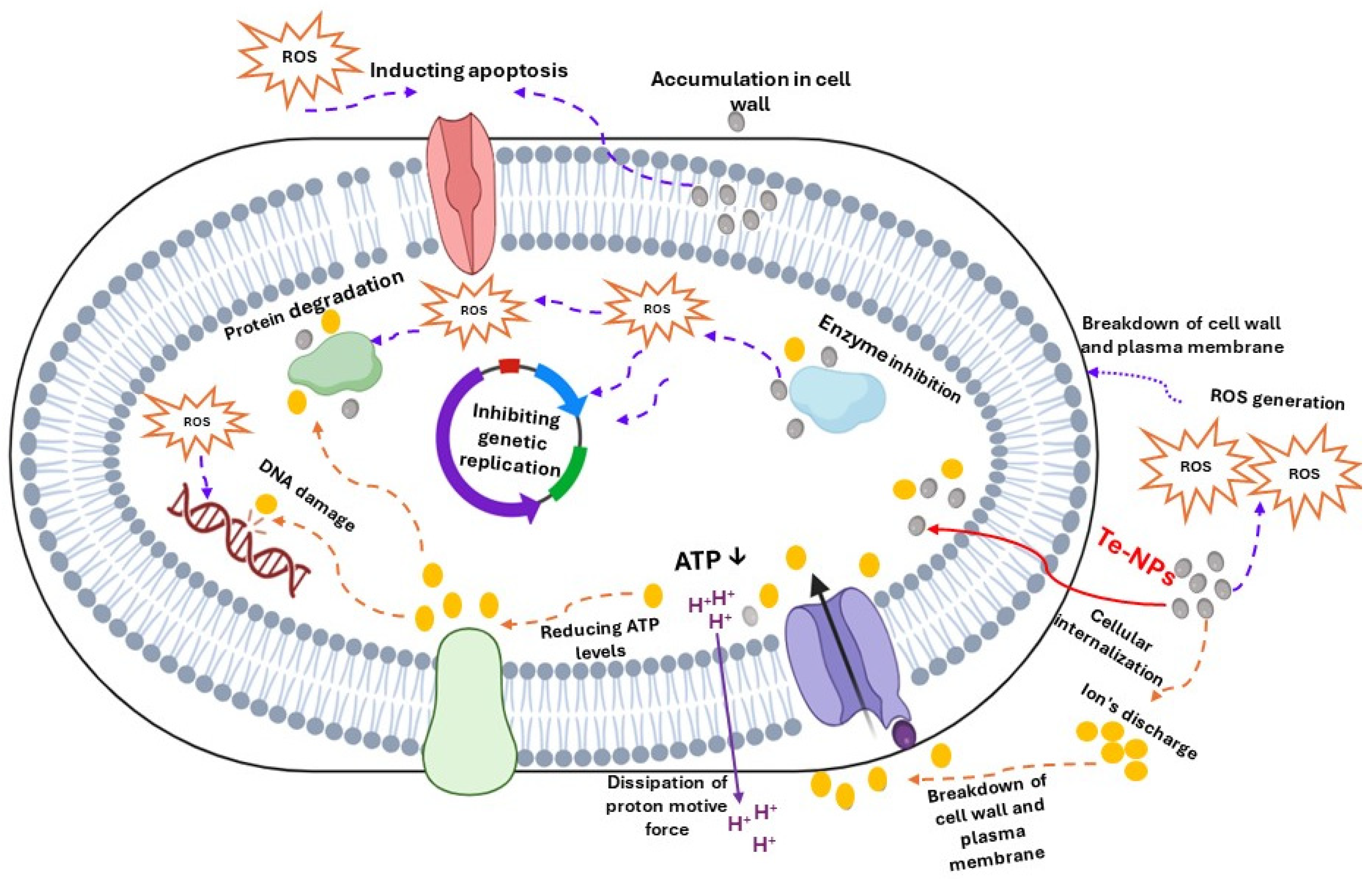
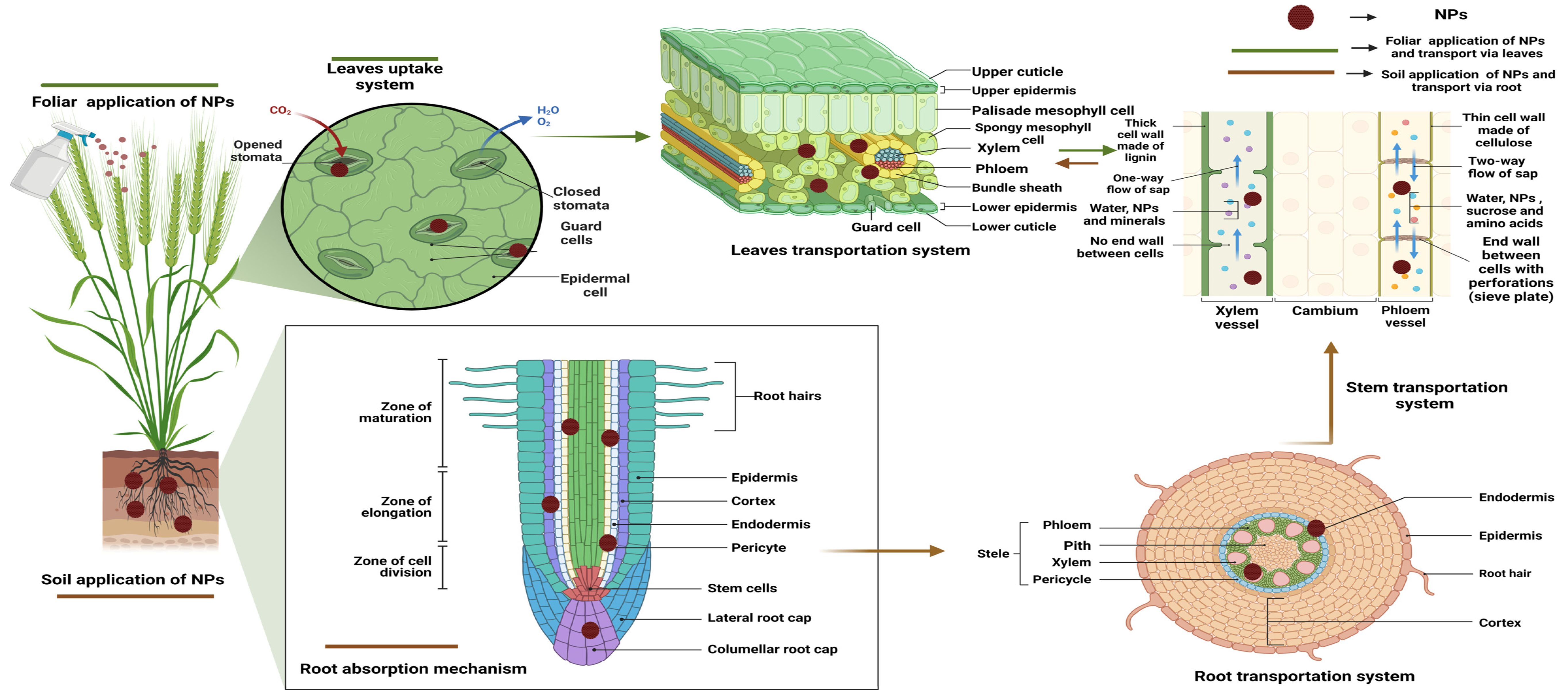
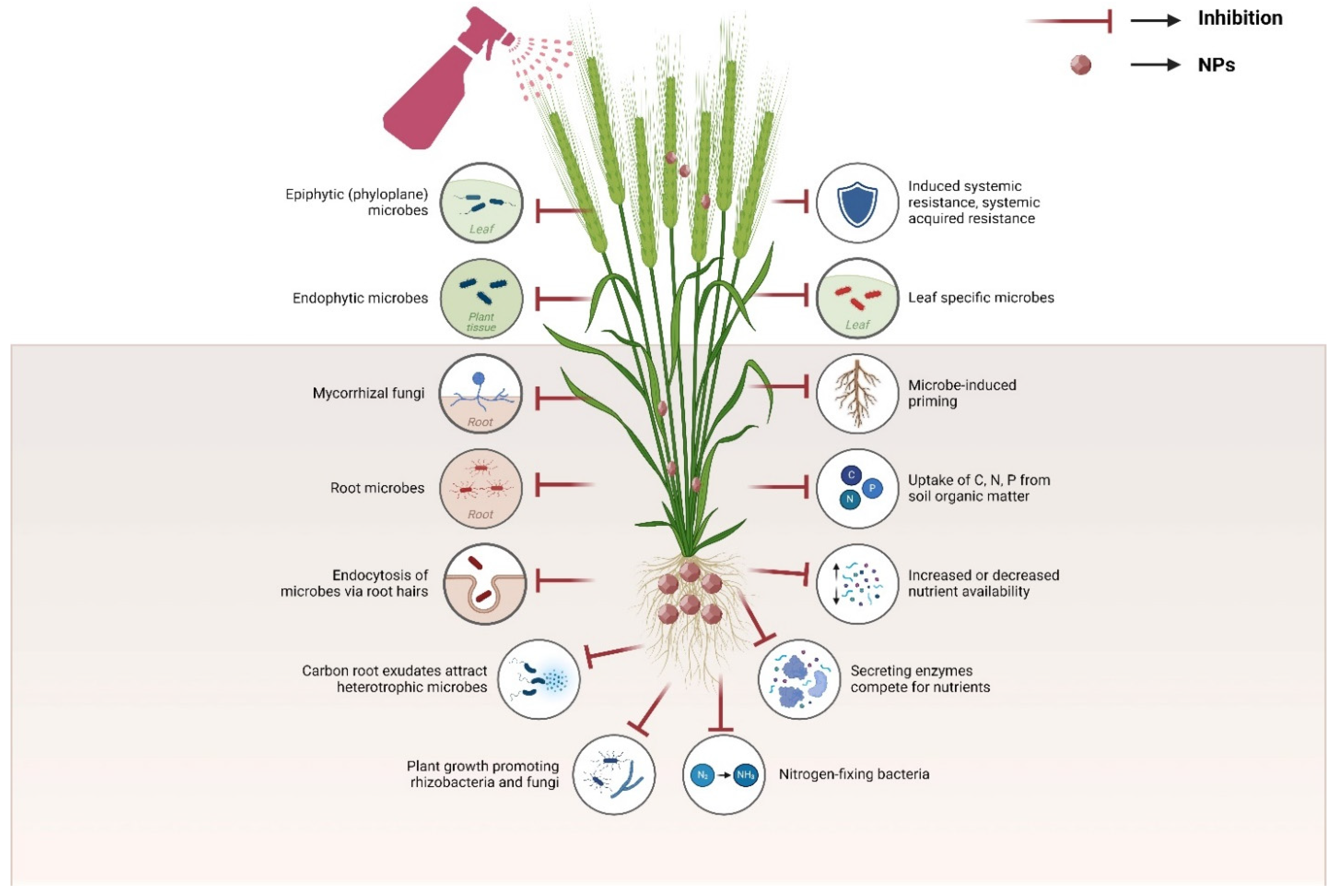
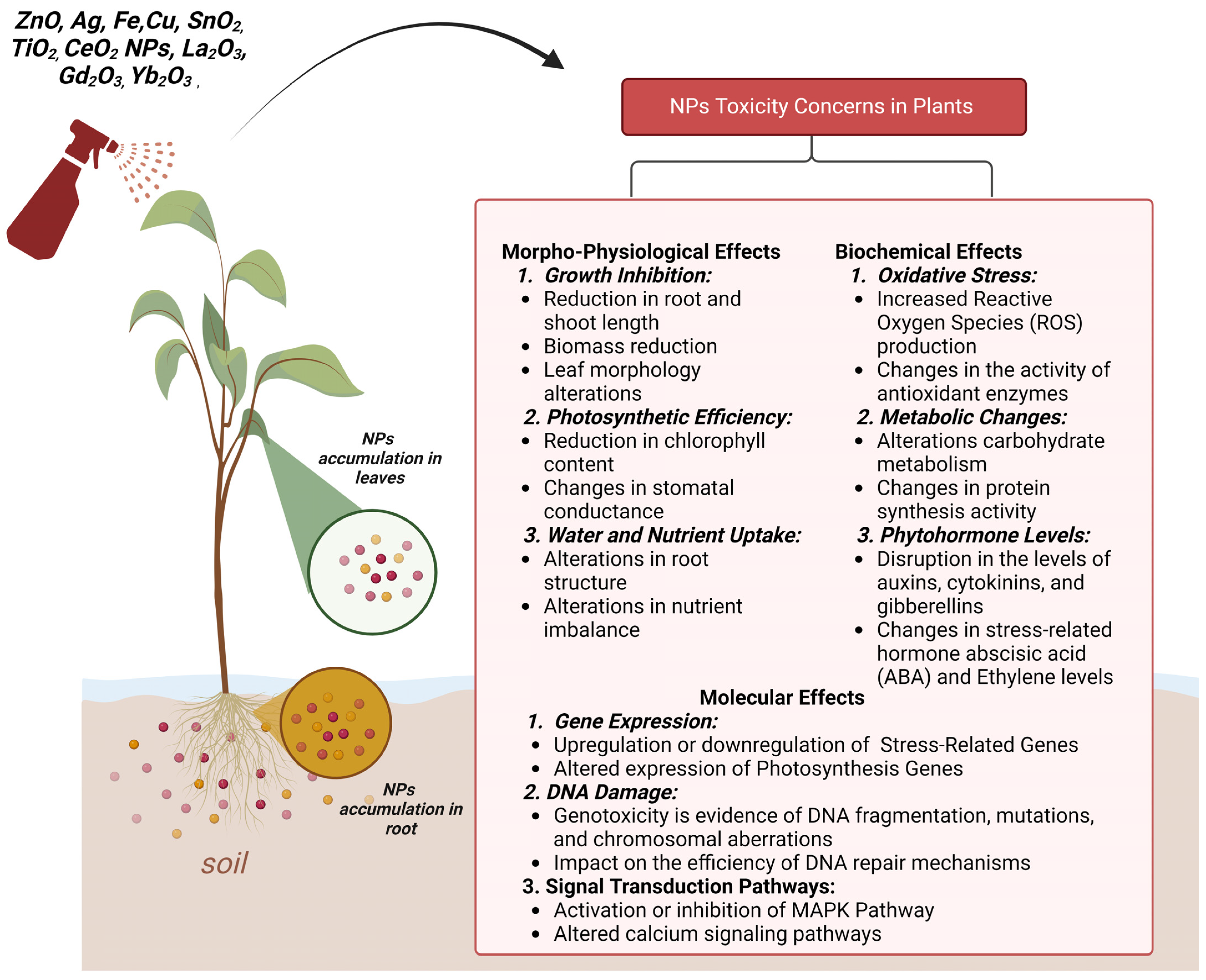
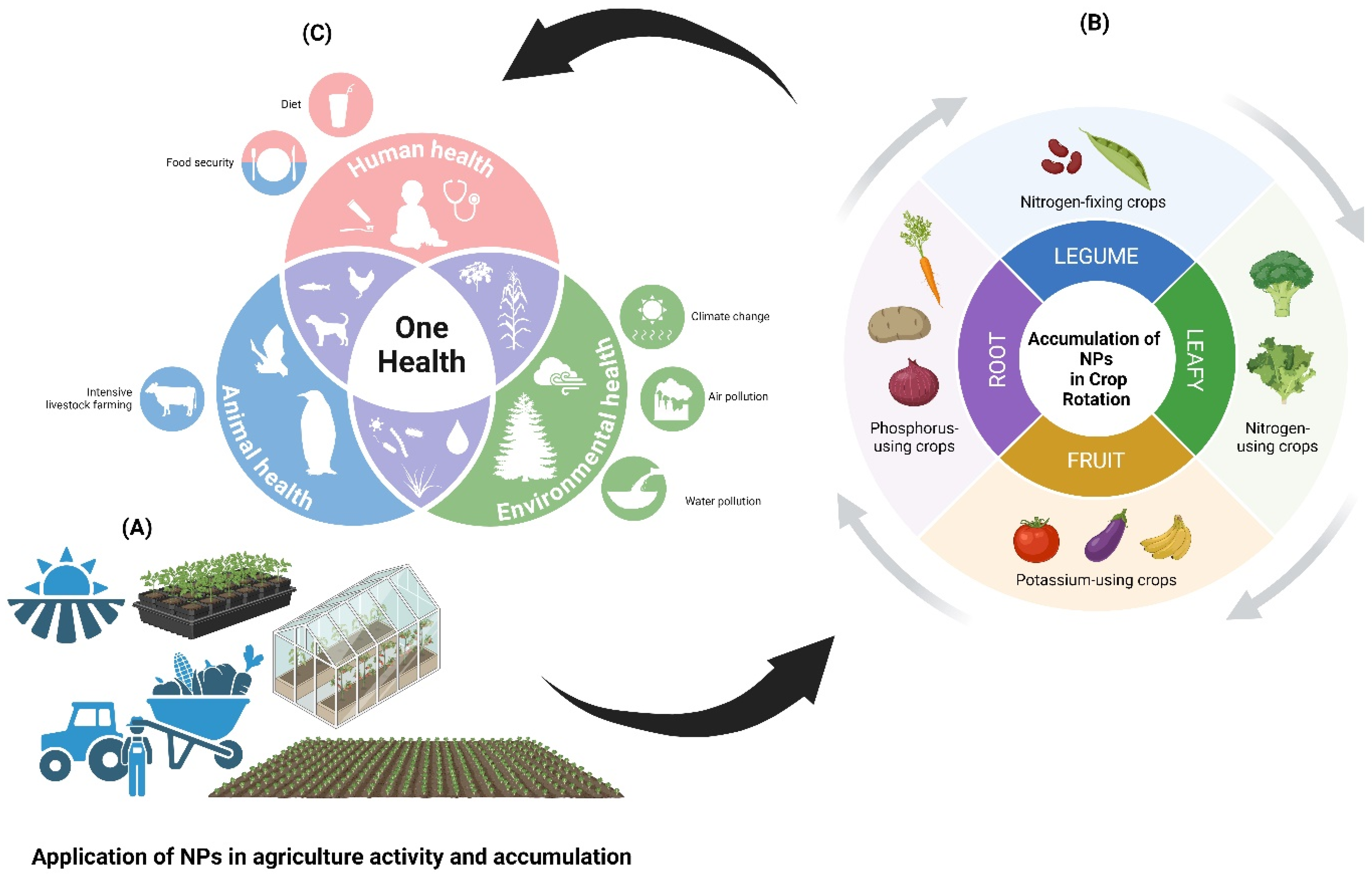
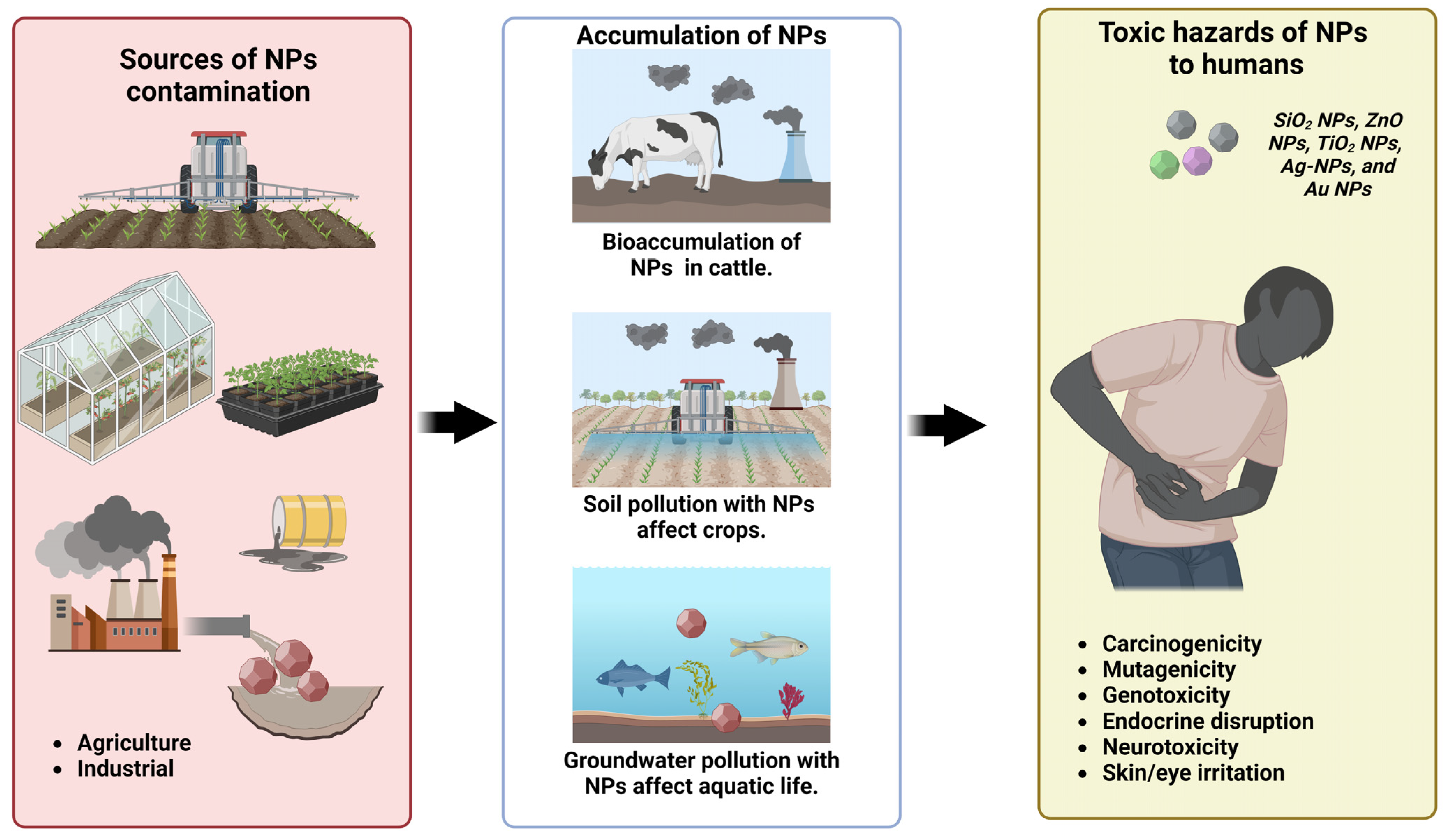
| Plant Species | Microbe(s) or Related Item | Soil Conditions | The Type of Relationship | Refs. |
|---|---|---|---|---|
| Plants hyper-accumulators | Pseudomonas aeruginosa, Maytenus bureaviana and Vibrio parahaemolyticus | Polluted soil with heavy metals (As, Cd, Hg, Cr, Cu, Pb) | Microbe–plant assisted bioremediation | [46] |
| Tomato (Solanum lycopersicum L.) | Firmicutes (Caldalkalibacillus, Bacillus) and Actinobacteria (Streptomyces) | Tomato 13C-residue decomposition under different CO2 levels | Plant residue C in plant/soil/microbe system | [54] |
| Maize, sesame, soybean, and sweet potato | Microbial biomass (fungal and bacterial), and enzyme activities | Sandy loam soil (11.30 g kg−1 SOM, 5.69 pH) | Intercropping system (different 4 crops) | [47] |
| Rice, rice, Chinese milk vetch rotations | Bacterial-derived C proportions for decomposition of organic amendments | Organic fertilization for a 40-year field experiment | Organic amendments under long-term in paddy soils | [48] |
| Eggplant, maize, cucumber, pumpkin, soybean | Soil bacterial communities under weed Ageratina adenophora L. | Soil samples were used in measure the total phenolic content | Allelopathic system in soil and gall-forming in plant tissues | [49] |
| Amaranthus hypochondriacus | Fungus-derived biochar and salt-tolerant bacterium-plant | Cd-polluted saline-alkali soils | Myco-plant remediation of polluted soils | [51] |
| Crop rotations, alternative, cover, and green crops | Microbial composition, abundance, diversity, functions and services | Soil quality, soil fertility, and soil microbiome functions | Plant–soil–microbe–anthropogenic activity nexus | [50] |
| Grasses besides soybean, wheat and maize | Soil heterotrophic bacterial communities along with soil respiration rate | Soil OM (3%), well-drained with sandy-loam texture | Land cover types, plant residues and soil microbes | [52] |
| Pak choi (Brassica chinensis L.) | Soil microbial biomass-C, -N, -P, and microbial stoichiometry | Soil pH 5.47, SOM 1.75%, available K 65.33 mg kg−1 | Soil microplastic pollution | [55] |
| Ginger (Zingiber officinale L.) | Soil rhizosphere microbial community, beside 16 S rRNA gene sequence | Co-polluted soil with ofloxacin and chromium | Polluted soil with antibiotics and heavy metals | [56] |
| Elymus nutans, Kobresia humilis, Kobresia pygmaea | Plant–soil nematode/bacterial and fungal linkages | Pastoral soils in alpine swampy meadows as peat bog | Grazing and plant–soil biota system | [57] |
| Gymnocarpos przewalskii L. | Arbuscular mycorrhizal fungal, fungal–bacterial ratio | Grey-brown desert soil and aeolian sandy soil | Soil microbial under hyper-arid desert | [58] |
| Conifer tree, broadleaf tree, and grasses | Airborne bacterial composition, urban greenspace microbiomes | Soil samples were collected using a metal soil corer | Air–phyllosphere–soil continuum | [59] |
| Microbe Species | Organic Precursors | Target Applications | Refs. |
|---|---|---|---|
| Staphylococcus aureus and Escherichia coli | Potato dextrose agar, and potato dextrose broth | Using the hydrothermal method at 200 °C for 24 h to detect bacteria, dead/live microbial differentiation dyes | [76] |
| Bacillus subtilis | Tea leaves | Green synthesis of CNDs from fermentation of tea leaves with Bacillus subtilis | [77] |
| Lactobacillus plantarum LLC-605 | L. plantarum LLC-605 was isolated from the traditional Chinese fermented food | Commercial dead/live microbial test dyes as a new type of anti-biofilm material by hydrothermal method at 200 °C for 24 h | [78] |
| Cyanobacteria | Cyanobacteria powder | CNDs was produces using hydrothermal methods having high photostability and low cytotoxicity | [75] |
| Bacillus cereus MYB41-22 cells | Yeast extract | Multicolor fluorescence bioimaging at 200 °C for 12 h | [79] |
| Lactobacillus acidophilus | Cell-free supernatant (CFS) of L. acidophilus | Producing functionalized nano-paper for UV and antimicrobial protective food active packaging | [80] |
| E. coli and S. aureus | Ampicillin sodium | Applied bioimaging with high sensitivity detecting Hg2+ ions in live/dead microbes at 200 °C for 6 h | [81] |
| Saccharomyces cerevisiae | 1% glucose, 2% yeast extract, and pH at 5.8 for 72 h | Homogeneous N and P-doped CDs (~4.1 nm) were hydrothermal synthesized (7 h at 200 °C) as non-toxic (˂3.5 mg/mL) to produce antimicrobial bacterial nano-cellulose membrane | [82] |
| Staphylococcus aureus | Vancomycin hydrochloride (VAN) | For detect poisonous tin (Sn4+) using VAN-CNDs (0.899 nm) through changes in the fluorescence intensity, with antibacterial low biological toxicity | [83] |
| Aspergillus flavus | Dry fungal biomass by hydrothermal method | Applied CQDs acted as a high-harvesting agent for improving the absorption of sunlight during photosynthesis through stimulating the enzymes | [84] |
| Plants | CND Details | Main Findings | Refs. |
|---|---|---|---|
| Rice seeds | (10–100 ppm) for 10 days (5–10 nm) | Nano-priming rice seeds using green CNDs promoted rice growth by increasing the aroma compound due to their high content of phenolic content as antioxidants | [90] |
| Curcumin (Curcuma longa L.) | 1–5 mg L−1 (7.34 nm) for 91 days | Plastic-derived CDs significantly enhanced enzymic antioxidants through nano-priming of seeds, besides content of chlorophyll and carbohydrate | [84] |
| Pea (Pisum sativum L.) seeds | (1.3–4.0 nm; up to 2 mg mL−1) | Plastic derived CDs significantly enhanced enzymic antioxidants through nano-priming of seeds, beside content of chlorophyll and carbohydrate | [91] |
| Tomato (Solanum lycopersicum L.) | 1–30 mg kg−1 (2–8 nm) for 15 days | Functional CNDs improved tomato growth, and under drought stress by activating chlorophyll forming, osmolyte synthesis, cell division, enzymatic activation, along with soil pH, organic matter, organic carbon and soil biological activities | [92] |
| Tomato (Solanum lycopersicum L.) | 8–16 mg kg−1 (4–15 nm) for 85 days of cultivation | Soil-applied functional CNDs ameliorated negative impacts of saline-alkali condition by up-regulation effects on soil properties (fertility, enzyme activity and decline both pH and salinity) and plant physiology (antioxidants, and nutrient uptake) along with fruit quality | [93] |
| Salvia miltiorrhiza | 0.5 g in 100 mL (0.8–7.2 nm) | The presence of CNDs during plant growth enhanced the adaptability to harsh environment without a reactive oxygen species burst | [2] |
| Soybeans | 1–50 mg kg−1 (for 30 days) | CNDs increased the growth of soybean plant under drought stress through the enhancement of nitrogen bioavailability | [86] |
| Tomato, mung beans | 0.015–0.13 mg mL−1 | CNDs improved seed germination under drought stress | [87] |
| Rice seedlings | 50–300 μg mL−1 for 16 days; 2.53 nm | Mg-N co-doped CNDs significantly increased the height (22.34%) and fresh biomass (70.60%) of rice plants | [89] |
| Arabidopsis thaliana | 4 mg L−1 (2–8 nm) for 13 days | Applied functional CNDs enhanced seedlings to be longer and stronger in their roots, bigger rosette and thicker leaves due to their easier uptake, generating more positive effects on plant | [94] |
| Soybean, tomato, eggplant | 0.14–2.24 mg mL−1 (for 10 days; 5 nm) | The degradable CNDs can effectively enhance the ribulose bisphosphate carboxylase oxygenase activity and then promote the dicotyledons’ growth | [95] |
| Rice plants | 0.14–2.24 mg mL−1 (for 10 days; 5 nm) | CNDs can penetrate all parts of rice plants, including the cell nuclei, which can enhance the disease resistance ability | [88] |
| Peanut plants | 180 mg L−1 (2–5 nm for 25 d) | Significantly improved stress-resistant properties of plants (at 180 ppm) by increasing antioxidants of SOD, CAT, and POD, and reducing MDA content | [85] |
| Pumpkin | 100–400 ppm (2–6 nm) for 7 days | CNDs could potentially trigger the antioxidant defense systems (SOD, CAT, and POD) in pumpkin seedlings with more impacts on roots than shoots of pumpkin plants | [96] |
| Plants | Applied Level (s) | Phytotoxicity Concerns | Refs. |
|---|---|---|---|
| Solanum nigrum L. | 5–15 mg kg−1 (2–6 nm) for 60 days cultivation | Functional CNDs improved soil nano-remediation by suppressing metal translocation (Cd, Pb) to shoots, activated enzymes (SOD, POD, and glutathione peroxidase), and microbial diversity in the rhizosphere | [101] |
| Arabidopsis thaliana | 24.93 and 53.55 µg mL−1 for 30 days | CNDs decreased the photosynthesis rates and gas exchange in plants | [97] |
| Allium cepa tubers | 20 µg mL−1, (5–10 nm), for 24 h | Sugar-terminated CNDs were found to be non-toxic as nanofertilizers can promote the growth of Vigna radiata seedlings under salt stress (up to 100 mM NaCl) | [102] |
| Water hyacinth (Eichhornia crassipes) | 4, 8, 16, 30 mg L−1 for 8 days | Functional CNDs improved removing of heavy metals (Cd, Pb) by nano-phytoremediation, regulate enzymatic levels, moderate their biotoxicity and inhibit their transfer | [103] |
| Citrus maxima | 600 and 900 ppm for 10 days | CNDs can be used as repair agents to mitigate the toxicity of Cd2+ to plants at the level of 900 ppm by mitigating the oxidative stress and reduce transported Cd2+ to leaves | [99] |
| Wheat seedlings | 50 and 75 mg L−1 for 10 days | The toxicity of Cd2+ was reduced with the uptake of CNDs by reducing Cd2+ uptake and increase root activity | [100] |
| Arabidopsis thaliana L. | 125–1000 ppm for 7 days | The phytotoxic CNDs was 1000 ppm, which led to increased activities of glutathione reductase in roots and shoots in contrast to control and reduced the metabolites | [94] |
| Materials Related to Te-NPs | Size and Morphology | Inhibited Microbes or Anti-Microbes | Refs. |
|---|---|---|---|
| Te-nanostructures of Au-, Ag-, and Au/Ag | Nanotube size (25–30 nm) wall thickness (5–6 nm) | E. coli, S. aureus, and S. enteritidis | [114] |
| Tellurium oxide NPs | Spheres diameter ~65 nm | S. aureus, K. pneumoniae, and E. coli | [115] |
| Tellurium nanorods | Rod-shaped size (22 nm), length 185 nm | Methicillin-resistant S. aureus, S. typhi (PTCC 1609), and P. aeruginosa (PTTC 1574) | [116] |
| Te-loaded polymeric fiber | Clusters ~20 µm | P. aeruginosa, S. aureus and E. coli | [117] |
| TeO2-NPs sols | Spheres diameter ~55 nm | Bacillus subtilis, S. enteritidis, and E. coli | [118] |
| Lime-mediated-Te-NPs, Orange-mediated-Te-NPs, Lemon-mediated-Te-NPs | Nanorods of orange Te-NPs (50–200 nm length), cubic shape in others (100–200 nm in length) | Multidrug-resistant (MDR) E. coli (ATCC BAA-2471) and methicillin-resistant S. aureus (MRSA) (ATCC 4330) | [119] |
| Chitosan-fabricated Te-NPs | Spheres diameter (37 nm) | L. monocytogenes, B. cereus, and S. aureus | [120] |
| Myco-synthesized Te-NPs | Spheres diameter ~60.8 nm | S. cerevisiae PTCC 5269, C. albicans ATCC 10231, and K. pneumoniae ATCC 10031 | [121] |
| Gallic acid-Te NPs | Spherical size 19.74 nm | Staphylococcus aureus, Salmonella enterica, and Escherichia coli | [120] |
| Nano-tellurium | Rod shape (size 21.4 nm) | Staphylococcal bacteremia and Staphylococcus aureus | [122] |
| Te–CeO2 nanocomposite | Nanofibrous sphere (nano wools 200 nm) | Klebsiella pneumoniae MTCC 3384 and Bacillus subtilis MTCC 441 | [123] |
| Te-doped ZnO nanoparticles | Nano-sheet at hexagonal pattern (13 mm) | Anti-bacterial (E. coli and S. aureus), and antifungal (C. albicans and E. salmonicolor) | [124] |
| Biological Te-NPs by Acinetobacter pittii | Rod-shaped (60–130 nm) | Escherichia coli BW25113 | [125] |
| Biological Te-NPs by Aromatoleum sp. CIB | Rod-shaped (200 nm) | Aromatoleum sp. CIB (pIZ2-0135) strain | [126] |
| Biological Te-NPs by Mortierella sp. AB1 | Rod-shaped (100–500 nm) | Shigella dysenteriae, E. coli, Enterobacter sakazakii, and Salmonella typhimurium | [127] |
| Biological Te-NPs by Haloferaxalexandrinus GUSF-1 | Rod-shaped (7–40 nm) | Pseudomonas aeruginosa ATCC 9027 | [128] |
| Biological Te-NPs by Gayadomonas sp. TNPM15 | Nanorods (15–23 nm) | Fusarium oxysporum AUMC 10313 and Alternaria alternata AUMC 3882 | [129] |
Disclaimer/Publisher’s Note: The statements, opinions and data contained in all publications are solely those of the individual author(s) and contributor(s) and not of MDPI and/or the editor(s). MDPI and/or the editor(s) disclaim responsibility for any injury to people or property resulting from any ideas, methods, instructions or products referred to in the content. |
© 2024 by the authors. Licensee MDPI, Basel, Switzerland. This article is an open access article distributed under the terms and conditions of the Creative Commons Attribution (CC BY) license (https://creativecommons.org/licenses/by/4.0/).
Share and Cite
Prokisch, J.; Nguyen, D.H.H.; Muthu, A.; Ferroudj, A.; Singh, A.; Agrawal, S.; Rajput, V.D.; Ghazaryan, K.; El-Ramady, H.; Rai, M. Carbon Nanodot–Microbe–Plant Nexus in Agroecosystem and Antimicrobial Applications. Nanomaterials 2024, 14, 1249. https://doi.org/10.3390/nano14151249
Prokisch J, Nguyen DHH, Muthu A, Ferroudj A, Singh A, Agrawal S, Rajput VD, Ghazaryan K, El-Ramady H, Rai M. Carbon Nanodot–Microbe–Plant Nexus in Agroecosystem and Antimicrobial Applications. Nanomaterials. 2024; 14(15):1249. https://doi.org/10.3390/nano14151249
Chicago/Turabian StyleProkisch, József, Duyen H. H. Nguyen, Arjun Muthu, Aya Ferroudj, Abhishek Singh, Shreni Agrawal, Vishnu D. Rajput, Karen Ghazaryan, Hassan El-Ramady, and Mahendra Rai. 2024. "Carbon Nanodot–Microbe–Plant Nexus in Agroecosystem and Antimicrobial Applications" Nanomaterials 14, no. 15: 1249. https://doi.org/10.3390/nano14151249


