Article

MIL-Derived Hollow Tubulous-Shaped In2O3/ZnIn2S4 Z-Scheme Heterojunction for Efficient Antibacterial Performance via In Situ Composite

1
Key Laboratory of Advanced Marine Materials, Key Laboratory of Marine Environmental Corrosion and Bio-Fouling, Institute of Oceanology, Chinese Academy of Sciences, Qingdao 266071, China
2
University of Chinese Academy of Sciences, 19 (Jia) Yuquan Road, Beijing 100049, China
*
Authors to whom correspondence should be addressed.
These authors contributed equally to this work.
Nanomaterials 2024, 14(16), 1366; https://doi.org/10.3390/nano14161366
Submission received: 22 July 2024 / Revised: 15 August 2024 / Accepted: 18 August 2024 / Published: 21 August 2024
(This article belongs to the Special Issue Heterogeneous Photocatalysts Based on Nanocomposites)

Abstract

:
In this study, a hollow tubulous-shaped In2O3 derived from MIL (MIL-68 (In)) exhibited an enhanced specific surface area compared to MIL. To further sensitize In2O3, ZnIn2S4 was grown in situ on the derived In2O3. The 40In2O3/ZnIn2S4 composite (1 mmol ZnIn2S4 loaded on 40 mg In2O3) exhibited degradation rates of methyl orange (MO) under visible light (80 mW·cm−2, 150 min) that were 17.9 and 1.4 times higher than those of the pure In2O3 and ZnIn2S4, respectively. Moreover, the 40In2O3/ZnIn2S4 exhibited an obviously improved antibacterial performance against Pseudomonas aeruginosa, with an antibacterial rate of 99.8% after visible light irradiation of 80 mW cm−2 for 420 min. The 40In2O3/ZnIn2S4 composite showed the highest photocurrent density, indicating an enhanced separation of photogenerated charge carriers. Electron spin resonance results indicated that the 40In2O3/ZnIn2S4 composite generated both ·O2 and ·OH radicals under visible light, whereas ·OH radicals were almost not detected in ZnIn2S4 alone, suggesting the presence of a Z-scheme heterojunction between In2O3 and ZnIn2S4, thereby enhancing the degradation and antibacterial capabilities of the composite. This offers fresh perspectives on designing effective photocatalytic materials for use in antibacterial and antifouling applications.

1. Introduction

Biofilm formed by marine microorganisms and their metabolites on the surfaces of marine facilities serve as carriers for the attachment of macroorganisms, leading to marine biofouling [1]. Marine biofouling is a significant factor affecting and constraining the development of marine industries. Currently, antifouling coatings containing biocides are the most commonly used method. However, the slow release of biocides may pose threats to the safety of marine ecological environments [2]. Therefore, the development of novel environmentally friendly antimicrobial and antifouling materials to replace toxic alternatives has been recognized as an inevitable choice for sustainable development [3]. Photocatalysis generates highly oxidative reactive radicals capable of effectively degrading organic compounds and disrupting microbial cell structures. Moreover, photocatalytic sterilization is environmentally friendly and does not pose issues related to biological resistance [4,5]. Since sunlight can penetrate up to 200 m in seawater, it can be fully received in the upper layers of the ocean where biofouling occurs. Therefore, photocatalytic technology holds significant potential for applications in marine antimicrobial and antifouling fields.
Indium oxide (In2O3) is a semiconductor material with a relatively narrow bandgap, attracting attention due to its good stability and conductivity [6,7]. However, conventional In2O3 photocatalysts often suffer from limitations such as a simple structure, small surface area, weak light response, and high recombination rate of photogenerated charge carriers, which restrict their further application in photocatalysis [8]. To enhance the photocatalytic performance of monolithic In2O3, researchers have explored various methods including elemental doping [9,10], morphology control [11], and the construction of heterojunctions [12,13], achieving significant improvements. Recently, In2O3 photocatalysts prepared via the pyrolysis of metal-organic frameworks (MOFs) have garnered significant attention. Liu et al. achieved the formation of rhombohedral corundum/cubic In2O3 by the direct annealing of NH2-MIL-68(In) in air atmosphere. Spectroscopic and photoelectrochemical tests demonstrated that this unique structure effectively accelerates the separation and transfer of photogenerated charges within In2O3 [14]. Yang et al. employed a simple oil bath method to grow CdZnS on shuttle-shaped mesoporous In2O3 derived from NH2-MIL-68. Under visible light irradiation, the photocatalytic hydrogen evolution rate was significantly enhanced. This improvement can be attributed to several factors: the derived In2O3 possessing mesopores which increase active sites, the introduction of ultrafine CdZnS nanoparticles reducing the bandgap of material, thereby enhancing the visible light response, and the construction of Type II heterojunctions which enhance the separation efficiency of photogenerated charge carriers in the composite material [15]. Research indicates that MOFs-derived In2O3 can retain the original framework structure of MOFs, while the resulting hollow structures and pores provide additional reactive sites for photocatalytic reactions, thereby enhancing photocatalytic activity. In situ composite semiconductor heterojunctions constructed with multi-site hollow framework MOFs derivatives can further improve photocatalytic activity [13,16].
Ternary metal sulfides, characterized by narrow bandgaps, good stability, and excellent electrical and optical properties, are considered promising photocatalytic materials with high performance and potential applications [17]. Among these, ternary metal sulfides with an AB2X4 structure exhibit excellent photocatalytic stability. Within the AB2X4 series, ZnIn2S4 stands out due to its narrower bandgap and absence of toxic metal ions, making it an excellent candidate for photocatalytic applications in the energy and environmental fields. ZnIn2S4 is commonly used to sensitize wide-bandgap semiconductor materials, enhancing the charge carrier separation efficiency of monolithic semiconductor materials through the construction of heterojunction-based multi-component photocatalytic systems. Moreover, the facile preparation method of ZnIn2S4 enhances its appeal as an excellent candidate for modified materials [18,19].
In this paper, the derived hollow tubulous-like In2O3 was synthesized through calcination. Compared to the MIL, the derived In2O3 exhibited a reduced bandgap, increased optical absorption threshold, and enhanced photoelectric response. The composite material based on hollow tubulous-like In2O3 with in situ-grown ZnIn2S4 exhibits significantly enhanced photosensitivity compared to individual In2O3 and ZnIn2S4. Among them, the 40In2O3/ZnIn2S4 (1 mmol ZnIn2S4 loaded on 40 mg In2O3) composite shows the highest performance. The application potential of In2O3/ZnIn2S4 composites for marine antibacterial and antifouling purposes was evaluated by measuring their capability to degrade MO dye and eradicate Pseudomonas aeruginosa (P. aeruginosa). The 40In2O3/ZnIn2S4 composite degraded 99.5% of 10 ppm MO under simulated visible light irradiation for 150 min, which is 17.9 times higher than In2O3 and 1.4 times higher than ZnIn2S4 alone. Additionally, the composite achieved a 99.8% antibacterial rate against P. aeruginosa after 420 min of light exposure, demonstrating a significant enhancement in photocatalytic performance compared to the individual photocatalytic materials. Electron Spin Resonance (ESR) studies indicate that both In2O3 and ZnIn2S4 can generate ·O2 radicals, attributed to their conduction band potentials (ECB = −0.59 V (In2O3), ECB = −0.75 V (ZnIn2S4)) being more negative than the reduction potential of O2/·O2 (−0.33 V vs. NHE). Additionally, only In2O3 can generate ·OH radicals due to its valence band potential (EVB = 2.75 V) being more positive than the oxidation potential of H2O/·OH (EVB = 2.40 V vs. NHE). However, the composite material 40In2O3/ZnIn2S4 exhibits increased production of both ·O2 and ·OH radicals, suggesting the formation of a Z-Scheme heterojunction structure between In2O3 and ZnIn2S4, thereby enhancing the separation of photogenerated charge carriers. The composite material, prepared by the in situ growth of ZnIn2S4 on MIL-derived hollow tubular In2O3, demonstrates obviously enhanced degradation and antibacterial capabilities compared to pure In2O3 and ZnIn2S4, further expanding the possibilities for practical applications. Meanwhile, this advancement provides new insights for the development and design of novel, efficient photocatalytic antibacterial and antifouling materials.

2. Experimental Section

2.1. Synthesis of Rod-Shaped MIL-68 (MIL)

Using a solvothermal method, 5 mmol of indium nitrate (In(NO3)3) was dissolved in 35 mL of N,N-Dimethylformamide (DMF) to form solution A, while 6 mmol of phthalic acid (C8H6O4) was dissolved in 35 mL of DMF to form solution B. Solution A was then added dropwise to solution B under stirring at room temperature for 30 min. The resulting mixture was transferred to a high-pressure reaction vessel and subjected to a hydrothermal reaction at 100 °C for 12 h. Afterward, the product was centrifuged, washed several times with ethanol, and dried under vacuum at 80 °C for 12 h, yielding a white powder of MIL.

2.2. Synthesis of MIL-Derived In2O3 Photocatalyst

A certain amount of MIL powder was spread evenly in a crucible. The temperature was then ramped up at a rate of 10 °C/min until reaching 500 °C and maintained for 2 h, resulting in off-white In2O3 powder.

2.3. Synthesis of ZnIn2S4 Photocatalyst

In total, 1 mmol of ZnCl2, 2 mmol of InCl3, and 4 mmol of thiourea (TAA) were dissolved in 100 mL of deionized water. The mixture was stirred at room temperature for 1 h. Subsequently, the solution was heated and stirred in a water bath at 80 °C for 2 h. After cooling, the resulting mixture was centrifuged and washed several times with deionized water and ethanol. Finally, the material was dried under vacuum at 80 °C for 12 h to obtain the ZnIn2S4 photocatalyst.

2.4. Synthesis of In2O3/ZnIn2S4 Composites

In total, 30, 40, 50, 60, and 300 mg of In2O3 were individually weighed and dispersed in 100 mL of deionized water with stirring for 30 min to ensure thorough dispersion. Subsequently, 1 mmol of ZnCl2, 2 mmol of InCl3, and 4 mmol of thiourea (TAA) were added sequentially to each dispersion, followed by stirring at room temperature for 1 h. The mixtures were then subjected to constant-temperature reactions at 80 °C in a water bath for 2 h. After cooling, the reaction products were collected by centrifugation, washed several times with deionized water and ethanol, and finally dried under vacuum at 80 °C for 12 h. The resulting materials were named as follows based on the amount of In2O3 used: 30In2O3/ZnIn2S4, 40In2O3/ZnIn2S4, 50In2O3/ZnIn2S4, 60In2O3/ZnIn2S4, and 300In2O3/ZnIn2S4. The synthesis steps of the materials are shown in the Figure 1 below.

2.5. Characterization

X-ray diffraction spectra was used to characterize the crystal structure of the prepared composites (XRD, Smart Lab, Rigaku Co., Tokyo, Japan); Scanning electron microscopy was used to observe the microstructure of the samples (SEM, JSM-7601F, NEC Co., Tokyo, Japan); and the species and distribution of elements were determined by energy dispersive X-ray spectroscopy (EDS, Oxford INCAx-sight, Oxford, UK). The microscopic bonding of the composites was further observed using transmission electron microscopy (TEM, JEOL 2100F, NEC Co., Tokyo, Japan); Ultraviolet-visible diffuse reflectance spectroscopy (UV-Vis DRS, U-3900H, Shimadzu, Kyoto, Japan) was used to observe the optical properties of the composites and calculate their bandgap information; The valence states of reactive elements were determined by X-ray photoelectron spectroscopy (XPS, ESCALAB 250Xi, Thermo, Waltham, MA, USA). The production of free radicals was detected by electron paramagnetic resonance spectroscopy (EPR, EMXplus, Bruker Co., Billerica, MA, USA; Ettlingen, Germany).

2.6. Photoelectrochemical Test

The photoelectrochemical testing was conducted using a CHI660E electrochemical workstation (CHI660E, Shanghai Chenhua Instrument Co. Ltd., Shanghai, China). A three-electrode system was employed, with an Ag/AgCl electrode as the reference electrode, a Pt electrode as the counter electrode, and the prepared photoelectrode as the working electrode. The experiments were carried out in a 3.5 wt% NaCl solution to simulate seawater conditions. In addition, a xenon lamp (PLS-SXE300D, Beijing Perfect Light Co., Ltd., Beijing, China) was used as the simulated solar light illumination. The light was filtered through an AM 1.5 filter to obtain the simulated solar light and then adjusted to an intensity of 100 mW·cm−2. For the Mott–Schottky measurements, the open circuit potential of the system was first determined. Then, the Mott–Schottky plot was tested over a range of ±0.6 V with a frequency of 1000 Hz and an AC voltage amplitude of 10 mV.

2.7. Photocatalytic Degradation and Photocatalytic Antibacterial Performance

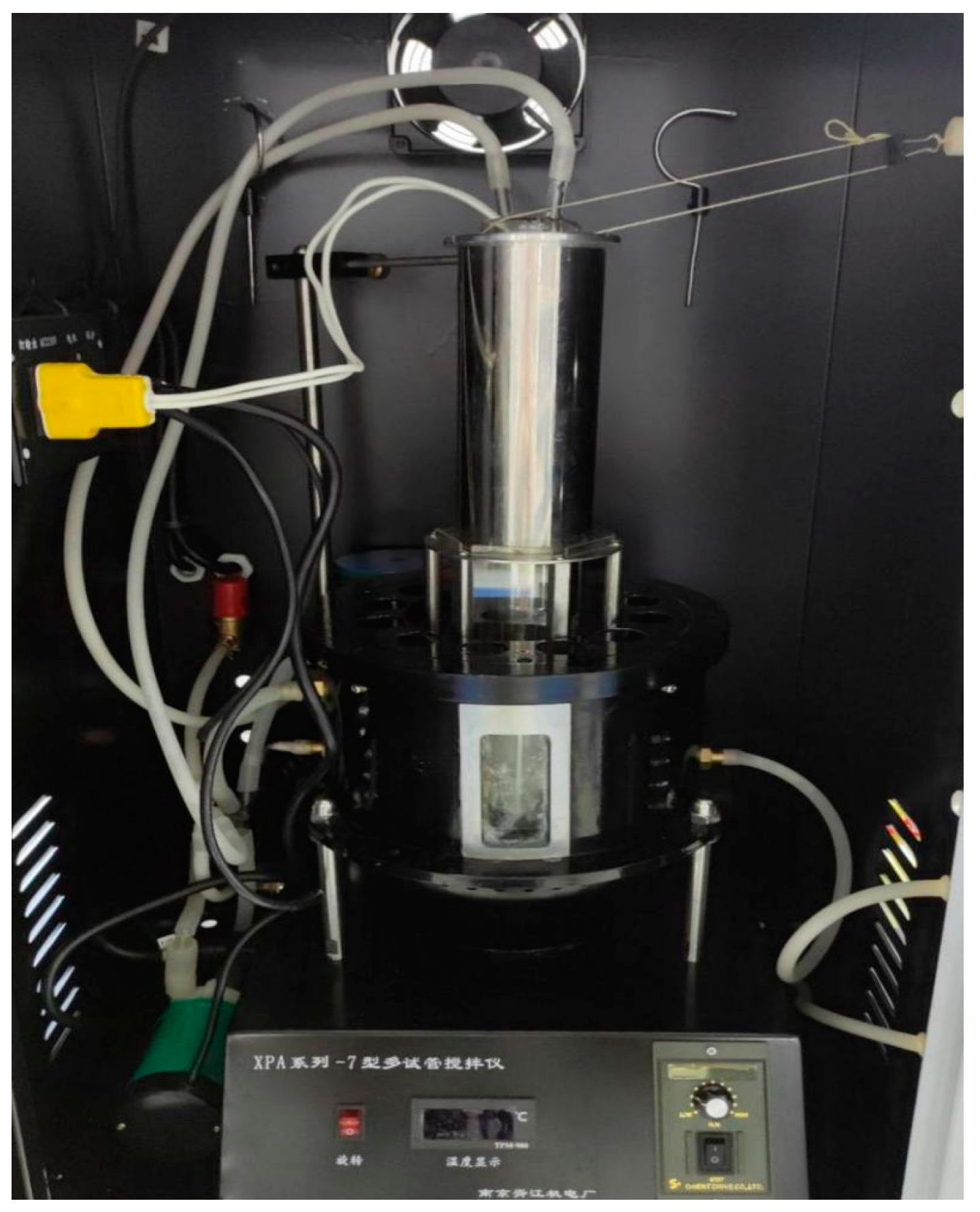
For the photocatalytic degradation of methyl orange (MO), 15 mg of the sample was weighed in a 50 mL quartz tube, followed by adding 50 mL of a 10 ppm MO solution; each sample was tested three times. The quartz tube was then placed into a photocatalytic reactor (Figure 2) and stirred for 60 min to achieve adsorption–desorption equilibrium. Subsequently, photocatalytic degradation reactions were conducted under 800 W xenon lamp irradiation (with a 420 nm cut-off wavelength filter and a light intensity of 80 mW·cm−2). Then, 1 mL solution of each sample was taken every 30 min during the reaction, and finally, the changes in MO absorbance were measured using an enzyme-labeler (YP-96C, Youyunpu, Weifang, China) to calculate the degradation rate.
The photocatalytic antibacterial experiments used P. aeruginosa (BNCC186070), commonly found in marine fouling biofilm. In total, 50 mg of the photocatalyst was added to a 50 mL quartz tube, followed by adding 49.5 mL of sterilized 0.1 M phosphate buffer saline (PBS), and each sample requires three parallel experiments. This was inoculated with 500 μL of an appropriately diluted bacterial suspension (5.9 × 10⁸ cfu·mL−1). The experiments were also conducted in a photocatalytic reactor with the same simulated light conditions. Prior to light exposure, the mixture was stirred for 60 min to achieve adsorption–desorption equilibrium in the dark condition. Then, 100 μL of the bacteria solution from the quartz tube was collected every 60 min. The antibacterial efficiency was determined by counting viable bacteria using the colony-counting method on a solid LB medium plate. The equipment used for photocatalysis is as follows:

3. Results and Discussion

3.1. Analysis of Physical Properties

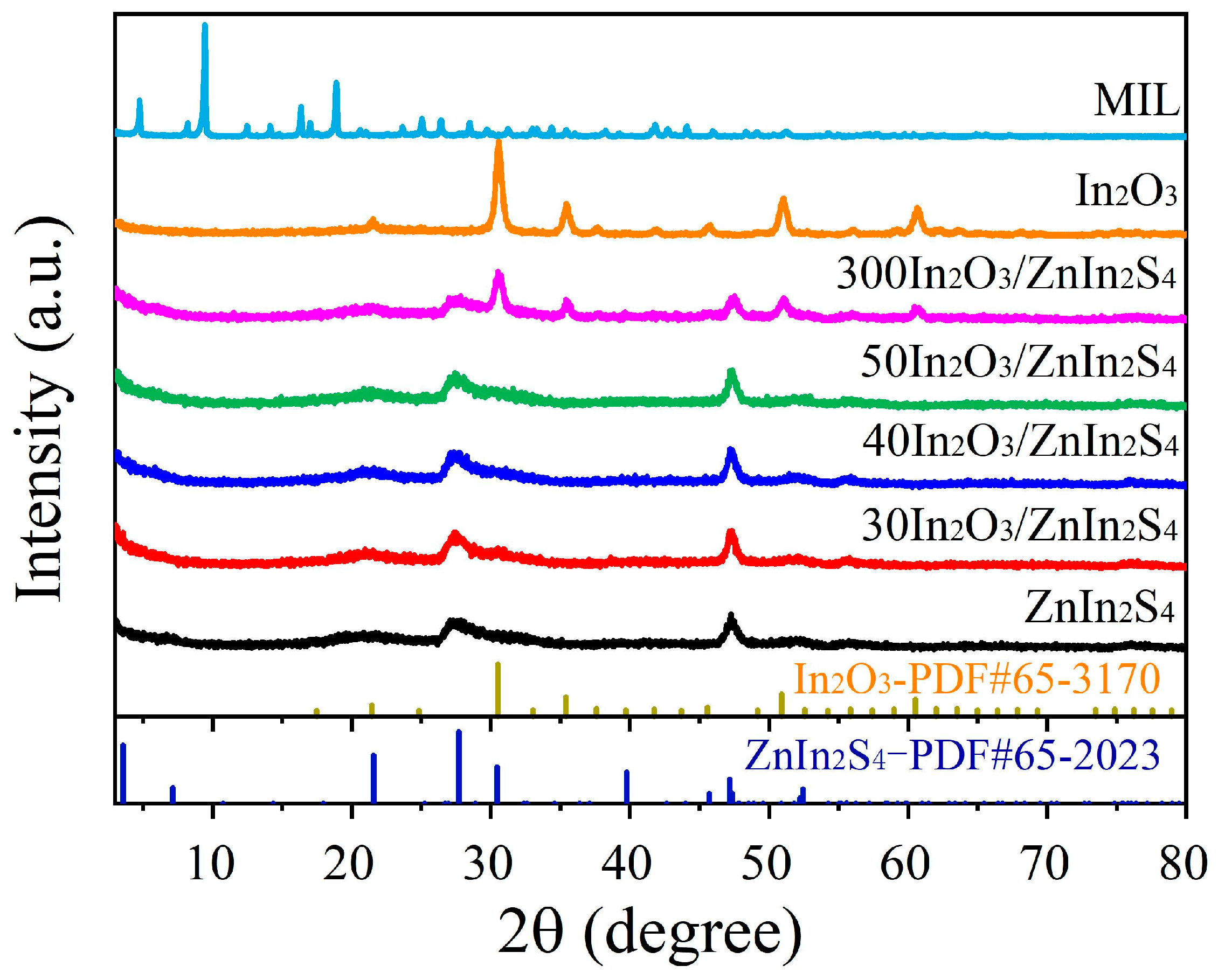
The XRD patterns of MIL, derived In2O3, ZnIn2S4, and different In2O3/ZnIn2S4 composites were determined, and the results are shown in Figure 3. It is evident that the peaks of the rod-like MIL template are sharp, indicating good crystallinity, and the peak patterns correspond well with those reported in the literature [20]. The diffraction peaks of the derived In2O3 at 2θ = 21.5°, 30.5°, 35.4°, 45.6°, 50.9°, and 60.6° correspond to the (211), (222), (400), (400), (440), and (622) crystal planes of cubic-phase In2O3 (PDF#65-3170), indicating that the material derived from MIL after high-temperature calcination is In2O3. The characteristic peaks at 21.6°, 27.7°, and 47.2° in the synthesized ZnIn2S4 correspond to the (006), (102), and (110) diffraction peaks of hexagonal-phase ZnIn2S4 (PDF#65-2023), confirming the successful synthesis of ZnIn2S4. The XRD analysis of the 300In2O3/ZnIn2S4 composite shows characteristic diffraction peaks of both In2O3 and ZnIn2S4, indicating the simultaneous presence of both materials. However, in the XRD spectra of 30In2O3/ZnIn2S4, 40In2O3/ZnIn2S4, and 50In2O3/ZnIn2S4, no distinct In2O3 diffraction peaks are observed. This suggests that In2O3 is heavily loaded with ZnIn2S4, resulting in the characteristic peaks of In2O3 being less pronounced.
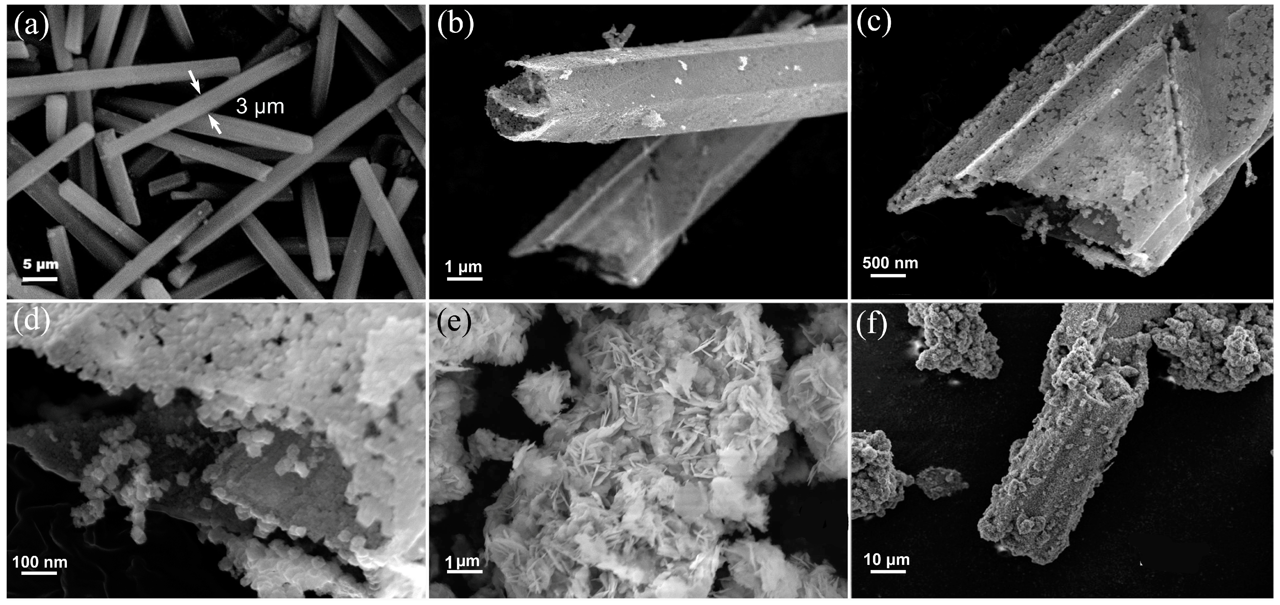
The microstructures of MIL, MIL-derived In2O3, ZnIn2S4, and 40In2O3/ZnIn2S4 composite were characterized using SEM. As shown in Figure 4a, MIL exhibits a rod-like structure with a smooth and uniform surface, and the rod width is approximately 3 μm. Figure 4b–d show the SEM images of the derived In2O3 products after calcination. The In2O3 retains the rod-like morphology of MIL but develops a hollow structure with abundant surface voids, possibly due to the removal of organic ligands during combustion [15]. Figure 4e displays the SEM image of ZnIn2S4, revealing a flower-ball structure composed of nanoplates. This nanoplate structure endows ZnIn2S4 with strong light absorption performance and provides numerous active sites [21]. Figure 4f shows the SEM image of the 40In2O3/ZnIn2S4 composite, where the surface of In2O3 becomes roughened following the in situ growth of ZnIn2S4. Combined with Figure 4g, it is evident that a multi-layered ZnIn2S4 nanoplate was loaded on the surface of In2O3. Figure 4h presents the EDS spectra of the 40In2O3/ZnIn2S4 composite, indicating the uniform distribution of In, O, Zn, and S elements on the surface, confirming the successful composite of ZnIn2S4 on the hollow tubulous-like In2O3.
XPS was employed to analyze the surface composition and chemical states of the derived In2O3, ZnIn2S4, and 40In2O3/ZnIn2S4. Figure 5a presents the XPS survey spectra of the three samples. It can be observed that both the prepared ZnIn2S4 and 40In2O3/ZnIn2S4 exhibit characteristic peaks corresponding to Zn2p, In3d, and S2p. In the In3d orbital XPS spectra (Figure 5b), all three samples show two distinct peaks. For ZnIn2S4, the peaks at 452.7 eV and 445.2 eV are attributed to the In3d3/2 and In3d5/2 orbitals of ZnIn2S4, respectively. Similarly, for 40In2O3/ZnIn2S4, peaks corresponding to In3d3/2 and In3d5/2 appear at the same binding energies as ZnIn2S4, indicating the substantial in situ growth of ZnIn2S4 on the In2O3 carrier. In contrast, the binding energies of In3d3/2 and In3d5/2 in pure In2O3 are located at 451.9 eV and 444.3 eV, respectively, indicating the presence of In3+ in the samples. Figure 5c displays the orbital peaks of O1s. In In2O3, the O1s peak appears at a binding energy of 529.8 eV, corresponding to lattice oxygen. In contrast, the high binding energy peak at 532.0 eV for the In2O3/ZnIn2S4 composite is typically attributed to chemisorbed surface oxygen [22,23]. As shown in Figure 5d, Zn2p in ZnIn2S4 exhibits two characteristic peaks: Zn2p1/2 at 1045.5 eV and Zn2p3/2 at 1022.5 eV. For the 40In2O3/ZnIn2S4, the Zn2p orbital peaks are observed at 1045.6 eV (Zn2p1/2) and 1022.6 eV (Zn2p3/2), indicating a slight shift towards higher binding energies of 0.1 eV compared to ZnIn2S4. From the high-resolution spectra of S2p (Figure 5e), it can be observed that for ZnIn2S4, the S2p1/2 and S2p3/2 orbital peaks are located at 163.0 eV and 161.7 eV, respectively. For the 40In2O3/ZnIn2S4, the characteristic peaks of S2p1/2 and S2p3/2 orbitals are found at 162.9 eV and 161.8 eV, respectively. Sulfur exists in the S2- state in both ZnIn2S4 and 40In2O3/ZnIn2S4 [22]. For the 40In2O3/ZnIn2S4, there is a negative shift in the binding energy of S compared to that of ZnIn2S4. The shift in binding energies of the Zn and S orbital peaks indicates strong interactions between In2O3 and ZnIn2S4 [24,25]. Simultaneously, it suggests that due to the interfacial coupling between In2O3 and ZnIn2S4, there is charge transfer occurring between the two phases to achieve a new equilibrium. Interfacial tight binding can promote the separation and migration of photogenerated charge carriers, reduce the electron–hole recombination rate, and enhance the charge transfer rate in photocatalytic reactions, thereby improving the activity of In2O3/ZnIn2S4 composite photocatalysts [26]. XPS analysis confirms the chemical states of In, Zn, S, and O elements in the samples as In3+, Zn2+, S2−, and O2−, respectively, verifying the presence of In2O3 and ZnIn2S4 in the composite.

3.2. Analysis of Photocatalytic Degradation of MO and Sterilization of P. aeruginosa

The photocatalytic activity of the composite was evaluated by assessing the degradation performance of MO solution under visible light irradiation. As shown in Figure 6a, the photocatalytic material reached adsorption–desorption equilibrium after 1 h. Under simulated visible light irradiation for 150 min, MIL, In2O3, and ZnIn2S4 exhibited degradation rates of 33.0%, 5.6%, and 69.7% respectively. The 40In2O3/ZnIn2S4 composite showed the highest degradation efficiency, reaching 99.5%, which was 3.0, 17.9, and 1.4 times higher than that of the pure MIL, In2O3, and ZnIn2S4, respectively. Other ratios of composites also significantly enhanced the degradation of MO, indicating that the combination of In2O3 and ZnIn2S4 facilitates the separation of photogenerated charge carriers, thereby improving its degradation performance. As depicted in Figure 6b, under visible light exposure, the absorbance of MO at different illumination times decreased gradually for the 40In2O3/ZnIn2S4, particularly at the peak intensity around 465 nm, indicating the effective degradation of MO.
To assess the stability of the 40In2O3/ZnIn2S4 composite in degrading MO solution through recycling tests of recovered samples, as shown in Figure 6c, its degradation efficiency remained consistently high after three cycles of testing. This indicates excellent stability during the photocatalytic process and demonstrates good repeatability. Additionally, the XRD diffraction peaks of the 40In2O3/ZnIn2S4 composite showed no significant changes before and after degradation, suggesting the robust structural stability of the composite photocatalyst before and after the reaction.
To evaluate the antibacterial and antifouling performance of In2O3, ZnIn2S4, and In2O3/ZnIn2S4 composites, their antibacterial efficiency against the typical marine fouling microorganism P. aeruginosa (5.9 × 10⁶ cfu·mL−1) under visible light was assessed. In Figure 7a, it is evident that there was minimal change in the bacterial count in the control group, indicating that light exposure had a minimal effect on the activity of P. aeruginosa. Compared to the individual photocatalysts MIL, In2O3, and ZnIn2S4, the In2O3/ZnIn2S4 composite exhibited enhanced antibacterial performance, with 40In2O3/ZnIn2S4 performing the best. As shown in Figure 7b, after visible light irradiation of 80 mW cm−2 for 420 min, 40In2O3/ZnIn2S4 achieved an antibacterial rate of 99.8% against P. aeruginosa, which is significantly higher compared to that of MIL, In2O3, and ZnIn2S4 by 45.1%, 18.9%, and 17.7%, respectively. The antibacterial rates of the 30In2O3/ZnIn2S4 and 50In2O3/ZnIn2S4 composites were 96.6% and 95.5%, respectively, also notably higher than those of the individual materials. The excellent antibacterial efficacy of the In2O3/ZnIn2S4 composite demonstrates its potential for highly effective antibacterial action in real marine environments.

3.3. Mechanism Analysis of the Promotion of Photocatalytic Performance

Figure 8a shows the UV–Vis diffuse reflectance spectra of MIL, In2O3, ZnIn2S4, and In2O3/ZnIn2S4 composite. It can be observed that pure MIL has an absorption threshold at 321 nm, while the derived In2O3 shows an increased absorption threshold at 447 nm, indicating enhanced light absorption performance due to high-temperature derivation from MIL. ZnIn2S4 exhibits a larger absorption threshold at 547 nm, which further increases the threshold of In2O3 to 530 nm, leading to enhanced light absorption intensity. Figure 8b presents the band gaps (Eg) of MIL, In2O3, ZnIn2S4, and 40In2O3/ZnIn2S4 calculated from the Tauc plot [27]. It can be seen that the band gaps of MIL, In2O3, ZnIn2S4, and 40In2O3/ZnIn2S4 are 4.00 eV, 3.34 eV, 2.45 eV, and 2.62 eV, respectively. Compared to MIL, In2O3 has a lower band gap, indicating increased sensitivity to light. After the composite with ZnIn2S4, the band gap of In2O3 further decreases, enhancing its photosensitivity, consistent with the diffuse reflectance spectroscopy (DRS) results. The Mott–Schottky test results indicate that MIL, In2O3, and ZnIn2S4 samples all exhibit positive slopes, indicating that these materials are n-type semiconductors. Therefore, the intersection of the tangent to the curve with the X-axis gives the flat band potential (Efb) of the materials [26,27]. Figure 8c shows the flat band potentials (Efb) for MIL, In2O3, ZnIn2S4, and 40In2O3/ZnIn2S4, which are −0.62 V, −0.79 V, −0.95 V, and −1.05 V (vs. Ag/AgCl), respectively. For n-type semiconductors, Efb is approximately equal to ECB. Thus, the ECB values for MIL, In2O3, ZnIn2S4, and 40In2O3/ZnIn2S4 are −0.62 V, −0.79 V, −0.95 V, and −1.05 V (vs. Ag/AgCl), which correspond to −0.42 V, −0.59 V, −0.75 V, and −0.85 V (vs. NHE), respectively. According to the EVB = Eg + ECB equation, the valence band edges (EVB) for MIL, In2O3, ZnIn2S4, and 40In2O3/ZnIn2S4 are 3.58 V, 2.75 V, 1.70 V, and 1.77 V (vs. NHE), respectively.
Figure 8d shows the photocurrent densities of different samples in 3.5 wt% NaCl solution under simulated sunlight irradiation of 100 mW·cm−2. It can be observed that the photocurrent density of the electrode made from pure MIL powder is relatively low, whereas the derived In2O3 shows a moderate increase in photocurrent density. The In2O3/ZnIn2S4 composite exhibits significantly enhanced photocurrent density compared to both In2O3 and ZnIn2S4 alone, with 40In2O3/ZnIn2S4 showing the highest photoresponse. The increased photocurrent density of the composites indicates that the In2O3/ZnIn2S4 heterojunction promotes the efficient separation of photogenerated charge carriers.
To investigate the electron transfer mechanism between In2O3 and ZnIn2S4, ESR was used to further explore the generation of radicals in In2O3, ZnIn2S4, and 40In2O3/ZnIn2S4 [28]. The results, as shown in Figure 8e,f, reveal that after 20 min of visible light irradiation, 40In2O3/ZnIn2S4 exhibits higher peaks in ·O2 and ·OH signals compared to In2O3 and ZnIn2S4 individually, indicating that the heterojunction formed by In2O3 and ZnIn2S4 promotes the separation of photogenerated electrons and holes. ESR testing confirms that ZnIn2S4 does not generate ·OH radicals under visible light irradiation, which correlates with its valence band being lower than the potential of H2O/ OH. In contrast, In2O3 can generate both ·O2 and ·OH radicals under light exposure.
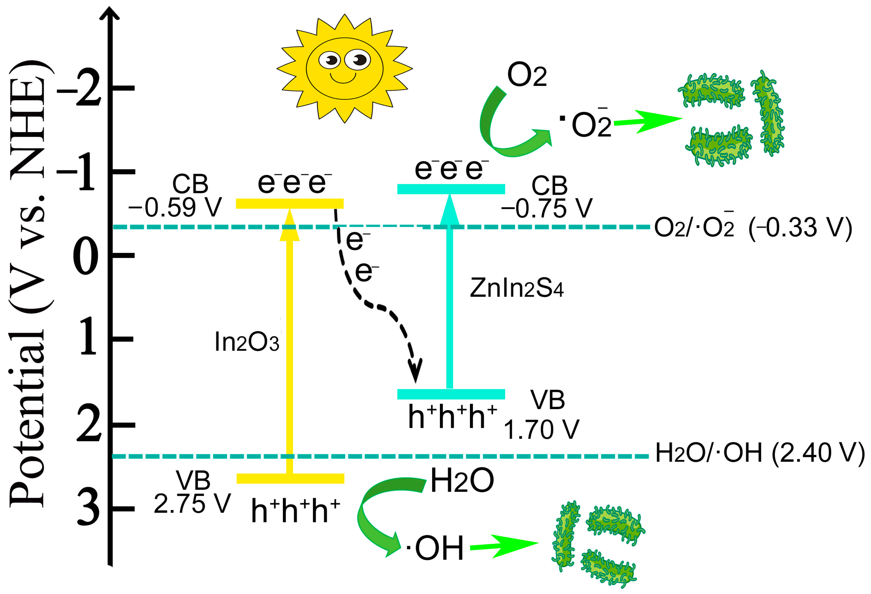
Based on the types and intensities of the generated radicals, the ·O2 and ·OH signals of the 40In2O3/ZnIn2S4 composite are stronger compared to those of In2O3 and ZnIn2S4 alone. This indicates that the heterojunction formed between In2O3 and ZnIn2S4 enhances the separation of photogenerated charge carriers. In ESR results, In2O3 can simultaneously produce ·O2 and ·OH under light exposure because its valence band (EVB = 2.75 V vs. NHE) is higher than the potential of H2O/·OH (EVB = 2.40 V vs. NHE), and its conduction band (ECB = −0.59 V vs. NHE) is lower than the potential of O2/·O2 (ECB = −0.33 V vs. NHE). Conversely, the valence band of ZnIn2S4 (EVB = 1.70 V vs. NHE) is lower than the potential of H2O/·OH; thus, it cannot generate ·OH radicals but can produce ·O2 due to its lower conduction band (ECB = −0.75 V), capable of reducing O2 to generate radicals. Therefore, based on these deductions, it is more likely that a Z-scheme heterojunction forms between In2O3 and ZnIn2S4. This Z-scheme heterojunction preserves the highly reductive photogenerated electrons of ZnIn2S4 and highly oxidative photogenerated holes of In2O3. Then, this retains the high reducibility of ZnIn2S4 while leveraging the strong oxidizing capability of In2O3, enhancing the degradation and sterilization performance. Below is a schematic diagram illustrating the potential charge transfer pathways in the In2O3/ZnIn2S4 composite, as shown in Figure 9:
According to the above conclusion (Figure 9), a possible mechanism [29] of the bactericidal action of In2O3/ZnIn2S4 is as follows:
In2O3 → In2O3 (e + h+)     ZnIn2S4 → ZnIn2S4 (e + h+)
In2O3 (e + h+) + ZnIn2S4 (e + h+) → In2O3 (h+) + ZnIn2S4 (e)
ZnIn2S4 (e) + O2 → ·O2
In2O3 (h+) + H2O → ·OH
Live bacteria +·O2 + ·OH → Dead bacteria

4. Conclusions

The hollow tubulous-shaped In2O3 derived from metal-organic framework (MOF) structure MIL was obtained via calcination, which exhibits an increased specific surface area, reactive sites, light absorption performance, and photoelectric response performance. Adopting this derived In2O3 as a template, nanoflower-shaped ZnIn2S4 was in situ-composited on it and obtained the In2O3/ZnIn2S4 composite, resulting in enhancing performance in MO degradation and P. aeruginosa sterilization. The optimal 40In2O3/ZnIn2S4 composite exhibited degradation rates of MO under visible light (80 mW·cm−2, 150 min) that were 17.9 and 1.4 times higher than those of the pure In2O3 and ZnIn2S4, respectively. And the 40In2O3/ZnIn2S4 shows the highest antibacterial performance against P. aeruginosa, with an antibacterial rate of 99.8% after visible light irradiation of 80 mW cm−2 for 420 min. Even after three cycles of reuse, the 40In2O3/ZnIn2S4 maintained high degradation activity without significant structural changes.
SEM, TEM, and XPS characterizations confirmed the tight contact between In2O3 and ZnIn2S4. The enhanced photocurrent density further indicates the presence of heterojunction. The ESR analysis of free radicals indicated that the 40In2O3/ZnIn2S4 generated both ·O2 and ·OH radicals under visible light, whereas ·OH radicals were not detected in ZnIn2S4 alone, suggesting the presence of a Z-scheme heterojunction between In2O3 and ZnIn2S4. The Z-scheme heterojunction preserves the highly reductive photogenerated electrons of ZnIn2S4 and highly oxidative photogenerated holes of In2O3, simultaneously enhancing the separation of photogenerated charge carriers.
This in situ growth of ZnIn2S4 on MIL-derived In2O3 produces an efficient composite for photocatalytic degradation and sterilization, offering guidance for applications in photocatalytic antimicrobial and antifouling treatments. Additionally, the synthesis conditions of the materials in this article are relatively mild, the raw materials are widely sourced, and only a small amount is needed to achieve significant degradation and bactericidal effects. At the same time, the physicochemical properties are stable, allowing for multiple cycles of use, which better meets the cost control requirements in practical applications and demonstrates considerable potential for application.

Author Contributions

Conceptualization, J.Z. and J.D. (Jiao Duan); methodology, J.D. (Jiao Duan), H.Z. and M.S.; software, J.D. (Jiao Duan), H.Z. and M.S.; validation, J.D. (Jiao Duan), H.Z. and M.S.; formal analysis, J.D. (Jiao Duan), H.Z. and M.S.; investigation, J.Z., J.D. (Jiao Duan) and M.S.; resources, J.Z., M.S. and J.D. (Jizhou Duan); data curation, J.Z., J.D. (Jiao Duan), H.Z. and M.S.; writing—original draft preparation, J.D. (Jiao Duan); writing—review and editing, J.Z., H.Z. and M.S.; visualization, J.D. (Jiao Duan), H.Z. and M.S.; supervision, J.Z., M.S. and J.D. (Jizhou Duan); project administration, J.Z., J.D. (Jiao Duan) and M.S.; funding acquisition, J.Z. and M.S. All authors have read and agreed to the published version of the manuscript.

Funding

This research was funded by the Major Basic Research Project of the Natural Science Foundation of Shandong Province, China (No. ZR2023ZD31), the National Natural Science Foundation of China (No. 42076043, 42176049), the Shandong Provincial Natural Science Foundation, China (No. ZR2022YQ39), and the Key Research Program of Frontier Sciences, Chinese Academy of Sciences (No. ZDBS-LY-DQC025).

Data Availability Statement

The data are contained within the article.

Conflicts of Interest

The authors declare no conflicts of interest.

References

  1. Vuong, P.; McKinley, A.; Kaur, P. Understanding biofouling and contaminant accretion on submerged marine structures. NPJ Mater. Degrad. 2023, 7, 50. [Google Scholar] [CrossRef]
  2. Callow, J.A.; Callow, M.E. Trends in the development of environmentally friendly fouling-resistant marine coatings. Nat. Commun. 2011, 2, 244. [Google Scholar] [CrossRef]
  3. Li, S.; Feng, K.; Li, J.; Li, Y.; Li, Z.; Yu, L.; Xu, X. Marine antifouling strategies: Emerging opportunities for seawater resource utilization. Chem. Eng. J. 2024, 486, 149859. [Google Scholar] [CrossRef]
  4. Hassaan, M.A.; El-Nemr, M.A.; Elkatory, M.R.; Ragab, S.; Niculescu, V.-C.; El Nemr, A. Principles of Photocatalysts and Their Different Applications: A Review. Top. Curr. Chem. 2023, 381, 31. [Google Scholar] [CrossRef]
  5. Ma, J.; Shi, Y.; An, D.; Chen, Y.; Guo, J.; Qian, Y.; Wang, S.; Lu, J. Inactivation mechanism of E. coli in water by enhanced photocatalysis under visible light irradiation. Sci. Total Environ. 2023, 866, 161450. [Google Scholar] [CrossRef] [PubMed]
  6. Luo, L.; Fu, L.; Liu, H.; Xu, Y.; Xing, J.; Chang, C.-R.; Yang, D.-Y.; Tang, J. Synergy of Pd atoms and oxygen vacancies on In2O3 for methane conversion under visible light. Nat. Commun. 2022, 13, 2930. [Google Scholar] [CrossRef] [PubMed]
  7. Wang, L.; Dong, Y.; Yan, T.; Hu, Z.; Ali, F.M.; Meira, D.M.; Duchesne, P.N.; Loh, J.Y.Y.; Qiu, C.; Storey, E.E.; et al. Black indium oxide a photothermal CO2 hydrogenation catalyst. Nat. Commun. 2020, 11, 2432. [Google Scholar] [CrossRef] [PubMed]
  8. Ji, Y.; Cui, M.; Wei, J.; Shi, Y.; Bao, L.; Tian, Z.; Hu, X.; Zhang, X.; Li, C. Interfacial oxygen vacancy engineering and built-in electric field mediated Z-scheme In2O3/Ag3PO4 heterojunction for boosted photocatalytic doxycycline degradation. Chem. Eng. J. 2024, 489, 151216. [Google Scholar] [CrossRef]
  9. Liu, J.; Guo, C.; Wu, N.; Li, C.; Qu, R.; Wang, Z.; Jin, R.; Qiao, Y.; He, Z.; Lu, J.; et al. Efficient photocatalytic degradation of PFOA in N-doped In2O3/simulated sunlight irradiation system and its mechanism. Chem. Eng. J. 2022, 435, 134627. [Google Scholar] [CrossRef]
  10. Wang, Q.; Chen, Y.; Liu, X.; Li, L.; Du, L.; Tian, G. Sulfur doped In2O3-CeO2 hollow hexagonal prisms with carbon coating for efficient photocatalytic CO2 reduction. Chem. Eng. J. 2021, 421, 129968. [Google Scholar] [CrossRef]
  11. Li, Z.; Zhang, P.; Shao, T.; Li, X. In2O3 nanoporous nanosphere: A highly efficient photocatalyst for decomposition of perfluorooctanoic acid. Appl. Catal. B-Environ. 2012, 125, 350–357. [Google Scholar] [CrossRef]
  12. Cao, S.W.; Liu, X.F.; Yuan, Y.P.; Zhang, Z.Y.; Liao, Y.-S.; Fang, J.; Loo, S.C.J.; Sum, T.C.; Xue, C. Solar-to-fuels conversion over In2O3/g-C3N4 hybrid photocatalysts. Appl. Catal. B-Environ. 2014, 147, 940–946. [Google Scholar] [CrossRef]
  13. Mu, F.; Liu, C.; Xie, Y.; Zhou, S.; Dai, B.; Xia, D.; Huang, H.; Zhao, W.; Sun, C.; Kong, Y.; et al. Metal-organic framework-derived rodlike AgCl/Ag/In2O3: A plasmonic Z-scheme visible light photocatalyst. Chem. Eng. J. 2021, 415, 129010. [Google Scholar] [CrossRef]
  14. Han, L.; Jing, F.; Zhang, J.; Luo, X.-Z.; Zhong, Y.-L.; Wang, K.; Zang, S.-H.; Teng, D.-H.; Liu, Y.; Chen, J.; et al. Environment friendly and remarkably efficient photocatalytic hydrogen evolution based on metal organic framework derived hexagonal/cubic In2O3 phase-junction. Appl. Catal. B-Environ. 2021, 282, 119602. [Google Scholar] [CrossRef]
  15. Yang, H.; Tang, J.; Luo, Y.; Zhan, X.; Liang, Z.; Jiang, L.; Hou, H.; Yang, W. MOFs-derived fusiform In2O3 mesoporous nanorods anchored with ultrafine CdZnS nanoparticles for boosting visible-light photocatalytic hydrogen evolution. Small 2021, 17, 2102307. [Google Scholar] [CrossRef]
  16. Yu, J.; Yan, P.; Chen, F.; Jin, S.; Xu, X.; Qian, J. MgIn2S4@In2O3 hierarchical tubular heterostructures with expedited photocarrier separation for efficient visible-light-driven antimicrobial activity. Chem. Eng. J. 2023, 452, 139559. [Google Scholar] [CrossRef]
  17. Kulkarni, P.; Nataraj, S.K.; Balakrishna, R.G.; Nagaraju, D.H.; Reddy, M.V. Nanostructured binary and ternary metal sulfides: Synthesis methods and their application in energy conversion and storage devices. J. Mater. Chem. A 2017, 5, 22040–22094. [Google Scholar] [CrossRef]
  18. Meng, S.; Wu, H.; Cui, Y.; Zheng, X.; Wang, H.; Chen, S.; Wang, Y.; Fu, X. One-step synthesis of 2D/2D-3D NiS/Zn3In2S6 hierarchical structure toward solar-to-chemical energy transformation of biomass-relevant alcohols. Appl. Catal. B-Environ. 2020, 266, 118617. [Google Scholar] [CrossRef]
  19. Wang, D.; Xu, Y.; Jing, L.; Xie, M.; Song, Y.; Xu, H.; Li, H.; Xie, J. In situ construction efficient visible-light-driven three-dimensional Polypyrrole/Zn3In2S6 nanoflower to systematically explore the photoreduction of Cr(VI): Performance, factors and mechanism. J. Hazard. Mater. 2020, 384, 121480. [Google Scholar] [CrossRef]
  20. Qiu, C.; Qian, K.; Yu, J.; Sun, M.Z.; Cao, S.F.; Gao, J.Q.; Yu, R.X.; Fang, L.Z.; Yao, Y.W.; Lu, X.Q.; et al. MOF-Transformed In2O3-x@C Nanocorn Electrocatalyst for Efficient CO2 Reduction to HCOOH. Nano-Micro Lett. 2022, 14, 16. [Google Scholar] [CrossRef]
  21. Wang, J.; Sun, S.; Zhou, R.; Li, Y.; He, Z.; Ding, H.; Chen, D.; Ao, W. A review: Synthesis, modification and photocatalytic applications of ZnIn2S4. J. Mater. Sci. Technol. 2021, 78, 1–19. [Google Scholar] [CrossRef]
  22. Shen, Q.; Sun, L.; Zhuang, Y.; Zhan, W.; Wang, X.; Han, X. Hollow Dodecahedral Structure of In2O3-In2S3 Heterojunction Encapsulated by N-Doped C as an Excellent Visible-Light-Active Photocatalyst for Organic Transformation. Inorg. Chem. 2020, 59, 17650–17658. [Google Scholar] [CrossRef]
  23. Sun, Y.; Zhu, Q.; Bai, B.; Li, Y.; He, C. Novel all-solid-state Z-scheme SnO2/Pt/In2O3 photocatalyst with boosted photocatalytic performance on water splitting and 2,4-dichlorophenol degradation under visible light. Chem. Eng. J. 2020, 390, 124518. [Google Scholar] [CrossRef]
  24. Jiang, C.; Wang, H.; Wang, Y.; Ji, H. All solid-state Z-scheme CeO2/ZnIn2S4 hybrid for the photocatalytic selective oxidation of aromatic alcohols coupled with hydrogen evolution. Appl. Catal. B-Environ. 2020, 277, 119235. [Google Scholar] [CrossRef]
  25. Liu, C.; Wang, L.; Tang, Y.; Luo, S.; Liu, Y.; Zhang, S.; Zeng, Y.; Xu, Y. Vertical single or few-layer MoS2 nanosheets rooting into TiO2 nanofibers for highly efficient photocatalytic hydrogen evolution. Appl. Catal. B-Environ. 2015, 164, 1–9. [Google Scholar] [CrossRef]
  26. Wang, F.; Jin, Z.; Jiang, Y.; Backus, E.H.G.; Bonn, M.; Lou, S.N.; Turchinovich, D.; Amal, R. Probing the charge separation process on In2S3/Pt-TiO2 nanocomposites for boosted visible-light photocatalytic hydrogen production. Appl. Catal. B-Environ. 2016, 198, 25–31. [Google Scholar] [CrossRef]
  27. Ma, X.; Cheng, H.F. Synergy of nitrogen vacancies and intercalation of carbon species for enhancing sunlight photocatalytic hydrogen production of carbon nitride. Appl. Catal. B-Environ. 2022, 314, 121497. [Google Scholar] [CrossRef]
  28. Peng, Y.; Geng, M.; Yu, J.; Zhang, Y.; Tian, F.; Guo, Y.n.; Zhang, D.; Yang, X.; Li, Z.; Li, Z.; et al. Vacancy-induced 2H@1T MoS2 phase-incorporation on ZnIn2S4 for boosting photocatalytic hydrogen evolution. Appl. Catal. B-Environ. 2021, 298, 120570. [Google Scholar] [CrossRef]
  29. Li, P.; Liang, T.; Liu, H.; Li, J.; Duo, S.; Xu, X.; Qiu, L.; Wen, X.; Shi, R. In2O3 microspheres decorated with ZnIn2S4 nanosheets as core-shell hybrids for boosting visible-light photodegradation of organic dyes. Mater. Res. Express 2021, 8, 025505. [Google Scholar] [CrossRef]
Figure 1. Synthesis process of the In2O3/ZnIn2S4 composites.
Nanomaterials 14 01366 g001
Figure 2. Internal structure of the photochemical reaction apparatus.
Nanomaterials 14 01366 g002
Figure 3. XRD patterns of MIL, In2O3, ZnIn2S4, and In2O3/ZnIn2S4 composites.
Nanomaterials 14 01366 g003
Figure 4. SEM images of MIL (a), In2O3 (bd), ZnIn2S4 (e), and 40In2O3/ZnIn2S4 (f); TEM images of 40In2O3/ZnIn2S4 (f,g); (h) Elemental mapping of 40In2O3/ZnIn2S4 composite.
Nanomaterials 14 01366 g004aNanomaterials 14 01366 g004b
Figure 5. The full XPS survey spectra of In2O3, ZnIn2S4, and 40In2O3/ZnIn2S4 (a); high-resolution XPS spectra of In3d (b), O1s (c) Zn2p (d), and S2p (e).
Nanomaterials 14 01366 g005
Figure 6. (a) Photodegradation of MO by MIL, In2O3, ZnIn2S4, and In2O3/ZnIn2S4 composites; (b) the changes in UV absorbance of methyl orange during the degradation process by 40In2O3/ZnIn2S4; (c) cycling degradation of 40In2O3/ZnIn2S4; (d) XRD patterns of 40In2O3/ZnIn2S4 after three cycles under visible light irradiation; (d) the changes in the XRD pattern of 40In2O3/ZnIn2S4 after degrading methyl orange.
Nanomaterials 14 01366 g006
Figure 7. (a) Survival rates and (b) antimicrobial rates of MIL, In2O3, ZnIn2S4, and In2O3/ZnIn2S4 composites against P. aeruginosa.
Nanomaterials 14 01366 g007
Figure 8. (a) UV–Vis diffuse reflection absorbance spectra, (b) Tauc plots, (c) Mott–Schottky curves of MIL, In2O3, ZnIn2S4, and 40In2O3/ZnIn2S4, (d) photocurrent response curves of MIL, In2O3, ZnIn2S4, and In2O3/ZnIn2S4 composites, (e,f) EPR spectra of In2O3, ZnIn2S4, and 40In2O3/ZnIn2S4.
Nanomaterials 14 01366 g008
Figure 9. Possible charge transfer mechanism of In2O3/ZnIn2S4.
Nanomaterials 14 01366 g009
Disclaimer/Publisher’s Note: The statements, opinions and data contained in all publications are solely those of the individual author(s) and contributor(s) and not of MDPI and/or the editor(s). MDPI and/or the editor(s) disclaim responsibility for any injury to people or property resulting from any ideas, methods, instructions or products referred to in the content.