Article

Novel Synthesis Route of Plasmonic CuS Quantum Dots as Efficient Co-Catalysts to TiO2/Ti for Light-Assisted Water Splitting

1
Université Paris Cité, CNRS UMR-7086, ITODYS, 75205 Paris, France
2
Faculté des Sciences, Université 20-Août-1955-Skikda, Skikda 21000, Algeria
*
Author to whom correspondence should be addressed.
Nanomaterials 2024, 14(19), 1581; https://doi.org/10.3390/nano14191581
Submission received: 20 August 2024 / Revised: 20 September 2024 / Accepted: 27 September 2024 / Published: 30 September 2024
(This article belongs to the Special Issue Photofunctional Nanomaterials and Nanostructures)

Abstract

:
Self-doped CuS nanoparticles (NPs) were successfully synthesized via microwave-assisted polyol process to act as co-catalysts to TiO2 nanofiber (NF)-based photoanodes to achieve higher photocurrents on visible light-assisted water electrolysis. The strategy adopted to perform the copper cation sulfidation in polyol allowed us to overcome the challenges associated with the copper cation reactivity and particle size control. The impregnation of the CuS NPs on TiO2 NFs synthesized via hydrothermal corrosion of a metallic Ti support resulted in composites with increased visible and near-infrared light absorption compared to the pristine support. This allows an improved overall efficiency of water oxidation (and consequently hydrogen generation at the Pt counter electrode) in passive electrolyte (pH = 7) even at 0 V bias. These low-cost and easy-to-achieve composite materials represent a promising alternative to those involving highly toxic co-catalysts.

1. Introduction

The quest for sustainable and efficient energy solutions has led to significant interest in the development of advanced materials for water splitting applications. Among these, semiconductive nanostructures play a crucial role in the photoelectrochemical (PEC) water decomposition reactions, which harnesses solar energy to produce hydrogen fuel. TiO2 is among the most prolific materials for light-assisted water electrolysis thanks to its excellent chemical stability and strong photooxidative capabilities, with a band structure triggering water redox potentials [1,2,3]. Nevertheless, TiO2 presents significant challenges due to its wide bandgap (approximately 3.2 eV for anatase), which limits its light absorption to the UV region, a small fraction of the solar spectrum. Moreover, TiO2 suffers from the rapid recombination of photogenerated electron-hole pairs, which severely impacts its photocatalytic efficiency, limiting electron transfer from the TiO2 conduction band (CB) to the external circuit and then to the cathode materials to be scavenged for proton reduction and hydrogen generation.
A wide variety of strategies was developed to overcome these limitations, the elaboration of titania-based semiconductive hetero-nanostructures being the object of particularly intensive research [4,5]. In such studies, TiO2-based structures are combined to one (or more) sensitizer, a semiconductor with fine-tuned properties that allow to compensate for the TiO2 shortcomings and improve the overall efficiency of the resulting system. The chosen semiconductor must possess appropriate electronic band structures with small bandgap energy and a high absorption coefficient in order to harvest sufficient solar photons. In addition, the energetic levels of its band structure need to straddle the redox potential of the desired water redox reactions, satisfying both the thermodynamics and kinetics requirements for conducting efficient photocatalytic reactions. Furthermore, chemical robustness and photostability are also essential for the selected semiconductors to be considered for such applications to allow for long-term, stable activity in photocatalytic processes [4]. Finally, such materials must be of low toxicity to avoid any deleterious effects on human operators as well as any environmental contamination.
The photocatalytic efficiency of the resulting semiconductive hetero-nanostructures is highly dependent on both the intrinsic photon absorption capability and charge transfer dynamics of the two (or more) photoanode components. Light absorption and charge transfer capabilities are mostly inherent to the band gap of the selected materials and their doping, including self-doping. The narrower the band gap, the higher the photogenerated charge density. As the doping rate increases, the conductivity increases. So, in a standard photoelecrochemical (PEC) cell, in ideal conditions, upon illumination, the photogenerated hole carriers are transported from the bulk of the semiconductor to its electrolyte interface, where they may participate in a water oxidation reaction. Conversely, the photogenerated electron carriers are transported from the bulk of the semiconductor to its titanium interface to be collected through the external circuit by the cathode counterpart to be finally involved in proton reduction into hydrogen.
According to the generalized Marcus theory [6,7,8], the driving force of electron transfer from a donor to an acceptor (in this case, the CB of the semiconductor to the CB of titania) is determined by the difference in their energetic levels. The logarithm of the rate of charge transfer is defined by a quadratic function with respect to the term of charge transfer driving force [4]. By enlarging the energetic difference between these energies, interfacial charge transfer can be boosted.
By aggregating all these requirements, it becomes evident why metal chalcogenides are particularly well-suited for forming semiconductive hetero-nanostructures with TiO2 [9], especially when the metal chalcogenide nanostructures consist of nanometal sulfides. Most nanocrystalline metal sulfide compounds exhibit remarkable visible light responsiveness, possess a sufficient number of active sites, and have appropriate reduction and/or oxidation potentials to function as effective photocatalysts [10]. Additionally, their quantum size effects allow for tunable properties such as rapid charge transfer and extended excited-state lifetimes [11].
Among the various combinations with TiO2, cadmium sulfide (CdS) nanocrystals have been the most extensively studied [12,13,14,15,16,17,18], despite concerns about their acute toxicity [19,20,21]. Our group, along with several others worldwide, has already prepared CdS-TiO2 composites. We demonstrated that replacing pristine TiO2 with CdS-TiO2 as the photoanode in a standard photoelectrochemical (PEC) cell significantly enhances photocurrent generation, even at 0 V bias [12,22].
In practice, by using a controlled hydrothermal corrosion process followed by air calcination at 400 °C of metallic titanium sheets [1], we successfully produced TiO2/Ti substrates. The resulting titania consisted of interconnected nanofibers (NFs), each about tens of nanometers in diameter and several hundreds of nanometers in length, uniformly covering the metal surface. These nanofibers crystallized in the anatase phase, exhibiting strong UV-range absorption. Using metallic Ti plates as both the support and the precursor for TiO2 NFs allowed us to streamline the production process while ensuring optimal contact between the photoactive and conductive elements of the photoanode, minimizing current losses [1,23,24,25].
By selecting a heating temperature below 500 °C, we were able to promote the formation of the anatase phase, which is more suitable for our intended application compared to other titania allotropes [26,27]. Additionally, preformed CdS nanoparticles (NPs), approximately 2–3 nm in size, were deposited onto and between the TiO2 NFs through a simple ethanol-based impregnation method. This process resulted in a valuable CdS-TiO2/Ti composite architecture for the targeted application [12,22]. Impregnation is considered as the simplest and most sustainable approach for constructing semiconductive hetero-nanostructures [28,29,30].
Despite these promising photoelectrochemical (PEC) results, replacing CdS NPs with less toxic yet equally effective metal sulfide NPs nanoparticles remained a key objective of our work. Our goal was to reduce risks to workers, consumers, and the environment during production and handling, ensuring that neither safety nor innovation is compromised, with sustainability playing a central role in our approach.
In this context, we chose to test CuS-TiO2/Ti using a similar simple, low-cost material processing method. Copper sulfide compounds, such as Cu2S and Cu7S4, are well known for their unique optical and electrical properties. Specifically, CuS has a narrow band gap with an energy range between 2.0 and 2.2 eV [31,32]. It can exhibit either p-type (predominantly) or n-type conductivity depending on the nature of its self-doping, making it highly promising for heterojunction design. CuS is often non-stoichiometric, meaning that variations in the oxidation state of its components can result in either excess electrons in its CB or holes in its otherwise filled valence band (VB). This property allows nanosized CuS to be classified as a plasmonic semiconductor due to its self-doping characteristics [33,34].
Plasmonic semiconductors, including CuS, possess extraordinary optoelectronic properties, particularly related to localized surface plasmon resonances (LSPRs) in the near-infrared (NIR) spectral region [35,36]. These characteristics make CuS an excellent candidate for photoelectrochemical (PEC) applications, such as light-assisted water splitting, either as a standalone material [37] or when coupled with titania [38].
Such self-doped particles with controlled morphology were previously prepared through wet synthesis routes with variable degrees of success (see, for instance [39,40,41,42,43]). Achieving the desired non-stoichiometry without contamination from foreign phases required several strategies. Typically, the redox properties of the reaction medium were carefully adjusted to attain the appropriate mixed valence states of copper and/or sulfur while avoiding the formation of impurities such as CuO or metallic Cu. Post-synthesis treatments, such as ion exchange or exposure to a redox atmosphere, were also employed to modify the oxidation states of the copper and sulfur elements [38,40,41,42,43,44,45].
Given that one of the primary goals of this study was the straightforward and reproducible production of uniformly sized, well-crystallized, and self-doped CuS NPs, we opted for microwave (MW)-assisted polyol synthesis. By varying the MW heating power and time and using either thioacetamide (TAA) or thiourea (ThU) as sulfur sources, we successfully optimized the synthesis to obtain the desired nanostructures. In this method, Cu2+ and S2− precursors were dissolved in a polyol solvent, and rapid heating was applied to the reaction medium. This facilitated nucleophilic substitution and condensation reactions while preventing complete reduction, thereby avoiding the contamination of Cu⁰ [46], and allowing partial reduction of Cu2+ to Cu+ for the desired covellite CuS self-doping.
Additionally, the adsorption of polyol molecules on the surface of the primary particles inhibited their growth and aggregation, enabling effective size control [47]. Finally, using a simple ethanol-based impregnation process, the CuS NPs were deposited onto and between the TiO2 nanofibers (NFs), resulting in the desired CuS-TiO2/Ti architectures.
Structural, optical, and electrochemical characterizations of the resulting CuS-TiO2/Ti composite confirmed the effectiveness of this material and processing approach. These results contribute to the advancement of photoelectrochemical (PEC) technology, highlighting its potential for sustainable hydrogen production.

2. Experiments

2.1. Material Synthesis

TiO2/Ti sheets were prepared by hydrothermal corrosion of commercial Ti plates. In brief, 0.8 × 2 cm Ti metallic plates (Goodfellow, >97%, 1 mm of thickness) were mechanically polished (intermediate polishing) with sandpaper of different granulometries to ensure the removal of the pre-existing oxide layers and eventual contaminants. The samples were washed in ultrasound in water, ethanol, and acetone (10 min each) and air-dried before being submitted to chemical polishing. In practice, the plates were immersed in an oxalic acid aqueous solution (5% w/w, equivalent to 0.6 mol·L−1) and heated at 100 °C for 2 h. They were then washed in water and air dried before their controlled hydrothermal surface oxidation according to already optimized operating conditions [1]: The plates were placed in a Teflon®-lined 120 mL autoclave in an equivolumetric mixture (10 mL total) of H2O2 (30%) and NaOH (10 mol·L−1). The closed autoclave was placed in an oven at 80 °C for 24 h. The plates were rinsed with deionized water, protonated HCL solution (0.1 mol·L−1), and again with deionized water and dried at 80 °C. Finally, a calcination in air took place at a 45 min heating ramp, with a target temperature of 400 °C kept constant for 60 min. Scanning electron microscopy (SEM) confirmed the 1D porous network titania morphology covering the entire titanium sheet (between 0.5 and 1.0 µm in thickness), with ropes of an average diameter of 20–50 nm, interweaving between each other, leading to a highly porous hierarchical structure (Figure S1). Transmission electron microscopy (TEM) evidences the veil-type structure on individual titania NFs, each veil being folded on itself, resulting in a fiber structure with a large specific area (Figure S2) and then a large interaction surface, which is an advantage for the desired catalytic application.
CuS particle synthesis was performed by the MW-assisted polyol process under different conditions. In the presence of sulfide nucleophilic agents, microwave heating allows shortening the reaction, promoting sulfidation instead of total reduction [46]. Two sulfide sources were used (ThU and TAA), and two different operating conditions were explored: low heating power, typically 200 W, for relatively long reaction times (25 to 30 min), and high heating power, namely 1200 W, for very short reaction times (1 to 3 min). In practice, 6.15 × 10⁻² mol·L−1 of copper (II) acetate were dispersed in 80 mL of ethyleneglycol (EG) with either TAA or ThU (TAA or ThU/copper molar ratio being equal to 1.2). The mixture was vigorously agitated and submitted to intense ultrasound for at least 30 min. The mixture was then transferred to a microwave-adapted reactor and heated in a multiwave Anton Paar microwave oven under constant radiation power. The resulting particles were recovered by centrifugation and washed with ethanol at least three times. They were finally dried at 60 °C overnight in air. The list of the prepared samples is summarized in the supporting information section (Table S1), in which each sample is referenced by adding to CuS the type of sulfur source, microwave power, and heating time. For instance, CuS-ThU-1200-1 corresponds to particles prepared with ThU for a heating time of 1 min under a heating power of 1200 W. All the produced particles are from the covellite structure, as confirmed by Rietveld refinements on all the recorded X-ray diffraction (XRD) patterns (Figure S3 and Table S2). The smallest CuS particles were selected for the final photoanode preparation step to take advantage of their large specific surface area. According to TEM micrographs of ethanolic suspensions containing the variously prepared particles (Figure S4), using a low microwave power of 200 W with a long reaction time (25 min) resulted in larger particle sizes (up to ~40 nm). In contrast, a higher power of 1200 W with a shorter reaction time (1 min) significantly reduced the particle size (down to ~7 nm). Additionally, for a given reaction time, particles synthesized with the thiourea (ThU) precursor were consistently smaller than those produced with thioacetamide (TAA), due to the faster decomposition of ThU in the reaction medium [47]. As a result, the CuS-ThU-1200-1 particles, with a typical size of 7–8 nm, were chosen for the fabrication of the CuS-TiO2/Ti photoanode.
The previously prepared TiO2/Ti substrates were fully immersed in a dilute CuS impregnation solution (3 mg of CuS in 4 mL of ethanol), sonicated for 10 min, and then left to rest overnight. This low concentration was deliberately chosen to allow for a performance comparison between our engineered CuS-TiO2/Ti photoanode and a similarly prepared photoanode in which the less toxic CuS co-catalysts were replaced with the more toxic CdS ones [12,22]. After impregnation, the plates were rinsed with ethanol, dried at 80 °C for 1 h, and stored under standard conditions without requiring any special handling.

2.2. Material Characterization

The structure of all the prepared samples was examined by XRD using two diffractometers (Panalytical, Almelo, Netherlands), an Empyrean equipped with a Cu Kα X-ray source (1.5418 Å) operating in the w-2θ (w = 1°) geometry for the plates and an X’pert Pro equipped with a Co Kα X-ray source (1.7889 Å) operating in the θ-θ geometry for the powders. The collected patterns were analyzed thanks to Highscore+ software version 5.2.0 (PANAYTICAL©, Almelo, The Netherlands).
The chemical composition was investigated by X-ray photoelectron spectroscopy (XPS) on an Escalab250 instrument (Thermo-VG, East Grinstead, UK) equipped with an Al-KαX-ray source (1486.6 eV). The pass energy was maintained at 200 eV for the survey scan (step size = 1 eV) and at 80 eV for the high-resolution spectra (step size = 0.1 eV). The spectra were calibrated against the (C-C/C-H) C 1s component set at 285 eV, and their analysis was achieved thanks to Avantage software, version 5.9902 (Thermo Scientific™, Boston, MA, USA).
The exact morphology of CuS NPs and TiO2 NFs was checked by TEM using a JEM 2100 Plus microscope (JEOL, Tokyo, Japan) operating at 200 kV. Additionally, SEM was carried out on the as-produced pristine TiO2/Ti and composite CuS-TiO2/Ti photoanodes, using a Gemini SEM 360 microscope (ZEISS, Jena, Germany) operating at 5 kV to check their general morphology. The microscope is also equipped with an Oxford Instrument (Abingdon, UK) energy-dispersive X-ray spectroscopy (EDX) detector (Ultim Max 170 mm2 detector), allowing chemical analysis, including chemical mapping. All The recorded micrographs were analyzed by ImageJ software version 1.54 j (open source).

2.3. Photoelectrochemical Assays

Each prepared photoanode, TiO2/Ti or CuS-TiO2/Ti, was employed as a working electrode (WE) in a home-made quartz single-compartment PEC cell (Figure 1), using an Ag/AgCl reference electrode (RE), a Pt wire counter electrode (CE), and a Na2SO4 aqueous electrolyte ([SO4] = 0.5 M, pH = 7). In practice, the I-V curves, thanks to a AUTOLAB PGSTAT12 scanning potentiostat (Metrohm Instrument, Herisau, Switzerland), were collected. Prior to all experiments, the electrolyte was purged by Argon from dissolved dioxygen. To simulate a solar light exposition, a 150 W Xenon lamp (ORIEL instruments, Bozeman, MO, USA) was used, fixing the area of WE illumination to 0.7 × 1.0 cm2.
Prior to PEC measurements, the UV-visible diffuse reflectance spectra of the produced composites were recorded on a Lambda 1050 spectrophotometer (PerkinElmer, Shelton, CT, USA) equipped with a PTFE-coated integration sphere.

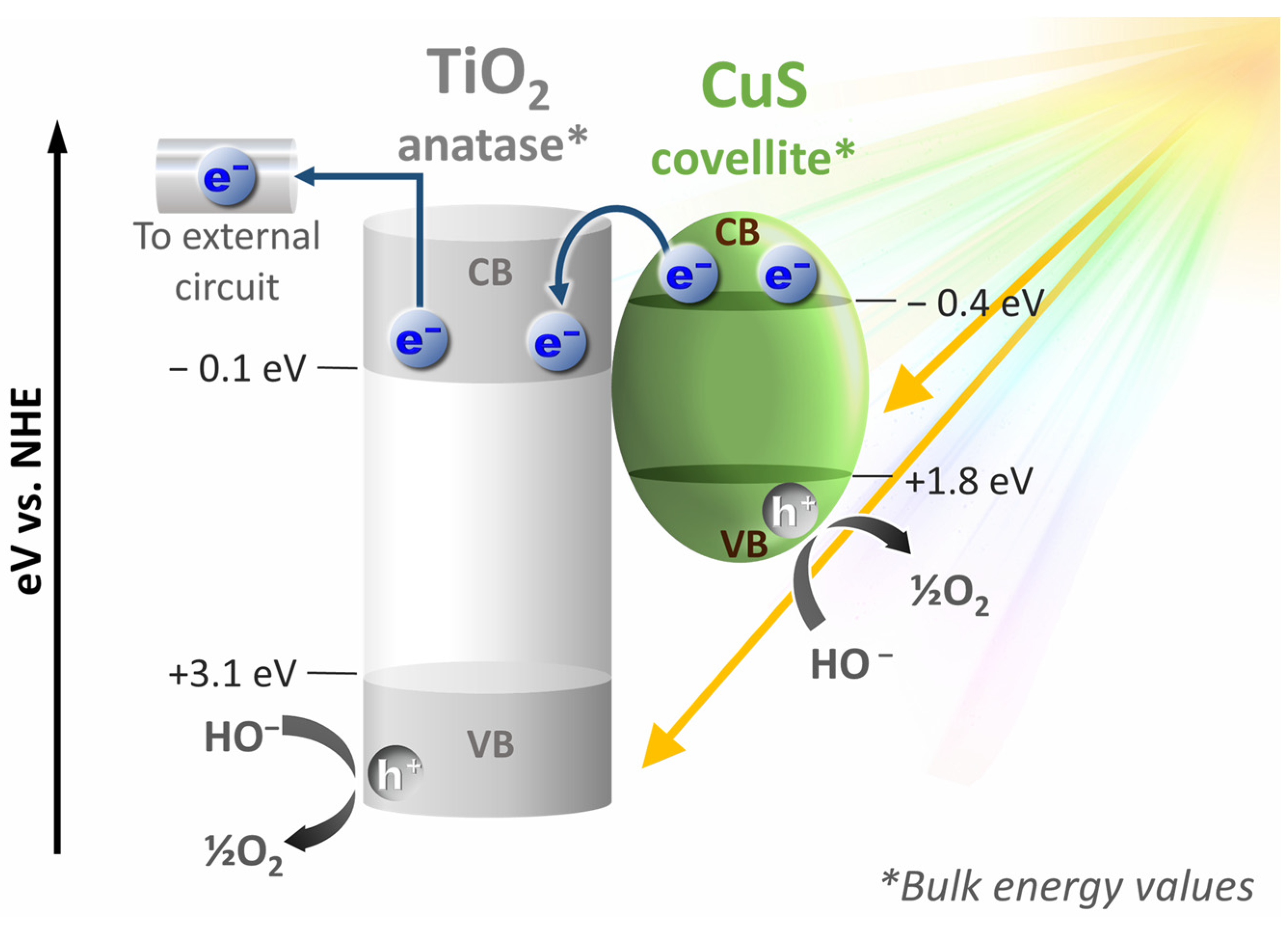
3. Results and Discussion

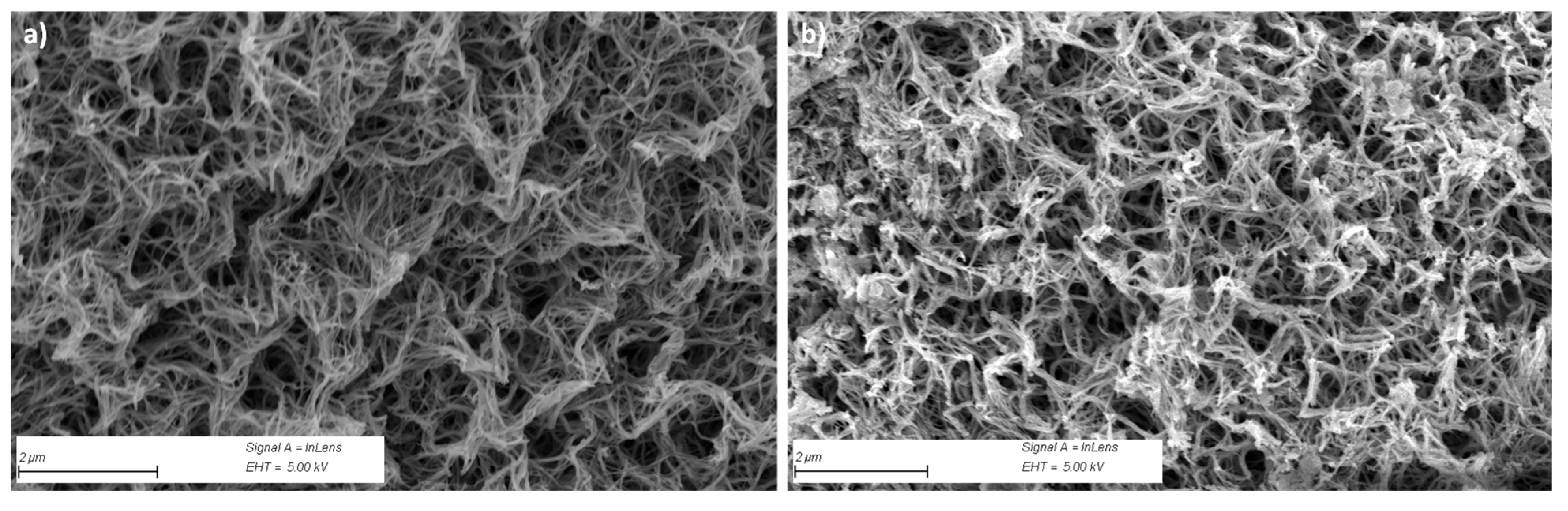
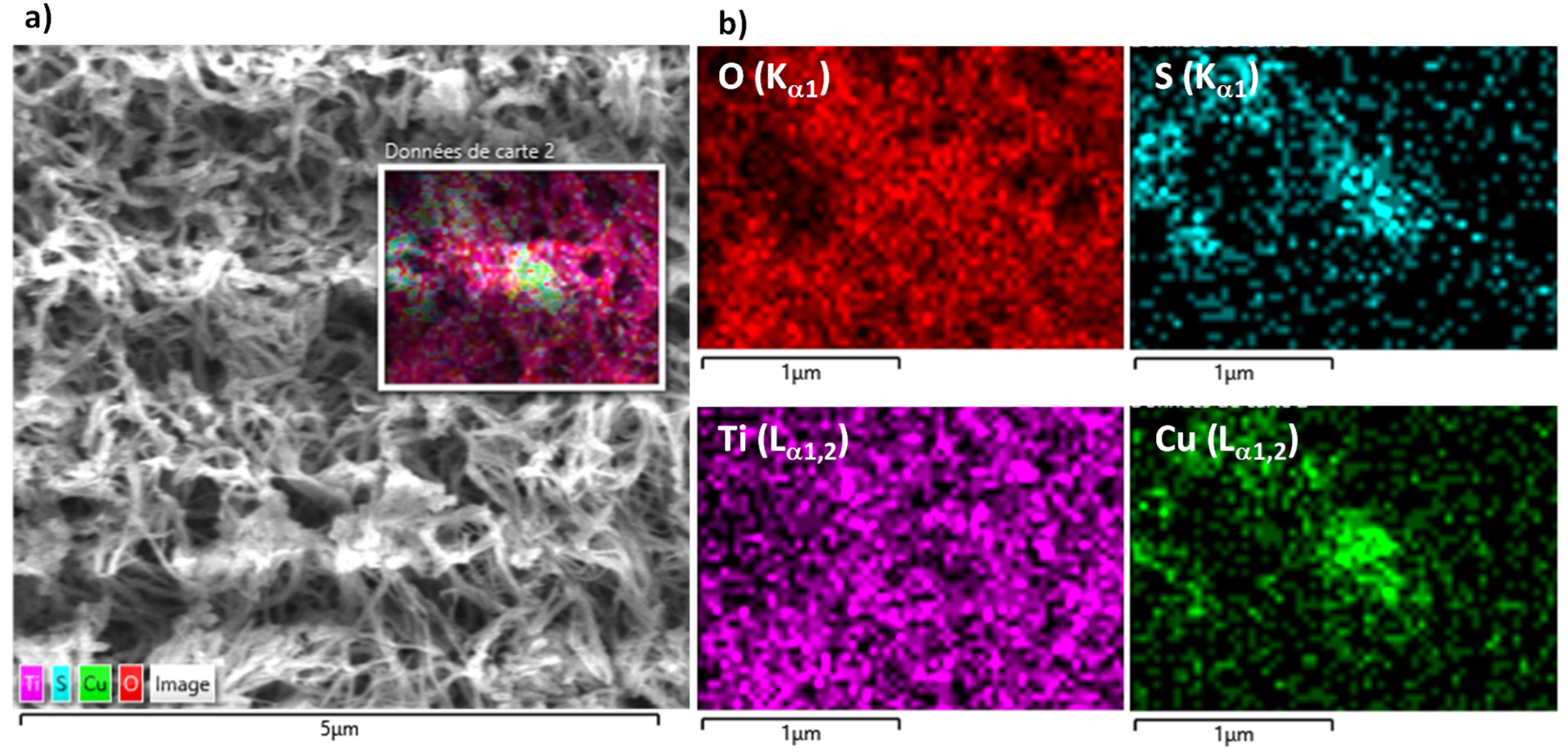
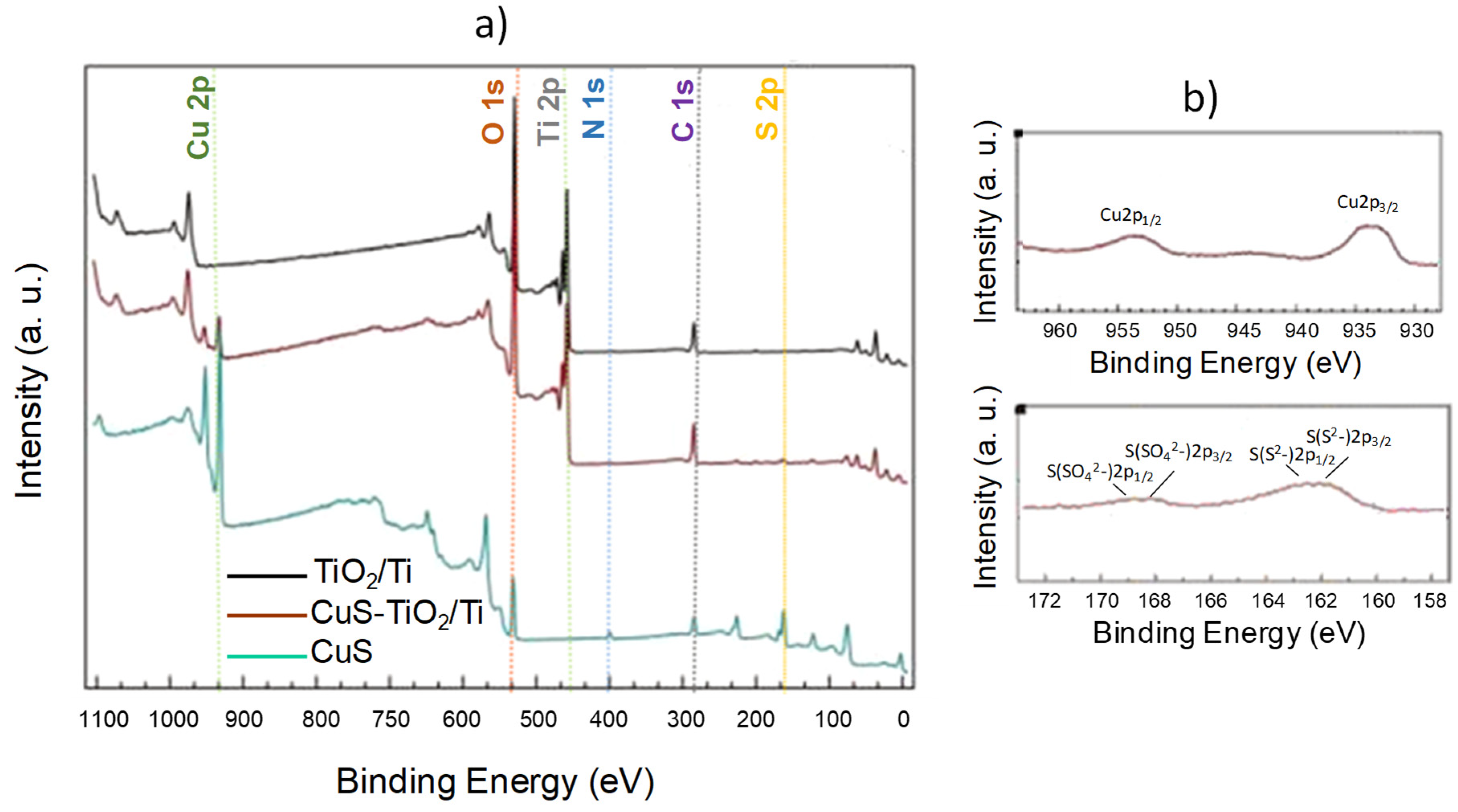
3.1. Photoanode Engineering

The elaboration of the CuS-TiO2/Ti substrates consisted of three main steps involving Ti plate-controlled corrosion to produce a well-adherent thick, porous anatase coating on a conductive substrate, polyol CuS particle synthesis optimization to obtain ultrafine co-catalysts (less than 10 nm in size), and an easy-to-achieve impregnation route, tacking advantage from the abundance of pores and the high surface-to-volume ratio of pristine TiO2/Ti.
The efficiency of the photoanode material processing was first checked by XRD analysis (Figure 2). The recorded pattern of CuS-TiO2/Ti matched very well with that of pure TiO2 and Ti phases. Indeed, all the diffraction peaks were fully indexed in the tetragonal anatase structure (ICDD No. 00-021-1272) and the hexagonal titanium one (ICDD No. 00-044-1294) without clear evidence of CuS signature due to its low content and/or its ultrasmall crystal size.
To confirm the presence of CuS particles, the engineered photoanode was observed by SEM, and the recorded SEM micrographs were compared to those collected on pristine TiO2/Ti. A simple contrast lecture of the two types of images evidenced some differences on some titania fiber nodes (Figure 3). Focusing on such a zone, EDS chemical mapping confirmed the simultaneous presence of copper and sulfur elements at this area at almost the same concentration (Figure 4).
A semi-quantitative EDS analysis of CuS-TiO2/Ti confirmed the presence of copper and sulfur elements at very low but non-zero contents compared to titanium and oxygen elements (Figure S5), leading to a whole Cu/Ti content of about 0.5 at.-%. Such an atomic ratio aligns well with the low concentration of the CuS impregnation solution used in the photoanode preparation. This ratio is also comparable to the Cd/Ti atomic ratio in our previously studied CdS-TiO2/Ti photoanode, making a performance comparison between the two systems in terms of PEC efficiency both relevant and meaningful.
Additionally, a comparison of the Cu/Ti atomic ratio from EDS with that inferred from XPS analysis confirmed that a significant portion of the impregnated CuS particles reside on the outer surface of the TiO2 fibers (15.4 at.% vs. 0.5 at.%), which is advantageous for our intended application. The survey XPS spectrum of CuS-TiO2/Ti, compared to those of pristine TiO2/Ti and CuS (Figure 5a), confirms the presence of all expected elements—Ti and O for the TiO2 phase, and Cu and S for the CuS phase. While there were no notable differences in the respective bonding energies between samples, a significant variation in the Cu2p and S2p peak intensities was observed. Specifically, the surface Cu concentration on CuS-TiO2/Ti was 2.9 at.%, compared to 23.2 at.% on the surface of pristine CuS.
A focus on the high-resolution Cu 2p signal recorded on CuS-TiO2/Ti (Figure 5b) compared to that of pristine CuS (Figure S6) confirms that copper is at the particle surface divalent with Cu 2p1/2 and Cu 2p3/2 binding energies of 952.5 and 932.4 eV, respectively, close to the values reported in the literature for CuS [48,49] and Cu2S [50,51] phases. CuS also exhibits a small shake-up or multiplet splitting structure, while Cu2S does not [48,49,50,51,52]. This feature agrees with the formation of CuS without excluding the presence of Cu+ species. Additionally, the Cu LMM peaks (Figure S7) recorded on all the prepared CuS particles, including those used for the preparation of CuS-TiO2/Ti, exhibit a peak shape completely different from that usually observed on Cu0 species [52], confirming the absence of copper metal. Moreover, the slight non-stoichiometry measured by XPS on the CuS particles before and after their attachment by impregnation to the pristine TiO2/Ti (Table 1) agrees fairly with self-doping, which may result from a partial substitution of Cu2+ cations by monovalent Cu+ ones within the covellite lattice.
Assuming that all these features are representative of the whole volume of all CuS particles (the 7–8 nm average size of CuS particles is smaller than the 10–12 nm XPS analysis depth), one may conclude in favor of their self-doping, giving then of the properties of plasmonic semiconductors.
Also, the S2p high-resolution XPS spectra of both CuS (Figure S6) and CuS-TiO2/Ti (Figure 5b) are quite similar. Their total intensities are of course different, but both exhibit a doublet at 163.5 (2p1/2) and 162.3 eV (2p3/2) characteristics of sulfide S2− species in CuS [48,49] or Cu2S [50,51] phases. Interestingly, both exhibit a supplementary contribution: a broad and small in intensity peak at 168.6 eV usually attributed to sulfate SO42− anions, suggesting a weak surface oxidation with the production of a thin CuSO4 passivation layer. In other words, the composition of the analyzed copper sulfide particles is consistent with a CuS@CuSO4 core-shell nanostructure. Comparing the intensity of the S2− 2p3/2 and SO42− 2p3/2 XPS peaks allows us to estimate that, approximatively, the fourth of the involved sulfur atoms are in the form of sulfate, in agreement with a very thin protective copper sulfate layer.
If the former XPS analysis confirmed the presence of CuS particles on the surface of the CuS-TiO2/Ti sample, it also suggested that the chosen impregnation route did not affect the chemical state of titanium cations on the surface of the titania coating. Indeed, there are no significant differences between the Ti 2p XPS profiles of TiO2/Ti and CuS-TiO2/Ti, as well as between their O 1s XPS profiles (Figure S6), agreeing very well with the TiO2 oxide nature of the outer layer of the titanium plates [1,5,53].
Finally, the optical absorption spectrum of the engineered CuS-TiO2/Ti photoanode was measured in diffuse reflectance and compared to that of pristine TiO2/Ti, recorded in diffuse reflectance as well, and that of pristine CuS, recorded in a transmission scheme (Figure 6). Regarding the semiconducting nature of TiO2/Ti, the typical anatase band-to-band signature at around 300 nm was identified, with a band-gap value inferred from Tauc plots of about 3.2 eV in pristine TiO2/Ti and 2.6 eV in CuS-TiO2/Ti. The last small value is not at all the consequence of a gap decrease but is the consequence of a more complex composite band diagram. Indeed, the CuS-TiO2/Ti spectrum is the combination of those of pristine TiO2/Ti and CuS, with absorption capabilities ranging from UV to NIR spectral ranges, due to the photo-excitation of both titania and copper sulfite semiconductors. The anatase and the covellite band-to-band absorptions (around 300 [1,5] and 400 nm [30], respectively) superposed to the self-doped CuS LSPR absorption (around 1200 nm [31,32]) explain together the optical properties of our engineered photoanode. Clearly, the amount of CuS particles deposited on TiO2/Ti by impregnation, even small, appeared large enough to induce a widened light absorption, which is fruitful for improved PEC responses.

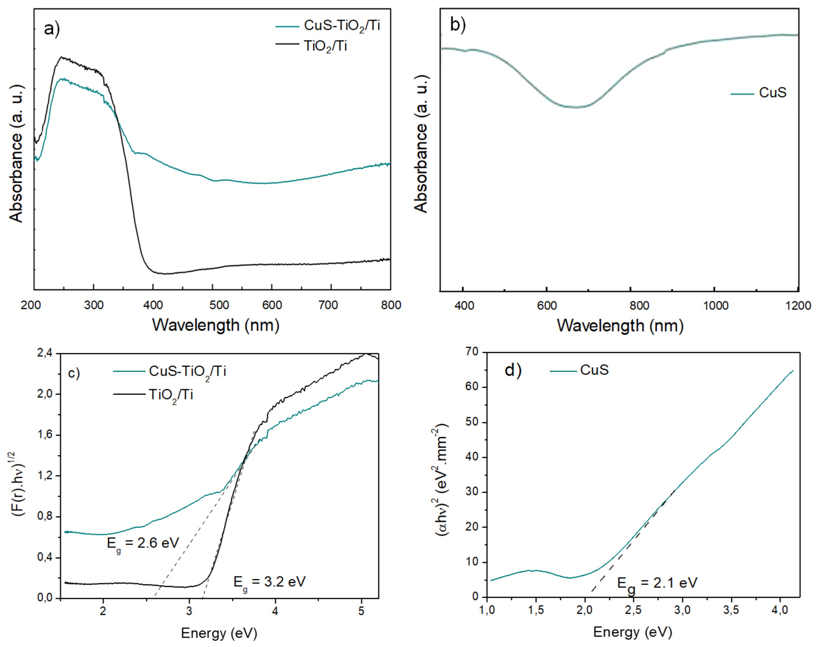
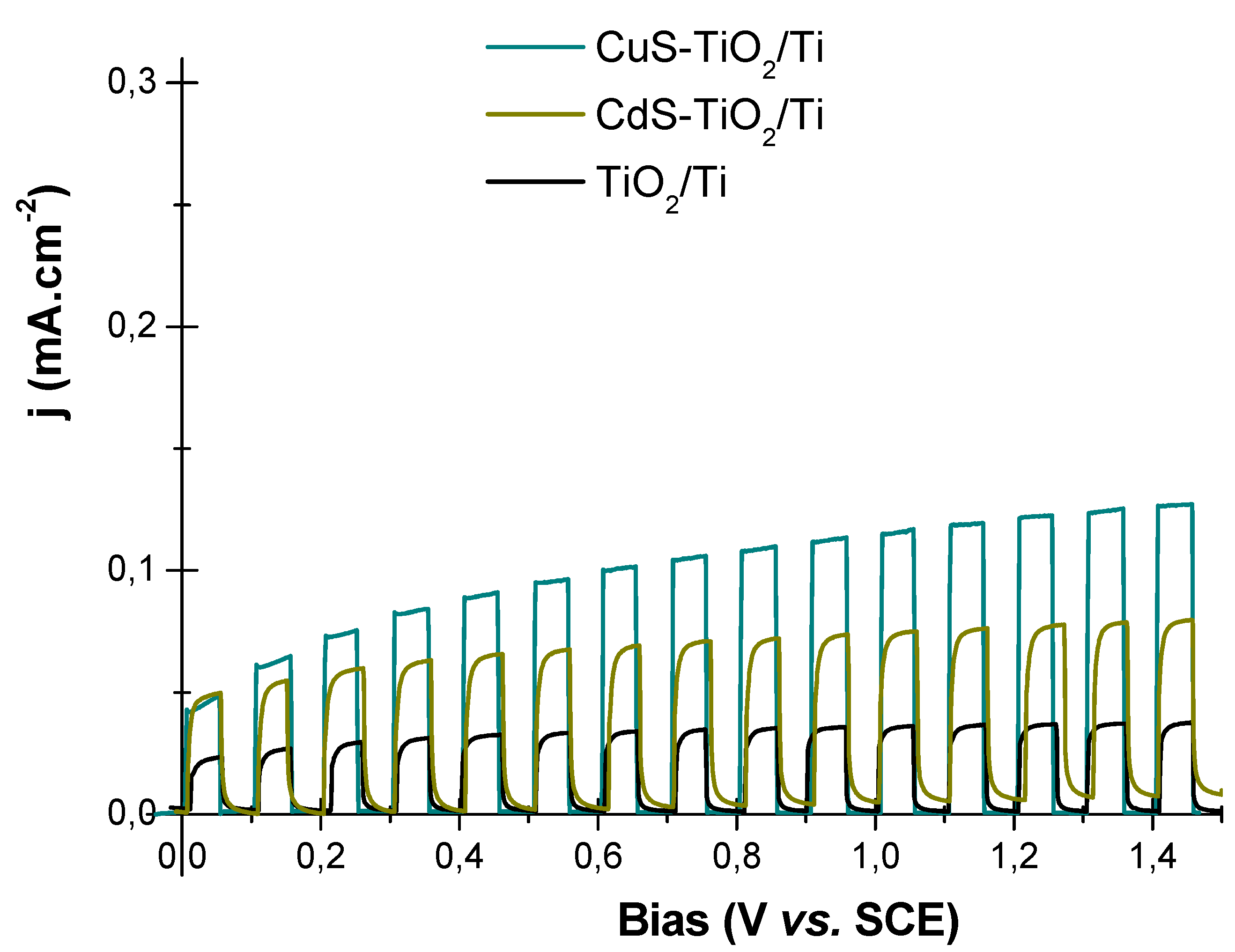
3.2. Photoanode PEC Properties

Hydrogen photo-generation activity of the as-synthetized CuS-TiO2/Ti photoanode and its TiO2/Ti parent was carried out under a Xenon lamp irradiation using a passive and neutral electrolyte. Interestingly, operating in a passive electrolyte, an intermittent illumination of CuS-TiO2/Ti provides a higher photocurrent than pristine TiO2/Ti, whatever the applied bias (Figure 7). The chronoamperometry under intermittent lighting (black arrows indicating the beginning of dark periods and the orange arrows indicating the beginning of illuminated periods) shows that the sensible increase in the photogenerated current is stable at 1.23 V (vs. Ag/AgCl), and while there is a decrease in the photocurrent during the first minute of illuminated periods, the original values are restored after a dark period.
By comparing the behavior of the bare and impregnated photoanodes under 0 V and 1.23 V bias, we can infer that the improved photocurrent is due to a higher amount of photogenerated charge carriers, favored by the increase of absorbed photons, promoted by CuS NPs. CuS-TiO2/Ti absorbs more light, in a wider spectral range, than pristine TiO2/Ti, creating a sufficient number of electron−hole pairs. The electrons can then be transferred from CuS CB to that of TiO2 as summarized hereafter:
CuS + hv → CuS(e + h+)
e(CBCuS) + TiO2 → TiO2(e)
h+(VBTiO2) + H2O → 2H+ + ½O2
h+(VBTiO2) + CuS → CuS(h+)
h+(VBCuS) + H2O → 2H+ + ½O2
The collected electrons in TiO2 CB were then transferred through the external circuit to the Pt cathode to achieve the reduction of aqueous protons into hydrogen gas (Figure 8).
These findings align with results from a few research groups studying CuS-TiO2 systems, which remain relatively underexplored in the literature on photocatalytic hydrogen generation compared to metal chalcogenide-based titania nanocomposites, such as the CdS-TiO2 system. This is despite the well-documented acute toxicity of (see, for instance, [19,20,21] and the references therein).
To the best of our knowledge, notable results have been reported by Chandra et al. [56], who prepared their composites by a hydrothermal and a solution-based process. Operating by photocatalysis (PC) in a sacrificial Na2S (0.25 M)-Na2SO3 (0.25 M) electrolyte, they succeeded in producing 1262 µmol of H2 per hour and per gram of catalyst, more than 10 and 9 times higher than that by pristine TiO2 and pristine CuS powders under Xe lamp irradiation, respectively. There are also results reported by Jia et al. [57], who decorated TiO2 nanowire arrays grown on conductive substrate by CuS nanoclusters by successive ionic layer adsorption and reaction (SILAR method). Operating by photo-electrocatalysis (PEC) in a passive Na2SO4 (1.00 M) electrolyte, they demonstrated an increased light absorption and an efficient charge separation leading to an improved photocurrent. They succeeded in obtaining within the same setup a photocurrent density 5 times higher than that of pristine TiO2 at a bias of 0.35 V. One may also cite the results of Liu et al. [58], who successfully constructed a CuS/TiO2 heterojunction using metal-organic framework (MOF)-derived TiO2 as a substrate. They pointed out that CuS/TiO2 exhibited excellent bifunctional PC activity without noble metal cocatalysts. They typically evidenced H2 production and benzylamine oxidation in a coupled experiment, with a H2 evolution activity of the CuS/TiO2 17.1 and 29.5 times higher than that of TiO2 and CuS, respectively. Wang et al. [59] also investigated CuS/TiO2 photocatalysts, prepared via a high-temperature hydrothermal method, and evaluated their photocatalytic activity. They demonstrated that loading TiO2 with 1 wt.-% CuS significantly enhanced its photocatalytic performance for water decomposition to hydrogen in a methanol aqueous solution under Xe lamp irradiation. The CuS/TiO2 photocatalysts produced approximately 570 µmol of H2 per hour, which is 32 times higher than that produced by pristine TiO2.
Clearly, in all these studies and in others (Table S3), widened light absorption and an efficient charge separation were systematically reported. All converged, placing CuS as one of the most interesting metal chalcogenide titania co-catalysts for water splitting.
To support these scientific advances, we compared the performance of the CuS-TiO2/Ti photoanode with that of a similarly prepared CdS-TiO2/Ti photoanode [12,22]. The main difference between the two is the size of the particles, with CdS having a particle size of 3 nm. Both photoanodes were tested using the same photoelectrochemical (PEC) setup. Interestingly, the photocurrent measured for the CuS-TiO2/Ti photoanode was consistently higher than that for the CdS-TiO2/Ti photoanode. This is attributed to the broader light absorption range of CuS (Figure 9), indicating that CuS is a more effective co-catalyst compared to CdS.

4. Conclusions

In conclusion, the integration of CuS nanoparticles (NPs) with TiO2 nanofibers (NFs) has proven to be a promising approach for achieving efficient photoelectrochemical (PEC) responses in water splitting and hydrogen generation, in good alignment with the relevant literature. The synergistic properties of these nanomaterials enable excellent light absorption, effective charge separation, and efficient electron transport, leading to significant improvements in PEC performance.
By optimizing the microwave-assisted polyol process conditions, self-doped covellite CuS particles with sizes of 7–8 nm, which absorb in the visible and near-infrared (NIR) spectral ranges, were successfully produced without foreign contaminants. These particles were effectively integrated with TiO2 NFs supported on a titanium substrate, which was prepared through controlled metal plate corrosion (hydrothermal treatment followed by calcination). The simple ethanol-based impregnation method proved sufficient for creating the CuS-TiO2/Ti semiconductive hetero-nanostructures.
A CuS concentration as low as 0.5 at.-% in the composite was sufficient to achieve photocurrents of 0.030 and 0.122 mA/cm2 at 0 V and 1.23 V, respectively. In comparison, the photocurrents measured for pristine TiO2/Ti under the same PEC conditions were 0.020 and 0.051 mA/cm2. Notably, the photocurrent with the CuS co-catalyst was comparable to that obtained with toxic CdS at 0 V and significantly higher at 1.23 V (0.122 vs. 0.082 mA/cm2). These results highlight that the selected materials and the employed synthetic approaches offer a novel and effective pathway for developing sustainable hydrogen production systems.

Supplementary Materials

The following supporting information can be downloaded at: https://www.mdpi.com/article/10.3390/nano14191581/s1, Figure S1: SEM (a) top view and (b) cross view of the as-prepared TiO2/Ti sheet confirming the 1D morphology of the formed titania, covering all the Ti substrate surface; Figure S2: TEM micrographs recorded on an (a) assembly and an (b) individual representative TiO2 NF separated by sonication from the as-produced TiO2/Ti sheets in an ethanolic solution. Titania fibers appear as veils folded on themselves; Figure S3: (a) XRD patterns of CuS particles prepared using TAA (up) and ThU (bottom) reagents while applying a microwave heating power of 200 and 1200 W for a total reaction time of 25 and 3 min, respectively. (b) Results of Rietveld refinements (using MAUD software) performed on the XRD pattern of CuS-ThU-1200-1 particles to illustrate the quality of the fits: the experimental pattern (black scatter) and the calculated one (green line) are perfectly superposed with a residue curve, defined as the difference between the experimental and calculated diffractograms, close to zero (blue line). The inferred crystallite shape is also given for information [60]; Figure S4: SEM images recorded on (a) CuS-TAA-200-25 and (b) CuS-ThU-200-25 particles. TEM images of (c) CuS-TAA61200-3 and (d) CuS-1200-ThU-1200-3 particles. (e) TEM micrograph of CuS-ThU-1200-1 particles and (f) HRTEM image of some representative CuS-ThU-1200-1 particles; Figure S5: SEM-EDS analysis of CuS-TiO2/Ti, focusing on TiO2 fiber nodes, where CuS particles seem to accumulate, leading to an average Cu/Ti atomic ratio of 0.5 at.-%; Figure S6. Ti 2p and O 1s high-resolution XPS spectra recorded on pristine TiO2/Ti (black) and Cu2P and S 2p high-resolution XPS spectra recorded on pristine CuS (blue-green line); Figure S6: Ti 2p and O 1s high-resolution XPS spectra recorded on pristine TiO2/Ti (black) and Cu2P and S 2p high-resolution XPS spectra recorded on pristine CuS (blue-green line); Figure S7: S 2p, Cu 2p, and Cu LMM high-resolution XPS spectra recorded on CuS NPs produced using ThU or TAA sulfur source for a microwave heating power of 1200 W along 1 min of reaction time; Table S1: List of prepared CuS samples and their main synthesis, MW-assisted polyol synthesis; Table S2: Main Rietveld refined structural parameters and their related reliability fit factors. The overall fit quality is described by a weighted profile (Rwp), expected profile (Rexp), and Bragg R-value (RB) close to 1; Table S3: Comparison of the PEC performances of our engineered photoanode with those of CuS-TiO2-based literature [38,56,57,61,62,63,64,65,66,67].

Author Contributions

L.C.: Conceptualization, Data curation, Formal analysis, Investigation, Methodology, Visualization, Writing—review & editing; S.C.: Conceptualization, Data curation, Formal analysis, Writing—review & editing; B.D.: Data curation, Formal analysis (he prepared and characterized CuS partucles); S.G.-D.: Data curation and Formal analysis (she performed SEM-EDS measurements and analyzed the collected data and images); S.N.: Data curation, Formal analysis (she performed XRD experiments and analyzed the collected data); F.M.: Conceptualization, Supervision, Visualization, Writing—review & editing; S.A.: Conceptualization, Supervision, Methodology, Visualization, Validation, Writing—review & editing, Funding acquisition, Project administration, Writing—original draft. All authors have read and agreed to the published version of the manuscript.

Funding

ANR (Agence Nationale de la Recherche) and CGI (Commissariat à l’Investissement d’Avenir) are gratefully acknowledged for financial support of this work through Labex SEAM (Science and Engineering for Advanced Materials and devices), ANR-10-LABX-096, and ANR-18-IDEX-0001 grants.

Data Availability Statement

The raw data supporting the conclusions of this article will be made available by the authors on request.

Acknowledgments

The authors would like to acknowledge Guillaume Thoraval and Tom Chevry (Université Paris Cité), who crafted the glassware necessary for the PEC experiments. They are also grateful to Patricia Beaunier (Sorbonne Université), who managed all TEM experiments.

Conflicts of Interest

The authors declare no conflict of interest.

References

  1. Chaguetmi, S.; Achour, S.; Mouton, L.; Decorse, P.; Nowak, S.; Costentin, C.; Mammeri, F.; Ammar, S. TiO2 nanofibers supported on Ti sheets prepared by hydrothermal corrosion: Effect of the microstructure on their photochemical and photoelectrochemical properties. RSC Adv. 2015, 5, 95038–95046. [Google Scholar] [CrossRef]
  2. Lianos, P. Review of recent trends in photoelectrocatalytic conversion of solar energy to electricity and hydrogen. Appl. Catal. B Environ. 2017, 210, 235–254. [Google Scholar] [CrossRef]
  3. Egerton, T.A. Does photoelectrocatalysis by TiO2 work? J. Chem. Technol. Biotechnol. 2011, 86, 1024–1031. [Google Scholar] [CrossRef]
  4. Tsao, C.-W.; Fang, M.-J.; Hsu, Y.-J. Modulation of interfacial charge dynamics of semiconductor heterostructures for advanced photocatalytic applications. Coord. Chem. Rev. 2021, 438, 213876. [Google Scholar] [CrossRef]
  5. Lettieri, S.; Pavone, M.; Fioravanti, A.; Amato, L.S.; Maddalena, P. Charge Carrier Processes and Optical Properties in TiO2 and TiO2-Based Heterojunction Photocatalysts: A Review. Materials 2021, 14, 1645. [Google Scholar] [CrossRef] [PubMed]
  6. Marcus, R.A. On the theory of electron-transfer reactions. VI. Unified treatment for homogeneous and electrode reactions. J. Chem. Phys. 1965, 43, 679–701. [Google Scholar] [CrossRef]
  7. Gao, Y.Q.; Marcus, R.A. On the theory of electron transfer reactions at semiconductor/liquid interfaces. II. A free electron model. J. Chem. Phys. 2000, 113, 6351–6360. [Google Scholar] [CrossRef]
  8. Tvrdy, K.; Frantsuzov, P.A.; Kamat, P.V. Photoinduced electron transfer from semiconductor quantum dots to metal oxide nanoparticles. Proc. Natl. Acad. Sci. USA 2010, 108, 29–34. [Google Scholar] [CrossRef]
  9. Moridon, S.N.F.; Arifin, K.; Yunus, R.M.; Minggu, L.J.; Kassim, M.B. Photocatalytic water splitting performance of TiO2 sensitized by metal chalcogenides: A review. Ceram. Int. 2022, 48, 5892–5907. [Google Scholar] [CrossRef]
  10. Mamiyev, Z.; Balayeva, N.O. Metal Sulfide Photocatalysts for Hydrogen Generation: A Review of Recent Advances. Catalysts 2022, 12, 1316. [Google Scholar] [CrossRef]
  11. Keimer, B.; Moore, J.E. The physics of quantum materials. Nat. Phys. 2017, 13, 1045–1055. [Google Scholar] [CrossRef]
  12. Chaguetmi, S.; Mammeri, F.; Pasut, M.; Nowak, S.; Lecoq, H.; Decorse, P.; Costentin, C.; Achour, S.; Ammar, S. Synergetic effect of CdS quantum dots and TiO2 nanofibers for photoelectrochemical hydrogen generation. J. Nanopart. Res. 2013, 15, 2140. [Google Scholar] [CrossRef]
  13. Khatamian, M.; Oskoui, M.S.; Haghighi, M.; Darbandi, M. Visible-light response photocatalytic water splitting over CdS/TiO2 and CdS-TiO2/metalosilicate composites. Int. J. Energy Res. 2014, 38, 1712–1726. [Google Scholar] [CrossRef]
  14. Manchwari, S.; Khatter, J.; Chauhan, R. Enhanced photocatalytic efficiency of TiO2/CdS nanocomposites by manipulating CdS suspension on TiO2 nanoparticles. Inorg. Chem. Commun. 2022, 146, 110082. [Google Scholar] [CrossRef]
  15. Nguyen, V.N.; Doan, M.T.; Nguyen, M.V. Photoelectrochemical water splitting properties of CdS/TiO2 nanofibers-based photoanode. J. Mater. Sci. Mater. Electron. 2019, 30, 926–932. [Google Scholar] [CrossRef]
  16. Shen, J.; Meng, Y.; Xin, G. CdS/TiO2 nanotubes hybrid as visible light driven photocatalyst for water splitting. Rare Met. 2011, 30, 280–283. [Google Scholar] [CrossRef]
  17. Li, C.; Yuan, J.; Han, B.; Jiang, L.; Shangguan, W. TiO2 nanotubes incorporated with CdS for photocatalytic hydrogen production from splitting water under visible light irradiation. Int. J. Hydrogen Energy 2010, 35, 7073–7079. [Google Scholar] [CrossRef]
  18. Sharma, K.; Hasija, V.; Malhotra, M.; Verma, P.K.; Khan, A.A.P.; Thakur, S.; Van Le, Q.; Quang, H.H.P.; Nguyen, V.-H.; Singh, P.; et al. A review of CdS-based S-scheme for photocatalytic water splitting: Synthetic strategy and identification techniques. Int. J. Hydrogen Energy 2024, 52, 804–818. [Google Scholar] [CrossRef]
  19. Varmazyari, A.; Taghizadehghalehjoughi, A.; Sevim, C.; Baris, O.; Eser, G.; Yildirim, S.; Hacimuftuoglu, A.; Buha, A.; Wallace, D.R.; Tsatsakis, A.; et al. Cadmium sulfide-induced toxicity in the cortex and cerebellum: In vitro and in vivo studies. Toxicol. Rep. 2020, 7, 637–648. [Google Scholar] [CrossRef]
  20. Hossain, S.T.; Mukherjee, S.K. Toxicity of cadmium sulfide (CdS) nanoparticles against Escherichia coli and HeLa cells. J. Hazard. Mater. 2013, 260, 1073–1082. [Google Scholar] [CrossRef] [PubMed]
  21. Mahan, N.K.; Hassan, M.A.M.; Mohammed, A.H.; Khalee, R.I. Neuronal Toxicity of CdS Nanoparticles Prepared by Laser Ablation and their Effect on Liver. Mater. Sci. Forum 2021, 1039, 537–556. [Google Scholar] [CrossRef]
  22. Chaguetmi, S.; Chaperman, L.; Nowak, S.; Schaming, D.; Lau-Truong, S.; Decorse, P.; Beaunier, P.; Costentin, C.; Mammeri, F.; Achour, S.; et al. Photoelectrochemical properties of ZnS- and CdS-TiO2 nanostructured photocatalysts: Aqueous sulfidation as a smart route to improve catalyst stability. J. Photochem. Photobiol. A Chem. 2018, 356, 489–501. [Google Scholar] [CrossRef]
  23. Liu, Q.; Liu, Y.; Lei, T.; Tan, Y.; Wu, H.; Li, J. Preparation and characterization of nanostructured titanate bioceramic coating by anodization–hydrothermal method. Appl. Surf. Sci. 2015, 328, 279–286. [Google Scholar] [CrossRef]
  24. Wang, H.; Lai, Y.-K.; Zheng, R.-Y.; Bian, Y.; Lin, C.-J.; Zhang, K.-Q. Tuning the surface microstructure of titanate coatings on titanium implants for enhancing bioactivity of implants. Int. J. Nanomed. 2015, 10, 3887–3896. [Google Scholar] [CrossRef] [PubMed]
  25. Wu, Y.; Long, M.; Cai, W.; Dai, S.; Chen, C.; Wu, D.; Bai, J. Preparation of photocatalytic anatase nanowire films by in situ oxidation of titanium plate. Nanotechnology 2009, 20, 185703–185711. [Google Scholar] [CrossRef] [PubMed]
  26. Feng, T.; Yam, F. The influence of hydrothermal treatment on TiO2 nanostructure films transformed from titanates and their photoelectrochemical water splitting properties. Surf. Interfaces 2023, 38, 102767. [Google Scholar] [CrossRef]
  27. Xu, Y.; Zhang, M.; Zhang, M.; Lv, J.; Jiang, X.; He, G.; Song, X.; Sun, Z. Controllable hydrothermal synthesis, optical and photocatalytic properties of TiO2 nanostructures. Appl. Surf. Sci. 2014, 315, 299–306. [Google Scholar] [CrossRef]
  28. van Dillen, A.; Terörde, R.J.; Lensveld, D.J.; Geus, J.W.; de Jong, K.P. Synthesis of supported catalysts by impregnation and drying using aqueous chelated metal complexes. J. Catal. 2003, 216, 257–264. [Google Scholar] [CrossRef]
  29. Campelo, J.M.; Luna, D.; Luque, R.; Marinas, J.M.; Romero, A.A. Sustainable Preparation of Supported Metal Nanoparticles and Their Applications in Catalysis. ChemSusChem 2009, 2, 18–45. [Google Scholar] [CrossRef]
  30. Jang, J.S.; Kim, H.G.; Lee, J.S. Heterojunction semiconductors: A strategy to develop efficient photocatalytic materials for visible light water splitting. Catal. Today 2012, 185, 270–277. [Google Scholar] [CrossRef]
  31. Isik, M.; Terlemezoglu, M.; Gasanly, N.; Parlak, M. Structural, morphological and temperature-tuned bandgap characteristics of CuS nano-flake thin films. Phys. E Low-Dimens. Syst. Nanostruct. 2022, 144, 115407. [Google Scholar] [CrossRef]
  32. Mohammed, K.A.; Ahmed, S.M.; Mohammed, R.Y. Investigation of structure, optical, and electrical properties of CuS thin Films by CBD technique. Crystals 2020, 10, 684. [Google Scholar] [CrossRef]
  33. Kwon, Y.-T.; Lim, G.-D.; Kim, S.; Ryu, S.H.; Lim, H.-R.; Choa, Y.-H. Effect of localized surface plasmon resonance on dispersion stability of copper sulfide nanoparticles. Appl. Surf. Sci. 2019, 477, 204–210. [Google Scholar] [CrossRef]
  34. Nishi, H.; Asami, K.; Tatsuma, T. CuS nanoplates for LSPR sensing in the second biological optical window. Opt. Mater. Express 2016, 6, 1043–1048. [Google Scholar] [CrossRef]
  35. Agrawal, A.; Cho, S.H.; Zandi, O.; Ghosh, S.; Johns, R.W.; Milliron, D.J. Localized Surface Plasmon Resonance in Semiconductor Nanocrystals. Chem. Rev. 2018, 118, 3121–3207. [Google Scholar] [CrossRef] [PubMed]
  36. Comin, A.; Manna, L. New materials for tunable plasmonic colloidal nanocrystals. Chem. Soc. Rev. 2014, 43, 3957–3975. [Google Scholar] [CrossRef] [PubMed]
  37. Fu, W.; Liu, M.; Xue, F.; Wang, X.; Diao, Z.; Guo, L. Facile polyol synthesis of CuS nanocrystals with a hierarchical nanoplate structure and their application for electrocatalysis and photocatalysis. RSC Adv. 2016, 6, 80361–80367. [Google Scholar] [CrossRef]
  38. El-Gendy, R.A.; El-Bery, H.M.; Farrag, M.; Fouad, D.M. Metal chalcogenides (CuS or MoS2)-modified TiO2 as highly efficient bifunctional photocatalyst nanocomposites for green H2 generation and dye degradation. Sci. Rep. 2023, 13, 7994. [Google Scholar] [CrossRef] [PubMed]
  39. Ye, H.; Tang, A.; Hou, Y.; Yang, C.; Teng, F. Tunable near-infrared localized surface plasmon resonances of heterostructured Cu1.94S-ZnS nanocrystals. Opt. Mater. Express 2014, 4, 220. [Google Scholar] [CrossRef]
  40. Ding, Y.; Lin, R.; Xiong, S.; Zhu, Y.; Yu, M.; Duan, X. The Effect of Copper Sulfide Stoichiometric Coefficient and Morphology on Electrochemical Performance. Molecules 2023, 28, 2487. [Google Scholar] [CrossRef]
  41. Heydari, H.; Moosavifard, S.E.; Shahraki, M.; Elyasi, S. Facile synthesis of nanoporous CuS nanospheres for high-performance supercapacitor electrodes. J. Energy Chem. 2017, 26, 762–767. [Google Scholar] [CrossRef]
  42. Naveed, M.; Younas, W.; Zhu, Y.; Rafai, S.; Zhao, Q.; Tahir, M.; Mushtaq, N.; Cao, C. Template free and facile microwave-assisted synthesis method to prepare mesoporous copper sulfide nanosheets for high-performance hybrid supercapacitor. Electrochim. Acta 2019, 319, 49–60. [Google Scholar] [CrossRef]
  43. Savarimuthu, I.; Susairaj, M.J.A.M. CuS Nanoparticles Trigger Sulfite for Fast Degradation of Organic Dyes under Dark Conditions. ACS Omega 2022, 7, 4140–4149. [Google Scholar] [CrossRef]
  44. Ahmad, N.; Alshehri, A.; Ahmad, I.; Shkir, M.; Hasan, P.; Melaibari, A.A. In doping effect on the structural, morphological, optical and enhanced antimicrobial activity of facilely synthesized novel CuS nanostructures. Surf. Interfaces 2021, 27, 101536. [Google Scholar] [CrossRef]
  45. Rudra, S.; Bhar, M.; Mukherjee, P. Post-synthetic modification of semiconductor nanoparticles can generate lanthanide luminophores and modulate the electronic properties of preformed nanoparticles. 4open 2023, 6, 8. [Google Scholar] [CrossRef]
  46. Fiévet, F.; Ammar-Merah, S.; Brayner, R.; Chau, F.; Giraud, M.; Mammeri, F.; Peron, J.; Piquemal, J.-Y.; Sicard, L.; Viau, G. The polyol process: A unique method for easy access to metal nanoparticles with tailored sizes, shapes and compositions. Chem. Soc. Rev. 2018, 47, 5187–5233. [Google Scholar] [CrossRef] [PubMed]
  47. Longo, A.V.; Notebaert, B.; Gaceur, M.; Patriarche, G.; Sciortino, A.; Cannas, M.; Messina, F.; von Bardeleben, H.J.; Battaglini, N.; Ammar, S. Photo-Activated Phosphorescence of Ultrafine ZnS:Mn Quantum Dots: On the Lattice Strain Contribution. J. Phys. Chem. C 2022, 126, 1531–1541. [Google Scholar] [CrossRef]
  48. Hussain, S.; Patil, S.A.; Memon, A.A.; Vikraman, D.; Naqvi, B.A.; Jeong, S.H.; Kim, H.-S.; Kim, H.-S.; Jung, J. CuS/WS2 and CuS/MoS2 heterostructures for high performance counter electrodes in dye-sensitized solar cells. Sol. Energy 2018, 171, 122–129. [Google Scholar] [CrossRef]
  49. Karikalan, N.; Karthik, R.; Chen, S.-M.; Karuppiah, C.; Elangovan, A. Sonochemical Synthesis of Sulfur Doped Reduced Graphene Oxide Supported CuS Nanoparticles for the Non-Enzymatic Glucose Sensor Applications. Sci. Rep. 2017, 7, 2494. [Google Scholar] [CrossRef]
  50. Biesinger, M.C.; Hart, B.R.; Polack, R.; Kobe, B.A.; Smart, R.S. Analysis of mineral surface chemistry in flotation separation using imaging XPS. Miner. Eng. 2007, 20, 152–162. [Google Scholar] [CrossRef]
  51. Tang, A.; Qu, S.; Li, K.; Hou, Y.; Teng, F.; Cao, J.; Wang, Y.; Wang, Z. One-pot synthesis and self-assembly of colloidal copper(I) sulfide nanocrystals. Nanotechnology 2010, 21, 285602. [Google Scholar] [CrossRef] [PubMed]
  52. Biesinger, M.C. Advanced Analysis of Copper X-ray Photoelectron (XPS) Spectra. Surf. Interface Anal. 2017, 49, 1325–1334. [Google Scholar] [CrossRef]
  53. Zhu, L.; Lu, Q.; Lv, L.; Wang, Y.; Hu, Y.; Deng, Z.; Lou, Z.; Hou, Y.; Teng, F. Ligand-free rutile and anatase TiO2 nanocrystals as electron extraction layers for high performance inverted polymer solar cells. RSC Adv. 2017, 7, 20084–20092. [Google Scholar] [CrossRef]
  54. Marschall, R. Semiconductor composites: Strategies for enhancing charge carrier separation to improve photocatalytic activity. Adv. Funct. Mater. 2014, 24, 2421–2440. [Google Scholar] [CrossRef]
  55. Huerta-Flores, A.M.; Torres-Martínez, L.M.; Moctezuma, E.; Singh, A.P.; Wickman, B. Green synthesis of earth-abundant metal sulfides (FeS2, CuS, and NiS2) and their use as visible-light active photocatalysts for H2 generation and dye removal. J. Mater. Sci. Mater. Electron. 2018, 29, 11613–11626. [Google Scholar] [CrossRef]
  56. Chandra, M.; Bhunia, K.; Pradhan, D. Controlled Synthesis of CuS/TiO2 Heterostructured Nanocomposites for Enhanced Photocatalytic Hydrogen Generation through Water Splitting. Inorg. Chem. 2018, 57, 4524–4533. [Google Scholar] [CrossRef] [PubMed]
  57. Jia, S.; Li, X.; Zhang, B.; Yang, J.; Zhang, S.; Li, S.; Zhang, Z. TiO2/CuS heterostructure nanowire array photoanodes toward water oxidation: The role of CuS. Appl. Surf. Sci. 2019, 463, 829–837. [Google Scholar] [CrossRef]
  58. Liu, J.; Sun, X.; Fan, Y.; Yu, Y.; Li, Q.; Zhou, J.; Gu, H.; Shi, K.; Jiang, B. P-N Heterojunction Embedded CuS/TiO2 Bifunctional Photocatalyst for Synchronous Hydrogen Production and Benzylamine Conversion. Small 2024, 20, e2306344. [Google Scholar] [CrossRef]
  59. Wang, Q.; An, N.; Bai, Y.; Hang, H.; Li, J.; Lu, X.; Liu, Y.; Wang, F.; Li, Z.; Lei, Z. High photocatalytic hydrogen production from methanol aqueous solution using the photocatalysts CuS/TiO2. Int. J. Hydrogen Energy 2013, 38, 10739–10745. [Google Scholar] [CrossRef]
  60. Lutterotti, L.; Matthies, S.; Wenk, H.R. MAUD: A friendly Java program for material analysis using diffraction. IUCr Newsl. Comm. Powder Diffr. 1999, 21, 14–15. [Google Scholar]
  61. Yu, B.; Meng, F.; Zhou, T.; Fan, A.; Khan, M.W.; Wu, H.; Liu, X. Construction of hollow TiO2/CuS nanoboxes for boosting full-spectrum driven photocatalytic hydrogen evolution and environmental remediation. Ceram. Int. 2021, 47, 8849–8858. [Google Scholar] [CrossRef]
  62. Zhao, X.; Zhao, K.; Su, J.; Sun, L. TiO2/CuS core-shell nanorod arrays with aging-induced photoelectric conversion enhancement effect. Electrochem. Commun. 2020, 111, 106648. [Google Scholar] [CrossRef]
  63. Huang, Z.; Wen, Z.X.; Xiao, X. Photoelectrochemical Properties of CuS-TiO2 Composite Coating Electrode and Its Preparation via Electrophoretic Deposition. J. Electrochem. Soc. 2011, 158, H1247–H1251. [Google Scholar] [CrossRef]
  64. Tang, Y.; Chai, Y.; Liu, X.; Li, L.; Yang, L.; Liu, P.; Zhou, Y.; Ju, H.; Cheng, Y. A photoelectrochemical aptasensor constructed with core-shell CuS-TiO2 heterostructure for detection of microcystin-LR. Biosens. Bioelectron. 2018, 117, 224–231. [Google Scholar] [CrossRef] [PubMed]
  65. Wang, Y.; Bai, L.; Wang, Y.; Qina, D.; Shan, D.; Lu, X. Ternary nanocomposites of Au/CuS/TiO2 for ultrasensitive photoelectrochemical non-enzymatic glucose sensor. Analyst 2018, 143, 1699–1704. [Google Scholar] [CrossRef] [PubMed]
  66. Ngaotrakanwiwat, P.; Heawphet, P.; Rangsunvigit, P. Enhancement of photoelectrochemical cathodic protection of copper in Marine Condition by Cu-Doped TiO2. Catalysts 2020, 10, 146. [Google Scholar] [CrossRef]
  67. Liu, W.; Ji, H.; Wang, J.; Zheng, X.; Lai, J.; Ji, J.; Li, T.; Ma, Y.; Li, H.; Zhao, S.; et al. Synthesis and Photo-Response of CuS thin films by an in situ multi-deposition process at room temperature: A Facile & eco-friendly approach. NANO Brief Rep. Rev. 2015, 10, 1550032. [Google Scholar]
Figure 1. Home-made single-compartment quartz PEC cell, working in a classic 3-electrode configuration, using Ag/AgCl RE and Pt CE.
Nanomaterials 14 01581 g001
Figure 2. XRD patterns of as-prepared CuS-TiO2/Ti, TiO2/Ti, and CuS. The peak positions of TiO2 (anatase), Ti (α), and CuS (covellite) references are given for information.
Nanomaterials 14 01581 g002
Figure 3. Top view SEM micrographs of TiO2/Ti (a) before and (b) after CuS impregnation, highlighting the presence of an additional contrast at some TiO2 fiber nodes.
Nanomaterials 14 01581 g003
Figure 4. SEM-EDX analysis of the CuS-TiO2/Ti sample: (a) Z-contrasting top view SEM micrograph highlighting a TiO2 fiber noddle on which an assembly of CuS particles is aggregated, (b) EDS chemical mapping confirming the copper and sulfur element co-concentration in the selected area, in agreement with the presence of CuS particles.
Nanomaterials 14 01581 g004
Figure 5. (a) Survey XPS spectra of CuS-TiO2/Ti (brown line), TiO2/Ti (black line), and CuS (blue-green line). (b) Cu 2p and S 1s XPS high-resolution spectra of CuS-TiO2/Ti (brown line).
Nanomaterials 14 01581 g005
Figure 6. UV-Vis-NIR absorption spectra of (a) CuS-TiO2/Ti and TiO2/Ti recorded in total reflectance mode compared to (b) that of pristine CuS recorded in transmission. (c,d) The Tauc plots inferred from the previous data are given for band-gap determination. The lamp change from UV to visible range during spectra acquisition proceeded at 320 nm.
Nanomaterials 14 01581 g006
Figure 7. (a) Linear sweep voltammetry (10 mV.s−1) and (b) chronoamperometry of TiO2/Ti (black line) are CuS-TiO2/Ti (blue-green line) in a passive Na2SO4 (0.5 M) electrolyte.
Nanomaterials 14 01581 g007
Figure 8. General scheme of the energy band diagram of bulk TiO2 anatase and CuS covellite versus the normal hydrogen electrode (NHE), highlighting the reaction of their VB holes with water molecules to produce O2 and the collection of their CB electrons for their transfer to the external circuit in a standard PEC cell. To build this diagram, band gap energies and band positions versus NHE of anatase TiO2 and covellite CuS were inferred from [54,55], respectively.
Nanomaterials 14 01581 g008
Figure 9. Linear sweep voltammetry (10 mV.s−1) of CuS-TiO2/Ti (blue-green line), CdS-TiO2/Ti (green line), and TiO2/Ti (black line) in a passive Na2SO4 (0.5 M) electrolyte, focusing on the 0 to 1.5 V bias range.
Nanomaterials 14 01581 g009
Table 1. Recapitulative table of binding energies and atomic compositions for CuS-TiO2/Ti photoanodes and their pristine TiO2/Ti and CuS counterparts.
Binding Energy (eV)Content (at.- %)
TiO2/TiCuS-TiO2/TiCuSTiO2/TiCuS-TiO2/TiCuS
C 1s (C-C/C-H)284.8284.8284.817.619.013.4
C 1s (C-O)286.4286.5286.53.94.95.9
C 1s (C=O)288.8288.5289.12.02.02.0
Cu LMM-565.3568.9---
Cu 2p-933.6932.2-2.923.2
N 1s400.1399.7399.80.40.64.2
O 1s529.8530.0532.253.749.523.7
S2− 2p-162.2162.5-1.721.6
SO42− 2p-168.5168.9-0.45.8
Ti 2p458.5458.6-22.419.0-
Disclaimer/Publisher’s Note: The statements, opinions and data contained in all publications are solely those of the individual author(s) and contributor(s) and not of MDPI and/or the editor(s). MDPI and/or the editor(s) disclaim responsibility for any injury to people or property resulting from any ideas, methods, instructions or products referred to in the content.

