Article

Additive Manufacturing of Binary and Ternary Oxide Systems Using Two-Photon Polymerization and Low-Temperature Sintering

Univ. Lille, CNRS, UMR 8523—Physique des Lasers Atomes et Molécules (PhLAM), F-59000 Lille, France
*
Authors to whom correspondence should be addressed.
Nanomaterials 2024, 14(23), 1977; https://doi.org/10.3390/nano14231977
Submission received: 8 November 2024 / Revised: 2 December 2024 / Accepted: 6 December 2024 / Published: 9 December 2024
(This article belongs to the Section Synthesis, Interfaces and Nanostructures)

Abstract

:
Multicomponent oxide systems have many applications in different fields such as optics and medicine. In this work, we developed new hybrid photoresists based on a combination of an organic acrylate resin and an inorganic sol, suitable for 3D printing via two-photon polymerization (2PP). The inorganic sol contained precursors of a binary SiO2-CaO or a ternary SiO2-CaO-P2O5 system. Complex microstructures were 3D printed using these hybrid photoresists and 2PP. The obtained materials were characterized using thermogravimetric analysis (TGA), Fourier transform infrared spectroscopy (FTIR), and scanning electron microscopy (SEM) techniques. Our results revealed that the produced microstructures were able to endure sintering at 700 °C without collapsing, leading to scaffolds with 235 and 355 nm resolution and pore size, respectively. According to the TGA analysis, there was no significant mass loss beyond 600 °C. After sintering at 500 °C, the FTIR spectra showed the disappearance of the characteristic bands associated with the organic phase, and the presence of bands characteristic of the binary and ternary oxide systems and carbonate groups. The SEM images showed different morphologies of agglomerated nanoparticles with mean sizes of about 20 and 60 nm for ternary and binary systems, respectively. Our findings open the way towards precise control of bioglass scaffold fabrication with tremendous design flexibility.

1. Introduction

Glass additive manufacturing (AM) technologies are introducing new opportunities for the fabrication of photonic and optoelectronic devices. One of the most promising methods is two-photon polymerization (2PP) 3D printing. Indeed, it offers several advantages, such as the ability to produce glasses with high resolution up to 23 nm and good optical quality in flexible and complex geometries [1,2,3,4,5,6,7]. This technique involves the photopolymerization of a photoresist, where at least two photons are absorbed simultaneously. Photopolymerization reactions occur exclusively within the focal volume of a pulsed laser, due to the nonlinearity of this process [8]. Silica glass is among the most studied systems, thanks to its unique physico–chemical properties such as high chemical purity, thermal resistance, excellent optical transmission, and chemical durability [1]. The 2PP 3D print process for silica glasses involves the use of photocurable nanocomposite resins. At least, these consist of a liquid organic monomer containing a silica source and a photoinitiator. The most used silica sources are prefabricated nanoparticles [9,10] or molecular sol–gel precursors [4,11]. After 3D printing, a thermal treatment is required to convert the nanocomposite microstructures into silica glasses [4,9,10,12]. The recovered silica glasses are suitable for different applications such as micro-sensing [4] and optoelectronics [12]. However, various network-forming oxides or modifiers, such as P2O5 or CaO, can be incorporated inside the silica matrix to modify its properties [13,14]. These compositions offer opportunities for applications in various fields of biomedicine [15], including tissue engineering [16,17], dentistry [18,19], and bone regeneration in medicine [20,21,22]. These types of glasses have previously been fabricated in powder form using techniques such as sol–gel and melting processes [23,24,25,26]. However, it is difficult to form complex structure with bioglasses due to their stuffiness and brittleness [27]. AM presents great potential for scaffold fabrication with specific designs and tailored properties. Some bioglasses have already been made using different 3D printing techniques: fused deposition modeling (FDM), powder extrusion deposition (PED), direct ink writing (DIW), selective laser sintering (SLS), digital light processing (DLP), and stereolithography (SLA) [14,28,29,30,31,32]. Nevertheless, these 3D printing techniques have produced scaffold with a resolution of about 10 µm at best. However, improving bioglass AM resolution is still necessary to enhance the precision of scaffold manufacturing. A spatial resolution down to submicrometer scale is beneficial for precise control of scaffold pore structures [14]. Despite the great potential of 2PP 3D printing, to the best of our knowledge, such glasses have not yet been made using this technique. In the present study, we aim to demonstrate as proof of concept the possibility of 2PP 3D printing of scaffolds with submicron features, using two renowned oxide systems, namely SiO2-CaO and SiO2-CaO-P2O5. Here, we stress that the study of the bioactivity of such materials is out of the scope of this paper.

2. Materials and Methods

2.1. Syntheses of Organic Resin and Hybrid Photoresists

All the chemicals used were purchased from Sigma Aldrich (Sigma-Aldrich, Saint Quentin Fallavier, France) and were used as received. The sols of the SiO2-CaO and SiO2-CaO-P2O5 systems were synthesized by the sol–gel process using the following molecular precursors: tetraethyl orthosilicate (TEOS, ≥99%), calcium nitrate tetrahydrate (Ca(NO3)3·4H2O, ≥99%), and triethyl phosphate (TEP, >99%). We started by mixing TEOS, ethanol (EtOH, ≥99%), and deionized water in the following molar ratio: 1:2.4:1.45, under magnetic stirring for 30 min at room temperature (RT). Then, for the SiO2-CaO system, the calcium nitrate tetrahydrate was added and the mixture was stirred for 1 h to achieve a homogeneous solution. In the case of the SiO2-CaO-P2O5 system, the TEP was added secondly and the obtained solution was stirred for 30 min. Subsequently, the calcium nitrate tetrahydrate was introduced and the solution was stirred for 1 h. Finally, under stirring, 20 µL of nitric acid (HNO3 (2M), ≥99.99%) was added to each solution system to catalyze the sol–gel reactions. After 15 min, the recovered sols were clear, homogeneous, and transparent.
The organic resin was synthesized by mixing the pentaerythritol tetracrylate (PETA) (98 wt%) and 4,4’-Bis(diethylamino) benzophenone (BDEB) (2 wt%) under magnetic stirring at room temperature. The PETA was used as a tetrafunctional acrylate monomer and the BDEB as an organic polymerization photoinitiator (Figure 1).
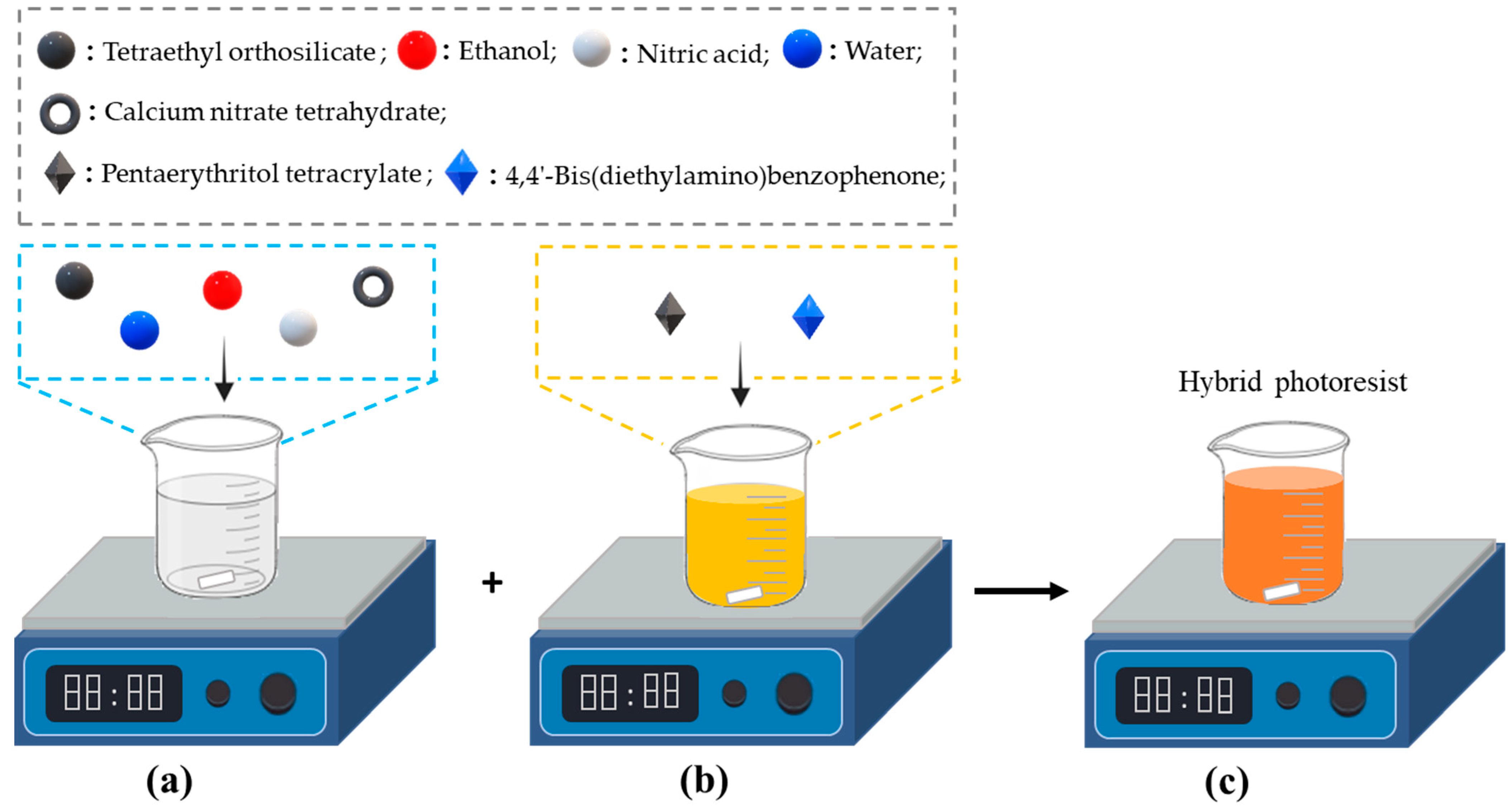
The hybrid photoresist was prepared by solution mixing the SiO2-CaO sol (20 wt%) with the organic resin (80 wt%) under stirring for 15 min. A homogeneous, transparent, dark yellow solution was obtained (Figure 2). Following the same route, the hybrid photoresist with SiO2-CaO-P2O5 sol was also prepared.

2.2. Two-Photon Polymerization 3D Printing

The 3D printing of the microstructures was performed using the 2PP technique on fused silica substrates (3D SF DiLL, 25 mm × 25 mm × 0.7 mm, from Nanoscribe GmbH, Eggenstein-Leopoldshafen, Germany). After designing the 3D model, the file (.STL) was imported into Describe software (version 2.7, Nanoscribe GmbH, Karlsruhe, Germany). The 3D printing parameters were adjusted in Describe to generate the job file (.GWL). Figure 3 shows the different steps of our 3D printing process. Firstly, a drop of the hybrid photoresist was placed on the fused silica substrate (Figure 3a). Then, the microstructures were printed using a Photonic Professional GT+ commercial printer (Nanoscribe GmbH, Karlsruhe, Germany) in the dip-in laser lithography (DiLL) configuration (Figure 3b). When the 3D printing was complete (Figure 3c), a development step was required to remove the unpolymerized photoresist. This consisted of immersing the substrate in propylene glycol monomethyl ether acetate (PGMEA) for 10 min followed by isopropyl alcohol (IPA) for 5 min, to recover the green microstructures (Figure 3d). Finally, the 3D-printed microstructures were heat treated under air at 700 °C, as detailed hereinafter. The 3D printer employed a femtosecond fiber laser source with a wavelength centered at 780 nm, a mean power of about 50 mW, a pulse duration of 100 fs, and a repetition rate of 80 MHz. A 63x/NA1.4 objective lens was used (from Zeiss society, Oberkochen, Germany).

2.3. Heat Treatment Process

The heat treatment was performed under air from RT to 700 °C in an LHTCT 01/16 furnace with lift Door (Nabertherm GmbH, Lilienthal, Germany). The samples were heat treated according to the stepwise heating protocol detailed in Figure 4. Different heating rates were used: 150 °C (2.2 °C/min), 320 °C (1.4 °C/min), 400 °C (0.7 °C/min), 500 °C (0.8 °C/min), and 700 °C (3.3 °C/min). After that, the substrates were removed from the furnace and left to cool down to room temperature.

2.4. Structural and Morphological Characterizations

For thermogravimetric analysis (TGA), a drop of the hybrid photoresist was deposited on a silica glass substrate and exposed to UV radiation. After UV curing using a Form Cure (Formlabs, Somerville, MA, USA), the powdered sample was recovered from the substrate. The TGA was carried out under air atmosphere using a Netzsch STA 2500 instrument (Netzsch, Selb, Germany), from RT to 1000 °C at a heating rate of 5 °C/min. The FTIR spectra of the UV-cured hybrid photoresists before and after heat treatment at 500 and 700 °C were recorded using a Nicolet 300 spectrometer (Thermo Fisher Scientific Inc., Waltham, MA, USA) equipped with a single reflection diamond ATR accessory. The scanning electron microscopy (SEM) images of the 3D-printed microstructures were obtained using two Field Emission Scanning Electron Microscopes (FE-SEM): SU8230 and SU5000 (Hitachi High Technologies, Tokyo, Japan). The optical images were obtained using Eclipse LV100 microscope (Nikon Corporation, Tokyo, Japan).

3. Results and Discussion

3.1. 3D Printed Microstructures

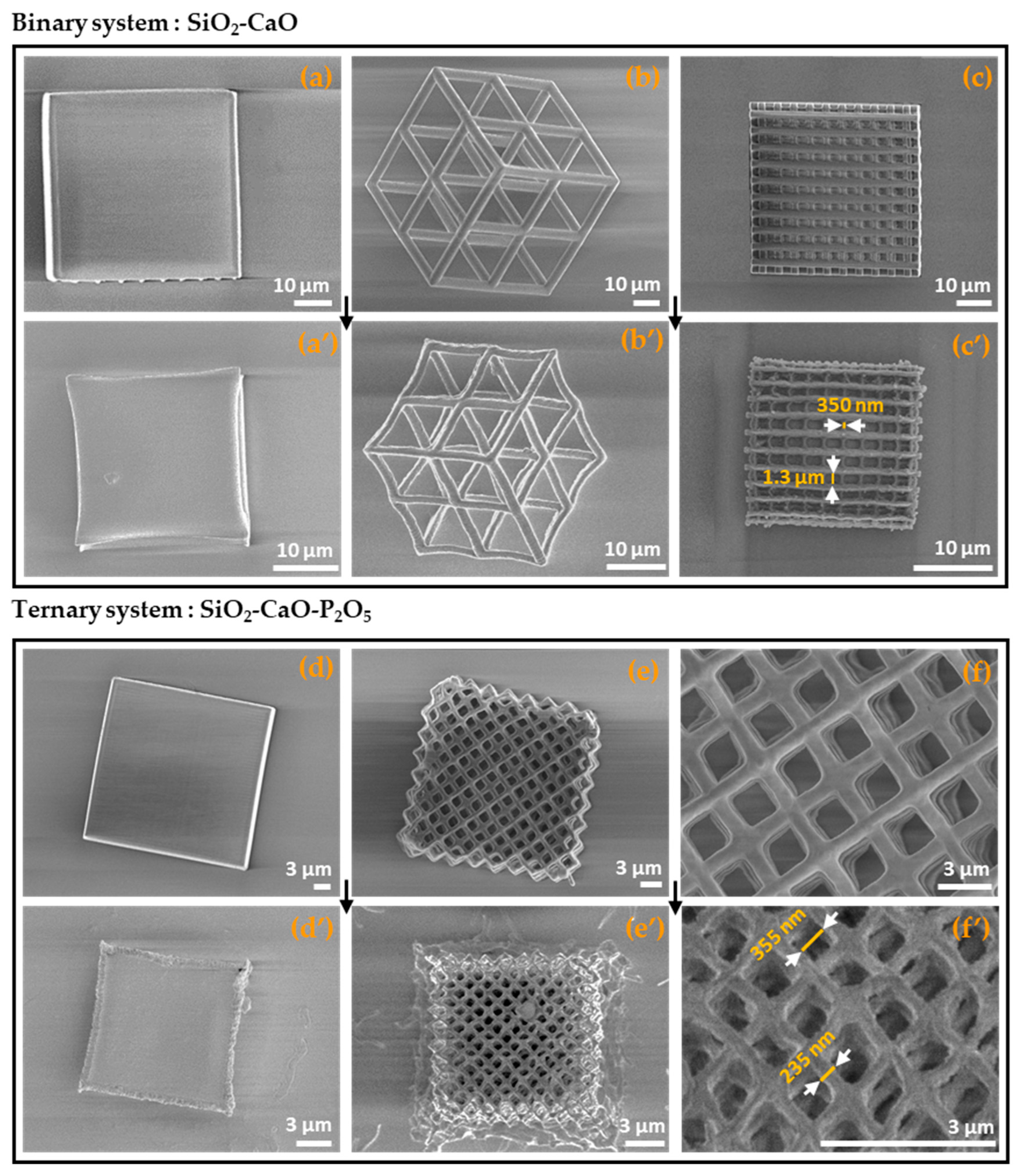
Figure 5 shows the 3D printed microstructures using both binary (a–c) and ternary (d–f) hybrid photoresists, before and after heat treatment at 700 °C. All the microstructures were 3D printed according to the following printing parameters: laser power (100%), scan speed (10,000 μm/s), and slicing/hatching distances (0.3 and 0.2 μm, respectively). The choice of sintering temperature was based on the results of the TGA analysis (see the Section 3.3). Although the thermograms showed a stabilization of mass loss at 600 °C due to the degradation of the organic phase, the microstructures were heat treated up to 700 °C to test their thermal resistance.
We also successfully 3D printed microcubes (Figure 5a,d) and more complex microstructures such as superlattice (Figure 5b), woodpile (Figure 5c), and cube–vertex–centroid (Figure 5e). After sintering at 700 °C, we observed that all the microstructures retained their shapes (Figure 5a′–f′) with slight deformation. Furthermore, we stress that binary microstructures showed higher thermal resistance to sintering at 700 °C compared with those of the ternary system. That was why we heat treated the microstructures for 1 h and 3 h for the ternary and binary systems, respectively. This result could be related to the presence of phosphorus in the composition of the ternary system. Furthermore, 3D objects with submicrometer features were produced, as shown in Figure 5c′,f′. According to this Figure, the resolutions reached for the binary and ternary systems were 350 nm and 235 nm, respectively. We note that such resolutions could be improved through further optimization of the resin composition and 3D printing parameters. Moreover, a scaffold pore size of about 355 nm was also obtained (Figure 5f′), which has never been achieved using other 3D-printing techniques, to the best of our knowledge. These results show that our approach can be used to develop adapted 3D scaffold structures for biomedical applications where scaffolds with small pore sizes are needed [33,34,35,36,37].
In addition, using optical microscopy to measure the size of printed microcubes before and after sintering at 700 °C, we observed that both the binary and ternary systems presented a close shrinkage rate of about 55–57% (Table 1). We note here that both the SiO2-CaO and SiO2-CaO-P2O5-based photoresists contained similar amounts of the organic resin. The obtained results show that the shrinkage was determined by the amount of the organic resin in the hybrid photoresist.

3.2. Morphological and Structural Analyses

3.2.1. Morphological Analysis

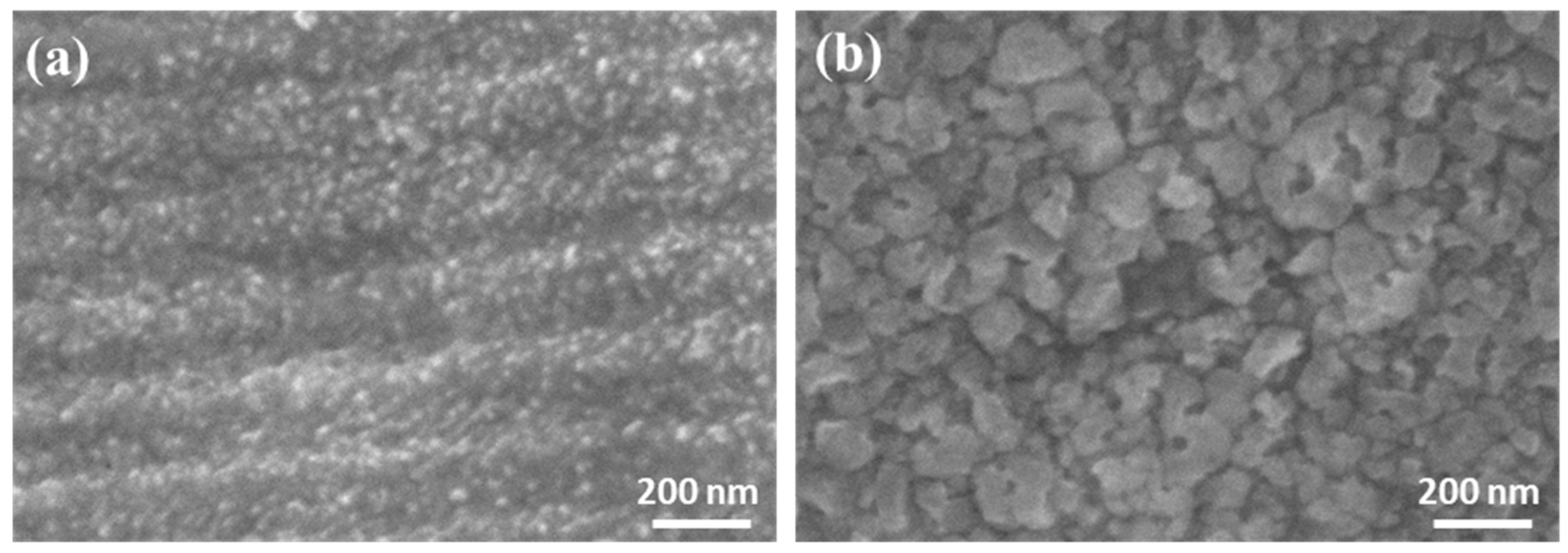
SEM micrographs were taken from the surfaces of 3D-printed microcubes of the binary and ternary systems after sintering at 700 °C. The images are shown in Figure 6. They reveal different morphologies depending on the composition of the material. Indeed, in the case of the ternary system (Figure 6a), the SEM image showed that it was made of an agglomeration of finer particles with an average particle size of about 20 nm. However, regarding the binary system (Figure 6b), the SEM micrograph revealed the presence of agglomerated particles with irregular shapes and a rough texture with an average particle size of about 60 nm. The presence of smaller nanoparticles (20 nm) in the case of the ternary system compared with the binary (60 nm), could be attributed to the presence of phosphorus in the composition of the ternary system.

3.2.2. FTIR Analysis

The FTIR spectra of the hybrid photoresists before and after heat treatment at 500 and 700 °C are shown in Figure 7. Absorption bands associated with the organic phase in the UV-treated hybrid photoresists were observed, as summarized in Table 2. Moreover, the wide band peaking at 3500 cm−1 originated from the -O-H stretching vibrations of hydroxyl groups and the stretching vibrations of free silanol groups [38]. After heat treatment at 500 °C, we observed the disappearance of absorption bands associated with the organic phase, and the FTIR spectra showed bands characteristic of the SiO2-CaO binary or SiO2-CaO-P2O5 ternary systems (Figure 7a,b). The typical bands of silica-based systems at around 798 and 1046 cm−1 correspond, respectively, to the symmetric and asymmetric stretching modes of Si–O–Si [4,39]. The observed vibrational band at 940 cm−1 was assigned to the stretching vibrations of the nonbridging Si-O (Si-O-NBO) and associated with the presence of calcium ions [38]. With regard to the ternary system, we do not exclude the presence of a band associated with the P–O bond around 1045–1090 cm−1, although this was masked by the broad silicate band [40]. We noted the appearance of two absorption bands peaking around 1420 and 1520 cm−1, attributed to the anti-symmetric stretching of C-O in carbonate groups CO32- [38,39,40]. The presence of these groups was associated with calcium and its reaction with the residual carbone [38] or the atmospheric CO2 [40]. The incorporation of carbonate groups provides the advantage of improving the solubility and resorbability processes that occur in bone [41,42]. The double peak around 2340 and 2363 cm−1 can be attributed to adsorbed CO2, as observed in similar systems [39,43,44]. After the heat treatment at 700 °C, compared with 500 °C, we noted that the band attributed to carbonate groups decreased while that associated with the absorbed CO2 increased. Morever, the band at 3500 cm−1 completely disappeared after heat treatment at 700 °C, which was due to the elimination of hydroxyl groups.

3.3. TGA

Figure 8 shows the TGA curves of the UV-cured hybrid photoresists; note that both SiO2-CaO and SiO2-CaO-P2O5 systems present similar TGA profiles. Three stages of mass loss were observed in the TGA curves. The first mass loss of about 8% ranged from 25 to 200 °C and was attributed to the removal of residual water and ethanol [38,47,48]. In the second range, 200–550 °C, the weight loss was about 87.4%, which was assigned to thermo–oxidative degradation of highly cross-linked acrylic polymers and the thermal decomposition of residual nitrates [4,47]. The weight loss shown in thermogram (b) was slightly delayed compared with that in (a), which could have been related to the presence of phosphorus. In the range 550–700 °C, the third mass loss was about 0.4%, which could have been associated with the thermal degradation of residual organics and condensation reactions. For this reason, we stopped the thermal treatment of our samples at 700 °C.

4. Conclusions

In summary, new hybrid photoresists suitable for 2PP additive manufacturing of multicomponent oxide systems were successfully synthesized via a combination of organic resin and sol–gel sols. It was found that the 3D-printed microstructures endure a sintering temperature up to 700 °C leading to 3D scaffolds with submicrometer features and a resolution of about 235 nm with pore size of about 355 nm. After sintering at 700 °C, it was shown that the obtained binary (SiO2-CaO) and ternary (SiO2-CaO-P2O5) oxide systems, incorporating carbonate groups, present different morphology. They were formed of agglomerated nanoparticles with size of about 60 and 20 nm, for the binary and ternary system, respectively. Our findings open new opportunities for the precise fabrication of complex multicomponent oxide structures such as multiscale interconnected porous scaffolds for bone regeneration in the biomedical field.

Author Contributions

Conceptualization, H.E.A. and H.E.H.; methodology, H.E.A. and H.E.H.; validation, H.E.A. and H.E.H.; formal analysis, H.E.A. and H.E.H.; investigation, H.E.A., H.E.H. and M.D.; resources, H.E.A. and H.E.H.; data curation, H.E.A. and H.E.H.; writing—original draft preparation, H.E.A. and H.E.H.; writing—review and editing, H.E.A., H.E.H., M.D. and Y.Q.; visualization, H.E.A. and H.E.H.; supervision, H.E.H. and M.D.; project administration, H.E.H., M.D. and Y.Q.; funding acquisition, M.D. All authors have read and agreed to the published version of the manuscript.

Funding

This work was supported in part by the Agence Nationale de la Recherche through the Add4P Equipex+ project (ANR-21-ESRE-0007) and 3DGlassens project (ANR-21-CE08-0035-01). The present work was also carried out within the framework of the Contrat de Plan Etat-Region (CPER) WaveTech@HdF, which is supported by the Ministry of Higher Education and Research, the Hauts-de-France (HdF) Regional council, the Lille European Metropolis (MEL), the Institute of Physics of the French National Centre for Scientific Research (CNRS), and the European Regional Development Fund (ERDF).

Data Availability Statement

The data presented in this study are available on request from the corresponding author.

Acknowledgments

This work has been supported by the IRCICA institute and the FiberTech Lille platform of University of Lille (https://fibertech.univ-lille.fr/en/ (accessed on 7 November 2024)).

Conflicts of Interest

The authors declare no conflicts of interest.

References

  1. Xin, C.; Li, Z.; Hao, L.; Li, Y. A Comprehensive Review on Additive Manufacturing of Glass: Recent Progress and Future Outlook. Mater. Des. 2023, 227, 111736. [Google Scholar] [CrossRef]
  2. Doualle, T.; André, J.-C.; Gallais, L. 3D Printing of Silica Glass through a Multiphoton Polymerization Process. Opt. Lett. 2021, 46, 364–367. [Google Scholar] [CrossRef] [PubMed]
  3. Zhu, D.; Zhang, J.; Xu, Q.; Li, Y. Two-Photon Polymerization of Silica Glass Diffractive Micro-Optics with Minimal Lateral Shrinkage. Opt. Express 2023, 31, 36037–36047. [Google Scholar] [CrossRef] [PubMed]
  4. El Aadad, H.; El Hamzaoui, H.; Brévalle-Wasilewski, G.; Bernard, R.; Kinowski, C.; Quiquempois, Y.; Douay, M. Solmers: Versatile Hybrid Resins for Nanometric 3D Printing of Silica-Based Photonic Components. Mater. Today Adv. 2024, 22, 100500. [Google Scholar] [CrossRef]
  5. Ye, P.; Hong, Z.; Loy, D.A.; Liang, R. Solvent-Free Silsesquioxane Self-Welding for 3D Printing Multi-Refractive Index Glass Objects. Adv. Opt. Mater. 2024, 12, 2400783. [Google Scholar] [CrossRef]
  6. Lai, L.-L.; Huang, P.-H.; Stemme, G.; Niklaus, F.; Gylfason, K.B. 3D Printing of Glass Micro-Optics with Subwavelength Features on Optical Fiber Tips. ACS Nano 2024, 18, 10788–10797. [Google Scholar] [CrossRef]
  7. Wang, H.; Pan, C.-F.; Li, C.; Menghrajani, K.S.; Schmidt, M.A.; Li, A.; Fan, F.; Zhou, Y.; Zhang, W.; Wang, H. Two-Photon Polymerization Lithography for Imaging Optics. Int. J. Extrem. Manuf. 2024, 6, 42002. [Google Scholar] [CrossRef]
  8. Wang, H.; Zhang, W.; Ladika, D.; Yu, H.; Gailevičius, D.; Wang, H.; Pan, C.; Nair, P.N.S.; Ke, Y.; Mori, T. Two-photon Polymerization Lithography for Optics and Photonics: Fundamentals, Materials, Technologies, and Applications. Adv. Funct. Mater. 2023, 33, 2214211. [Google Scholar] [CrossRef]
  9. Kotz, F.; Quick, A.S.; Risch, P.; Martin, T.; Hoose, T.; Thiel, M.; Helmer, D.; Rapp, B.E. Two-photon Polymerization of Nanocomposites for the Fabrication of Transparent Fused Silica Glass Microstructures. Adv. Mater. 2021, 33, 2006341. [Google Scholar] [CrossRef]
  10. Wen, X.; Zhang, B.; Wang, W.; Ye, F.; Yue, S.; Guo, H.; Gao, G.; Zhao, Y.; Fang, Q.; Nguyen, C.; et al. 3D-Printed Silica with Nanoscale Resolution. Nat. Mater. 2021, 20, 1506–1511. [Google Scholar] [CrossRef]
  11. Hong, Z.; Ye, P.; Loy, D.A.; Liang, R. High-Precision Printing of Complex Glass Imaging Optics with Precondensed Liquid Silica Resin. Adv. Sci. 2022, 9, 2105595. [Google Scholar] [CrossRef] [PubMed]
  12. Bauer, J.; Crook, C.; Baldacchini, T. A Sinterless, Low-Temperature Route to 3D Print Nanoscale Optical-Grade Glass. Science 2023, 380, 960–966. [Google Scholar] [CrossRef] [PubMed]
  13. Simorgh, S.; Alasvand, N.; Khodadadi, M.; Ghobadi, F.; Kebria, M.M.; Milan, P.B.; Kargozar, S.; Baino, F.; Mobasheri, A.; Mozafari, M. Additive Manufacturing of Bioactive Glass Biomaterials. Methods 2022, 208, 75–91. [Google Scholar] [CrossRef] [PubMed]
  14. He, L.; Yin, J.; Gao, X. Additive Manufacturing of Bioactive Glass and Its Polymer Composites as Bone Tissue Engineering Scaffolds: A Review. Bioengineering 2023, 10, 672. [Google Scholar] [CrossRef]
  15. Cannio, M.; Bellucci, D.; Roether, J.A.; Boccaccini, D.N.; Cannillo, V. Bioactive Glass Applications: A Literature Review of Human Clinical Trials. Materials 2021, 14, 5440. [Google Scholar] [CrossRef]
  16. Santos, K.W.; Costa, K.; Gonçalves, I.S.; Alves, M.; Lauda, D.P.; Vasconcellos, L.M.R.; Campos, T.M.B.; Oliveira, I.R. Influence of Manufacturing Parameters on Bioactive Glass 45S5: Structural Analysis and Applications in Bone Tissue Engineering. Ceram. Int. 2024, 50, 51043–51054. [Google Scholar] [CrossRef]
  17. Gul, H.; Zahid, S.; Kaleem, M. Bioglass, A New Trend Towords Clinical Bone Tissue Engineering. Pak. Oral Dent. J. 2015, 35, 706–712. [Google Scholar]
  18. Meskher, H.; Sharifianjazi, F.; Tavamaishvili, K.; Irandoost, M.; Nejadkoorki, D.; Makvandi, P. Limitations, Challenges and Prospective Solutions for Bioactive Glasses-Based Nanocomposites for Dental Applications: A Critical Review. J. Dent. 2024, 150, 105331. [Google Scholar] [CrossRef]
  19. Profeta, A.C.; Prucher, G.M. Bioactive-Glass in Periodontal Surgery and Implant Dentistry. Dent. Mater. J. 2015, 34, 559–571. [Google Scholar] [CrossRef]
  20. Kowalska, K.J.; Czechowska, J.P.; Zima, A. Novel Phosphate Bioglasses and Bioglass-Ceramics for Bone Regeneration. Ceram. Int. 2024, 50, 45976–45985. [Google Scholar] [CrossRef]
  21. Hench, L.L.; Polak, J.M. Third-Generation Biomedical Materials. Science 2002, 295, 1014–1017. [Google Scholar] [CrossRef] [PubMed]
  22. Balamurugan, A.; Sockalingum, G.; Michel, J.; Fauré, J.; Banchet, V.; Wortham, L.; Bouthors, S.; Laurent-Maquin, D.; Balossier, G. Synthesis and Characterisation of Sol Gel Derived Bioactive Glass for Biomedical Applications. Mater. Lett. 2006, 60, 3752–3757. [Google Scholar] [CrossRef]
  23. Shoushtari, M.S.; Hoey, D.; Biak, D.R.A.; Abdullah, N.; Kamarudin, S.; Zainuddin, H.S. Sol–Gel-templated Bioactive Glass Scaffold: A Review. Res. Biomed. Eng. 2024, 40, 281–296. [Google Scholar] [CrossRef]
  24. Bouhazma, S.; Adouar, I.; Chajri, S.; Herradi, S.; Khaldi, M.; Ouammou, A.; El Bali, B.; Mansori, M.; Lachkar, M. A Sol-Gel Synthesis, Characterization and in Vitro Bioactivity of Binary, Ternary and Quaternary Bioglasses with High Mechanical Strength. Mediterr. J. Chem. 2020, 10, 310–318. [Google Scholar] [CrossRef]
  25. Vichery, C.; Nedelec, J.-M. Bioactive Glass Nanoparticles: From Synthesis to Materials Design for Biomedical Applications. Materials 2016, 9, 288. [Google Scholar] [CrossRef]
  26. Sepulveda, P.; Jones, J.R.; Hench, L.L. Characterization of Melt-derived 45S5 and Sol-gel–Derived 58S Bioactive Glasses. J. Biomed. Mater. Res. 2001, 58, 734–740. [Google Scholar] [CrossRef]
  27. Boccaccini, A.R.; Blaker, J.J. Bioactive Composite Materials for Tissue Engineering Scaffolds. Expert Rev. Med. Devices 2005, 2, 303–317. [Google Scholar] [CrossRef]
  28. Martelli, A.; Bellucci, D.; Cannillo, V. Additive Manufacturing of Polymer/Bioactive Glass Scaffolds for Regenerative Medicine: A Review. Polymers 2023, 15, 2473. [Google Scholar] [CrossRef]
  29. Yuan, X.; Zhu, W.; Yang, Z.; He, N.; Chen, F.; Han, X.; Zhou, K. Recent Advances in 3D Printing of Smart Scaffolds for Bone Tissue Engineering and Regeneration. Adv. Mater. 2024, 36, 2403641. [Google Scholar] [CrossRef]
  30. Baino, F.; Gaido, F.; Gabrieli, R.; Alidoost, D.; Schiavi, A.; Mohammadi, M.; Schwentenwein, M.; Tulyaganov, D.; Verné, E. Vat Photopolymerization of Ultra-Porous Bioactive Glass Foams. Open Ceram. 2024, 20, 100690. [Google Scholar] [CrossRef]
  31. Diao, Q.; Zeng, Y.; Chen, J. The Applications and Latest Progress of Ceramic 3D Printing. Addit. Manuf. Front. 2024, 3, 200113. [Google Scholar] [CrossRef]
  32. Aykora, D.; Uzun, M. Bone Tissue Engineering for Osteointegration: Where Are We Now? Polym. Bull. 2024, 81, 8595–8605. [Google Scholar] [CrossRef]
  33. Ben-Arfa, B.A.E.; Pullar, R.C. A Comparison of Bioactive Glass Scaffolds Fabricated by Robocasting from Powders Made by Sol-Gel and Melt-Quenching Methods. Processes 2020, 8, 615. [Google Scholar] [CrossRef]
  34. Varma, M.V.; Kandasubramanian, B.; Ibrahim, S.M. 3D Printed Scaffolds for Biomedical Applications. Mater. Chem. Phys. 2020, 255, 123642. [Google Scholar] [CrossRef]
  35. Chocholata, P.; Kulda, V.; Babuska, V. Fabrication of Scaffolds for Bone-Tissue Regeneration. Materials 2019, 12, 568. [Google Scholar] [CrossRef]
  36. Mohd, N.; Razali, M.; Fauzi, M.B.; Abu Kasim, N.H. In Vitro and in Vivo Biological Assessments of 3D-Bioprinted Scaffolds for Dental Applications. Int. J. Mol. Sci. 2023, 24, 12881. [Google Scholar] [CrossRef]
  37. Singh, G.; Soundarapandian, S. Effect of Particle Size and Freezing Conditions on Freeze-Casted Scaffold. Ceram. Int. 2019, 45, 11633–11638. [Google Scholar] [CrossRef]
  38. Zagrajczuk, B.; Dziadek, M.; Olejniczak, Z.; Cholewa-Kowalska, K.; Laczka, M. Structural and Chemical Investigation of the Gel-Derived Bioactive Materials from the SiO2–CaO and SiO2-CaO-P2O5 Systems. Ceram. Int. 2017, 43, 12742–12754. [Google Scholar] [CrossRef]
  39. Pappas, G.S.; Liatsi, P.; Kartsonakis, I.A.; Danilidis, I.; Kordas, G. Synthesis and Characterization of New SiO2–CaO Hollow Nanospheres by Sol–Gel Method: Bioactivity of the New System. J. Non. Cryst. Solids 2008, 354, 755–760. [Google Scholar] [CrossRef]
  40. Luz, G.M.; Mano, J.F. Preparation and Characterization of Bioactive Glass Nanoparticles Prepared by Sol–Gel for Biomedical Applications. Nanotechnology 2011, 22, 494014. [Google Scholar] [CrossRef]
  41. Wopenka, B.; Pasteris, J.D. A Mineralogical Perspective on the Apatite in Bone. Mater. Sci. Eng. C 2005, 25, 131–143. [Google Scholar] [CrossRef]
  42. Kumar, K.C.V.; Subha, T.J.; Ahila, K.G.; Ravindran, B.; Chang, S.W.; Mahmoud, A.H.; Mohammed, O.B.; Rathi, M.A. Spectral Characterization of Hydroxyapatite Extracted from Black Sumatra and Fighting Cock Bone Samples: A Comparative Analysis. Saudi J. Biol. Sci. 2021, 28, 840–846. [Google Scholar] [CrossRef] [PubMed]
  43. Petkova, V.; Stoyanov, V.; Kostova, B. Phase Transformation in the CaO-SiO2-P2O5 Natural System during High Temperature Heat Treatment. J. Chem. Technol. Metall. 2022, 57, 614–621. [Google Scholar]
  44. Mihailova, I.; Radev, L.; Aleksandrova, V.; Colova, I.; Salvado, I.M.M.; Fernandes, M.H. V Carbonate-Apatite Forming Ability of Polyphase Glass-Ceramics in the CaO-MgO-SiO. J. Chem. Technol. Metall. 2015, 50, 502–511. [Google Scholar]
  45. Bertoluzza, A.; Fagnano, C.; Morelli, M.A.; Gottardi, V.; Guglielmi, M. Raman and Infrared Spectra on Silica Gel Evolving toward Glass. J. Non. Cryst. Solids 1982, 48, 117–128. [Google Scholar] [CrossRef]
  46. Doménech-Carbó, M.T.; Doménech-Carbó, A.; Gimeno-Adelantado, J.V.; Bosch-Reig, F. Identification of Synthetic Resins Used in Works of Art by Fourier Transform Infrared Spectroscopy. Appl. Spectrosc. 2001, 55, 1590–1602. [Google Scholar] [CrossRef]
  47. Wen, C.; Bai, N.; Luo, L.; Ye, J.; Zhan, X.; Zhang, Y.; Sa, B. Structural Behavior and in Vitro Bioactivity Evaluation of Hydroxyapatite-like Bioactive Glass Based on the SiO2-CaO-P2O5 System. Ceram. Int. 2021, 47, 18094–18104. [Google Scholar] [CrossRef]
  48. Meiszterics, A.; Sinkó, K. Sol–Gel Derived Calcium Silicate Ceramics. Colloids Surf. A Physicochem. Eng. Asp. 2008, 319, 143–148. [Google Scholar] [CrossRef]
Figure 1. Chemical structures of pentaerythritol tetracrylate (a) and 4,4’-Bis(diethylamino) benzophenone (b) used for the preparation of organic resin.
Nanomaterials 14 01977 g001
Figure 2. Schematic illustration of the synthesis procedure of the hybrid photoresist based on SiO2-CaO sol: (a) inorganic sol, (b) organic resin, and (c) hybrid photoresist.
Nanomaterials 14 01977 g002
Figure 3. Different steps of the 3D printing process: (a) depositing a drop of hybrid photoresist, (b) 3D printing using dip-in laser lithography configuration, (c) end of 3D printing, (d) development, (e) heat treatment of the printed microstructure.
Nanomaterials 14 01977 g003
Figure 4. The multi-step heat treatment protocol.
Nanomaterials 14 01977 g004
Figure 5. 3D-printed microstructures of binary (ac) and ternary (df) systems before (af) and after heat treatment at 700 °C (a′f′), respectively; (f) zoomed image of (e) and (f′) zoomed image of (e′).
Nanomaterials 14 01977 g005
Figure 6. Surface morphology SEM images of SiO2-CaO-P2O5 (a) and SiO2-CaO (b) oxide system microcubes after heat treatment at 700 °C.
Nanomaterials 14 01977 g006
Figure 7. FTIR spectra of UV-treated hybrid photoresists before and after heat treatment at 500 and 700 °C for 1 h: (a) SiO2-CaO and (b) SiO2-CaO-P2O5.
Nanomaterials 14 01977 g007
Figure 8. TGA curves of the UV-cured hybrid photoresists comprising (a) binary SiO2-CaO and (b) ternary SiO2-CaO-P2O5 systems.
Nanomaterials 14 01977 g008
Table 1. 3D-printed microcube dimensions for binary and ternary systems before and after heat treatment at 700 °C, and their shrinkage rates.
SystemSide Length (RT)Side Length (700 °C)Shrinkage Rate (%)
Binary system30.2 µm13.0 µm57
Ternary system30.5 µm13.7 µm55
Table 2. FTIR bands assigned to the organic phase in the UV-treated hybrid photoresists [4,45,46].
Binary System (cm−1)Ternary System (cm−1)Attribution
29642964ν (C-H)
29002902ν (C-H)
17221722ν (C=O)
16341634ν(C=C)
16351635ν (C=C)
16191918ν (C=C)
14721470δ (C-H)
14081407δ (C-H)
12961294γtwisting (CH2)
12631263ν (C-O-CH3)ester
11691169ν (C-C)
10591059ν (C-O)
982981ν (Si-OH)
806806δ (=C-H)
Disclaimer/Publisher’s Note: The statements, opinions and data contained in all publications are solely those of the individual author(s) and contributor(s) and not of MDPI and/or the editor(s). MDPI and/or the editor(s) disclaim responsibility for any injury to people or property resulting from any ideas, methods, instructions or products referred to in the content.