
Improving the Adhesion of Multi-Walled Carbon Nanotubes to Titanium by Irradiating the Interface with He+ Ions: Atomic Force Microscopy and X-ray Photoelectron Spectroscopy Study

 ,
,  , , ,
, , ,
Abstract
1. Introduction
2. Materials and Methods
3. Results and Discussion
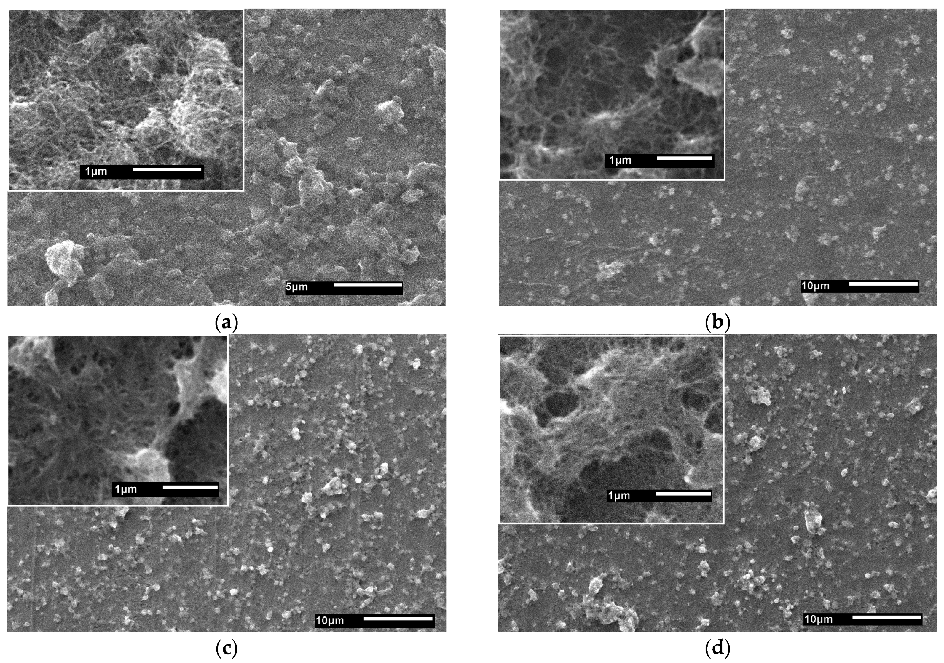
3.1. SEM
3.2. EDS
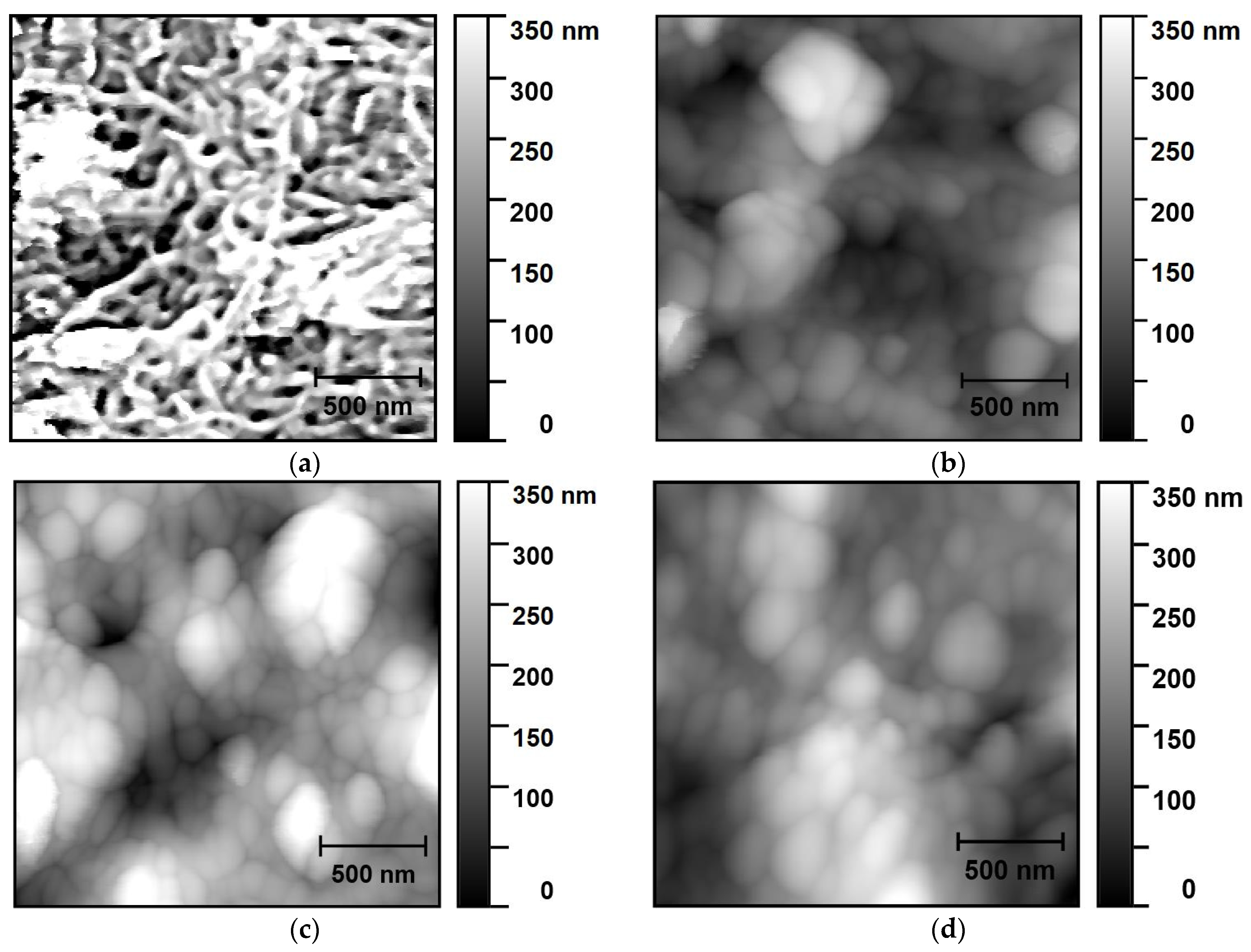
3.3. AFM
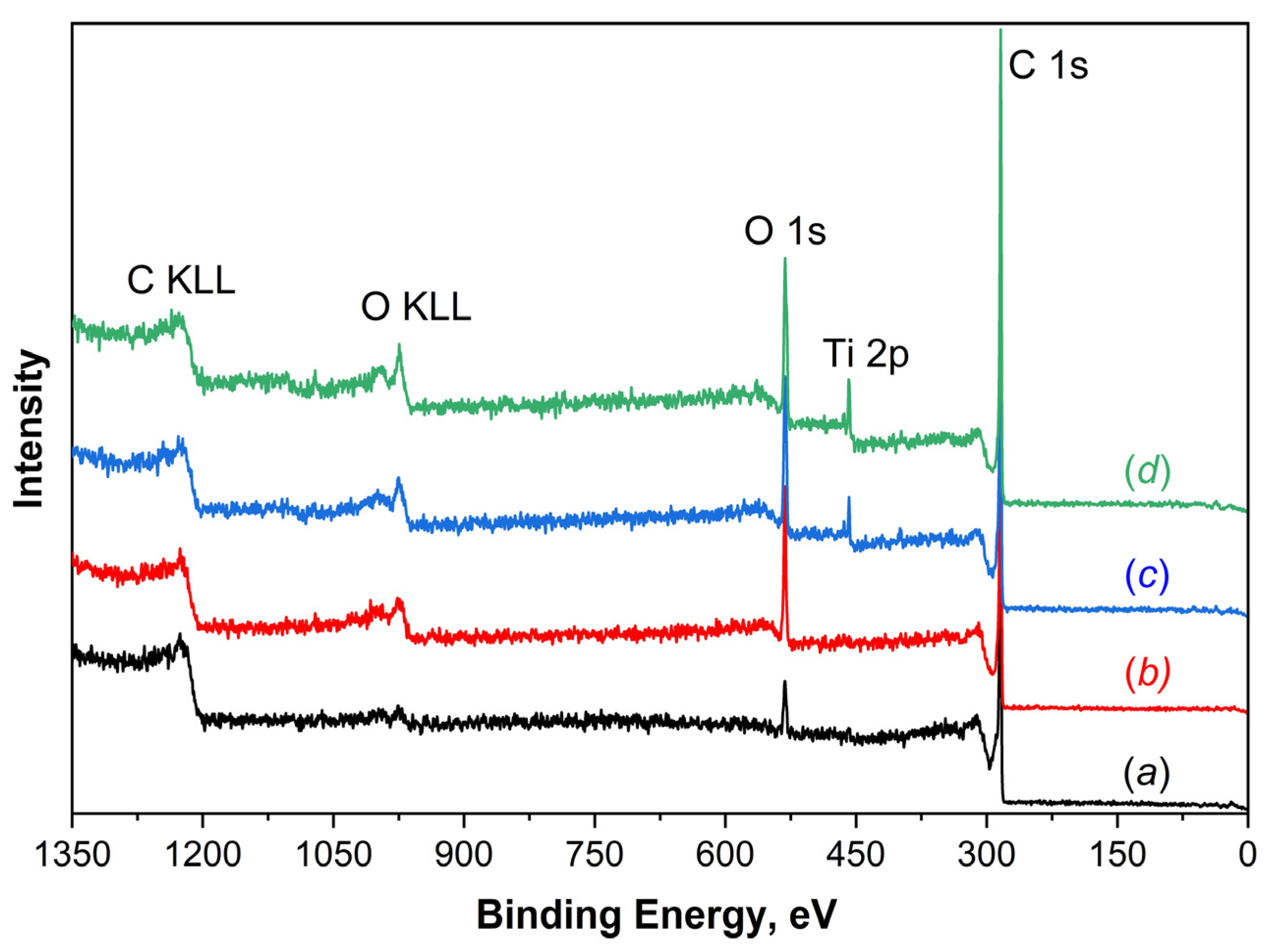
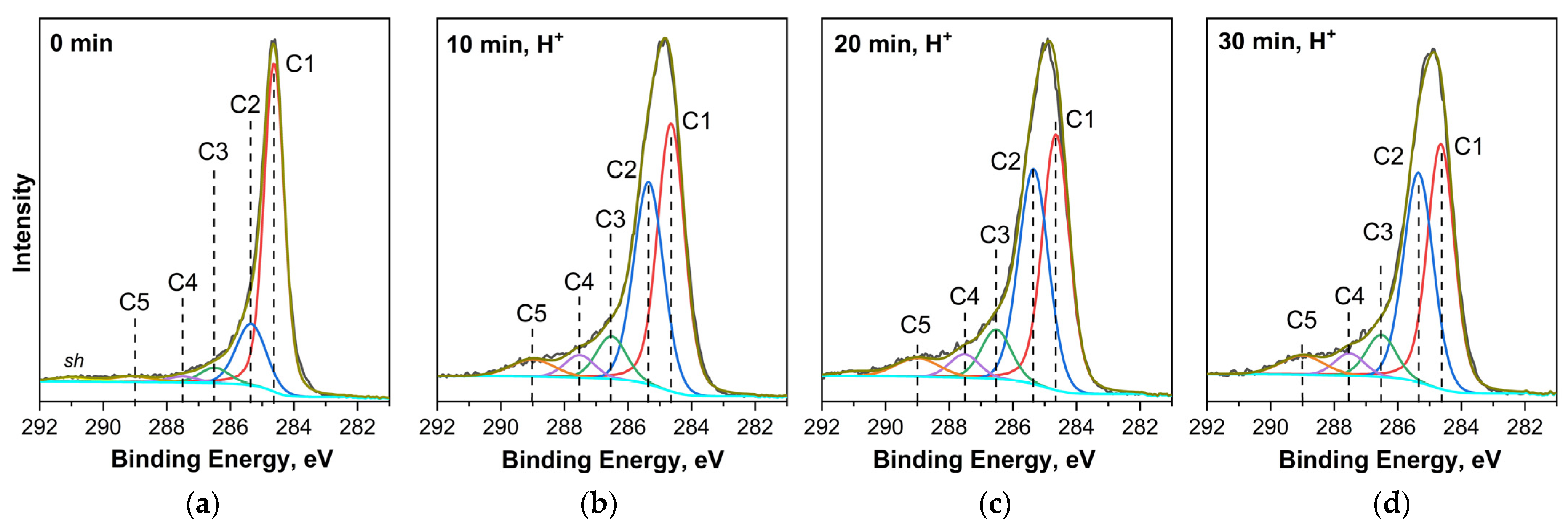
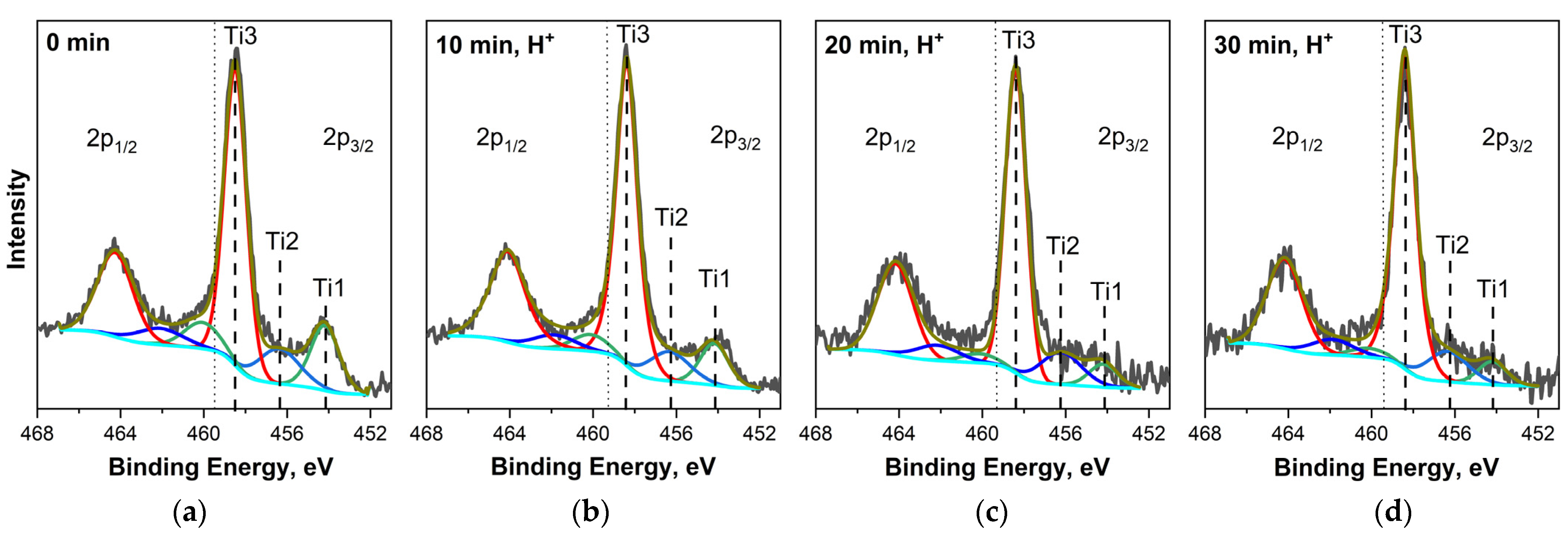
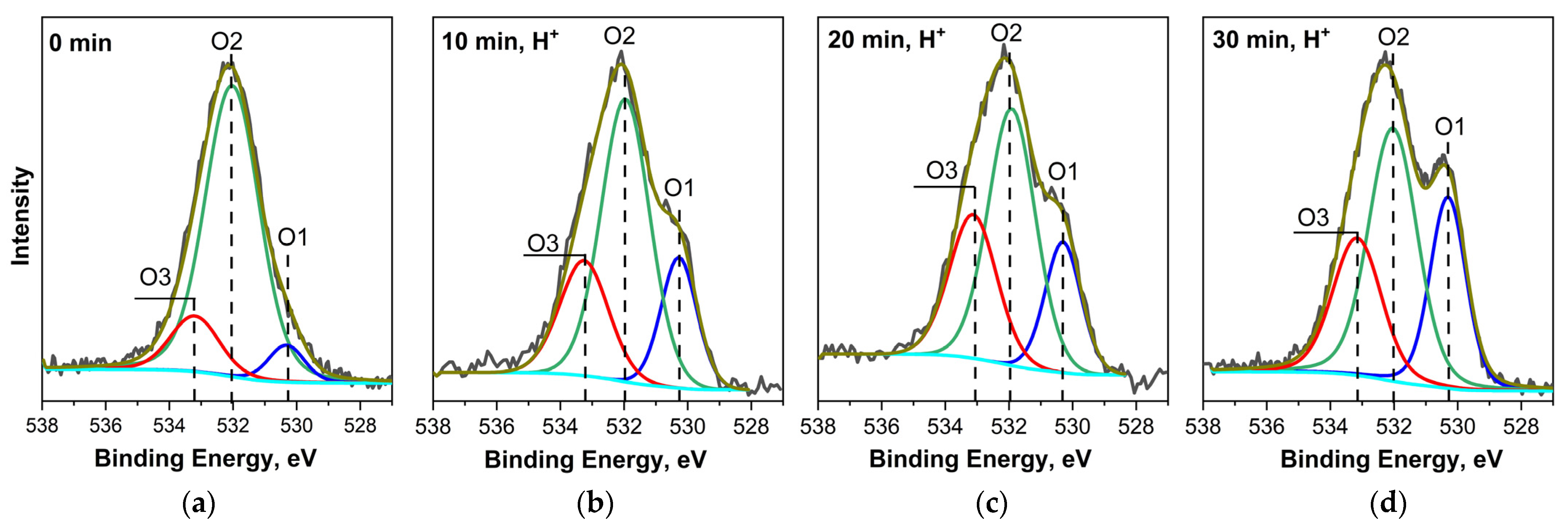
3.4. XPS
4. Conclusions
Author Contributions
Funding
Data Availability Statement
Acknowledgments
Conflicts of Interest
References
- Polozhentseva, Y.A.; Novozhilova, M.V.; Bykov, V.A.; Karushev, M.P. Modification of Porous Carbon Material with Polymeric Cobalt Complex with a Schiff Base of Salen-Type for Electrodes of Electrochemical Supercapacitors. Tech. Phys. Lett. 2020, 46, 913–915. [Google Scholar] [CrossRef]
- Kompan, M.E.; Malyshkin, V.G.; Kuznetsov, V.P.; Krivchenko, V.A. Specifics of Energy Storage in a Double Layer on the Surface of a Graphene Material. Tech. Phys. Lett. 2017, 43, 177–179. [Google Scholar] [CrossRef]
- Ahirrao, D.J.; Jha, N. Polyaniline-Manganese Dioxide Nanorods Nanocomposite as an Electrode Material for Supercapacitors. AIP Conf. Proc. 2017, 1832, 050168. [Google Scholar]
- Li, X.; Li, J.; Kang, F. Enhanced Electrochemical Performance of Salen-Type Transition Metal Polymer with Electron-Donating Substituents. Ionics 2019, 25, 1045–1055. [Google Scholar] [CrossRef]
- Bresser, D.; Buchholz, D.; Moretti, A.; Varzi, A.; Passerini, S. Alternative Binders for Sustainable Electrochemical Energy Storage—The Transition to Aqueous Electrode Processing and Bio-Derived Polymers. Energy Environ. Sci. 2018, 11, 3096–3127. [Google Scholar] [CrossRef]
- Istomina, A.S.; Bushkova, O.V. Polymer Binders for the Electrodes of Lithium Batteries Part 1. Polyvinylidene Fluoride, Its Derivatives and Other Commercialized Materials. Electrochem. Energetics 2020, 20, 115–131. [Google Scholar] [CrossRef]
- Eliseeva, S.N.; Kamenskii, M.A.; Tolstopyatova, E.G.; Kondratiev, V.V. Effect of Combined Conductive Polymer Binder on the Electrochemical Performance of Electrode Materials for Lithium-Ion Batteries. Energies 2020, 13, 2163. [Google Scholar] [CrossRef]
- Knyazev, E.V.; Korusenko, P.M.; Makushenko, R.V.; Nesov, S.N.; Povoroznyuk, S.N.; Ivlev, K.E.; Sivkov, D.V.; Petrova, O.V.; Vinogradov, A.S. Enhancing Interfacial Adhesion at the Carbon Nanotube/Titanium Interface Using Ion Beam Irradiation. Tech. Phys. Lett. 2024; in press. [Google Scholar]
- Santidrián, A.; Sanahuja, O.; Villacampa, B.; Diez, J.L.; Benito, A.M.; Maser, W.K.; Muñoz, E.; Ansón-Casaos, A. Chemical Postdeposition Treatments to Improve the Adhesion of Carbon Nanotube Films on Plastic Substrates. ACS Omega 2019, 4, 2804–2811. [Google Scholar] [CrossRef]
- Lahiri, I.; Lahiri, D.; Jin, S.; Agarwal, A.; Choi, W. Carbon Nanotubes: How Strong Is Their Bond with the Substrate? ACS Nano 2011, 5, 780–787. [Google Scholar] [CrossRef]
- Lee, S.W.; Kim, K.K.; Cui, Y.; Lim, S.C.; Cho, Y.W.; Kim, S.M.; Lee, Y.H. Adhesion Test of Carbon Nanotube Film Coated onto Transparent Conducting Substrates. Nano 2010, 05, 133–138. [Google Scholar] [CrossRef]
- Ishikawa, M.; Harada, R.; Sasaki, N.; Miura, K. Adhesion and Peeling Forces of Carbon Nanotubes on a Substrate. Phys. Rev. B 2009, 80, 193406. [Google Scholar] [CrossRef]
- Bouhamed, A.; Kia, A.M.; Naifar, S.; Dzhagan, V.; Müller, C.; Zahn, D.R.T.; Choura, S.; Kanoun, O. Tuning the Adhesion between Polyimide Substrate and MWCNTs/Epoxy Nanocomposite by Surface Treatment. Appl. Surf. Sci. 2017, 422, 420–429. [Google Scholar] [CrossRef]
- Marrero Rosa, R.E.; Corr, D.J.; Espinosa, H.D.; Shah, S.P. Characterization of Adhesion Strength between Carbon Nanotubes and Cementitious Materials. Cem. Concr. Compos. 2023, 138, 104953. [Google Scholar] [CrossRef]
- Jang, C.W.; Byun, Y.T.; Woo, D.H.; Lee, S.; Jhon, Y.M. Enhanced Adhesion between Carbon Nanotubes and Substrate Surfaces by Low-Temperature Annealing. J. Korean Phys. Soc. 2012, 61, 2096–2099. [Google Scholar] [CrossRef]
- Lim, S.C.; Choi, H.K.; Jeong, H.J.; Song, Y.I.; Kim, G.Y.; Jung, K.T.; Lee, Y.H. A Strategy for Forming Robust Adhesion with the Substrate in a Carbon-Nanotube Field-Emission Array. Carbon 2006, 44, 2809–2815. [Google Scholar] [CrossRef]
- Su, H.-C.; Chen, C.-H.; Chen, Y.-C.; Yao, D.-J.; Chen, H.; Chang, Y.-C.; Yew, T.-R. Improving the Adhesion of Carbon Nanotubes to a Substrate Using Microwave Treatment. Carbon 2010, 48, 805–812. [Google Scholar] [CrossRef]
- Ageev, O.A.; Blinov, Y.F.; Il’ina, M.V.; Il’in, O.I.; Smirnov, V.A.; Tsukanova, O.G. Study of Adhesion of Vertically Aligned Carbon Nanotubes to a Substrate by Atomic-Force Microscopy. Phys. Solid State 2016, 58, 309–314. [Google Scholar] [CrossRef]
- Il’in, O.I.; Il’ina, M.V.; Rudyk, N.N.; Fedotov, A.A. Adhesive Coatings Based on Aligned Arrays of Carbon Nanostructures. IOP Conf. Ser. Mater. Sci. Eng. 2018, 443, 012009. [Google Scholar] [CrossRef]
- Strus, M.C.; Zalamea, L.; Raman, A.; Pipes, R.B.; Nguyen, C.V.; Stach, E.A. Peeling Force Spectroscopy: Exposing the Adhesive Nanomechanics of One-Dimensional Nanostructures. Nano Lett. 2008, 8, 544–550. [Google Scholar] [CrossRef]
- Krasnikov, D.V.; Shmakov, A.N.; Kuznetsov, V.L.; Ishchenko, A.V. Towards the Optimization of Carbon Nanotube Properties via in Situ and Ex Situ Studies of the Growth Mechanism. J. Struct. Chem. 2016, 57, 1436–1443. [Google Scholar] [CrossRef]
- Ziegler, J.F.; Ziegler, M.D.; Biersack, J.P. SRIM—The Stopping and Range of Ions in Matter (2010). Nucl. Instrum. Methods Phys. Res. B 2010, 268, 1818–1823. [Google Scholar] [CrossRef]
- Fairley, N.; Fernandez, V.; Richard-Plouet, M.; Guillot-Deudon, C.; Walton, J.; Smith, E.; Flahaut, D.; Greiner, M.; Biesinger, M.; Tougaard, S.; et al. Systematic and Collaborative Approach to Problem Solving Using X-ray Photoelectron Spectroscopy. Appl. Surf. Sci. Adv. 2021, 5, 100112. [Google Scholar] [CrossRef]
- Korusenko, P.M.; Nesov, S.N.; Iurchenkova, A.A.; Fedorovskaya, E.O.; Bolotov, V.V.; Povoroznyuk, S.N.; Smirnov, D.A.; Vinogradov, A.S. Comparative Study of the Structural Features and Electrochemical Properties of Nitrogen-Containing Multi-Walled Carbon Nanotubes after Ion-Beam Irradiation and Hydrochloric Acid Treatment. Nanomaterials 2021, 11, 2163. [Google Scholar] [CrossRef] [PubMed]
- Korusenko, P.; Kharisova, K.; Knyazev, E.; Levin, O.; Vinogradov, A.; Alekseeva, E. Surface Engineering of Multi-Walled Carbon Nanotubes via Ion-Beam Doping: Pyridinic and Pyrrolic Nitrogen Defect Formation. Appl. Sci. 2023, 13, 11057. [Google Scholar] [CrossRef]
- Nesov, S.N.; Korusenko, P.M.; Sachkov, V.A.; Bolotov, V.V.; Povoroznyuk, S.N. Effects of Preliminary Ion Beam Treatment of Carbon Nanotubes on Structures of Interfaces in MOx/Multi-Walled Carbon Nanotube (M=Ti,Sn) Composites: Experimental and Theoretical Study. J. Phys. Chem. Solids 2022, 169, 110831. [Google Scholar] [CrossRef]
- Belhadj, H.; Moulefera, I.; Sabantina, L.; Benyoucef, A. Effects of Incorporating Titanium Dioxide with Titanium Carbide on Hybrid Materials Reinforced with Polyaniline: Synthesis, Characterization, Electrochemical and Supercapacitive Properties. Fibers 2022, 10, 46. [Google Scholar] [CrossRef]
- Ivanovskaya, M.; Ovodok, E.; Kotsikau, D.; Azarko, I.; Micusik, M.; Omastova, M.; Golovanov, V. Structural Transformation and Nature of Defects in Titanium Carbide Treated in Different Redox Atmospheres. RSC Adv. 2020, 10, 25602–25608. [Google Scholar] [CrossRef] [PubMed]
- Olowoyo, J.O.; Kumar, M.; Jain, S.L.; Babalola, J.O.; Vorontsov, A.V.; Kumar, U. Insights into Reinforced Photocatalytic Activity of the CNT–TiO2 Nanocomposite for CO2 Reduction and Water Splitting. J. Phys. Chem. C 2019, 123, 367–378. [Google Scholar] [CrossRef]
- Eda, Y.; Manaka, T.; Hanawa, T.; Chen, P.; Ashida, M.; Noda, K. X-ray Photoelectron Spectroscopy-based Valence Band Spectra of Passive Films on Titanium. Surf. Interface Anal. 2022, 54, 892–898. [Google Scholar] [CrossRef]
- Zhang, X.; Zhou, J.; Song, H.; Chen, X.; Fedoseeva, Y.V.; Okotrub, A.V.; Bulusheva, L.G. “Butterfly Effect” in CuO/Graphene Composite Nanosheets: A Small Interfacial Adjustment Triggers Big Changes in Electronic Structure and Li-Ion Storage Performance. ACS Appl. Mater. Interfaces 2014, 6, 17236–17244. [Google Scholar] [CrossRef] [PubMed]
- Sivkov, D.V.; Petrova, O.V.; Nekipelov, S.V.; Vinogradov, A.S.; Skandakov, R.N.; Isaenko, S.I.; Ob’edkov, A.M.; Kaverin, B.S.; Vilkov, I.V.; Korolev, R.I.; et al. The Identification of Cu–O–C Bond in Cu/MWCNTs Hybrid Nanocomposite by XPS and NEXAFS Spectroscopy. Nanomaterials 2021, 11, 2993. [Google Scholar] [CrossRef] [PubMed]
- Wu, C.-H.; Kuo, C.-Y.; Chen, S.-T. Synergistic Effects between TiO2 and Carbon Nanotubes (CNTs) in a TiO2/CNTs System under Visible Light Irradiation. Environ. Technol. 2013, 34, 2513–2519. [Google Scholar] [CrossRef] [PubMed]
- Felten, A.; Suarez-Martinez, I.; Ke, X.; Van Tendeloo, G.; Ghijsen, J.; Pireaux, J.; Drube, W.; Bittencourt, C.; Ewels, C.P. The Role of Oxygen at the Interface between Titanium and Carbon Nanotubes. ChemPhysChem 2009, 10, 1799–1804. [Google Scholar] [CrossRef] [PubMed]
- Zikalala, S.A.; Chabalala, M.B.; Gumbi, N.N.; Coville, N.J.; Mamba, B.B.; Mutuma, B.K.; Nxumalo, E.N. Microwave-Assisted Synthesis of Titania–Amorphous Carbon Nanotubes/Amorphous Nitrogen-Doped Carbon Nanotubes Nanohybrids for Photocatalytic Degradation of Textile Wastewater. RSC Adv. 2021, 11, 6748–6763. [Google Scholar] [CrossRef] [PubMed]
- Ye, W.; Chi, Q.; Zhou, H.; Gao, P. Ball-Milling Preparation of Titanium/Graphene Composites and Its Enhanced Hydrogen Storage Ability. Int. J. Hydrogen Energy 2018, 43, 19164–19173. [Google Scholar] [CrossRef]


| Sample | Concentration, at. % | ||
|---|---|---|---|
| [C] | [O] | [Ti] | |
| Initial MWCNT/Ti | 40.8 | 5.3 | 54.0 |
| MWCNT/Ti, irradiated for 10 min | 35.7 | 4.2 | 60.1 |
| MWCNT/Ti, irradiated for 20 min | 29.7 | 7.4 | 62.9 |
| MWCNT/Ti, irradiated for 30 min | 25.7 | 5.4 | 68.9 |
| Sample | Initial MWCNT/Ti | MWCNT/Ti, Irradiated for 10 min | MWCNT/Ti, Irradiated for 20 min | MWCNT/Ti, Irradiated for 30 min | |
|---|---|---|---|---|---|
| Adhesion Strength | |||||
| F, μN | 10.5 ± 0.5 | 10.5 ± 0.5 | 18.3 ± 1.0 | 14.4 ± 1.3 | |
| Sample | Concentration, at. % | ||
|---|---|---|---|
| [C] | [O] | [Ti] | |
| Initial MWCNT/Ti | 93.80 | 5.21 | 0.99 |
| MWCNT/Ti, irradiated for 10 min | 85.88 | 13.38 | 0.74 |
| MWCNT/Ti, irradiated for 20 min | 83.01 | 14.93 | 2.06 |
| MWCNT/Ti, irradiated for 30 min | 80.38 | 16.45 | 3.17 |
| Sample | Relative Component Intensity, % | Concentration, at.% | |||||
|---|---|---|---|---|---|---|---|
| O1 | O2 | O3 | Otot | O1 | O2 | O3 | |
| Initial MWCNT/Ti | 6.7 | 79.6 | 13.7 | 5.21 | 0.35 | 4.14 | 0.72 |
| MWCNT/Ti, irradiated for 10 min | 19.6 | 56.5 | 23.9 | 13.38 | 2.62 | 7.56 | 3.20 |
| MWCNT/Ti, irradiated for 20 min | 19.8 | 51.8 | 28.4 | 14.93 | 2.95 | 7.74 | 4.24 |
| MWCNT/Ti, irradiated for 30 min | 27.8 | 46.5 | 25.7 | 16.45 | 4.57 | 7.65 | 4.23 |
Disclaimer/Publisher’s Note: The statements, opinions and data contained in all publications are solely those of the individual author(s) and contributor(s) and not of MDPI and/or the editor(s). MDPI and/or the editor(s) disclaim responsibility for any injury to people or property resulting from any ideas, methods, instructions or products referred to in the content. |
© 2024 by the authors. Licensee MDPI, Basel, Switzerland. This article is an open access article distributed under the terms and conditions of the Creative Commons Attribution (CC BY) license (https://creativecommons.org/licenses/by/4.0/).
Share and Cite
Korusenko, P.M.; Knyazev, E.V.; Petrova, O.V.; Sokolov, D.V.; Povoroznyuk, S.N.; Ivlev, K.E.; Bakina, K.A.; Gaas, V.A.; Vinogradov, A.S. Improving the Adhesion of Multi-Walled Carbon Nanotubes to Titanium by Irradiating the Interface with He+ Ions: Atomic Force Microscopy and X-ray Photoelectron Spectroscopy Study. Nanomaterials 2024, 14, 699. https://doi.org/10.3390/nano14080699
Korusenko PM, Knyazev EV, Petrova OV, Sokolov DV, Povoroznyuk SN, Ivlev KE, Bakina KA, Gaas VA, Vinogradov AS. Improving the Adhesion of Multi-Walled Carbon Nanotubes to Titanium by Irradiating the Interface with He+ Ions: Atomic Force Microscopy and X-ray Photoelectron Spectroscopy Study. Nanomaterials. 2024; 14(8):699. https://doi.org/10.3390/nano14080699
Chicago/Turabian StyleKorusenko, Petr M., Egor V. Knyazev, Olga V. Petrova, Denis V. Sokolov, Sergey N. Povoroznyuk, Konstantin E. Ivlev, Ksenia A. Bakina, Vyacheslav A. Gaas, and Alexander S. Vinogradov. 2024. "Improving the Adhesion of Multi-Walled Carbon Nanotubes to Titanium by Irradiating the Interface with He+ Ions: Atomic Force Microscopy and X-ray Photoelectron Spectroscopy Study" Nanomaterials 14, no. 8: 699. https://doi.org/10.3390/nano14080699
APA StyleKorusenko, P. M., Knyazev, E. V., Petrova, O. V., Sokolov, D. V., Povoroznyuk, S. N., Ivlev, K. E., Bakina, K. A., Gaas, V. A., & Vinogradov, A. S. (2024). Improving the Adhesion of Multi-Walled Carbon Nanotubes to Titanium by Irradiating the Interface with He+ Ions: Atomic Force Microscopy and X-ray Photoelectron Spectroscopy Study. Nanomaterials, 14(8), 699. https://doi.org/10.3390/nano14080699