Review

State-of-the-Art Advances of Nanomedicine for Diagnosis and Treatment of Bladder Cancer

1
Department of Urology, The Third Affiliated Hospital of Shenzhen University, Shenzhen University, Shenzhen 518000, China
2
Graduate School, Shanghai University of Traditional Chinese Medicine, Shanghai 201203, China
3
Department of Urology, The Affiliated South China Hospital of Shenzhen University, Shenzhen University, Shenzhen 518000, China
*
Author to whom correspondence should be addressed.
These authors contributed equally to this work.
Biosensors 2022, 12(10), 796; https://doi.org/10.3390/bios12100796
Submission received: 17 August 2022 / Revised: 17 September 2022 / Accepted: 20 September 2022 / Published: 27 September 2022
(This article belongs to the Special Issue Advances in Nanomedicines for Disease Diagnosis and Therapeutics)

Abstract

:
Bladder cancer is a common malignant tumor of the urinary system. Cystoscopy, urine cytology, and CT are the routine diagnostic methods. However, there are some problems such as low sensitivity and difficulty in staging, which must be urgently supplemented by novel diagnostic methods. Surgery, intravesical instillation, systemic chemotherapy, and radiotherapy are the main clinical treatments for bladder cancer. It is difficult for conventional treatment to deal with tumor recurrence, progression and drug resistance. In addition, the treatment agents usually have the defects of poor specific distribution ability to target tumor tissues and side effects. The rapid development of nanomedicine has brought hope for the treatment of bladder cancer in reducing side effects, enhancing tumor inhibition effects, and anti-drug resistance. Overall, we review the new progression of nano-platforms in the diagnosis and treatment of bladder cancer.

1. Introduction

Bladder cancer is the sixth most common cancer in the United States. Approximately 80,470 new cases of urinary bladder cancer (61,700 men and 18,770 women) were diagnosed in the United States in 2019 with 17,670 deaths (12,870 men and 4,800 women) occurring during this same period [1,2]. In general, bladder cancer can be classified into non-muscle-invasive bladder cancer (NMIBC) and muscle-invasive bladder cancer (MIBC) based on how extensive the tumor is [3]. Approximately 70% of the patients are diagnosed with NMIBC and the rest are MIBC and advanced bladder cancer [4]. Moreover, bladder cancer costs per patient range from 40,000 USD to more than 170,000 USD in the USA, which causes a huge financial burden for patients [5,6].
Currently, the mainstream diagnosis methods are cystoscopy, biopsy, and computer tomography (CT) scanning. It is difficult to identify the oblate abnormal tissue and tumor at an early stage by cystoscopy. Therefore, the accuracy of cystoscopy is decided by the examiner’s experience. In this regard, it is useful to evaluate the tumor location and tumor invasion of the surrounding tissue by CT when the tumor is large. Nevertheless, it is difficult to distinguish the T2, T2, and T3 stages precisely [7,8,9]. The treatment and administration of bladder cancer mainly depend on the stage of tumor and the degree of metastasis (Figure 1). For NMIBC, the current standard therapy is complete transurethral resection of the bladder cancer (TURBT) to remove as much of the tumor as possible and then conduct intravesical therapy to prevent recurrence or delay progression to a higher stage [10]. As for MIBC and the advanced stage of bladder tumor, systemic and intraperitoneal chemotherapy, adjuvant radiotherapy, photodynamic therapy as well as targeted drugs are commonly used in clinic to prolong survival time [11]. However, even after taking the full course of standard therapy and enduring all the adverse effects, the recurrence rate of bladder patients is still unsatisfying [12,13,14,15,16]. For example, 40% of NMIBC patients’ stage progressed after BCG instillation [17]. Moreover, the latest study suggested that the effect of chemotherapy for treating advanced bladder cancer was still far from satisfactory. About 44% of patients suffer recurrence or progression despite taking chemo-drugs instillation after TURBT [10,18]. Therefore, there is an unfulfilled need for more precise diagnostic methods and more effective treatments.
At present, there are many difficulties in the application of drugs which are commonly used in the treatment of bladder cancer. For example, many chemotherapy drugs have problems such as large side effects, low bioavailability, poor solubility, and tumor resistance, though with the continuous progress of drug research, some problems have been solved. In clinical practice, both doctors and patients are still expecting better drugs.
Having emerged in the past decades, nanomedicine is the application of nanotechnologies in medicine to improve diagnosis as well as therapy. More specifically, nanobiotechnologies play an important role in the discovery of biomarkers and molecular diagnostics and facilitate the integration of diagnosis and therapy. Typically, nanomaterials have been widely developed for cancer treatment. The flexibility in size, structure, and feasibility of precise release make nanomaterials a promising drug delivery tool. For instance, by packaging with a well-designed nano-shell, the diagnostic agents as well as therapeutic agents can attach to the tumor cell, so as to increase the concentration and prolong the exposure time. Moreover, using a nano-vehicle with a biphasic structure can increase the solubility of hydrophobic agents and improve the drug distribution in the bladder [19,20]. Moreover, solid tumor tissues are rich in blood vessels, wide in vascular wall space, poor in structural integrity, and lack lymphatic return, resulting in high permeability and retention of nanoparticles. That is known as the enhanced permeability and retention (EPR) effect. The EPR effect promotes the selective distribution of macromolecular substances in tumor tissue as well as preventing the internal agents from prematurely exposing to the body fluid or the normal tissue, so as to protect the agents from degradation and decrease the immunogenicity of agents [20]. It is noted that multifunctional nanomaterials have received intense attention recently. These kinds of novel particles can realize the purpose of diagnosis and treatment simultaneously [21,22,23,24]. This review mainly discusses the current nanotechnologies in the diagnosis and treatment of bladder cancer.

2. The Current Diagnostic Methods

An accurate diagnostic method is critical for a comprehensive assessment of bladder cancer progression and prognosis. Prior to the treatment, the main diagnostic methods of bladder cancer include cystoscopy and imaging examination, which are used for the qualitative diagnosis, location diagnosis, and staging diagnosis of bladder cancer. When a patient has the most typical symptoms of painless hematuria, endoscopic biopsy or biopsy and histopathologic examination are the basis for the diagnosis of bladder cancer (Figure 2). In addition, the clinical stage before treatment can be determined according to the CT examination of the whole body. If liver metastasis, lymph node metastasis, and bone metastasis are suspected, MRI and PET/CT can provide an additional reference in addition to CT results. Additionally, symptom assessment, urine cytology, and biomarkers detection are also commonly used in clinic.
Cystoscopy and biopsy are the gold standards for the diagnosis and recurrence monitoring of bladder cancer. However, frequent cystoscopy is often required, and cystoscopy is an invasive procedure. Therefore, it is possible to cause urinary tract infection, urethral stricture, and other complications [25]. In addition, the detection rate of bladder cancer is closely related to the doctors’ experience. Recently, a fluorescence cystoscope has been applied to diagnose bladder cancer by intravesical instilling 5-aminolevulinic acid (ALA) of hexaminolaevulinic acid (HLA). Compared with a normal cystoscope, a fluorescence cystoscope is more likely to detect tumors, especially carcinoma in situ. However, inflammation, recent TURBT or BCG instillation may cause false positive results. Therefore, there is an urgent need for new technical advances used to assist and improve cystoscopy.
Urine cytology and biomarkers tests are routine methods for the diagnosis of bladder cancer. It is not only non-invasive and has high specificity for the detection of bladder cancer, but sensitivity is related to the type of bladder cancer [26,27,28]. For example, the cytology sensitivity of low-grade bladder cancer is only 16% [29], which limits the clinical application. As for biomarkers tests, numerous proteins and RNA were found related to bladder cancer. However, the FDA only approved six biomarkers for urine tests and no marker test for blood or tissue due to the lack of high-quality prospective trials and low level of clinical evidence [30]. Nevertheless, none of them were sufficiently effective when used in isolation. Further randomized clinical trials are still needed in the future. Therefore, though clinical urine cytology and urine biomarkers may use in individual therapy guidelines, they are still auxiliary means of cystoscopy in clinical practice (Table 1).
Abdominal and pelvic CT is always the first choice of imaging examination for bladder cancer [33]. It can effectively detect bladder cancer and determine its location and stage [7]. For T4 stage bladder cancer, it is possible to accurately determine the location, extent, and depth of invasion of tumor via enhanced CT scan and routine scan combined with multiplanar image reconstruction. However, CT scan cannot illustrate the structure of the bladder wall. Therefore, it is hard to distinguish T1, T2, and T3a stages only using CT scans [9]. MRI is often used to assess the preoperative stage and lymph node metastasis. Generally speaking, using MRI to diagnose a T2 or T3 stage bladder tumor is much better than using CT scan. Especially when diffusion-weighted imaging and dynamic enhancement are used, MRI can better distinguish NMIBC and MIBC [11]. However, to some extent, the imaging quality of bladder wall and tumor depends on whether the bladder is filling. In addition, it is difficult to perform enhanced CT/MRI when patients are allergic to iodine contrast agents or have renal insufficiency [34,35]. Clinically, ultrasound imaging is widely used in the screening of patients with hematuria, especially in patients with iodine contrast medium allergy and renal insufficiency [36]. However, the accuracy and detection rate of ultrasonography are not satisfactory [37].

3. The Current Therapies

Current clinic therapies for bladder cancer are illustrated in Figure 3. For NMIBC, TURBT combined with single-dose immediate intravesical chemotherapy (SI) is the current standard therapy [38]. SI with gemcitabine and mitomycin can effectively prevent the implantation of residual tumor cells and reduce the risk of recurrence [13,38]. Furthermore, patients are recommended intravesical chemotherapy and BCG according to their stages. Epirubicin and mitomycin are usually used for intravesical chemotherapy while BCG is another commonly used intravesical agent for NMIBC. BCG was utilized for bladder cancer treatment as early as 1976 [39] and was finally approved by the FDA for NMIBC in 1990. Although decades have passed, intravesical instillation of BCG is still the most mature and widely used immunological therapy for NMIBC. According to the 2020 version of the NCCN (National Comprehensive Cancer Network) clinical practice guidelines on bladder cancer, BCG intravesical instillation is suggested for the induction intravesical instillation of medium-risk patients and should be prioritized for the induction of high-risk patients after TURBT. As for maintenance therapy, BCG is preferred for all medium/high-risk patients rather than chemotherapeutic drugs [33]. After instilling BCG, the fibronectin attachment protein (FAP) expressed on the mycobacterium wall combines with fibronectin expressed on urothelium, which helps BCG attach to the urothelium [40,41,42]. Bladder cancer cells then internalize BCG through micropinocytosis while it is much more difficult for normal urothelium cells to uptake it [43,44,45]. Then, BCG could activate multiple kinds of immune cells and initiate innate immune responses as well as adaptive immune responses [46,47,48]. In addition, BCG could cause bladder cancer cell death directly through apoptosis [49,50,51] and cellular oxidative stress [52,53,54].
As for MIBC and advanced bladder cancer, the standard therapy is neoadjuvant chemotherapy followed by radical cystectomy. Usually, advanced bladder cancer is more sensitive to platinum-based combination chemotherapy. Therefore, the major regimens are methotrexate, vinblastine, doxorubicin and cisplatin (MVAC), and gemcitabine cisplatin/carboplatin (GC regimen). In addition, adjuvant and palliative radiotherapy can be used as treatments for patients who are not suitable for cystectomy to increase the local tumor control rate and prolong the overall survival of the patients [55,56,57]. Immunotherapy and targeted therapy have developed rapidly in recent years. Compared with conventional chemotherapy, PD-1/L1 mAb-based immunotherapy has significantly improved the overall survival rate for advanced bladder cancer [58,59,60]. FGFR inhibitors as well as HER2 inhibitors have been approved for the treatment for advanced bladder cancer [61,62], which brings new hope to patients with chemotherapy failure.
Because the bladder is a hollow organ, the location of bladder tumors, especially NMIBC, is generally more superficial than that of other organs. Therefore, in addition to normal surgical treatment and chemotherapy, it is easier to implement local treatment, such as photosensitizer-based photodynamic therapy (PDT). Researchers have studied photosensitizers since the beginning of last century. These photosensitizers are usually dyers, which are able to accumulate in tumor tissues and are sensitive to light. After the oxygen molecule activation by light, reactive oxygen species (ROS) generate and therefore induce cytotoxic effects. Based on this mechanism, researchers successfully shrunk rats’ malignant tumors by using a photosensitizer with visible light in 1972 [63]. Following up on this study, PDT was widely developed and confirmed to have therapeutic effects on various cancers. The utilization of traditional photosensitizers to eradicate superficial bladder tumors has also been discussed. Regrettably, a limited number of photosensitizers have been approved for clinical use for bladder cancer despite various photosensitizers, mainly including the porphyrins, chlorins, and phthalocyanines as well as their derivatives, having been described in the literature. At present, only photofrin, Levulan, and Deuteporfin have been approved for superficial bladder cancer by the FDA. However, all of the current approved PDT agents have great limitations [64]. The long drug metabolism and increasing skin photosensitivity caused by photosensitizers may cause cutaneous toxicities, thus prolonged light-avoid time is needed. Moreover, the poor tumor selectivity of traditional photosensitizers also limits their use in bladder cancer. Photofrin/profimer sodium-PDT can lesion the adjacent tissue in the muscle layer of the bladder, thereby leading to irreversible bladder fibrosis and contracture [65,66]. In addition, it is necessary to have ample oxygen for ROS generation. Paradoxically, most malignant tumor displays a hypoxic environment within and the PDT therapy itself can cause hypoxia, which eventually reduces the curative effect. Moreover, by activating cellular immunity, PDT therapy can up-regulate the expression of PD-L1 [67,68,69], which leads to immune escape. Due to these drawbacks, PDT has not yet been recommended for bladder cancer treatment in the clinical guidelines.

4. Nanotechnology in Diagnosis

4.1. Nanotechnology in Light-Based Imaging

It is difficult to detect tiny or flat cancerous tissue accurately with the naked eye, which often results in a high recurrence rate after TURBT. Combining cystoscopes with bladder tumor-specific imaging agents is a good solution for improving diagnostic methods. Semiconductor nanocrystals, also called quantum dots (QD), have gained more attention than before for their unique properties in optics and electronics. Being able to emit fluorescence and carry tumor-targeting molecular agents makes semiconductor nanocrystals promising tumor tracers. It has been reported that, after intravesical instillation of the complex of QD and CD47 antibodies, this nanoparticle significantly concentrated in bladder tumors [70]. The combination of anti-CD47-QD and blue light cystoscopy showed satisfactory diagnostic accuracy for bladder cancer [71]. However, it should be noted that core materials such as cadmium (Cd) and selenium (Se) of QD might have long-term tissue toxicity, though the toxicity of heavy metals has not been observed in vivo. The prostate stem cell antigen (PSCA) is also a biomarker highly expressed on the surface of bladder tumor cells [72]. Yuan et al. simply conjugated the PSCA mAb with QD605 to develop a useful targeted fluorescent probe [73]. This probe could recognize bladder cancer cells specifically and emit stable and long-duration fluorescence. Carbonic anhydrase is a kind of tumor biomarker associated with tumor progression and metastasis [74]. Researchers developed CdSe/ZnS QD nanoprobes modified with carbonic anhydrase inhibitors. Notably, the nanoprobes were coated with DHLA-EDADA ligand and glucose residues, ensuring the stability of CdSe/ZnS QD, as well as providing good biocompatibility and dispersion [75].
Heteroatom-doped graphene quantum dots (GQDs) might be a promising luminescent material that could emit white light and broad excitation-dependent full-color photoluminescence from 463 nm to 672 nm. In addition, it exhibited a prohibiting growth and adhering effect when treating bladder cell lines [76]. However, the growth of tumor cells in vivo is different from that in culture dishes, and the result of inhibiting bladder cancer cell proliferation may be difficult to repeat in animal models.
Though the existing fluorescent cystoscope can target bladder cancer cells more precisely, given the molecular heterogeneity of tumors, it is unable to detect 100% of tumor cells with a single target. Therefore, Davis et al. developed a new endoscope system based on surface-enhanced Raman scattering nanoparticles [77]. This Raman nanoparticle could simultaneously carry a variety of antibodies which enabled the multiplexed detection of multiple molecular targets. With the help of multiple antibody-based targeting as well as EPR effect of nanoparticles, Raman nanoparticles could accumulate in bladder tumor tissue and enable multiple imaging (Figure 4).
Imaging cancer cells by specifically targeting overexpressed biomarkers is a good prospect in tumor diagnosis. However, the spectrum used for optical imaging should have the characteristics of low light scattering, minimum tissue absorption, and low spontaneous fluorescence. Near-infrared fluorescence possesses the characteristics of low light scattering, low light absorption and low spontaneous fluorescence when irradiating tissue. Therefore, it has a better tissue penetration ability and enables the imaging of deeper tissues. In order to diagnose tumors with near-infrared rays (NIR), NIR probes and imaging systems that can target tumors are necessary. Cho et al. developed nanoclusters of up-conversion nanoparticle (UCNP) conjunct with antibodies to epidermal growth factor (EGFR). This UCNP could emit visible light under NIR irritation and produce high-contrast images with no background fluorescence, hence accurately indicating the location of cancer cells [78].

4.2. Nanotechnology in Urine Test

Urine test for diagnosis of bladder cancer includes urine cytology and biomarkers detection. Urine exfoliative cytology detection mainly identifies abnormal cells in urine. Although it has a high sensitivity to advanced cancer, it possesses poor sensitivity to low-grade cancer. Therefore, it is much better to use high-sensitivity and -specificity nanotechnology to diagnose bladder cancer by detecting urine biomarkers.
A new background-eliminated fluorescence platform for sensitive and selective detection of cancer biomarkers has been built by combining feroxyhyte nanosheets (δ-FeOOH) with amino-functionalized silicon quantum dots (Si QDs). Using highly expressed hyaluronidase (Haase) as the target to detect the urine of bladder cancer patients, this platform possessed a similar accuracy compared to ELISA. The results showed the detection limit for Haase was 0.02 ng/mL (based on 3σ/S), which was three orders lower than most of the reported fluorescence biosensors (Table 2) [76]. Telomerase is an important biomarker in bladder cancer diagnosis. Ma et al. developed a highly sensitive ratiometric fluorescence paper sensor to detect telomerase in urine based on the color change of Rox-DNA functionalized QD. With the help of telomerase, H2O2 turned into H2O and O2, and the detected color, therefore, changed from red to yellow or green, which enabled visual semi-quantitative detection with naked eye. More importantly, the detection limit was 10 cells and response time was within an hour [79].

5. Nanotechnology in Treatment

5.1. Nano-Formulations for Chemotherapy

Paclitaxel (PTX) and docetaxel (DTX) are commonly used chemotherapy drugs. It is often used in systemic chemotherapy and neoadjuvant chemotherapy in the treatment of bladder cancer. However, the poor solubility in water and large systemic adverse reactions after intravenous dripping bring pain to the patients. Therefore, in order to improve the solubility and dispersion of PTX, accurate drug delivery and reduced adverse reactions via nanomaterials is very promising.
Nanoparticle albumin-bound (nab)-paclitaxel is a solvent-free formulation of paclitaxel approved for the treatment of metastatic breast cancer. Compared with standard PTX, nab-paclitaxel is better tolerated and demonstrated better efficacy than standard taxanes [80,81]. Nab-paclitaxel has also shown great efficacy on bladder cancer in the United States and Canada’s clinical trials [82,83] (Table 3).
Lumbrokinase (LK) is an enzyme complex with anti-thrombotic and antitumor effects, which may be promising to cure tumors with chemo drugs. Nevertheless, the toxicity and side effects of chemo drugs and LK limit clinical application. Hu et al. skillfully used block copolymer PEG-b-(PELG-g-(PZLL-r-PLL)) as a nanocarrier to deliver lumbrokinase (LK) with PTX and avoided the great toxicity and ensure sustained release in bladder. In addition, LK/PTX/PEG- b-(PELG-g-(PZLL-r-PLL)) successfully suppressed bladder tumor growth and induced bladder cancer cell death both in vivo and in vitro [84].
Pan et al. developed disulfide-crosslinked PLZ4 nanomicelles (DC-PNM) to deliver PTX. The loading efficiency of PTX was more than 99% and PTX was able to be released under the physiological concentration of glutathione. Interestingly, after being decrorated with PLZ4, the nanoparticles could target bladder tumors specifically but did not accumulate in the lung cancer xenografts, which could greatly reduce the systemic toxicity caused by PTX chemotherapy [85]. This nanomedicine has entered a phase 1 clinical trial in the United States (NCT05519241).
Liu et al. used chitosan (CS) nanosuspensions (NSs)-capsuled PTX, which exhibited a sustained and prolonged delivery as well as improved the curative effect of PTX intravesical bladder cancer. The positively charged properties enable the PTX/CS NSs to easily attach to the inner mucosa of the bladder through electrostatic adsorption and the nanoparticles could prolong the release of PTX for more than 10 days [86].
Ephrin receptor A2 (EphA2) is a kind of receptor tyrosine kinase that can regulate intercellular interactions and differentiation [87]. It has been found that EphA2 is highly expressed in bladder cancer patients and was associated with progression. EphA2 is a membrane-bound protein and is easy to internalize after antibody binding, which is a hopeful target for drug development. Walid Kamoun et al. developed a new EphA2 targeted antibody-directed nanotherapeutic drug (ADN), named EphA2-ILS-DTXp, which encapsulates the hydrolysis-sensitive prodrug of docetaxel (DTXp) [88]. EphA2-ILS-DTXp was designed to possess good pharmacokinetic properties to reduce plasma exposure while still maintaining the selective tumor exposure of DTX. The results showed that EphA2-ILS-DTXp was far more effective than a combination of free docetaxel and gemcitabine in the treatment of EphA2 positive patient-derived xenograft (PDX) model of bladder cancer [89].
MMC has good water solubility. Although it is convenient for intravesical instillation, it is difficult for it to penetrate the lipophilic cell membrane, therefore the curative effect is often unsatisfying. It has been reported to deliver MMC through a Mn:ZnS QD and chitosan-based nano-delivery system with excellent internalization efficiency which could improve the bioavailability of MMC [90].
Gemcitabine is one of the most commonly used chemo-drugs for bladder cancer. Fluorinated chitosan (FCS), gemcitabine, and H2 production catalyst were creatively assembled into a nano-drug delivery platform. In this research, fluorinated chitosan was used to enhance the ability to cross bladder mucosa and penetrate tumor cells. At the same time, under 660 nm laser irradiation, the hydrogen catalyst could effectively generate non-toxic hydrogen. The results showed that hydrogen could inhibit the mitochondrial function of bladder cancer cells, thus inhibiting the P-glycoprotein from excreting gemcitabine (Figure 5) [91].
Qiu et al. combined PAMAM, a kind of dendrimer, with PEG to form the PEG–PAMAM complex as a nanocarrier to deliver Doxorubicin (Dox). The results showed that PEG–PAMAM was a stable nanocarrier with a small size and good biosafety. Dox is released slowly from PEG–PAMAM at a neutral environment while released rapidly in an acidic microenvironment. As a drug carrier, PEG–PAMAM could effectively penetrate the urothelium of the mouse bladder, and increase the content of DOX in the bladder wall after intravesical instillation [92]. Their work provided a new idea for bladder cancer drug delivery.
Cisplatin is often used in systematic chemotherapy. Ding et al. [93] developed multifunctional BITT@BSA-DSP nanoparticles which could deliver cisplatin while producing NIR fluorescence imaging. In addition, with infrared irradiation, this multifunctional nanoparticle can be administrated precisely into the tumor. At the same time, it showed good photothermal and photodynamic effects, which enhanced the sensitivity of cancer cells to cisplatin and reduced the drug resistance in vivo.
In addition to the conventional chemotherapeutic drugs mentioned above, some natural components have also been reported to be used for chemotherapy. β−Elemene (β−E) is an anti-tumor chemical extracted from the herb Curcuma wenyujin [94]. So far, elemene emulsion injection as well as oral emulsion have been reported to be used to treat various cancers [95]. Zhai et al. developed an amino-terminal fragment (ATF) peptide-targeted liposome to carry β-elemene (ATF24-PEG-Lipo-β-E). ATF24-PEG-Lipo-β-E had stable shape and size. Additionally, urokinase-type plasminogen activator receptor (uPAR) tends to overexpress in high metastatic cancer cells and tumor stromal cells [96]. ATF peptide can compete to bind uPAR on the surface of tumor and endothelial cells, so as to inhibit tumor growth and angiogenesis, which therefore could help liposomes target delivery to tumors and stromal cells. The results suggested ATF 24−PEG−Lipo−β−E combined with cisplatin significantly inhibited the proliferation of bladder cancer cells in vivo [97].
Polybia-mastoparan I (MPI) is a kind of antibiotic peptide isolated from the venom of the social wasp Polybia paulista [98]. In previous studies, MPI showed potent antiproliferative effects and might act as a potential agent for bladder cancer treatment [99]. Li et al. synthesized fluorinated PEI, a transmucosal agent, and conjugated it with MPI to form a bladder instillation formula. Not only did this nano-formula possess great selectivity and biosafety, but also showed a great curative effect on bladder tumors [100].
Azurite ore is a kind of natural copper mineral with a potential anti-tumor effect. Xiong et al. synthetized Cuprous oxide nanoparticles (CONPs) with azurite ore and these water-soluble NPs showed remarkable anti-tumor effects in several cancers. After internalizing by bladder cancer cells, CONPs induced ROS generation, thus causing autophagy and apoptosis [101].
The enzyme/prodrug system is another method for cancer therapy. Indole-3-acetic acid (IAA) is well-known as a plant hormone. Significantly, IAA can be oxidized by horseradish peroxidase (HRP) and then induces the generation of ROS as well as lesions of DNA [102]. A CS/HA NP capsuled with IAA or HRP was developed and reduced the cell viability of the human bladder cancer cell line (T24) by 88% [103]. The hyaluronic acid (HA) mounted on the surface enables a specific adherence to bladder cancer cells. However, in order to avoid being oxidized prematurely, IAA and HRP were capsuled separately, and nano-formulation has not been tested in vivo yet (Table 4).

5.2. Nano-Formulations for Immune Therapy

Despite the positive outcomes of BCG in treating NMIBC, there are concerns regarding the side effects, including bladder inflammation, sepsis, and allergy. In addition, BCG-relapse and BCG-refractory have severely limited their application. Several studies successfully reduced the side effect of BCG therapy and prolonged BCG exposure time in the bladder via nanotechnologies. For example, Zhang et al. developed a magnetic thermosensitive hydrogel formulated with chitosan (CS), b-glycerophosphate (GP), and Fe3O4 magnetic nanoparticles (Fe3O4-MNP). This hydrogel could gelate quickly under room temperature and deliver BCG to the bladder mucosa. Under an applied magnetic field, it could prolong intravesical BCG residence time [104].
BCG cell wall skeleton (BCG-CWS) is the key component of BCG and a promising substitute for BCG, which has the same immunogenicity as BCG and would not cause tuberculosis infection [105]. However, the insolubility has limited its use. Harashima capsuled BCG-CWS nanoparticles (CWS-NP) by liposome evaporated emulsified lipid (LEEL). Both in vitro and vivo, the CWS-NP could be internalized by bladder cancer cells and inhibit cancerous cell growth [106,107]. Due to the excellent water dispersibility, it has been intravenously injected into advanced cancer patients as an immune adjuvant [108]. On this basis, Yoon et al. combined the CWP-NP with folic acid and Pep-1 peptide as targeting and cell-penetrating moieties. With help of these specific ligands, CWS-NP was absorbed intracellularly by bladder cancer cells to a significant level both in vitro and vivo. In addition, dual-ligand-functionalized liposome was also superior to single-ligand-functionalized liposomes when inhibiting cancer growth [109]. Later, Whang et al. revealed that CWS-NP induced bladder cancer cells by activating oxygen species (ROS) generation and AMPK activation [110]. Other studies found that the combination of nanoparticle BCG-CWS and octaarginine-modified liposomes could suppress bladder carcinogenesis. Moreover, compared with the normal BCG instillation, the smaller dose obtained better efficacy [111,112,113] (Table 5).

5.3. Nano-Formulations for Targeted Therapy

Small interfering RNA (siRNA) is widely used to inhibit the expression of oncogene because of the capacity of degrading mRNA. Several studies have demonstrated that the knockdown of oncogene can inhibit bladder cancer cell proliferation. However, the single-strand structure makes it easily degradable and the negative charge makes it difficult to penetrate cell membrane. Thus, an extra carrier is needed to protect siRNA from degradation.
Lipid or virus vehicles are the most commonly used siRNA delivery system. However, these vehicles are usually cytotoxic in humans. Chen et al. developed a Mg (II)-Catechin nano-gene delivery system to deliver siRNA EIF5A2. Both in the T24 cells and xenogenic model, Mg(II)-Cat/siEIF5A2 complex suppressed bladder cancer growth. In addition, this self-assembled, well-compatible Mg(II)-Catechin NPs showed a preferential uptake by bladder cancer and a good synergistic anti-tumor effect [114]. This study offered a new idea for NPs’ design by using natural anti-tumor compounds to enhance the NPs.
Survivin is an oncogene that can inhibit apoptosis and promote cell proliferation and the over-expression of survivin helps cancer cells to escape from cell cycle checkpoints and inhibits apoptosis [115,116,117]. Thus, it plays an important role in tumor growth and drug resistance [118,119]. Maria et al. used a poly (beta-aminoester) proprietary polymer to encapsulate siRNA survival and tested it in two bladder cancer cell lines. This nanoparticle has a C32 pBAE backbone, including 50% arginines and 50% lysines as terminal oligopeptides, with a coating of the protein bromelain, which makes it easier to cross the mucosal barriers. The bladder cancer cells showed a significant decrease in cell viability after treatment with siRNA NPs, however, the effectiveness of PTX is much better than that of siRNA NPs. Furthermore, a synergistic effect was not observed when using the two anti-tumor agents simultaneously, which indicated these siRNA NPs could not improve PTX resistance of bladder cancer cells [120].
It is difficult for drugs to penetrate bladder tumors due to the bladder permeability barrier (BPB), which consists of umbrella cells, tight junctions, and plaques. In order to solve this problem, Martin et al. used a poly (lactic-co-glycolic acid; PLGA) NP to transport siRNA survivin. Low-molecular-weight positively charged mucoadhesive chitosan was added to the surface. Chitosan can adhere to bladder urothelium hence prolonging the NPs’ retention time in the bladder [121]. With the enhancement of AP as well as low-molecular-weight chitosan, the intake rate of these NPs in mouse bladder and human ureter tissue was 10 times than the level of unmodified PLGA NPs. SiRNA release time was prolonged and the volume of the bladder tumor in the xenograft model shrunk significantly [122].
AIB1 was initially found to be over-expressed in many cancers. Subsequently, it was shown to be expressed in many human urothelial carcinomas of the bladder and is a new independent prognostic marker of patient survival [123,124]. Wei et al. used ACC/CaIP6 nanocomposite particles (NPACC/CaI--P6 to transfer siRNA AIB1 [125]. This novel nanoparticle was not cytotoxic. Both in vitro and in vivo, this ACC/CaIP6/siRNA complex cloud inhibits cell proliferation, induces apoptosis induction and down-regulates the PI3K/Akt pathway (Table 6).

5.4. Nano-Formulations for Light-Based Therapy

The novel nanotechnology may help the broader application of PDT therapy for the treatment of bladder cancer. Researchers developed various nanomaterials which possess much better selectivity to the tumor and then loaded these with photosensitizers. Hopefully, through these enhancements, dyes will be able to highly concentrate in tumors rather than adjunctive tissue and be retained in the bladder for a longer time. In general, these nanomaterials can be divided into two types. The first type, active targeting, can attach to bladder cancer cells or the surface receptors actively by modifying the nanomaterials with ligands and antibodies, etc. Because of the improvement of drug selectivity, the curative effect can be enhanced while the adverse events can be reduced in theory. The other type, passive targeting, mainly passively penetrates bladder cancer tissue by taking advantage of the enhanced permeability and retention (EPR) effect.
The synergistic effect of porphyrins and graphene-based nanomaterials in PDT has been reported, in which graphene oxide (GO) and graphene QDs are used as drug carriers [126,127]. These nanomaterials not only have a larger surface area to carry the photosensitizer, but also can improve the stability of the photosensitizer in liquid. Luca et al. non-covalently loaded three different photosensitizers (PS) (TMPyP, Zn-TMPyP, and P1-C5) on GO or GQD in one step. This porphyrin delivery system could respond to blue and red light simultaneously and showed great cytotoxic activity against T24 cell lines [128]. Moreover, this material can absorb NIR and generate heat, which can prompt further study into the possibility of their use in PTT.
The hypoxic environment caused by tumors and PDT can significantly decrease the effect of PDT. Wu et al. mixed the conventional antiparasitic agent nitazoxanide (NTZ) with chlorine e6 (Ce6)-conjugated human serum albumin (HSA), and HSA–Ce6 is able to form self-assembled HSA–Ce6/NTZ nanoparticles (NPs). Then, the HSA–Ce6/NTZ complexes further fabricate with FCS, the synthesized transmucosal carrier, to form a biocompatible nanoscale system HSA–Ce6/NTZ/FCS NPs. These novel nanoparticles could remarkably improve the hypoxic environment of tumors and therefore ensure the sustainability of PDT therapy [129].
The plaques of the glycosaminoglycan mucin layer, as well as the tight cell junctions on the mucosa layer of the bladder bring difficulties to drug penetration. The self-assembled CAT–Ce6/F-PEI nanoparticles, which were obtained by simply mixing fluorinated polyethyleneimine (F-PEI) with Chorin-e6-conjugated catalase (CAT–Ce6), had great membrane penetration ability. Furthermore, it could take advantage of the decomposition of tumor endogenous H2O2 by CAT–Ce6/F-PEI to improve the hypoxic environment, and enhance the PDT effect. The in vivo experiment proved the great antitumor activity, as well as the low systemic toxicity compared to the common photosensitizer [130]. Luo et al. constructed a nano-drug delivery platform poly (OEGMA)-PTX@Ce6 (NPs@Ce6) by combining photosensitizer Ce6 with a cathepsin B-sensitive polymer-paclitaxel (PTX) prodrug [131]. This system used a strategy called photochemical internalization (PCI), which basically contained the nano-capsuled photosensitizers. This system can take advantage of the ROS generated by photosensitizers to increase the permeability of cell membranes and then increase the uptake of nanoparticles [132,133]. The results both in T24 cell lines and the xenograft model suggested that photodynamic treatment at a low dose could promote cellular uptake of NPs@Ce6 and enhance their spheroid penetration [131].
It has been reported that persistent PDT can cause the regulation of PD-L1, which can help cancer cells escape, hence decreasing the effect of PDT. To mitigate the impact of PD-L1 upregulation, current studies have combined PDT with immune checkpoint drugs via nanotechnology. Metformin (Met) is much cheaper than PD-1/PD-L1 antibodies but can unexpectedly reduce the expression of PD-L1 [69,134,135]. In order to transport Met into bladder cancer cells and prolong drug retention time, researchers loaded Met and IR775 into a clinically usable liposome as a two-in-one nanoplatform (IR775@Met@Lip), successfully decreasing the expression of PD-L1 in a bladder cancer cell and reversing tumor hypoxia suppression simultaneously, therefore improving the curative effect of PDT [69]. Chin et al. also developed a special nanoparticle to conduct PDT therapy as well as immune activation, which includes lowering the PD-L1 signal [136]. Using Fe3O4 and iron chlorophyII (Chl/Fe) as photosensitizers showed a satisfying photodynamic effect. In addition, 4-carboxyphenylboronic acid (CPBA) on the surface enabled NPs to target the BC wall through glycoproteins and the iron-induced ferroptosis of bladder cancer cells. The reduction in PD-L1, TGF-β, IDO-1andM2-like macrophages demonstrated the possibility of reprograming the TME from cold (immune depletion) to hot (immune activation).
Under the irradiation of a specific wavelength laser, photothermal agents can convert light into heat, raise the temperature, and thus kill tumor cells. Photothermal therapy (PTT) is simple, non-invasive, and safe, which makes it a promising therapy in tumor treatment. Various inorganic nanomaterials and polymers have been used for PTT. Intravesical instillation is often used in PTT. Although it is convenient, the short retention time of the drug and the poor permeability greatly affects the curative effect and application of PTT. Compared with visible light and near-infrared-I, near-infrared-II (NIR-II) significantly increases the tissue penetration depth of light-based therapy. Based on this, Zhang et al. developed FCS-capped Cu2-xSe nanoparticles which were prepared through the one pot aqueous method. In addition, due to the excellent mucosal penetration capacity of FCS, the FCS-enhanced photothermal conversion agent Cu2-xSe shows a better penetration effect, which remarkably improved the therapeutic effect of NIR-II PTT for orthotopic bladder cancer [137]. TiO2 is another commonly used metallic oxide material for PTT. It is reported that the black TiO2 with a narrow band gap of 2.32 eV from P25 reduction exhibited a great PTT effect under 808 nm light triggering [138].
Some nanoparticles can produce a large amount of ROS as well as heat under NIR, that is, taking advantage of PDT and PTT simultaneously. Yu et al. developed spherical pheophorbide a-hydrazone-doxorubicin nanoparticles (PhD) which carried DOX inside the nanospheres. After being injected into a vein, these NPs accumulate in tumor tissue through EPR effect. Disassembly ensued when NIR was irradiated or in the low pH environment. Meanwhile, Pheophorbide possessed photodynamic and photothermal effects under NIR. Together, not only did this triple-model therapy realize an anti-tumor activity in vivo and in vitro, but also demonstrated a well potential to overcome chemo-drug resistance [139].
Near-infrared photoimmunotherapy (NIR-PIT) is a kind of brand-new photodynamic therapy, which can recognize tumor cells by targeted photosensitizers. Only when the photosensitizer combines with tumor cells can it be activated by light and initiate the phototoxicity. Compared with the traditional photosensitizers, targeted photosensitizers can better distinguish tumor cells, hence PIT can destroy tumor cells more selectively without damaging normal tissues [140] (Figure 6).
A novel multi-responsive mesoporous polydopamine composite nanorods were designed to carry indocyanine green (ICG) and PD-L1 antibodies. The wheat germ agglutinin was added to the surface of this nanorod which enabled the specific adherence to epithelial glycocalyx of the bladder cancer cell, thus decreasing the injury of normal tissue. In addition, MnO2, the skeleton of this nanoparticle, could induce disintegration of H2O2 and therefore improve the hypoxia. The 2,3-dimethylmaleic anhydride on the nanoparticles brought out the slow release of PD-L1 antibodies under the low pH, therefore it possessed the PTT and anti-PD-L1 efficacy without damaging the normal tissue [141].
Guo et al. developed AuNRs&IONs@Gel, a multiple-effect nanoparticle, which consists of a gel delivery platform, embedded gold nanorods (AuNRs), and iron oxide nanoparticles (IONs), in order to relieve tumor drug resistance. Firstly, the gel delivery platform can target the glucan aldehyde of collagen which is overexpressed in tumor tissue. In this case, the AuNRs perform imaging and accurate photothermal transformation under near-infrared radiation to ablate tumor tissue. Then, a high concentration of ions can be absorbed and induce the iron death of tumor cells. Last but not least, tumor-associated macrophages, which are usually M2-like phenotypes, will be repolarized by ions into M1-like phenotypes, playing the roles of anti-tumor and antigen presentation agent [142] (Figure 7).
CD47 is also known as integrin-associated protein (IAP), which is highly expressed on the surface of bladder cancer cells and protects them from being phagocytized by interacting with the SIRP protein mounted on the NK cell surface. Under the NIR, Bernhard et al. significantly slowed bladder cancer cell growth with anti-CD47-IR700. Notably, the retention time of anti-CD47-IR700 in bladder tumor tissue was up to at least two weeks, which might reduce the frequency of bladder instillation and reduce the pain of it [143].
CD 44 (HA receptor) is highly expressed on the surface of the bladder cancer cells. Self-assembled, HA-enhanced nanomaterials were synthesized in order to carry IR780. Significantly, the NPs could be degraded by HAase when it was over-accumulated in normal bladder cells, therefore exhibiting excellent biocompatibility [144] (Table 7).

5.5. Nano-Formulations for Sonodynamic Therapy

With the application of PDT and PTT in tumor treatment, their shortcomings are gradually revealed, such as the poor penetration of visible light, and the phototoxicity caused by long-term retention of photosensitizer in skin tissue, etc. These shortcomings limit the use of light-based therapy in the tumor, while sound-based therapy overcomes the main limitations of light-based therapy to a great extent. Yumita et al. discovered the cytotoxicity of hematoporphyrin in the sound field for the first time and defined it as sonodynamic therapy (SDT) [145]. Sonodynamic therapy has the advantages of deeper tissue penetration, better precision, fewer adverse reactions, and good patient compliance. Nano-acoustic sensitizers can significantly improve the effect of acoustic dynamics in tumor treatment. Li et al. developed Catalase-Meso-tetra(4-carboxyphenyl) porphine-fluorinated chitosan nanoparticles (CAT-TCPP/FCS NPs), a self-assembled SDT formula that possesses remarkable tumor tissue penetration abilities and SDT effects. In addition to the penetration of FCS and the cytotoxicity of sonosensitizers, the catalase-catalyzed O2 generation from tumor endogenous H2O2 relieves hypoxia, enhancing the curative efficacy [146].
Recently, aggregation-induced emission (AIE) has been widely studied because of its better photostability and biocompatibility, and AIE-based sonosensitizers have been applied for SDT. Duo et al. combined microvesicles with AIE molecules to develop AIE-active sonosensitizers (AMVs) and demonstrated superior targeting and personalized SDT for bladder cancer [147]. Remarkably, their results demonstrated a personalized tumor targeting ability, which possessed possible applications for subsequent personalized SDT (Figure 8) (Table 8).

6. Conclusions and Perspectives

Nanomaterials can easily carry various tumor biomarkers or fluorescent materials, making biomarker detection no longer limited to ELIIS and immunohistochemistry, etc. In addition, compared with traditional cystoscopy, CT scan, and urine test, the nanomaterials for bladder cancer diagnosis have the advantages of excellent selectivity as well as in situ visualization. Therefore, the combination of common diagnostic methods and nanomaterials can greatly improve diagnostic efficiency. At the same time, the advanced nano-diagnostic probes enable doctors to conduct accurate visual treatment. In addition, the combination of therapeutic agents on a single nanoplatform can realize diagnosis, treatment and curative effect monitoring simultaneously, which emphasizes nanoparticles’ huge potential in bladder cancer diagnosis. However, current research studies on nanotechnology for bladder cancer diagnosis often lack data on sensitivity and specificity, which are critical indicators to evaluate the effectiveness of diagnostic methods. In addition, it has not been reported that the individual tumor characteristics, tumor stage and other parameters of nanomaterials can be identified. Thus, further research is needed to design nanoparticles that can diagnose the nature, size, and depth of tumors more accurately. Furthermore, compared with therapeutic nanoparticles, diagnostic nanomaterials are easier to test in clinical practice. Nevertheless, none of them have entered the United States clinical trials as of yet, hence the clinical application of diagnostic nanoparticles should be promoted.
Nano-treatment of bladder cancer depends on chemo-drugs, immune drugs, and targeted drugs, as well as photosensitizers and acoustic sensitizers. With the in-depth study of drug delivery, nano-drug delivery platforms stand out among various drug carriers. Their great advantages in drug release, stability, and biocompatibility have been widely recognized by researchers. Nanomaterials attach to the tumor mainly through EPR effect and active targeting. However, the heterogeneity of the vascular system, the different tumor types, as well as the variability of the tumor microenvironment, make EPR uncertain. For this reason, researchers have used new targeting strategies when designing nanoparticles, such as equipping the surface of nanoparticles with proteins that can specifically bind to tumor cells, using metal and magnetic particles to realize visible and controllable transport, etc.
In addition to increasing delivery efficiency and improving drug distribution, an excellent nano-drug delivery platform requires a controllable drug release process, which will also be of benefit to reducing the side effects of treatment. It is an effective strategy to design tumor environment response nanoparticles, so as to take advantage of the acidic, hypoxic environment, etc. Last but not least, the nano-platform can carry multiple therapeutic agents at the same time, and the integration of multiple therapeutic methods will undoubtedly ease the drug resistance of tumors. Therefore, the development of multiple anti-tumor effect nanoplatforms will be the trend in the future. The research on nanomedicine in treating bladder cancer has bloomed in the past decades. Some nanomedicines such as Nab-paclitaxel demonstrated promising results in other countries’ clinical trials, but only two nano-formulations entered the clinical trials in United States and none of them were approved by FDA.
In general, these interesting outcomes of experimental research as well as clinical tests reveal the broad prospects of nanotechnology. The command of efficient diagnostic nanomaterials and safe therapeutic nano-drugs will be consistently drawing attention for the years to come.

Author Contributions

Conceptualization, S.Z. and S.W.; investigation, C.K., S.Z. and Q.L.; writing—original draft preparation, C.K. and S.Z.; writing—review and editing, S.Z. and S.W.; visualization, C.K., S.Z. and Q.L.; supervision, S.W.; funding acquisition, S.Z. and S.W. All authors have read and agreed to the published version of the manuscript.

Funding

This work was supported by funding from the National Natural Science Foundation of China (Number: 82102171), Guangdong Basic and Applied Basic Research Foundation (Number: 2021A1515110402, 2021A1515220156), the China Postdoctoral Science Foundation Grant (Number: 2020M682948), the Shenzhen Excellent Science and Technology Innovation Talents Training Project (Number: RCBS20200714114857172), Shenzhen Scientific and technological Research Program (Number: JSGG20201103153801005).

Institutional Review Board Statement

Not applicable.

Informed Consent Statement

Not applicable.

Data Availability Statement

Not applicable.

Acknowledgments

The author S.Z. thanks the partial support from Shenzhen Excellent Science and Technology Innovation Talents Training Project. Additionally, the authors would like to thank Fanxin Song of Soochow University for his help in acquirement of copyright resources.

Conflicts of Interest

The authors declare no conflict of interest.

Abbreviations

Abbreviations are listed by the order of first letter.
AIEAggregation induced emission
AIEAggregation-induced emission
AIEgenAIE luminogens
ALA5-aminolevulinic acid
AMPKAdenosine phosphate activated protein kinase
APPenetratin
ATFAmino-terminal fragment
AUCArea under curve
AuNRsEmbedded gold nanorods
BCGBacillus calmette–guérin
BCG-CWSBCG cell wall skeleton
BSABovine serum albumin
CatCatechin
CdCadmium
CD47Cytokine
Ce6Chlorin e6
ChlChlorophy
CIConfidence interval
CSChitosan
CTComputer tomography
CWS-NPBCG-CWS nanoparticle
DoxDoxorubicin
DTXDocetaxel
EGFREpidermal growth factor
ELISAEnzyme linked immunosorbent assay
ELISAEnzyme linked immunosorbent assay;
EphA2Ephrin receptor A2
EPREnhanced permeability and retention
FAPFibronectin attach protein
FCSFluorinated chitosan
FDAFood and Drug Administration
FGFRFibroblast growth factor receptors
FISHFluorescence in situ hybridization
GC regimenGemcitabine cisplatin/carboplatin
GEMGemcitabine
GOGraphene oxide
GPB-glycerophosphate
HAHyaluronic acid
HAaseHyaluronidase
HLAHexaminolaevulinic acid
HSAHuman serum albumin
IAPIntegrin-associated protein
IONsIron oxide nanoparticles
LEELLiposome evaporated emulsified lipid
LKLumbrokinase
mAbMonoclonal antibody
MetMetformin
MIBCMuscle-invasive bladder cancer
MMCMitomycin
MnManganese
MNPMagnetic nanoparticles
MPIPolybia-mastoparan I
MRIMagnetic resonance imaging
mRNAMessenger RNA
MVACMethotrexate, vinblastine, doxorubicin and cisplatin
NCCNNational comprehensive cancer network
NIRNear infrared ray
NIR-IINear-infrared-II
NK cellNatural kill cell
NMIBCNon-muscle-invasive bladder cancer
NSsNanosupensions
NTZNitazoxanide
OEGMAPolyethylene glycol ester
PAMAMPoly amidoamine
PCIPhotochemical internalization
PD-1/L1Programmed cell death 1
PD-L1Programmed cell death 1 ligand 1
PDTPhotodynamic therapy
PDXXenograft
PEGPolyethylene glycol
PET / CTPositron emission tomography computed tomography
PLGAPoly (lactic-co-glycolic acid)
PSPhotosensitizers
PSCAProstate stem cell antigen
PTTPhotothermal therapy
PTXPaclitaxel
QDQuantum dot
ROCReceiver operating characteristic
ROSReactive oxygen species
SDTSonodynamic therapy
SeSelenium
SISinge-dose immediate intravesical chemotherapy
SiSilicon
siRNASmall interfering RNA
TCPPMeso-tetra(4-carboxyphenyl)porphine
TiO2Titanium dioxide
TMETumor microenvironment
TURBTTransurethral resection of the bladder cancer
UCNPUpconversion nanoparticle
β- EΒ- elemene
δ-FeOOHFeroxyhyte nanosheets

References

  1. Siegel, R.L.; Miller, K.D.; Jemal, A. Cancer statistics, 2019. CA Cancer J. Clin. 2019, 69, 7–34. [Google Scholar] [CrossRef] [PubMed]
  2. Patel, V.G.; Oh, W.K.; Galsky, M.D. Treatment of muscle-invasive and advanced bladder cancer in 2020. CA A Cancer J. Clin. 2020, 70, 404–423. [Google Scholar] [CrossRef] [PubMed]
  3. Grayson, M. Bladder cancer. Nature 2017, 551, S33. [Google Scholar] [CrossRef] [PubMed]
  4. Antoni, S.; Ferlay, J.; Soerjomataram, I.; Znaor, A.; Jemal, A.; Bray, F. Bladder Cancer Incidence and Mortality: A Global Overview and Recent Trends. Eur. Urol. 2017, 71, 96–108. [Google Scholar] [CrossRef] [PubMed]
  5. Richters, A.; Aben, K.K.H.; Kiemeney, L.A.L.M. The global burden of urinary bladder cancer: An update. World J. Urol. 2019, 38, 1895–1904. [Google Scholar] [CrossRef]
  6. Sloan, F.A.; Yashkin, A.P.; Akushevich, I.; Inman, B. The Cost to Medicare of Bladder Cancer Care. Eur. Urol. Oncol. 2019, 3, 515–522. [Google Scholar] [CrossRef]
  7. Schmid, S.C.; Zahel, T.; Haller, B.; Horn, T.; Metzger, I.; Holzapfel, K.; Seitz, A.K.; Gschwend, J.E.; Retz, M.; Maurer, T. Prognostic value of computed tomography before radical cystectomy in patients with invasive bladder cancer: Imaging predicts survival. World J. Urol. 2015, 34, 569–576. [Google Scholar] [CrossRef]
  8. Trinh, T.W.; Glazer, D.I.; Sadow, C.A.; Sahni, V.A.; Geller, N.L.; Silverman, S.G. Bladder cancer diagnosis with CT urography: Test characteristics and reasons for false-positive and false-negative results. Abdom. Radiol. 2018, 43, 663–671. [Google Scholar] [CrossRef]
  9. Mossanen, M.; Chang, S.L.; Kimm, S.; Sonpavde, G.P.; Kibel, A.S. Current Staging Strategies for Muscle-Invasive Bladder Cancer and Upper Tract Urothelial Cell Carcinoma. Urol. Clin. North Am. 2018, 45, 143–154. [Google Scholar] [CrossRef]
  10. Leow, J.J.; Martin-Doyle, W.; Rajagopal, P.S.; Patel, C.G.; Anderson, E.M.; Rothman, A.T.; Cote, R.J.; Urun, Y.; Chang, S.L.; Choueiri, T.K.; et al. Adjuvant Chemotherapy for Invasive Bladder Cancer: A 2013 Updated Systematic Review and Meta-Analysis of Randomized Trials. Eur. Urol. 2014, 66, 42–54. [Google Scholar] [CrossRef]
  11. Flaig, T.W.; Spiess, P.E.; Agarwal, N.; Bangs, R.; Johnson-Chilla, A. Bladder Cancer, Version 3. 2020, NCCN Clinical Practice Guidelines in Oncology. Journal of the National Comprehensive Cancer Network: JNCCN 2020, 18, 329–354. [Google Scholar]
  12. Bosschieter, J.; Nieuwenhuijzen, J.A.; van Ginkel, T.; Vis, A.N.; Witte, B.; Newling, D.; Beckers, G.M.; van Moorselaar, R.J.A. Value of an Immediate Intravesical Instillation of Mitomycin C in Patients with Non–muscle-invasive Bladder Cancer: A Prospective Multicentre Randomised Study in 2243 patients. Eur. Urol. 2017, 73, 226–232. [Google Scholar] [CrossRef] [PubMed]
  13. Messing, E.M.; Tangen, C.M.; Lerner, S.P.; Sahasrabudhe, D.M.; Koppie, T.M.; Wood, D.P.; Mack, P.C.; Svatek, R.S.; Evans, C.P.; Hafez, K.S.; et al. Effect of Intravesical Instillation of Gemcitabine vs Saline Immediately Following Resection of Suspected Low-Grade Non–Muscle-Invasive Bladder Cancer on Tumor Recurrence. JAMA 2018, 319, 1880–1888. [Google Scholar] [CrossRef]
  14. Chang, S.S. Re: EORTC Nomograms and Risk Groups for Predicting Recurrence, Progression, and Disease-Specific and Overall Survival in Non-Muscle-Invasive Stage Ta-T1 Urothelial Bladder Cancer Patients Treated with 1-3 Years of Maintenance Bacillus Calmette-Guérin. J. Urol. 2017, 198, 39–41. [Google Scholar] [CrossRef]
  15. Jo, A.; Mb, B.; Rs, C.; Ab, D.; Cvdb, E.; Gva, F.; Pg, G.; Wh, H.; Lt, I.; Sm, C. Final Results of an EORTC-GU Cancers Group Randomized Study of Maintenance Bacillus Calmette-Guérin in Intermediate- and High-risk Ta, T1 Papillary Carcinoma of the Urinary Bladder: One-third Dose Versus Full Dose and 1 Year Versus 3 Years of Maintenance. European urology 2013, 63, 462–472. [Google Scholar]
  16. Barocas, D.A.; Globe, D.R.; Colayco, D.C.; Onyenwenyi, A.; Bruno, A.S.; Bramley, T.J.; Spear, R.J. Surveillance and Treatment of Non-Muscle-Invasive Bladder Cancer in the USA. Adv. Urol. 2012, 2012, 1–8. [Google Scholar] [CrossRef] [PubMed]
  17. Ślusarczyk, A.; Zapała, P.; Zapała, Ł.; Piecha, T.; Radziszewski, P. Prediction of BCG responses in non-muscle-invasive bladder cancer in the era of novel immunotherapeutics. Int. Urol. Nephrol. 2019, 51, 1089–1099. [Google Scholar] [CrossRef]
  18. Bajorin, D.F.; Witjes, J.A.; Gschwend, J.E.; Schenker, M.; Valderrama, B.P.; Tomita, Y.; Bamias, A.; Lebret, T.; Shariat, S.F.; Park, S.H.; et al. Adjuvant Nivolumab versus Placebo in Muscle-Invasive Urothelial Carcinoma. New Engl. J. Med. 2021, 384, 2102–2114. [Google Scholar] [CrossRef] [PubMed]
  19. Yao, Y.; Zhou, Y.; Liu, L.; Xu, Y.; Chen, Q.; Wang, Y.; Wu, S.; Deng, Y.; Zhang, J.; Shao, A. Nanoparticle-Based Drug Delivery in Cancer Therapy and Its Role in Overcoming Drug Resistance. Front. Mol. Biosci. 2020, 7, 193. [Google Scholar] [CrossRef]
  20. Pan, C.-X.; Lin, T.-Y.; Zhang, H.; Luo, J.; Li, Y.; Gao, T.; Lara, P.N., Jr.; White, R.D.V.; Lam, K.S. Multifunctional targeting micelle nanocarriers with both imaging and therapeutic potential for bladder cancer. Int. J. Nanomed. 2012, 7, 2793–2804. [Google Scholar] [CrossRef]
  21. Tao, K.; Liu, S.; Wang, L.; Qiu, H.; Li, B.; Zhang, M.; Guo, M.; Liu, H.; Zhang, X.; Liu, Y.; et al. Targeted multifunctional nanomaterials with MRI, chemotherapy and photothermal therapy for the diagnosis and treatment of bladder cancer. Biomater. Sci. 2019, 8, 342–352. [Google Scholar] [CrossRef]
  22. Zhou, Y.; Chan, C.-F.; Kwong, D.W.J.; Law, G.-L.; Cobb, S.; Wong, W.-K. αvβ3-Isoform specific erbium complexes highly specific for bladder cancer imaging and photodynamic therapy. Chem. Commun. 2016, 53, 557–560. [Google Scholar] [CrossRef] [Green Version]
  23. Zhang, H.; Wang, T.; Liu, H.; Ren, F.; Qiu, W.; Sun, Q.; Yan, F.; Zheng, H.; Li, Z.; Gao, M. Second near-infrared photodynamic therapy and chemotherapy of orthotopic malignant glioblastoma with ultra-small Cu2−xSe nanoparticles. Nanoscale 2019, 11, 7600–7608. [Google Scholar] [CrossRef] [PubMed]
  24. Zhang, S.; Sun, C.; Zeng, J.; Sun, Q.; Wang, G.; Wang, Y.; Wu, Y.; Dou, S.; Gao, M.; Li, Z. Ambient Aqueous Synthesis of Ultrasmall PEGylated Cu2-xSe Nanoparticles as a Multifunctional Theranostic Agent for Multimodal Imaging Guided Photothermal Therapy of Cancer. Adv. Mater. 2016, 28, 8927–8936. [Google Scholar] [CrossRef] [PubMed]
  25. Babjuk, M.; Burger, M.; Compérat, E.M.; Gontero, P.; Mostafid, A.H.; Palou, J.; van Rhijn, B.W.G.; Roupret, M.; Shariat, S.F.; Sylvester, R.; et al. European Association of Urology Guidelines on Non-muscle-invasive Bladder Cancer (TaT1 and Carcinoma In Situ)—2019 Update. Eur. Urol. 2019, 76, 639–657. [Google Scholar] [CrossRef]
  26. Lotan, Y.; Roehrborn, C.G. Sensitivity and specificity of commonly available bladder tumor markers versus cytology: Results of a comprehensive literature review and meta-analyses. Urology 2003, 61, 109–118. [Google Scholar] [CrossRef]
  27. Daneshmand, S.; Patel, S.; Lotan, Y.; Pohar, K.; Trabulsi, E.; Woods, M.; Downs, T.; Huang, W.; Jones, J.S.; O’Donnell, M.; et al. Efficacy and Safety of Blue Light Flexible Cystoscopy with Hexaminolevulinate in the Surveillance of Bladder Cancer: A Phase III, Comparative, Multicenter Study. J. Urol. 2018, 199, 1158–1165. [Google Scholar] [CrossRef]
  28. Lotan, Y.; Oʼsullivan, P.; Raman, J.D.; Shariat, S.F.; Kavalieris, L.; Frampton, C.; Guilford, P.; Luxmanan, C.; Suttie, J.; Crist, H.; et al. Clinical comparison of noninvasive urine tests for ruling out recurrent urothelial carcinoma. Urol. Oncol. Semin. Orig. Investig. 2017, 35, 531.e15–531.e22. [Google Scholar] [CrossRef]
  29. Yafi, F.A.; Brimo, F.; Steinberg, J.; Aprikian, A.G.; Tanguay, S.; Kassouf, W. Prospective analysis of sensitivity and specificity of urinary cytology and other urinary biomarkers for bladder cancer. Urol. Oncol. Semin. Orig. Investig. 2015, 33, 66.e25–66.e31. [Google Scholar] [CrossRef]
  30. Soria, F.; Krabbe, L.-M.; Todenhöfer, T.; Dobruch, J.; Mitra, A.P.; Inman, B.A.; Gust, K.M.; Lotan, Y.; Shariat, S.F. Molecular markers in bladder cancer. World J. Urol. 2018, 37, 31–40. [Google Scholar] [CrossRef]
  31. Chou, R.; Gore, J.L.; Buckley, D.; Fu, R.; Gustafson, K.; Griffin, J.C.; Grusing, S.; Selph, S. Urinary Biomarkers for Diagnosis of Bladder Cancer. Ann. Intern. Med. 2015, 163, 922–931. [Google Scholar] [CrossRef]
  32. Mowatt, G.; Zhu, S.; Kilonzo, M.; Boachie, C.; Fraser, C.; Griffiths, T.R.L.; N’Dow, J.; Nabi, G.; Cook, J.; Vale, L. Systematic review of the clinical effectiveness and cost-effectiveness of photodynamic diagnosis and urine biomarkers (FISH, ImmunoCyt, NMP22) and cytology for the detection and follow-up of bladder cancer. Health Technol. Assess. 2010, 14, 1–331, iii. [Google Scholar] [CrossRef] [Green Version]
  33. Hong, S.B.; Lee, N.K.; Kim, S.; Son, I.W.; Ha, H.K.; Ku, J.Y.; Kim, K.H.; Park, W.Y. Vesical Imaging–Reporting and Data System for Multiparametric MRI to Predict the Presence of Muscle Invasion for Bladder Cancer. J. Magn. Reson. Imaging 2020, 52, 1249–1256. [Google Scholar] [CrossRef]
  34. Lin, W.-C.; Chen, J.-H. Pitfalls and Limitations of Diffusion-Weighted Magnetic Resonance Imaging in the Diagnosis of Urinary Bladder Cancer. Transl. Oncol. 2015, 8, 217–230. [Google Scholar] [CrossRef]
  35. Zhang, S.; Song, M.; Zhao, Y.; Xu, S.; Sun, Q.; Zhai, G.; Liang, D.; Wu, G.; Li, Z.-C. Radiomics nomogram for preoperative prediction of progression-free survival using diffusion-weighted imaging in patients with muscle-invasive bladder cancer. Eur. J. Radiol. 2020, 131, 109219. [Google Scholar] [CrossRef]
  36. Li, C.; Gu, Z.; Ni, P.; Zhang, W.; Yang, F.; Li, W.; Yao, X.; Chen, Y. The value of contrast-enhanced ultrasound and magnetic resonance imaging in the diagnosis of bladder cancer. Journal of cancer research and therapeutics 2021, 17, 1179–1185. [Google Scholar] [CrossRef]
  37. McKibben, M.J.; Woods, M.E. Preoperative Imaging for Staging Bladder Cancer. Curr. Urol. Rep. 2015, 16, 1–7. [Google Scholar] [CrossRef]
  38. Sylvester, R.J.; Oosterlinck, W.; Holmang, S.; Sydes, M.R.; Birtle, A.; Gudjonsson, S.; De Nunzio, C.; Okamura, K.; Kaasinen, E.; Solsona, E.; et al. Systematic Review and Individual Patient Data Meta-analysis of Randomized Trials Comparing a Single Immediate Instillation of Chemotherapy After Transurethral Resection with Transurethral Resection Alone in Patients with Stage pTa–pT1 Urothelial Carcinoma of the Bladder: Which Patients Benefit from the Instillation? Eur. Urol. 2016, 69, 231–244. [Google Scholar] [CrossRef]
  39. Morales, A.; Eidinger, D.; Bruce, A.W. Intracavitary Bacillus Calmette-guerin in the Treatment of Superficial Bladder Tumors. J. Urol. 1976, 116, 180–182. [Google Scholar] [CrossRef]
  40. Ratliff, T.L.; Kavoussi, L.R.; Catalona, W.J. Role of Fibronectin in Intravesical BCG Therapy for Superficial Bladder Cancer. J. Urol. 1988, 139, 410–414. [Google Scholar] [CrossRef]
  41. Sinn, H.W.; Elzey, B.D.; Jensen, R.J.; Zhao, X.; Zhao, W.; Ratliff, T.L. The fibronectin attachment protein of bacillus Calmette-Guerin (BCG) mediates antitumor activity. Cancer Immunol. Immunother. 2007, 57, 573–579. [Google Scholar] [CrossRef]
  42. Ratliff, T.L.; Palmer, J.O.; McGarr, J.A.; Brown, E.J. Intravesical Bacillus Calmette-Guérin therapy for murine bladder tumors: Initiation of the response by fibronectin-mediated attachment of Bacillus Calmette-Guérin. Cancer Res. 1987, 47, 1762–1766. [Google Scholar]
  43. Mora-Bau, G.; Platt, A.M.; Van Rooijen, N.; Randolph, G.J.; Albert, M.L.; Ingersoll, M.A. Macrophages Subvert Adaptive Immunity to Urinary Tract Infection. PLOS Pathog. 2015, 11, e1005044. [Google Scholar] [CrossRef]
  44. Ingersoll, M.A.; Albert, M.L. From infection to immunotherapy: Host immune responses to bacteria at the bladder mucosa. Mucosal Immunol. 2013, 6, 1041–1053. [Google Scholar] [CrossRef]
  45. Redelman-Sidi, G.; Iyer, G.; Solit, D.B.; Glickman, M.S. Oncogenic Activation of Pak1-Dependent Pathway of Macropinocytosis Determines BCG Entry into Bladder Cancer Cells. Cancer Res. 2013, 73, 1156–1167. [Google Scholar] [CrossRef]
  46. Bakhru, P.; Sirisaengtaksin, N.; Soudani, E.; Mukherjee, S.; Khan, A.; Jagannath, C. BCG vaccine mediated reduction in the MHC-II expression of macrophages and dendritic cells is reversed by activation of Toll-like receptors 7 and 9. Cell. Immunol. 2013, 287, 53–61. [Google Scholar] [CrossRef]
  47. Kamat, A.M.; Briggman, J.; Urbauer, D.L.; Svatek, R.; González, G.M.N.; Anderson, R.; Grossman, H.B.; Prat, F.; Dinney, C.P. Cytokine Panel for Response to Intravesical Therapy (CyPRIT): Nomogram of Changes in Urinary Cytokine Levels Predicts Patient Response to Bacillus Calmette-Guérin. Eur. Urol. 2015, 69, 197–200. [Google Scholar] [CrossRef]
  48. Oddens, J.R.; De Reijke, T.M. The Current State of Predicting Response on Bacillus Calmette-Guérin Treatment for Nonmuscle Invasive Bladder Cancer is Not Yet Useful for Patients but Attributes to Understanding Its Mechanisms of Action. Eur. Urol. 2018, 73, 749–750. [Google Scholar] [CrossRef]
  49. Ajili, F.; Kaabi, B.; Darouiche, A.; Tounsi, H.; Kourda, N.; Chebil, M.; Manai, M.; Boubaker, S. Prognostic Value of Bcl-2 and Bax Tumor Cell Expression in Patients with Non Muscle-Invasive Bladder Cancer Receiving Bacillus Calmette-Guerin Immunotherapy. Ultrastruct. Pathol. 2012, 36, 31–39. [Google Scholar] [CrossRef]
  50. Yu, D.; Wu, C.; Ping, S.; Keng, C.; Shen, K. Bacille Calmette-Guerin can induce cellular apoptosis of urothelial cancer directly through toll-like receptor 7 activation. Kaohsiung J. Med Sci. 2015, 31, 391–397. [Google Scholar] [CrossRef]
  51. See, W.A.; Zhang, G.; Chen, F.; Cao, Y.; Langenstroer, P.; Sandlow, J. Bacille-Calmette Guèrin induces caspase-independent cell death in urothelial carcinoma cells together with release of the necrosis-associated chemokine high molecular group box protein 1. Br. J. Urol. 2009, 103, 1714–1720. [Google Scholar] [CrossRef] [PubMed]
  52. Ryk, C.; Koskela, L.R.; Thiel, T.; Wiklund, N.P.; Steineck, G.; Schumacher, M.C.; de Verdier, P.J. Outcome after BCG treatment for urinary bladder cancer may be influenced by polymorphisms in the NOS2 and NOS3 genes. Redox Biol. 2015, 6, 272–277. [Google Scholar] [CrossRef] [PubMed]
  53. Severino, P.F.; Silva, M.; Carrascal, M.; Malagolini, N.; Chiricolo, M.; Venturi, G.; Astolfi, A.; Catera, M.; Videira, P.A.; Dall’Olio, F. Expression of sialyl-Tn sugar antigen in bladder cancer cells affects response to Bacillus Calmette Guérin (BCG) and to oxidative damage. Oncotarget 2017, 8, 54506–54517. [Google Scholar] [CrossRef] [Green Version]
  54. Severino, P.F.; Silva, M.; Carrascal, M.; Malagolini, N.; Chiricolo, M.; Venturi, G.; Forleo, R.B.; Astolfi, A.; Catera, M.; Videira, P.A.; et al. Oxidative damage and response to Bacillus Calmette-Guérin in bladder cancer cells expressing sialyltransferase ST3GAL1. BMC Cancer 2018, 18, 198. [Google Scholar] [CrossRef]
  55. Bayoumi, Y.; Heikal, T.; Darweish, H. Survival benefit of adjuvant radiotherapy in stage III and IV bladder cancer: Results of 170 patients. Cancer Manag. Res. 2014, 6, 459–465. [Google Scholar] [CrossRef]
  56. Tey, J.; Soon, Y.Y.; Cheo, T.; Ooi, K.H.; Ho, F.; Vellayappan, B.; Chia, D.; Tai, B.C. Efficacy of Palliative Bladder Radiotherapy for Hematuria in Advanced Bladder Cancer Using Contemporary Radiotherapy Techniques. Vivo 2019, 33, 2161–2167. [Google Scholar] [CrossRef]
  57. Tey, J.; Ho, F.; Koh, W.Y.; Chia, D.; Ooi, K.H.; Tuan, J.K.L.; Vellayappan, B.; Soon, Y.Y. Palliative radiotherapy for bladder cancer: A systematic review and meta-analysis. Acta Oncol. 2021, 60, 635–644. [Google Scholar] [CrossRef]
  58. Balar, A.V.; Castellano, D.; O’Donnell, P.H.; Grivas, P.; Vuky, J.; Powles, T.; Plimack, E.R.; Hahn, N.M.; de Wit, R.; Pang, L.; et al. First-line pembrolizumab in cisplatin-ineligible patients with locally advanced and unresectable or metastatic urothelial cancer (KEYNOTE-052): A multicentre, single-arm, phase 2 study. Lancet Oncol. 2017, 18, 1483–1492. [Google Scholar] [CrossRef]
  59. Balar, A.V.; Galsky, M.D.; Rosenberg, J.E.; Powles, T.; Petrylak, D.P.; Bellmunt, J.; Loriot, Y.; Necchi, A.; Hoffman-Censits, J.; Perez-Gracia, J.L.; et al. Atezolizumab as first-line treatment in cisplatin-ineligible patients with locally advanced and metastatic urothelial carcinoma: A single-arm, multicentre, phase 2 trial. Lancet 2017, 389, 67–76. [Google Scholar] [CrossRef]
  60. Van der Heijden, M.S.; Loriot, Y.; Durán, I.; Ravaud, A.; Retz, M.; Vogelzang, N.J.; Nelson, B.; Wang, J.; Shen, X.; Powles, T. Atezolizumab Versus Chemotherapy in Patients with Platinum-treated Locally Advanced or Metastatic Urothelial Carcinoma: A Long-term Overall Survival and Safety Update from the Phase 3 IMvigor211 Clinical Trial. Eur. Urol. 2021, 80, 7–11. [Google Scholar] [CrossRef]
  61. Loriot, Y.; Necchi, A.; Park, S.H.; Garcia-Donas, J.; Huddart, R.; Burgess, E.; Fleming, M.; Rezazadeh, A.; Mellado, B.; Varlamov, S.; et al. Erdafitinib in Locally Advanced or Metastatic Urothelial Carcinoma. New Engl. J. Med. 2019, 381, 338–348. [Google Scholar] [CrossRef]
  62. Sheng, X.; Yan, X.; Wang, L.; Shi, Y.-X.; Yao, X.; Luo, H.; Shi, B.; Liu, J.-Y.; He, Z.; Yu, G.; et al. Open-label, Multicenter, Phase II Study of RC48-ADC, a HER2-Targeting Antibody–Drug Conjugate, in Patients with Locally Advanced or Metastatic Urothelial Carcinoma. Clin. Cancer Res. 2021, 27, 43–51. [Google Scholar] [CrossRef]
  63. Diamond, I.; Mcdonagh, A.; Wilson, C.; Granelli, S.; Nielsen, S.; Jaenicke, R. Photodynamic therapy of malignant tumours. Lancet 1972, 300, 1175–1177. [Google Scholar] [CrossRef]
  64. Li, X.; Lovell, J.F.; Yoon, J.; Chen, X. Clinical development and potential of photothermal and photodynamic therapies for cancer. Nat. Rev. Clin. Oncol. 2020, 17, 657–674. [Google Scholar] [CrossRef]
  65. Jichlinski, P.; Leisinger, H.-J. Photodynamic therapy in superficial bladder cancer: Past, present and future. Urol. Res. 2001, 29, 396–405. [Google Scholar] [CrossRef]
  66. Lee, J.Y.; Diaz, R.R.; Cho, K.S.; Lim, M.S.; Chung, J.S.; Kim, W.T.; Ham, W.S.; Choi, Y.D. Efficacy and Safety of Photodynamic Therapy for Recurrent, High Grade Nonmuscle Invasive Bladder Cancer Refractory or Intolerant to Bacille Calmette-Guérin Immunotherapy. J. Urol. 2013, 190, 1192–1199. [Google Scholar] [CrossRef]
  67. Ayers, M.; Lunceford, J.; Nebozhyn, M.; Murphy, E.; Loboda, A.; Kaufman, D.R.; Albright, A.; Cheng, J.D.; Kang, S.P.; Shankaran, V.; et al. IFN-γ-related mRNA profile predicts clinical response to PD-1 blockade. J. Clin. Investig. 2017, 127, 2930–2940. [Google Scholar] [CrossRef]
  68. Sun, C.; Mezzadra, R.; Schumacher, T.N. Regulation and Function of the PD-L1 Checkpoint. Immunity 2018, 48, 434–452. [Google Scholar] [CrossRef]
  69. Xiong, W.; Qi, L.; Jiang, N.; Zhao, Q.; Chen, L.; Jiang, X.; Li, Y.; Zhou, Z.; Shen, J. Metformin Liposome-Mediated PD-L1 Downregulation for Amplifying the Photodynamic Immunotherapy Efficacy. ACS Appl. Mater. Interfaces 2021, 13, 8026–8041. [Google Scholar] [CrossRef]
  70. Pan, Y.; Volkmer, J.-P.; Mach, K.E.; Rouse, R.V.; Liu, J.-J.; Sahoo, D.; Chang, T.C.; Metzner, T.J.; Kang, L.; van de Rijn, M.; et al. Endoscopic molecular imaging of human bladder cancer using a CD47 antibody. Sci. Transl. Med. 2014, 6, 260ra148. [Google Scholar] [CrossRef]
  71. Pan, Y.; Chang, T.; Marcq, G.; Liu, C.; Kiss, B.; Rouse, R.; Mach, K.E.; Cheng, Z.; Liao, J.C. In vivo biodistribution and toxicity of intravesical administration of quantum dots for optical molecular imaging of bladder cancer. Sci. Rep. 2017, 7, 1–9. [Google Scholar] [CrossRef] [PubMed]
  72. Deng, S.; Ren, Z.J.; Jin, T.; Yang, B.; Dong, Q. Contribution of prostate stem cell antigen variation rs2294008 to the risk of bladder cancer. Medicine 2019, 98, e15179. [Google Scholar] [CrossRef] [PubMed]
  73. Yuan, R.; Rao, T.; Cheng, F.; Yu, W.; Ruan, Y.; Zhang, X. Quantum dot-based fluorescent probes for targeted imaging of the EJ human bladder urothelial cancer cell line. Exp. Ther. Med. 2018, 16, 4779–4783. [Google Scholar] [CrossRef] [Green Version]
  74. Yamali, C.; Gul, H.I.; Ozli, G.; Angeli, A.; Kirmizibayrak, P.B.; Tepedelen, B.E.; Sakagami, H.; Bua, S.; Supuran, C.T. Exploring of tumor-associated carbonic anhydrase isoenzyme IX and XII inhibitory effects and cytotoxicities of the novel N-aryl-1-(4-sulfamoylphenyl)-5-(thiophen-2-yl)-1H-pyrazole-3-carboxamides. Bioorganic Chem. 2021, 115, 105194. [Google Scholar] [CrossRef] [PubMed]
  75. Biagiotti, G.; Angeli, A.; Giacomini, A.; Toniolo, G.; Landini, L.; Salerno, G.; Mannelli, L.D.C.; Ghelardini, C.; Mello, T.; Mussi, S.; et al. Glyco-Coated CdSe/ZnS Quantum Dots as Nanoprobes for Carbonic Anhydrase IX Imaging in Cancer Cells. ACS Appl. Nano Mater. 2021, 4, 14153–14160. [Google Scholar] [CrossRef]
  76. Li, X.; Wu, T.; Fu, Y.; Ding, X.; Li, Z.; Zhu, G.; Fan, J. A high sensitivity background eliminated fluorescence sensing platform for hyaluronidase activity detection based on Si QDs/HA-δ-FeOOH nanoassembly. Biosens. Bioelectron. 2019, 150, 111928. [Google Scholar] [CrossRef]
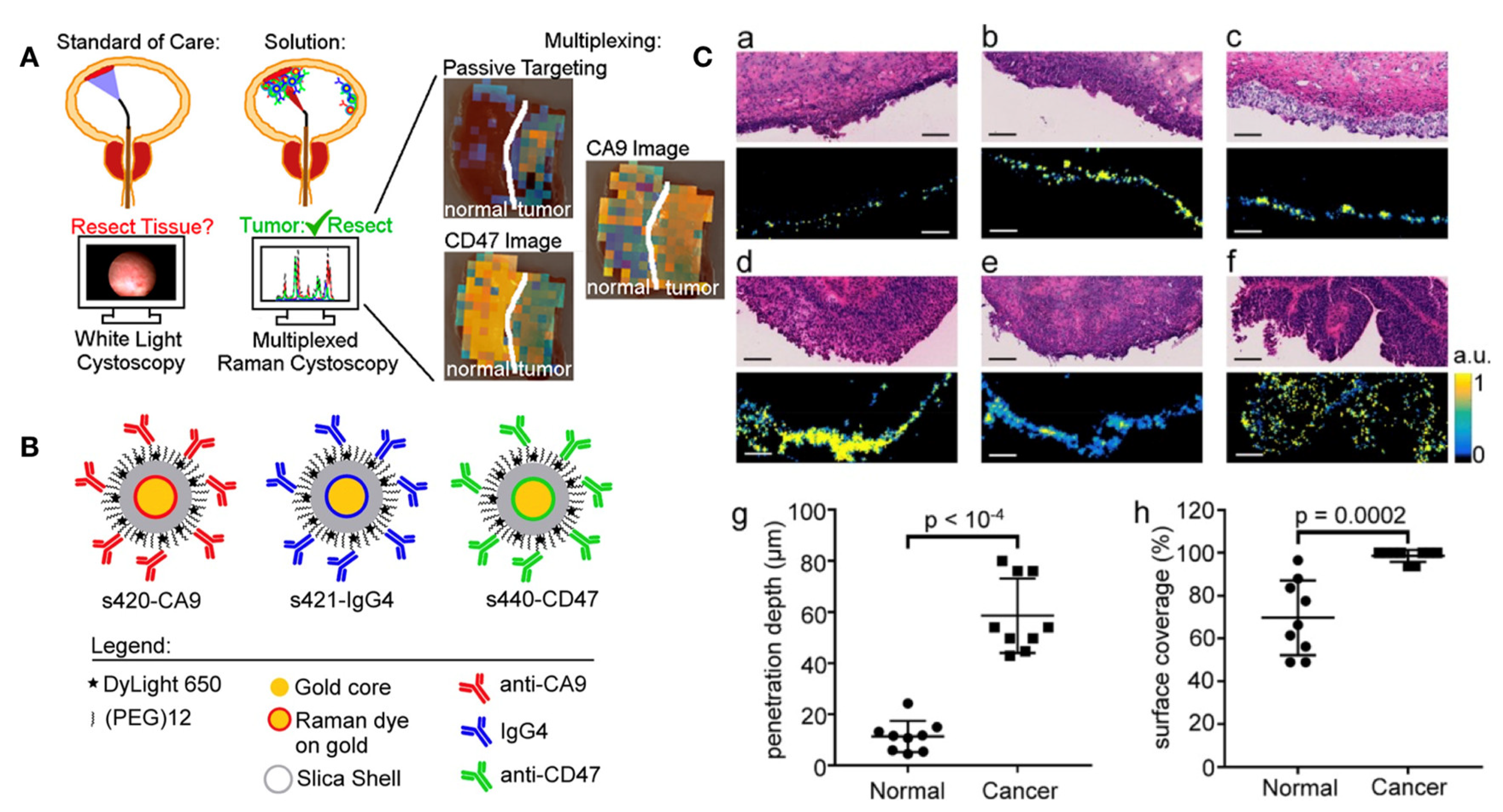
  77. Davis, R.M.; Kiss, B.; Trivedi, D.R.; Metzner, T.J.; Liao, J.C.; Gambhir, S.S. Surface-Enhanced Raman Scattering Nanoparticles for Multiplexed Imaging of Bladder Cancer Tissue Permeability and Molecular Phenotype. ACS Nano 2018, 12, 9669–9679. [Google Scholar] [CrossRef]
  78. Cho, S.K.; Su, L.-J.; Mao, C.; Wolenski, C.D.; Flaig, T.W.; Park, W. Multifunctional nanoclusters of NaYF4:Yb3+,Er3+ upconversion nanoparticle and gold nanorod for simultaneous imaging and targeted chemotherapy of bladder cancer. Mater. Sci. Eng. C 2018, 97, 784–792. [Google Scholar] [CrossRef]
  79. Ma, Y.; Mao, G.; Zhong, Y.; Wu, G.; Wu, W.; Zhan, Y.; He, Z.; Huang, W. Highly sensitive ratiometric fluorescent paper sensor for the urine assay of cancer. Talanta 2018, 194, 199–204. [Google Scholar] [CrossRef]
  80. Gennari, A.; Sun, Z.; Hasler-Strub, U.; Colleoni, M.; Kennedy, M.; Von Moos, R.; Cortés, J.; Vidal, M.; Hennessy, B.; Walshe, J.; et al. A randomized phase II study evaluating different maintenance schedules of nab-paclitaxel in the first-line treatment of metastatic breast cancer: Final results of the IBCSG 42-12/BIG 2-12 SNAP trial. Ann. Oncol. 2017, 29, 661–668. [Google Scholar] [CrossRef]
  81. Gradishar, W.J.; Krasnojon, D.; Cheporov, S.; Makhson, A.N.; Manikhas, G.M.; Clawson, A.; Bhar, P. Significantly Longer Progression-Free Survival With nab-Paclitaxel Compared With Docetaxel As First-Line Therapy for Metastatic Breast Cancer. J. Clin. Oncol. 2009, 27, 3611–3619. [Google Scholar] [CrossRef] [PubMed]
  82. Grivas, P.D.; Hussain, M.; Hafez, K.; Daignault-Newton, S.; Wood, D.; Lee, C.T.; Weizer, A.; Montie, J.E.; Hollenbeck, B.; Montgomery, J.S.; et al. A Phase II Trial of Neoadjuvant nab-paclitaxel, Carboplatin, and Gemcitabine (ACaG) in Patients With Locally Advanced Carcinoma of the Bladder. Urology 2013, 82, 111–117. [Google Scholar] [CrossRef] [PubMed]
  83. Ko, Y.-J.; Canil, C.M.; Mukherjee, S.D.; Winquist, E.; Elser, C.; Eisen, A.; Reaume, M.N.; Zhang, L.; Sridhar, S.S. Nanoparticle albumin-bound paclitaxel for second-line treatment of metastatic urothelial carcinoma: A single group, multicentre, phase 2 study. Lancet Oncol. 2013, 14, 769–776. [Google Scholar] [CrossRef]
  84. Hu, B.; Yan, Y.; Tong, F.; Xu, L.; Zhu, J.; Xu, G.; Shen, R. Lumbrokinase/paclitaxel nanoparticle complex: Potential therapeutic applications in bladder cancer. Int. J. Nanomed. 2018, 13, 3625–3640. [Google Scholar] [CrossRef]
  85. Pan, A.; Zhang, H.; Li, Y.; Lin, T.-Y.; Wang, F.; Lee, J.; Cheng, M.; Dall’Era, M.; Li, T.; White, R.D.; et al. Disulfide-crosslinked nanomicelles confer cancer-specific drug delivery and improve efficacy of paclitaxel in bladder cancer. Nanotechnology 2016, 27, 425103. [Google Scholar] [CrossRef]
  86. Liu, Y.; Wang, R.; Hou, J.; Sun, B.; Zhu, B.; Qiao, Z.; Su, Y.; Zhu, X. Paclitaxel/Chitosan Nanosupensions Provide Enhanced Intravesical Bladder Cancer Therapy with Sustained and Prolonged Delivery of Paclitaxel. ACS Appl. Bio Mater. 2018, 1, 1992–2001. [Google Scholar] [CrossRef]
  87. Xiao, T.; Xiao, Y.; Wang, W.; Tang, Y.Y.; Xiao, Z.; Su, M. Targeting EphA2 in cancer. J. Hematol. Oncol. 2020, 13, 1–17. [Google Scholar] [CrossRef]
  88. Kamoun, W.S.; Kirpotin, D.B.; Huang, Z.R.; Tipparaju, S.K.; Noble, C.O.; Hayes, M.E.; Luus, L.; Koshkaryev, A.; Kim, J.; Olivier, K.; et al. Antitumour activity and tolerability of an EphA2-targeted nanotherapeutic in multiple mouse models. Nat. Biomed. Eng. 2019, 3, 264–280. [Google Scholar] [CrossRef]
  89. Kamoun, W.; Swindell, E.; Pien, C.; Luus, L.; Cain, J.; Pham, M.; Kandela, I.; Huang, Z.R.; Tipparaju, S.K.; Koshkaryev, A.; et al. Targeting EphA2 in Bladder Cancer Using a Novel Antibody-Directed Nanotherapeutic. Pharmaceutics 2020, 12, 996. [Google Scholar] [CrossRef]
  90. Manan, F.A.A.; Yusof, N.A.; Abdullah, J.; Mohammad, F.; Nurdin, A.; Yazan, L.S.; Khiste, S.K.; Al-Lohedan, H.A. Drug Release Profiles of Mitomycin C Encapsulated Quantum Dots–Chitosan Nanocarrier System for the Possible Treatment of Non-Muscle Invasive Bladder Cancer. Pharmaceutics 2021, 13, 1379. [Google Scholar] [CrossRef]
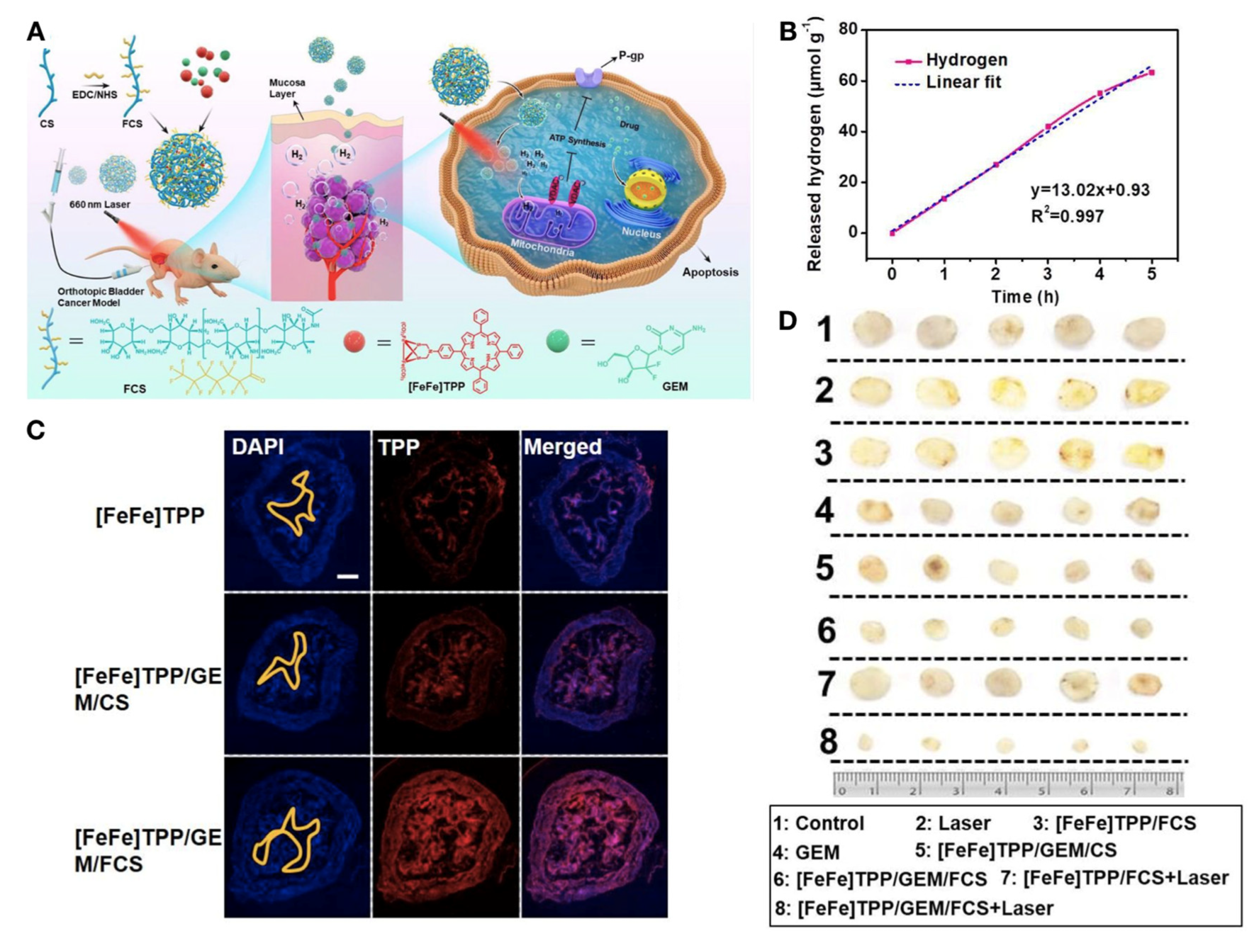
  91. Sun, R.; Liu, X.; Li, G.; Wang, H.; Luo, Y.; Huang, G.; Wang, X.; Zeng, G.; Liu, Z.; Wu, S. Photoactivated H2 Nanogenerator for Enhanced Chemotherapy of Bladder Cancer. ACS Nano 2020, 14, 8135–8148. [Google Scholar] [CrossRef] [PubMed]
  92. Qiu, X.; Cao, K.; Lin, T.; Chen, W.; Yuan, A.; Wu, J.; Hu, Y.; Guo, H. Drug delivery system based on dendritic nanoparticles for enhancement of intravesical instillation. Int. J. Nanomed. 2017, 12, 7365–7374. [Google Scholar] [CrossRef] [PubMed]
  93. Ding, K.; Wang, L.; Zhu, J.; He, D.; Huang, Y.; Zhang, W.; Wang, Z.; Qin, A.; Hou, J.; Tang, B.Z. Photo-Enhanced Chemotherapy Performance in Bladder Cancer Treatment via Albumin Coated AIE Aggregates. ACS Nano 2022, 16, 7535–7546. [Google Scholar] [CrossRef]
  94. Chang, Z.; Gao, M.; Zhang, W.; Song, L.; Jia, Y.; Qin, Y. Beta-elemene treatment is associated with improved outcomes of patients with esophageal squamous cell carcinoma. Surg. Oncol. 2017, 26, 333–337. [Google Scholar] [CrossRef]
  95. Zhai, B.; Zeng, Y.; Zeng, Z.; Zhang, N.; Li, C.; Zeng, Y.; You, Y.; Wang, S.; Chen, X.; Sui, X.; et al. Drug delivery systems for elemene, its main active ingredient β-elemene, and its derivatives in cancer therapy. Int. J. Nanomed. 2018, 13, 6279–6296. [Google Scholar] [CrossRef] [PubMed]
  96. Zhu, L.; Staley, C.; Kooby, D.; El-Rays, B.; Mao, H.; Yang, L. Current status of biomarker and targeted nanoparticle development: The precision oncology approach for pancreatic cancer therapy. Cancer Lett. 2016, 388, 139–148. [Google Scholar] [CrossRef]
  97. Zhai, E.A.B.; Chen, P.; Wang, W.; Liu, S.; Feng, J.; Duan, T.; Xiang, Y.; Zhang, R.; Zhang, M.; Han, X.; et al. An ATF24 peptide-functionalized β-elemene-nanostructured lipid carrier combined with cisplatin for bladder cancer treatment. Cancer Biol. Med. 2020, 17, 676–692. [Google Scholar] [CrossRef]
  98. Wang, K.-R.; Zhang, B.-Z.; Zhang, W.; Yan, J.-X.; Li, J.; Wang, R. Antitumor effects, cell selectivity and structure–activity relationship of a novel antimicrobial peptide polybia-MPI. Peptides 2008, 29, 963–968. [Google Scholar] [CrossRef]
  99. Oršolić, N. Bee venom in cancer therapy. Cancer Metastasis Rev. 2011, 31, 173–194. [Google Scholar] [CrossRef]
  100. Li, G.; Lei, Q.; Wang, F.; Deng, D.; Wang, S.; Tian, L.; Shen, W.; Cheng, Y.; Liu, Z.; Wu, S. Fluorinated Polymer Mediated Transmucosal Peptide Delivery for Intravesical Instillation Therapy of Bladder Cancer. Small 2019, 15, e1900936. [Google Scholar] [CrossRef]
  101. Xiong, Q.; Liu, A.; Ren, Q.; Xue, Y.; Yu, X.; Ying, Y.; Gao, H.; Tan, H.; Zhang, Z.; Li, W.; et al. Cuprous oxide nanoparticles trigger reactive oxygen species-induced apoptosis through activation of erk-dependent autophagy in bladder cancer. Cell Death Dis. 2020, 11, 1–13. [Google Scholar] [CrossRef] [PubMed]
  102. Folkes, L.K.; Wardman, P. Oxidative activation of indole-3-acetic acids to cytotoxic species— a potential new role for plant auxins in cancer therapy. Biochem. Pharmacol. 2001, 61, 129–136. [Google Scholar] [CrossRef]
  103. Pereira, F.M.; Melo, M.N.; Santos, K.M.; Oliveira, K.V.; Diz, F.M.; Ligabue, R.A.; Morrone, F.B.; Severino, P.; Fricks, A.T. Hyaluronic acid-coated chitosan nanoparticles as carrier for the enzyme/prodrug complex based on horseradish peroxidase/indole-3-acetic acid: Characterization and potential therapeutic for bladder cancer cells. Enzym. Microb. Technol. 2021, 150, 109889. [Google Scholar] [CrossRef]
  104. Zhang, D.; Sun, P.; Li, P.; Xue, A.; Zhang, X.; Zhang, H.; Jin, X. A magnetic chitosan hydrogel for sustained and prolonged delivery of Bacillus Calmette–Guérin in the treatment of bladder cancer. Biomaterials 2013, 34, 10258–10266. [Google Scholar] [CrossRef] [PubMed]
  105. Kato, T.; Bilim, V.; Yuuki, K.; Naito, S.; Yamanobe, T.; Nagaoka, A.; Yano, I.; Akaza, H.; Tomita, Y. Bacillus Calmette-Guerin and BCG cell wall skeleton suppressed viability of bladder cancer cells in vitro. Anticancer Res. 2010, 30, 4089–4096. [Google Scholar] [PubMed]
  106. Nakamura, T.; Fukiage, M.; Higuchi, M.; Nakaya, A.; Yano, I.; Miyazaki, J.; Nishiyama, H.; Akaza, H.; Ito, T.; Hosokawa, H.; et al. Nanoparticulation of BCG-CWS for application to bladder cancer therapy. J. Control. Release 2014, 176, 44–53. [Google Scholar] [CrossRef]
  107. Nakamura, T.; Fukiage, M.; Suzuki, Y.; Yano, I.; Miyazaki, J.; Nishiyama, H.; Akaza, H.; Harashima, H. Mechanism responsible for the antitumor effect of BCG-CWS using the LEEL method in a mouse bladder cancer model. J. Control. Release 2014, 196, 161–167. [Google Scholar] [CrossRef]
  108. Masuda, H.; Nakamura, T.; Noma, Y.; Harashima, H. Application of BCG-CWS as a systemic adjuvant by using nanoparticulation technology. Mol. Pharm. 2018, 15, 5762–5771. [Google Scholar] [CrossRef]
  109. Yoon, H.Y.; Yang, H.M.; Kim, C.H.; Goo, Y.T.; Hwang, G.Y.; Chang, I.H.; Whang, Y.M.; Choi, Y.W. Enhanced intracellular delivery of BCG cell wall skeleton into bladder cancer cells using liposomes functionalized with folic acid and pep-1 peptide. Pharmaceutics 2019, 11, 652. [Google Scholar] [CrossRef]
  110. Whang, Y.M.; Yoon, D.H.; Hwang, G.Y.; Yoon, H.; Park, S.I.; Choi, Y.W.; Chang, I.H. Liposome-Encapsulated Bacillus Calmette–Guérin Cell Wall Skeleton Enhances Antitumor Efficiency for Bladder Cancer In Vitro and In Vivo via Induction of AMP-Activated Protein Kinase. Cancers 2020, 12, 3679. [Google Scholar] [CrossRef]
  111. Joraku, A.; Homhuan, A.; Kawai, K.; Yamamoto, T.; Miyazaki, J.; Kogure, K.; Yano, I.; Harashima, H.; Akaza, H. Immunoprotection against murine bladder carcinoma by octaarginine-modified liposomes incorporating cell wall ofMycobacterium bovisbacillus Calmette-Guérin. Br. J. Urol. 2009, 103, 686–693. [Google Scholar] [CrossRef] [PubMed]
  112. Miyazaki, J.; Nishiyama, H.; Yano, I.; Nakaya, A.; Kohama, H.; Kawai, K.; Joraku, A.; Nakamura, T.; Harashima, H.; Akaza, H. The therapeutic effects of R8-liposome-BCG-CWS on BBN-induced rat urinary bladder carcinoma. Anticancer Res. 2011, 31, 2065–2071. [Google Scholar] [PubMed]
  113. Miyazaki, J.; Kawai, K.; Kojima, T.; Oikawa, T.; Joraku, A.; Shimazui, T.; Nakaya, A.; Yano, I.; Nakamura, T.; Harashima, H.; et al. The liposome-incorporating cell wall skeleton of Mycobacterium bovis bacillus Calmette-Guéin can directly enhance the susceptibility of cancer cells to lymphokine-activated killer cells through up-regulation of natural-killer group 2, member D ligands. Br. J. Urol. 2011, 108, 1520–1526. [Google Scholar] [CrossRef]
  114. Chen, Z.; Yu, T.; Zhou, B.; Wei, J.; Fang, Y.; Lu, J.; Guo, L.; Chen, W.; Liu, Z.-P.; Luo, J. Mg(II)-Catechin nanoparticles delivering siRNA targeting EIF5A2 inhibit bladder cancer cell growth in vitro and in vivo. Biomaterials 2015, 81, 125–134. [Google Scholar] [CrossRef] [PubMed]
  115. Groner, B.; Weiss, A. Targeting survivin in cancer: Novel drug development approaches. BioDrugs 2013, 28, 27–39. [Google Scholar] [CrossRef]
  116. García, D.M.; Manero-Rupérez, N.; Quesada, R.; Korrodi-Gregório, L.; Soto-Cerrato, V. Therapeutic strategies involving survivin inhibition in cancer. Med. Res. Rev. 2018, 39, 887–909. [Google Scholar] [CrossRef]
  117. Krafft, U.; Tschirdewahn, S.; Hess, J.; Harke, N.N.; Hadaschik, B.; Olah, C.; Krege, S.; Nyirády, P.; Szendröi, A.; Szücs, M.; et al. Validation of survivin and HMGA2 as biomarkers for cisplatin resistance in bladder cancer. Urol. Oncol. Semin. Orig. Investig. 2019, 37, 810.e7–810.e15. [Google Scholar] [CrossRef]
  118. Chen, L.; Liang, L.; Yan, X.; Liu, N.; Gong, L.; Pan, S.; Lin, F.; Zhang, Q.; Zhao, H.; Zheng, F. Survivin Status Affects Prognosis and Chemosensitivity in Epithelial Ovarian Cancer. Int. J. Gynecol. Cancer 2013, 23, 256–263. [Google Scholar] [CrossRef]
  119. Aliabadi, H.M.; Landry, B.; Mahdipoor, P.; Uludağ, H. Induction of Apoptosis by Survivin Silencing through siRNA Delivery in a Human Breast Cancer Cell Line. Mol. Pharm. 2011, 8, 1821–1830. [Google Scholar] [CrossRef]
  120. Arista-Romero, M.; Cascante, A.; Fornaguera, C.; Borrós, S. Role of Survivin in Bladder Cancer: Issues to Be Overcome When Designing an Efficient Dual Nano-Therapy. Pharmaceutics 2021, 13, 1959. [Google Scholar] [CrossRef]
  121. Muxika, A.; Etxabide, A.; Uranga, J.; Guerrero, P.; de la Caba, K. Chitosan as a bioactive polymer: Processing, properties and applications. Int. J. Biol. Macromol. 2017, 105, 1358–1368. [Google Scholar] [CrossRef] [PubMed]
  122. Martin, D.T.; Steinbach, J.M.; Liu, J.; Shimizu, S.; Kaimakliotis, H.Z.; Wheeler, M.A.; Hittelman, A.B.; Saltzman, W.M.; Weiss, R.M. Surface-Modified Nanoparticles Enhance Transurothelial Penetration and Delivery of Survivin siRNA in Treating Bladder Cancer. Mol. Cancer Ther. 2014, 13, 71–81. [Google Scholar] [CrossRef]
  123. Luo, J.-H.; Xie, D.; Liu, M.-Z.; Chen, W.; Liu, Y.-D.; Wu, G.-Q.; Kung, H.-F.; Zeng, Y.-X.; Guan, X.-Y. Protein expression and amplification of AIB1 in human urothelial carcinoma of the bladder and overexpression of AIB1 is a new independent prognostic marker of patient survival. Int. J. Cancer 2008, 122, 2554–2561. [Google Scholar] [CrossRef] [PubMed]
  124. Tong, Z.-T.; Wei, J.-H.; Zhang, J.-X.; Liang, C.-Z.; Liao, B.; Lu, J.; Fan, S.; Chen, Z.-H.; Zhang, F.; Ma, H.-H.; et al. AIB1 predicts bladder cancer outcome and promotes bladder cancer cell proliferation through AKT and E2F1. Br. J. Cancer 2013, 108, 1470–1479. [Google Scholar] [CrossRef] [PubMed]
  125. Wei, J.; Cheang, T.; Tang, B.; Xia, H.; Xing, Z.; Chen, Z.; Fang, Y.; Chen, W.; Xu, A.; Wang, S.; et al. The inhibition of human bladder cancer growth by calcium carbonate/CaIP6 nanocomposite particles delivering AIB1 siRNA. Biomaterials 2013, 34, 1246–1254. [Google Scholar] [CrossRef]
  126. Su, S.; Wang, J.; Vargas, E.; Wei, J.; Martinez-Zaguilan, R.; Sennoune, S.R.; Pantoya, M.L.; Wang, S.; Chaudhuri, J.; Qiu, J. Porphyrin Immobilized Nanographene Oxide for Enhanced and Targeted Photothermal Therapy of Brain Cancer. ACS Biomater. Sci. Eng. 2016, 2, 1357–1366. [Google Scholar] [CrossRef]
  127. Cao, Y.; Dong, H.; Yang, Z.; Zhong, X.; Chen, Y.; Dai, W.; Zhang, X. Aptamer-Conjugated Graphene Quantum Dots/Porphyrin Derivative Theranostic Agent for Intracellular Cancer-Related MicroRNA Detection and Fluorescence-Guided Photothermal/Photodynamic Synergetic Therapy. ACS Appl. Mater. Interfaces 2016, 9, 159–166. [Google Scholar] [CrossRef]
  128. Menilli, L.; Monteiro, A.R.; Lazzarotto, S.; Morais, F.M.P.; Gomes, A.T.P.C.; Moura, N.M.M.; Fateixa, S.; Faustino, M.A.F.; Neves, M.G.P.M.S.; Trindade, T.; et al. Graphene Oxide and Graphene Quantum Dots as Delivery Systems of Cationic Porphyrins: Photo-Antiproliferative Activity Evaluation towards T24 Human Bladder Cancer Cells. Pharmaceutics 2021, 13, 1512. [Google Scholar] [CrossRef]
  129. Wang, S.; Jin, S.; Li, G.; Xu, M.; Deng, D.; Xiao, Z.; Sun, H.; Zhang, S.; Zhang, E.; Xie, L.; et al. Transmucosal Delivery of Self-Assembling Photosensitizer–Nitazoxanide Nanocomplexes with Fluorinated Chitosan for Instillation-Based Photodynamic Therapy of Orthotopic Bladder Tumors. ACS Biomater. Sci. Eng. 2021, 7, 1485–1495. [Google Scholar] [CrossRef]
  130. Li, G.; Yuan, S.; Deng, D.; Ou, T.; Li, Y.; Sun, R.; Lei, Q.; Wang, X.; Shen, W.; Cheng, Y.; et al. Fluorinated Polyethylenimine to Enable Transmucosal Delivery of Photosensitizer-Conjugated Catalase for Photodynamic Therapy of Orthotopic Bladder Tumors Postintravesical Instillation. Adv. Funct. Mater. 2019, 29, 1901932. [Google Scholar] [CrossRef]
  131. Tan, P.; Cai, H.; Wei, Q.; Tang, X.; Zhang, Q.; Kopytynski, M.; Yang, J.; Yi, Y.; Zhang, H.; Gong, Q.; et al. Enhanced chemo-photodynamic therapy of an enzyme-responsive prodrug in bladder cancer patient-derived xenograft models. Biomaterials 2021, 277, 121061. [Google Scholar] [CrossRef] [PubMed]
  132. Yi, X.; Hu, J.-J.; Dai, J.; Lou, X.; Zhao, Z.; Xia, F.; Tang, B.Z. Self-Guiding Polymeric Prodrug Micelles with Two Aggregation-Induced Emission Photosensitizers for Enhanced Chemo-Photodynamic Therapy. ACS Nano 2021, 15, 3026–3037. [Google Scholar] [CrossRef] [PubMed]
  133. Wang, Y.; Wei, G.; Zhang, X.; Xu, F.; Xiong, X.; Zhou, S. A Step-by-Step Multiple Stimuli-Responsive Nanoplatform for Enhancing Combined Chemo-Photodynamic Therapy. Adv. Mater. 2017, 29, 1605357. [Google Scholar] [CrossRef] [PubMed]
  134. Cha, J.-H.; Yang, W.-H.; Xia, W.; Wei, Y.; Chan, L.-C.; Lim, S.-O.; Li, C.-W.; Kim, T.; Chang, S.-S.; Lee, H.-H.; et al. Metformin Promotes Antitumor Immunity via Endoplasmic-Reticulum-Associated Degradation of PD-L1. Mol. Cell 2018, 71, 606–620.e7. [Google Scholar] [CrossRef]
  135. Munoz, L.E.; Huang, L.; Bommireddy, R.; Sharma, R.; Monterroza, L.; Guin, R.N.; Samaranayake, S.G.; Pack, C.D.; Ramachandiran, S.; Reddy, S.J.; et al. Metformin reduces PD-L1 on tumor cells and enhances the anti-tumor immune response generated by vaccine immunotherapy. J. Immunother. Cancer 2021, 9, e002614. [Google Scholar] [CrossRef]
  136. Chin, Y.-C.; Yang, L.-X.; Hsu, F.-T.; Hsu, C.-W.; Chang, T.-W.; Chen, H.-Y.; Chen, L.Y.-C.; Chia, Z.C.; Hung, C.-H.; Su, W.-C.; et al. Iron oxide@chlorophyll clustered nanoparticles eliminate bladder cancer by photodynamic immunotherapy-initiated ferroptosis and immunostimulation. J. Nanobiotechnology 2022, 20, 1–18. [Google Scholar] [CrossRef]
  137. Zhang, S.; Li, G.; Deng, D.; Dai, Y.; Liu, Z.; Wu, S. Fluorinated Chitosan Mediated Synthesis of Copper Selenide Nanoparticles with Enhanced Penetration for Second Near-Infrared Photothermal Therapy of Bladder Cancer. Adv. Ther. 2021, 4, 2100043. [Google Scholar] [CrossRef]
  138. Ni, W.; Li, M.; Cui, J.; Xing, Z.; Li, Z.; Wu, X.; Song, E.; Gong, M.; Zhou, W. 808 nm light triggered black TiO2 nanoparticles for killing of bladder cancer cells. Mater. Sci. Eng. C 2017, 81, 252–260. [Google Scholar] [CrossRef]
  139. Self-Assembled Nanoparticle-Mediated Chemophototherapy Reverses the Drug Resistance of Bladder Cancers through Dual AKT/ERK Inhibition. Adv. Ther. 2020, 3, 2000032. [CrossRef]
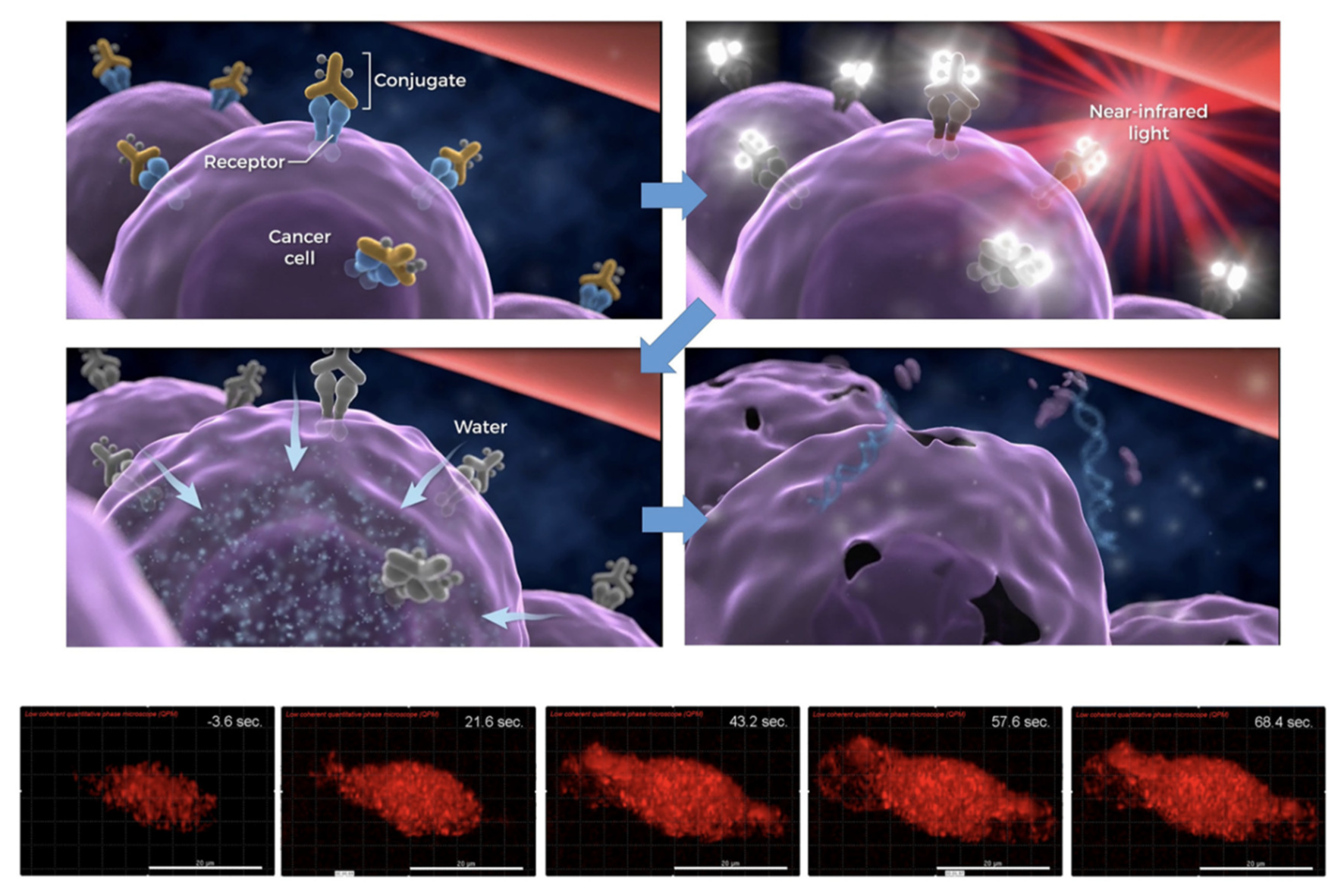
  140. Kobayashi, H.; Choyke, P.L. Near-Infrared Photoimmunotherapy of Cancer. Accounts Chem. Res. 2019, 52, 2332–2339. [Google Scholar] [CrossRef]
  141. Wu, X.; Wei, Y.; Lin, R.; Chen, P.; Hong, Z.; Zeng, R.; Xu, Q.; Li, T. Multi-responsive mesoporous polydopamine composite nanorods cooperate with nano-enzyme and photosensitiser for intensive immunotherapy of bladder cancer. Immunology 2022. early view. [Google Scholar] [CrossRef] [PubMed]
  142. Guo, P.; Wang, L.; Shang, W.; Chen, J.; Chen, Z.; Xiong, F.; Wang, Z.; Tong, Z.; Wang, K.; Yang, L.; et al. Intravesical In Situ Immunostimulatory Gel for Triple Therapy of Bladder Cancer. ACS Appl. Mater. Interfaces 2020, 12, 54367–54377. [Google Scholar] [CrossRef] [PubMed]
  143. Kiss, B.; Berg, N.S.V.D.; Ertsey, R.; McKenna, K.; Mach, K.E.; Zhang, C.A.; Volkmer, J.-P.; Weissman, I.L.; Rosenthal, E.L.; Liao, J.C. CD47-Targeted Near-Infrared Photoimmunotherapy for Human Bladder Cancer. Clin. Cancer Res. 2019, 25, 3561–3571. [Google Scholar] [CrossRef] [PubMed] [Green Version]
  144. Lin, T.; Yuan, A.; Zhao, X.; Lian, H.; Zhuang, J.; Chen, W.; Zhang, Q.; Liu, G.; Zhang, S.; Cao, W.; et al. Self-assembled tumor-targeting hyaluronic acid nanoparticles for photothermal ablation in orthotopic bladder cancer. Acta Biomater. 2017, 53, 427–438. [Google Scholar] [CrossRef] [PubMed]
  145. Yumita, N.; Nishigaki, R.; Umemura, K.; Umemura, S.-I. Synergistic Effect of Ultrasound and Hematoporphyrin on Sarcoma 180. Jpn. J. Cancer Res. 1990, 81, 304–308. [Google Scholar] [CrossRef]
  146. Li, G.; Wang, S.; Deng, D.; Xiao, Z.; Dong, Z.; Wang, Z.; Lei, Q.; Gao, S.; Huang, G.; Zhang, E.; et al. Fluorinated Chitosan To Enhance Transmucosal Delivery of Sonosensitizer-Conjugated Catalase for Sonodynamic Bladder Cancer Treatment Post-intravesical Instillation. ACS Nano 2020, 14, 1586–1599. [Google Scholar] [CrossRef]
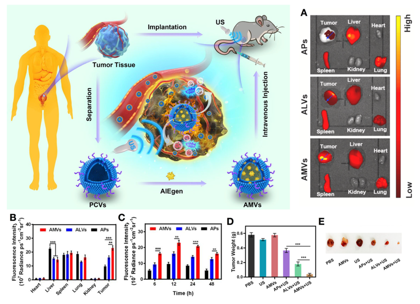
  147. Duo, Y.; Zhu, D.; Sun, X.; Suo, M.; Zheng, Z.; Jiang, W.; Tang, B.Z. Patient-derived microvesicles/AIE luminogen hybrid system for personalized sonodynamic cancer therapy in patient-derived xenograft models. Biomaterials 2021, 272, 120755. [Google Scholar] [CrossRef]
Figure 1. The stage and grade of bladder cancer. Bladder cancer is divided into five stages according to the degree of progression: T0 (Ta: non-invasive papilla-carcinoma and Tis: tumor in situ); T1 (Tumor invades the bladder mucosa); T2 (Tumor invades the bladder inner muscle); T3 (Tumor invades the bladder outer muscle); and T4 (Tumor metastasis). Non-muscle-invasive bladder cancer (NMIBC) refers to T0 and T1 stages, while muscle-invasive bladder cancer (MIBC) refers to ≥T2 stages.
Biosensors 12 00796 g001
Figure 2. Current strategies for the diagnosis of bladder cancer.
Biosensors 12 00796 g002
Figure 3. Current therapies of bladder cancer. TURBT: transurethral resection of the bladder cancer; BCG: bacillus Calmette-Guerin; CT: computer tomography.
Biosensors 12 00796 g003
Figure 4. Surface-enhanced Raman nanoparticle could simultaneously carry a variety of antibodies which enabled the multiplexed detection of multiple molecular targets. (A) Surface-enhanced Raman nanoparticle endoscope system with multiplexed molecular imaging of CD47 and CA9 enabled to classification of bladder tissue as normal or cancerous. Multiplexed molecular imaging of CD47 and Carbonic Anhydrase 9 tumor proteins. (B) The antibodies bound different color surface-enhanced Raman scattering (SERS) nanoparticles. The blue IgG4 nanoparticle is used as a negative experimental control for the active binding of CA9- and CD47-targeted SERS nanoparticles. (C) H&E stains (top) and s421-IgG4 Raman images (bottom) of normal bladder tissue (ac) and different grade of bladder cancer tissue (df). (gh): Passively targeted nanoparticles penetrated 5-fold deeper and bound to tumor tissue at 3.3-fold higher concentrations in cancer compared to normal bladder urothelium. Reprinted with permission from ref. [77]. Copyright 2018, ACS.
Biosensors 12 00796 g004
Figure 5. (A) A photoactivated H2 nanogenerator that comprises an FCS, gemcitabine, and a catalyst of H2 production ([FeFe]TPP). Under laser irradiation of 660 nm, the hydrogen catalyst could effectively generate non-toxic hydrogen which could enhance the effect of gemcitabine. (B) The gel could release hydrogen gradually. (C) After instillation with each formulation, the bladder frozen section experiment as performed (([FeFe]TPP): red fluorescence). ([FeFe]TPP)/GEM/FCS possessed a good tissue penetration ability. (D) The T24 cells were planted into the bladders of nude mice. After 4 weeks of treatment, the tumors were harvested. The tumors dramatically shrunk after treatment with ([FeFe]TPP)/GEM/FCS and the laser. Reprinted with permission from ref. [91]. Copyright 2020, ACS. FCS: Fluorinated chitosan; GEM: gemcitabine; TPP: tetraphenylporphyrin; FeFe: a hydrogenase subsite analogue [Fe2S2(CO)6].
Biosensors 12 00796 g005
Figure 6. The diagram of near-infrared photoimmunotherapy (NIR-PIT). The targeted photosensitizer can better distinguish tumor cells therefore PIT can destroy tumor cells more selectively under the NIR laser. Reprinted permission from ref. [140]. Copyright 2019, ACS.
Biosensors 12 00796 g006
Figure 7. Multiple-function AuNRs&IONs@Gel can target bladder tumors and perform accurate PTT with the help of nano-particle imaging. In addition, the ion can induce iron death as well as modulate TME, enhancing the curative effect. (A) Schematic illustration of how the AuNRs&IONs@Gel was synthesized. (B, C) AuNRs&IONs@Gel-induced ICD through triple therapy. Reprinted with permission from ref. [142]. Copyright 2020, ACS.
Biosensors 12 00796 g007
Figure 8. A patient-derived MVs/AIEgen hybrid system (AMVs) for personalized SDT in bladder cancer patient-derived xenograft (PDX) models. (A) Ex vivo images of excised organs and tumors of mice bearing PDX were examined at 12 h post-injection of APs, ALVs, and AMVs. (B) Fluorescence distribution profile on the specified blue arrows in tumors of (A). (C) Quantification of the time-dependent fluorescence intensity at the tumor site after various treatments. (D) Tumor weight and (E) tumor tissues were collected from the PDX mice after various treatments. APs, ALVs, and AMVs: PLGA/AIEgen nanoparticles; ALVs: MVs/AIEgen derived from a cell line. ** p < 0.01; *** p < 0.001. AMVs: patient-derived MVs/AIEgen hybrid system. Reprinted with the permission from ref. [147]. Copyright 2021, Elsevier. MVs: microvesicles; PDX: patient-derived xenograft; SDT: sonodynamic therapy; APs: PLGA/DCPy hybrid nanoparticle; ALVs: T24 cell-derived MVs/AIEgen hybrid system; AMVs: patient-derived MVs/AIEgen hybrid system; US: ultrasound.
Biosensors 12 00796 g008
Table 1. FDA-approved biomarkers.
Biomarkers and ManufacturerDetected BiomarkersAssay TypeSpecimenSensitivity (CI 95%)Specificity (CI 95%)Ref.
NMP22
(Matritech, Inc., Alere, Jena, Thuringia, Germany)
Nuclear mitotic apparatus proteinsELISAUrine62–75%70–83%[31]
NMP22
(Matritech, Inc., Alere, Jena, Thuringia, Germany)
Nuclear mitotic apparatus proteinsPoint- of-care test Urine52–59%87–89%[31]
BTA Stat
(Polymedco, Cortlandt, NY, USA)
Complement factor H-related protein and complement factor HPoint-of-care test Urine58–69%73–81%[31]
BTA TRAK
(Polymedco, Cortlandt, NY, USA)
Complement factor H-related protein and complement factor HELISAUrine54–75%64–82%[31]
UroVysion
(Abbott Vysis, Chicgo, Illinois, USA)
Alterations in chromosomes 3, 7, 17, and 9p21FISHUrine65–84%78–92%[32]
uCyt+/Immunocyt
(Scimedx, Inc., Dover, New Jersey, USA)
Bladder tumor cell associated mucins/carcinoembryonic antigenImmunocytochemistryUrine78–90%77–87%[31]
ELISA: enzyme-linked immunosorbent assay; FISH: fluorescence in situ hybridization.
Table 2. Nano-prognostic methods for bladder cancer.
NanomatierialsDetect TargetPropertiesSensitivity (CI 95%)Specificity (CI 95%)ApplcationsRef.
QD625CD47High sensitivity and specificity.82.9%90.5%Targeted fluorescent probe for cystoscope[70]
QD605PSCASpecifically targets BC cells and emits stable and long duration fluorescent--Targeted fluorescent probe for cystoscope[73]
CdSe/ZnS QDCarbonic anhydraseWell biocompatibility and dispersion.--Targeted fluorescent probe for cystoscope[74,75]
Heteroatom-doped graphene QDHaaseEmits white light and broad excitation-dependent full-color photoluminescence from 463 nm to 672 nm.--Targeted fluorescent probe for cystoscope[76]
Surface-enhanced Raman scattering nanoparticleCarbonic anhydrase9, CD47Multiple targets and imaging(ROC AUC: 0.95)-Targeted fluorescent probe for Raman endoscopy[77]
UCNPEGFRWell ability of tissue penetration--NIR probe and imaging system[78]
Si QDs/HA-δ-FeOOHHaaseDetection limit for Haase: 0.02 ng/mL (based on 3σ/S). RSD < 3% (Compared with ELISA method)Detection limit: 0.02 ng/mL-Fluorescence platform for urine test[76]
Rox-DNA functionalized QDTelomeraseEnabled visual semi-quantitative detection with naked eye. The detection limit was 10 cells and response time was within an hour.--Sensitive ratiometric fluorescence paper sensor[79]
CI: confidence interval; ROC: receiver operating characteristic; AUC: area under curve.
Table 3. Clinical trials for bladder cancer with nano-formulations in the United States.
NanoparticleTherapeutic AgentsConditionSponsor/CollaborationsStatesStudy StartNCT Number
Paclitaxel albumin-stabilized nanoparticle (Nab-paclitaxel)PTXRecurrent BC; Stage IV BCMayo Clinic/NCIPhase 2 (Withdrawn)June 2016NCT02718742
Paclitaxel albumin-stabilized nanoparticle (Nab-paclitaxel)PTXBladder cancerUniversity of Michigan Rogel Cancer Center/Celgene CorporationPhase 2December 2007NCT00585689
PLZ4-coated paclitaxel-loaded micelles (PPM)PTXNMIBCVA Office of Research and Development/University of California, DavisPhase 1 (Not yet recruiting)-NCT05519241
BC: bladder cancer; NCI: National Cancer Institute.
Table 4. Nano-formulations for chemotherapy.
NanoparticleSize (nm)Therapeutic AgentsLoading EfficiencyPropertiesApplicationRef.
Nab-paclitaxel150–200PTX10%Low side-effects; good solubility and biocompatibilityVein injection[80]
LK/PTX/PEGb- (PELG-g-(PZLL-r-PLL))89 ± 3LK, PTXLK (6.74%), PTX (4.13%)Increasing of the half-life and bioavailability of the drugsAbdominal subcutaneous injection[84]
DC-PNM-PTX23 ± 6PTX>99%Specifically targeting the bladder cancer PDXs; improvement of the cisplatin resistance; GSH-responsive releaseTail vein injection[85]
PTX/CS NSs194.48 ± 86.24PTX81.4%Attaching to mucosa of the bladder through electrostatic adsorptionIntravesical instillation[86]
EphA2-ILs-DTXp110 ± 10DTX prodrug90–99%Specific targeting to tumor; improvement of penetration;
minimal haematological toxicity
Tail vein injection[88,89]
MMC@CS -Mn:ZnS175MMC44.52 ± 1.05%Long retention time-[90]
[FeFe]TPP/GEM/FCS NPs220GEM; [FeFe]TPPGEM (6.9%); [FeFe]TPP (7.7%)Improvement of penetration capacity; H2 generation under 660nm laser irradiation; inhibition of drug transport capacity of cancer cellsIntravesical instillation[91]
PEG-PAMAM-DOX13DOX-pH-responsive releaseIntravesical instillation[92]
BITT@BSA-DSP70.2 ± 22.0DSP35%Visible drug delivery; photodynamic and photothermal effectIntravesical instillation[93]
ATF24-PEG-Lipo-β-E79.32 ± 1.282β-E98.37%Specific targeting to tumorIntravesical instillation[97]
MPI/F-PEI NPs260.67 ± 6.62MPI-Improved cross-membrane and transmucosal penetrationIntravesical instillation[100]
CONPs40~110CONPs-Activation of ERK-dependent autophagy; synergistic effect with chemo drugs. Intravesical instillation and in situ injection[101]
IAA-CS/HA NP and HRP-CS/HA NP170~200HRP, IAABoth > 90%Enzyme/prodrug system.In vitro (T24)[103]
Table 5. Nano-formulations for immune therapy.
NanoparticleSize (nm)Therapeutic AgentsLoading EfficiencyPropertiesApplicationRef.
Fe3O4-BCG-CS/GP gel-BCG1% (w/v)Response to magnetic field control; long retention timeIntravesical instillation[104]
CWS-NP/LEEL166BCG-CWS57%Good water solubilityIntravesical instillation[106,107]
CWS-FPL<200BCG-CWS60%Improvement of tumor targeting by folic acid; improvement of penetration by Pep-1 peptideIntravesical instillation[109]
R8-liposome-BCG-CW230BCG-CWS-Improvement of cell binding and internalizationIntravesical instillation[110]
Table 6. Nano-formulations for targeted therapy.
NanoparticleSize (nm)Therapeutic AgentsLoading EfficiencyPropertiesApplicationRef.
Mg(II)-Cat/siEIF5A210-20Catechin; siEIF5A2-Good biocompatibility and cellular uptake; inhibition of oncogene eukaryotic translation initiation factorTail vein injection[114]
Anti-survivin siRNA-1 pbae-NP150Survivin siRNA100%No synergistic effect with PTXIn virto (T24, RT4)[110]
NP-siSUR-CH2.5137 ± 51Survivin siRNA70%Long release time of sirnaIn situ injection[121,122]
NP-ACC/caip6/siAIB180–200siAIB1-Well ability of lysosome escape; good biocompatibilityIn situ injection[125]
Table 7. Nano-formulations for light-based therapy.
NanoparticleResponsive PartSize (nm)Therapeutic AgentsPropertiesApplicationRef.
Zn-TMPyP@GQDsAn-TMPyP, GQDs28.4-Blue light-responsive; good stability of porphyrins in aqueous solutions; multiple targets binding sites and possible photothermal effectIn vitro (T24)[128]
HSA-Ce6/NTZ/FCSCe6192NTZImprovement of tumor hypoxia and drug transmucosal deliveryIntravesical instillation[129]
CAT-Ce6/F-PEICe6220.3-Improvement of tumor hypoxia by catalase and drug transmucosal deliveryIntravesical instillation[130]
Poly (OEGMA)-PTX@Ce6 (NPs@Ce6)Ce6168.2 ± 1.12Polymer-PTX prodrugCombination of PCI effect and enhanced chemo-PDT In situ injection[131,132,133]
IR775@Met@LipIR775-MetforminImprovement of tumor hypoxia; down-regulate PD-L1Intravesical instillation[69]
Fe3O4@Chl/Fe CNPsChl/Fe12.8 ± 4.8-Photodynamic immunotherapy-initiated ferroptosis and immune stimulation.Intravesical instillation[136]
FCS-Cu2-xSeCu2-xSe30.1-Improvement of drug transmucosal delivery; NIR-II-responsiveIntravesical instillation[137]
Black TiO2 NPsTiO220–30-Absorption of visible light and near in-
frared
In vitro (T24)[138]
PhDPheophorbide a71DOXCombination of PDT, PTT and DOX; pH and NIR-responsive.Tail vein injection[139]
MPDIαWICG, MnO2120PD-L1 antibodyCombination of PTT and immunotherapy; specific adherence to bladder cancer cell; pH-responsiveIntravesical instillation[141]
AuNRs&IONs@GelAuNrs80–120Iron oxide nanoparticlesCombination of PTT, iron death, and macrophages re-polarization; targeting deliveryIn situ injection[142]
Anti-CD47-IR700IR700--Targeting delivery; long retention timeTail vein injection[143]
HA-IR780 NPsIR780171.3-Targeting delivery; good bioavailability and biocompatibilityTail vein injection[144]
Table 8. Nano-formulations for sonodynamic therapy.
NanoparticleResponsive PartSize (nm)Therapeutic AgentsPropertiesApplicationRef.
CAT-TCPP/FCS NPsTCPP190 ± 12-Improvement of tumor hypoxia by catalase and drug transmucosal deliveryIntravesical instillation[146]
AMVsAIEgen300-Good internalization and personalized tumor targeting abilityTail vein injection[147]
Publisher’s Note: MDPI stays neutral with regard to jurisdictional claims in published maps and institutional affiliations.

