Biosensors for the Detection of Enzymes Based on Aggregation-Induced Emission


Abstract
:1. Introduction
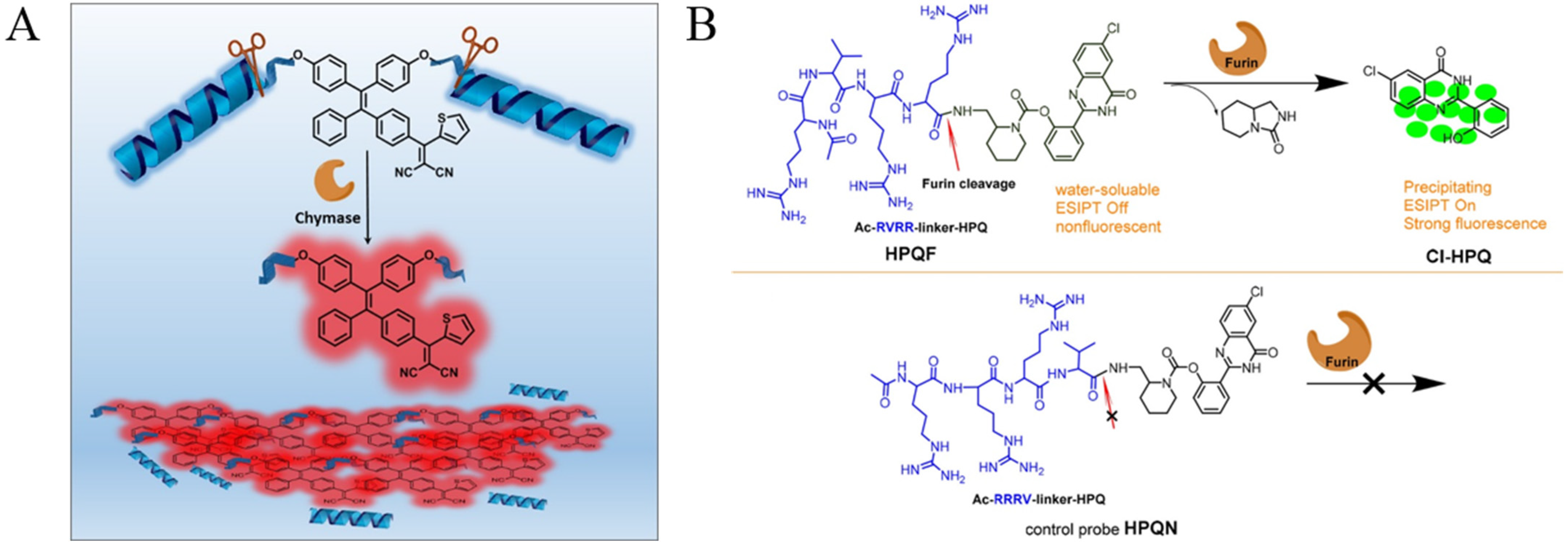
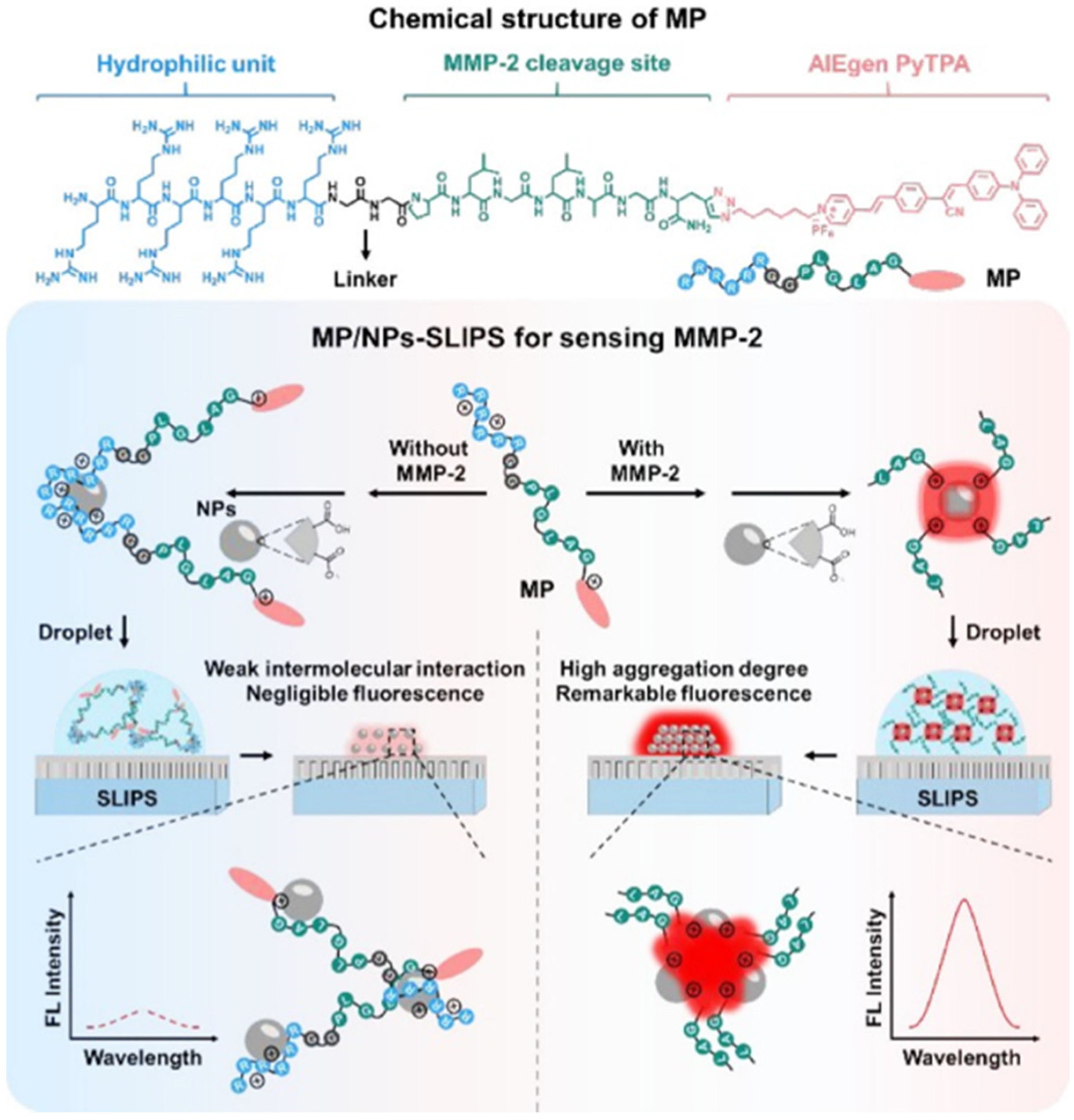
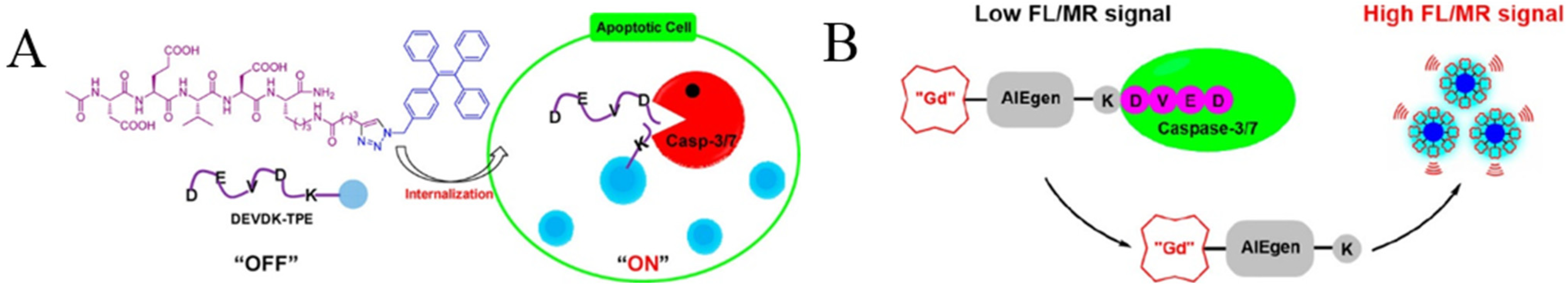
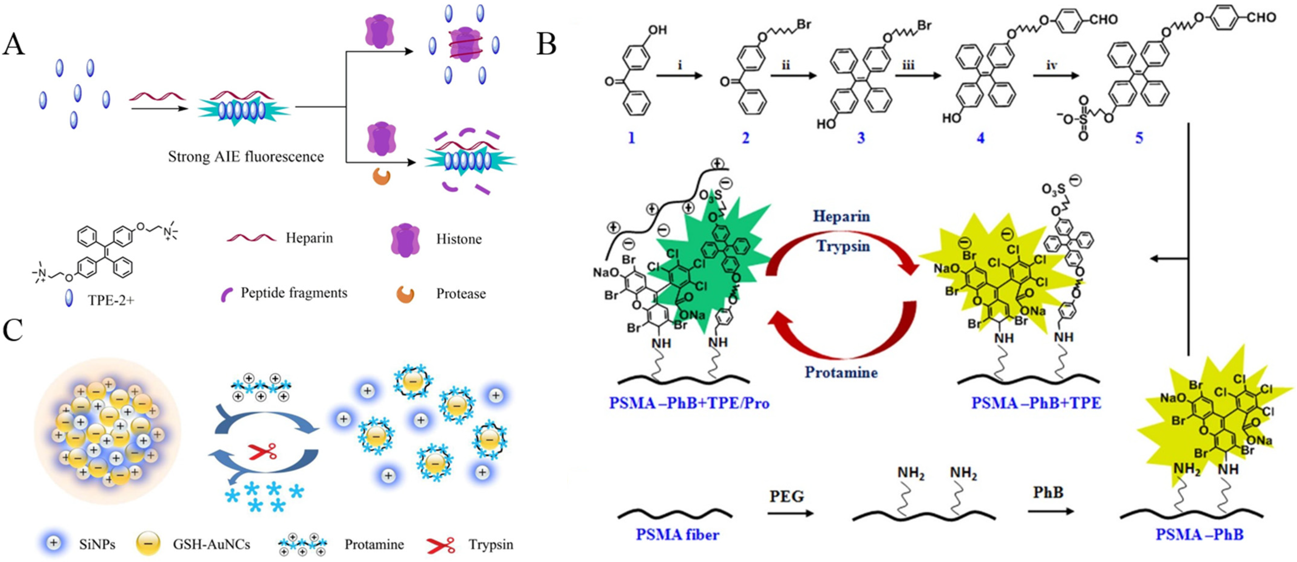
2. Proteases
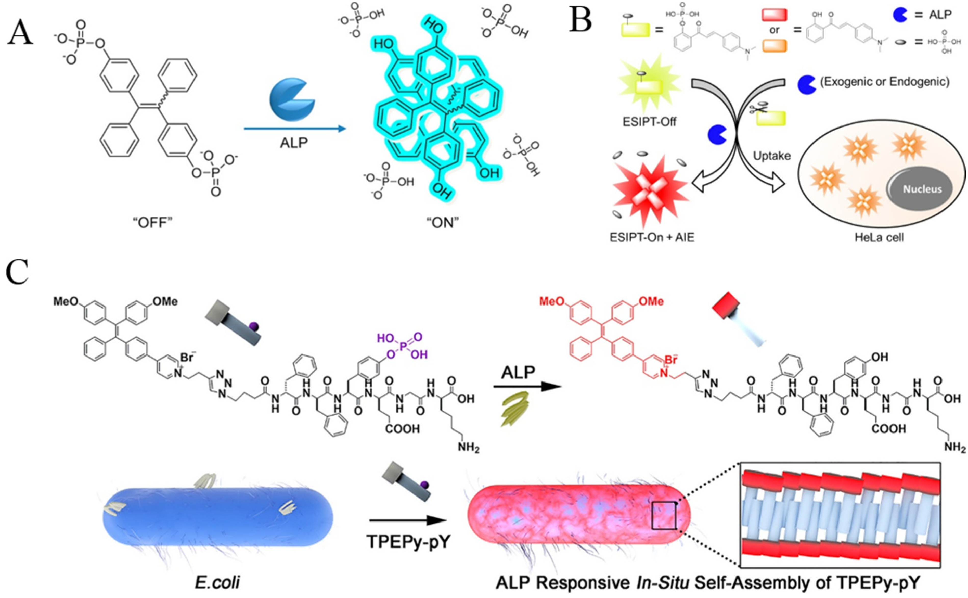
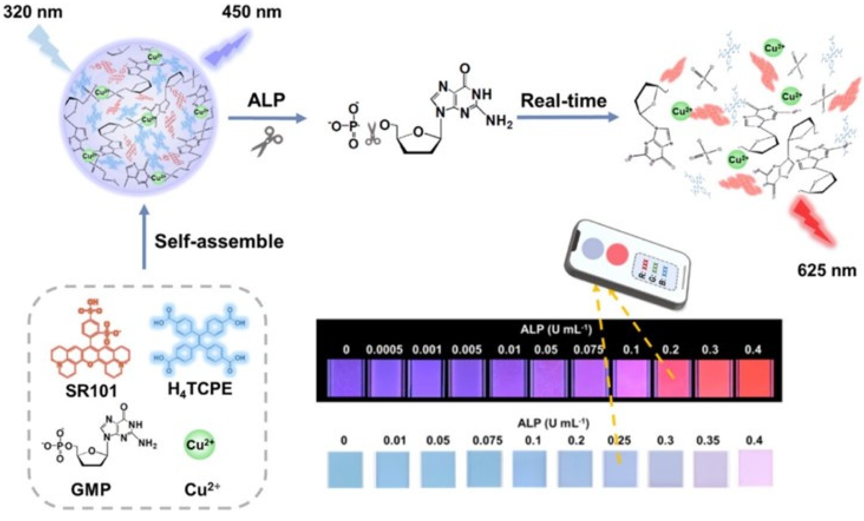
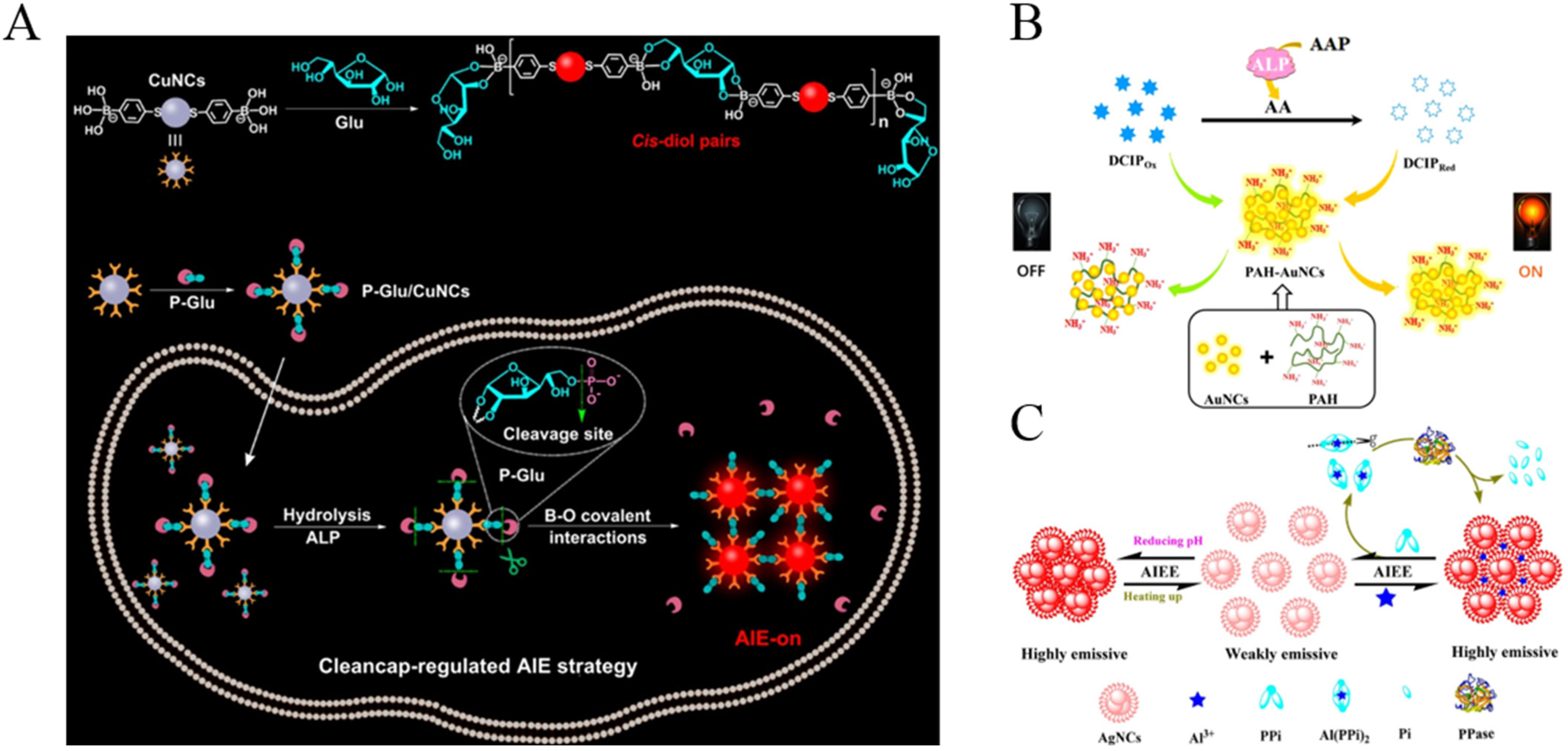
3. Phosphatases
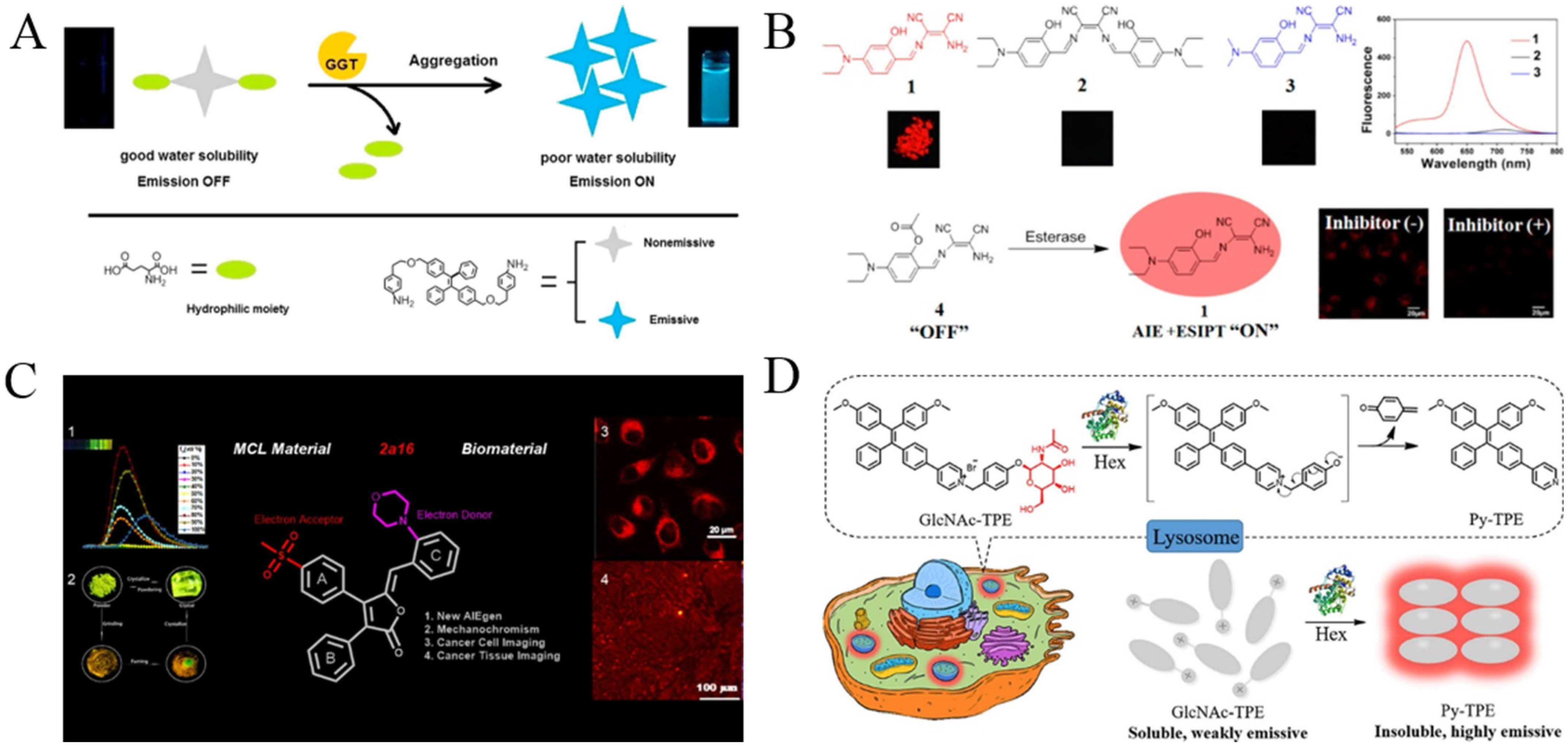
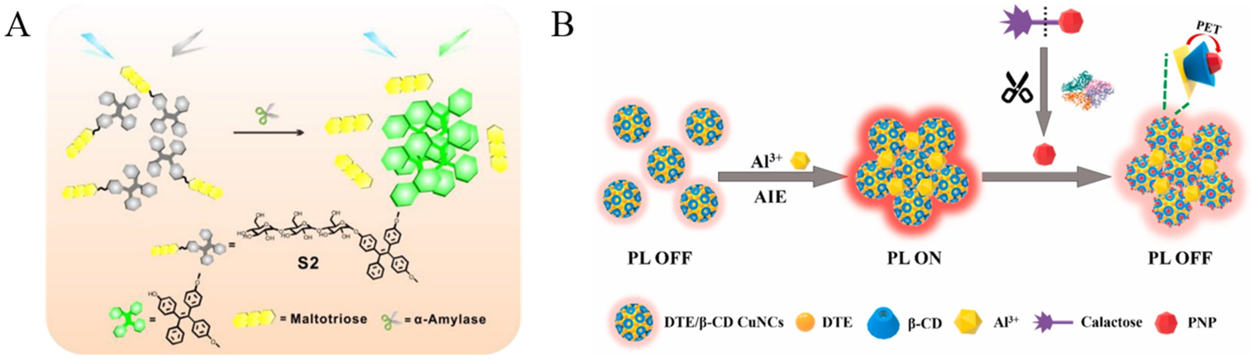
4. Glycosidases

| Targets | Probes | Linear Range | LOD | Ref. |
|---|---|---|---|---|
| β-gal | TPh-PyBz-βgal | 0.2~2 U/mL | 0.22 U/mL | [78] |
| β-gal | HBTTPAG | 0~3.15 U/mL | 3.7 mU/mL | [79] |
| β-gal | SA-βgal | 0~0.1 U/mL | 14 mU/mL | [80] |
| β-gal | QM-βgal | 0~6 U | 1 mU/mL | [81] |
| β-gal | TPE-DCM | 0~7 U/mL | 1.5 U/mL | [82] |
| α-amylase | TPE-maltotriose | 0~45.5 U/L | 0.14 U/L | [84] |
| β-gal | CuNCs | 0~200 U/L | 0.9 U/L | [85] |
| β-gal | CuNCs | 2.3~96 U/L | 0.7 U/L | [86] |
| β-gal | DTE/β-CD CuNCs | 0~50 U/L | 0.56 U/L | [87] |
| GUS | BTBP-Gluc | 0~7 U/mL | – | [88] |
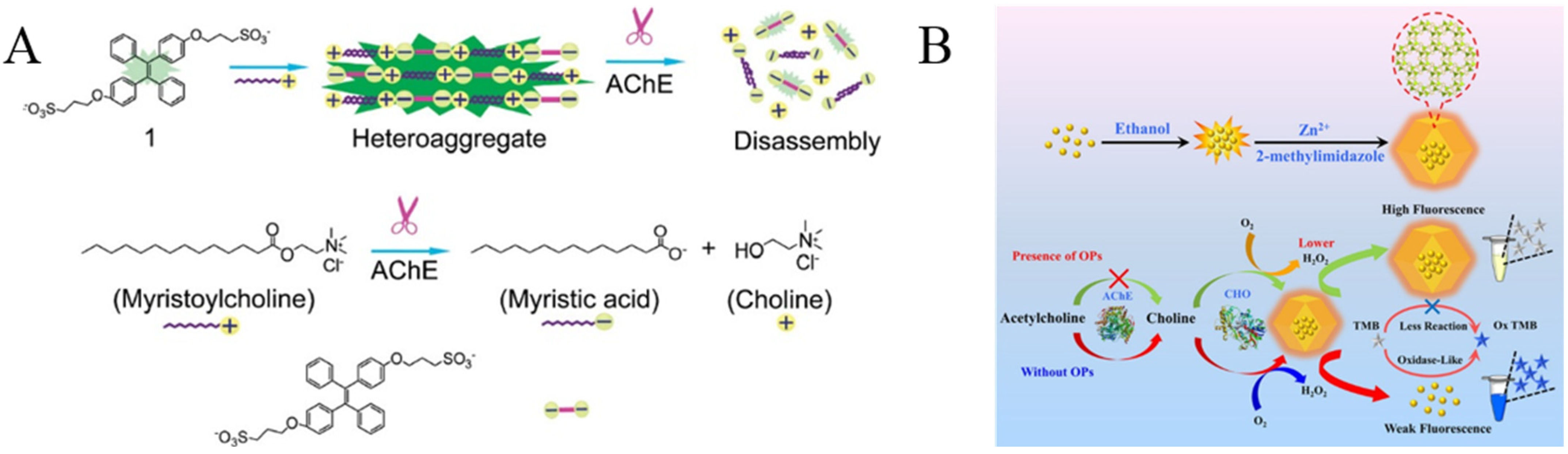
5. Cholinesterases
| Targets | Probes | Linear Range | LOD | Ref. |
|---|---|---|---|---|
| AChE | Sulfonated TPE | 0.5~2 U/mL | 0.5 U/mL | [89] |
| AChE | TPE-Maleimide | 0.3~3 mU/mL | 2.5 mU/mL | [90] |
| AChE | TPE-Leu | 0~100 mU | – | [91] |
| AChE | AIE-AuNPs | 0~8 mU/mL | 0.015 mU/mL | [92] |
| BChE | TB-BChE | 0~70 μg/mL | 39.24 ng/mL | [95] |
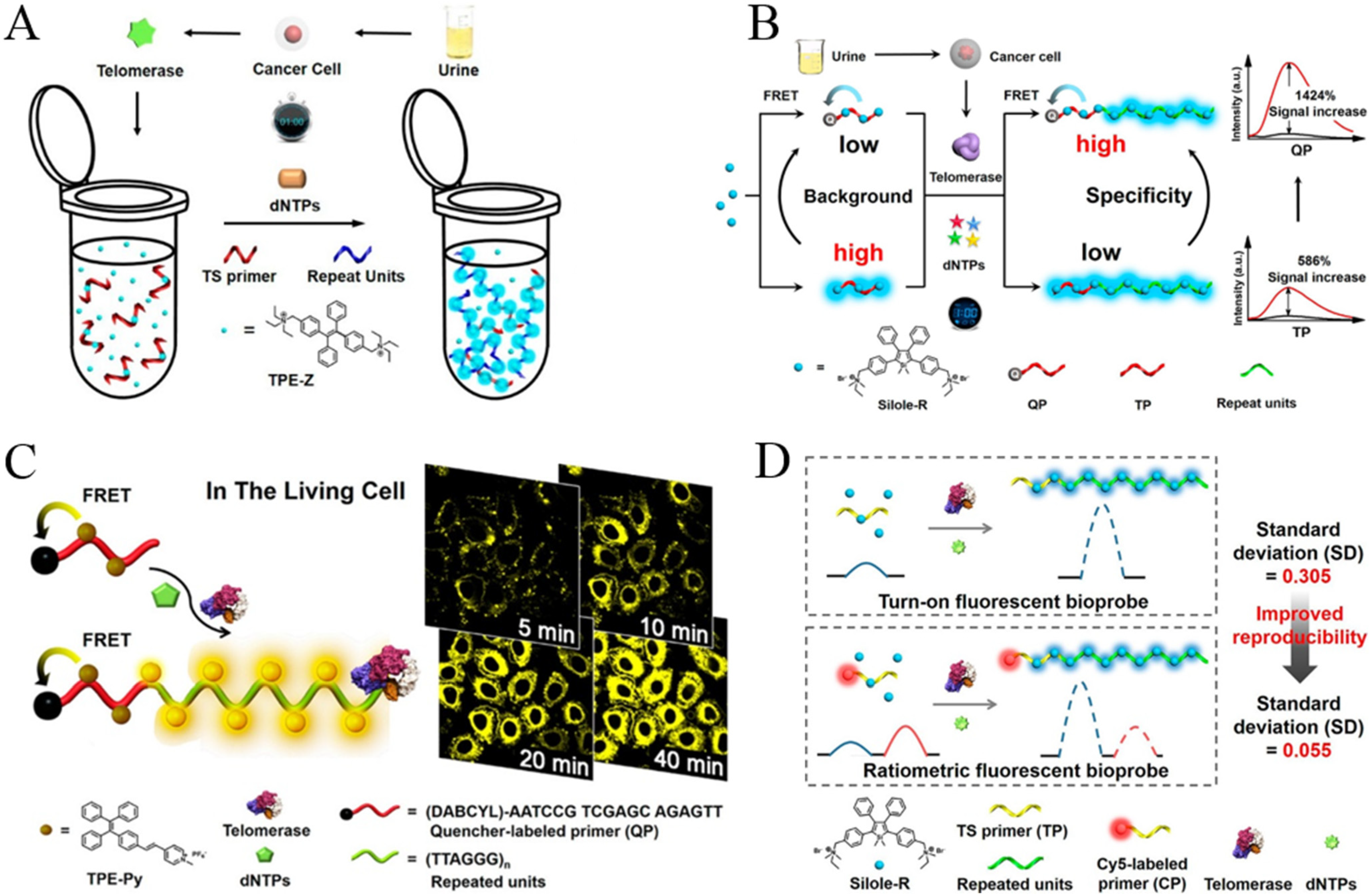
6. Telomerase
7. Others
| Targets | Probes | Linear Range | LOD | Ref. |
|---|---|---|---|---|
| GGT | ABTT-Glu | 10~90 U/L | 2.9 U/L | [101] |
| GGT | TPE | 0~80 U/L | 0.59 U/L | [102] |
| β-lactamase | DNBS-CSA | 0~10 mU/mL | 0.5 mU/mL | [103] |
| Lyso | AIE-Lyso-1 | 0.1~0.5 U/mL | 2.4 mU/mL | [104] |
| esterase | probe 4 | 0.01~0.15 U/mL | 5 mU/mL | [105] |
| lipase | TPE−COOC6H13 | 0.1~1.3 mg/mL | 0.1 mg/mL | [106] |
| lipase | benzophenone | 0.1~4 U/mL | 50 mU/mL | [107] |
| α-fuc | QM-NHαfuc | 0~1.75 U/mL | 10 mU/mL | [108] |
| Hex | GlcNAc-TPE | 0~0.2 U/mL | 3 mU/mL | [110] |
| MTase | TPE-Z | 0.5~100 U/mL | 0.16 U/mL | [112] |
| HAase | TPE-4N+ | 0.05~2 U/mL | 20 mU/mL | [114] |
| HAase | HA-AuNPs@AIEDs | 0.01~60 U/mL | 7.2 mU/mL | [115] |
8. Conclusions
Author Contributions
Funding
Conflicts of Interest
References
- Kwek, G.; Do, T.C.; Lu, X.; Lin, J.; Xing, B. Scratching the surface of unventured possibilities with in situ self-assembly: Protease-activated developments for imaging and therapy. ACS Appl. Bio. Mater. 2021, 4, 2192. [Google Scholar] [CrossRef] [PubMed]
- Yao, Y.; Zhang, Y.; Yan, C.; Zhu, W.H.; Guo, Z. Enzyme-activatable fluorescent probes for beta-galactosidase: From design to biological applications. Chem. Sci. 2021, 12, 9885. [Google Scholar] [CrossRef] [PubMed]
- Luo, S.; Zhang, Y.; Situ, B.; Zheng, L. Fluorescence sensing telomerase activity: From extracellular detection to in situ imaging. Sens. Actuat. B Chem. 2018, 273, 853. [Google Scholar] [CrossRef]
- Feng, G.; Liao, S.; Liu, Y.; Zhang, H.; Luo, X.; Zhou, X.; Fang, J. When AIE meets enzymes. Analyst 2022, 147, 3958. [Google Scholar] [CrossRef]
- Hu, Z.; Li, Y.; Hussain, E.; Huang, X.; Zhang, Y.; Niu, N.; Shahzad, S.A.; Yu, C. Black phosphorus nanosheets based sensitive protease detection and inhibitor screening. Talanta 2019, 197, 270. [Google Scholar] [CrossRef]
- Oliveira-Silva, R.; Sousa-Jeronimo, M.; Prazeres, D.M.F. Monitoring proteolytic activity in real time: A new world of opportunities for biosensors. Trends Biochem. Sci. 2020, 45, 604. [Google Scholar] [CrossRef]
- Balbaied, T.; Moore, E. Overview of optical and electrochemical alkaline phosphatase (ALP) biosensors: Recent approaches in cells culture techniques. Biosensors 2019, 9, 102. [Google Scholar] [CrossRef] [Green Version]
- Park, S.; Lim, S.Y.; Bae, S.M.; Kim, S.Y.; Myung, S.J.; Kim, H.J. Indocyanine-based activatable fluorescence turn-on probe for γ-glutamyltranspeptidase and its application to the mouse model of colon cancer. ACS Sens. 2016, 1, 579. [Google Scholar] [CrossRef]
- Xia, N.; Wang, Q.; Liu, L. Nanomaterials-based optical techniques for the detection of acetylcholinesterase and pesticides. Sensors 2015, 15, 499. [Google Scholar] [CrossRef] [Green Version]
- Hameed, A.M.; Lam, V.W.T.; Pleass, H.C. Significant elevations of serum lipase not caused by pancreatitis: A systematic review. HPB 2015, 17, 99. [Google Scholar] [CrossRef]
- Hata, R.; Nonaka, H.; Takakusagi, Y.; Ichikawa, K.; Sando, S. Design of a hyperpolarized molecular probe for detection of aminopeptidase N activity. Angew. Chem. Int. Ed. 2016, 55, 1765. [Google Scholar] [CrossRef] [PubMed]
- Niu, N.; Zhou, H.; Liu, N.; Jiang, H.; Hussain, E.; Hua, Z.; Yu, C. A smart perylene derived photosensitizer for lysosome-targeted and self-assessed photodynamic therapy. Chem. Commun. 2019, 55, 1036. [Google Scholar] [CrossRef] [PubMed]
- Eivazzadeh-Keihan, R.; Saadatidizaji, Z.; Maleki, A.; de la Guardia, M.; Mahdavi, M.; Barzegar, S.; Ahadian, S. Recent progresses in development of biosensors for thrombin detection. Biosensors 2022, 12, 767. [Google Scholar] [CrossRef] [PubMed]
- Hori, Y.; Kikuchi, K. Chemical tools with fluorescence switches for verifying epigenetic modifications. Acc. Chem. Res. 2019, 52, 2849. [Google Scholar] [CrossRef]
- Suzuki, Y.; Yokoyama, K. Development of functional fluorescent molecular probes for the detection of biological substances. Biosensors 2015, 5, 337. [Google Scholar] [CrossRef] [Green Version]
- Hu, Z.; Chen, J.; Li, Y.; Wang, Y.; Zhang, Q.; Hussain, E.; Yang, M.; Shahzad, S.A.; Yu, D.; Yu, C. Nucleic acid-controlled quantum dots aggregation: A label-free fluorescence turn-on strategy for alkaline phosphatase detection. Talanta 2017, 169, 64. [Google Scholar] [CrossRef]
- Yan, Y.; Zhang, J.; Yi, S.; Liu, L.; Huang, C. Lighting up forensic science by aggregation-induced emission: A review. Anal. Chim. Acta 2021, 1155, 238119. [Google Scholar] [CrossRef]
- Cai, X.; Liu, B. Aggregation-induced emission: Recent advances in materials and biomedical applications. Angew. Chem. Int. Ed. 2020, 59, 9868. [Google Scholar] [CrossRef]
- Suzuki, S.; Sasaki, S.; Sairi, A.S.; Iwai, R.; Tang, B.Z.; Konishi, G.I. Principles of aggregation-induced emission: Design of deactivation pathways for advanced AIEgens and applications. Angew. Chem. Int. Ed. 2020, 59, 9856. [Google Scholar] [CrossRef] [Green Version]
- Wang, X.; Hu, J.; Zhang, G.; Liu, S. Highly selective fluorogenic multianalyte biosensors constructed via enzyme-catalyzed coupling and aggregation-induced emission. J. Am. Chem. Soc. 2014, 136, 9890–9893. [Google Scholar] [CrossRef]
- Gao, Y.; He, Z.; He, X.; Zhang, H.; Weng, J.; Yang, X.; Meng, F.; Luo, L.; Tang, B.Z. Dual-color emissive AIEgen for specific and label-free double-stranded DNA recognition and single-nucleotide polymorphisms detection. J. Am. Chem. Soc. 2019, 141, 20097–20106. [Google Scholar] [CrossRef]
- Shi, J.; Li, Y.; Li, Q.; Li, Z. Enzyme-responsive bioprobes based on the mechanism of aggregation-induced emission. ACS Appl. Mater. Interfaces 2018, 10, 12278. [Google Scholar] [CrossRef] [PubMed]
- Gao, M.; Tang, B.Z. Fluorescent sensors based on aggregation-induced emission: Recent advances and perspectives. ACS Sens. 2017, 2, 1382. [Google Scholar] [CrossRef] [PubMed]
- Li, Y.; Zhou, H.; Chen, J.; Shahzad, S.A.; Yu, C. Controlled self-assembly of small molecule probes and the related applications in bioanalysis. Biosens. Bioelectron. 2016, 76, 38. [Google Scholar] [CrossRef]
- Shellaiah, M.; Sun, K.-W. Pyrene-based AIE active materials for bioimaging and theranostics applications. Biosensors 2022, 12, 550. [Google Scholar] [CrossRef] [PubMed]
- Dai, D.; Yang, J.; Yang, Y.W. Supramolecular assemblies with aggregation-induced emission properties for sensing and detection. Chem. Eur. J. 2022, 28, e202103185. [Google Scholar] [PubMed]
- Li, X.; Zha, M.; Li, Y.; Ni, J.; Min, T.; Kang, T.; Yang, G.; Tang, H.; Li, K.; Jiang, X. Sub-10 nm aggregation-induced emission quantum dots assembled by microfluidics for enhanced tumor targeting and reduced retention in the liver. Angew. Chem. Int. Ed. 2020, 59, 21899. [Google Scholar] [CrossRef]
- Mao, L.; Liu, Y.; Yang, S.; Li, Y.; Zhang, X.; Wei, Y. Recent advances and progress of fluorescent bio-/chemosensors based on aggregation-induced emission molecules. Dyes Pigments 2019, 162, 611. [Google Scholar] [CrossRef]
- Wei, W.; Qiu, Z. Diagnostics and theranostics of central nervous system diseases based on aggregation-induced emission luminogens. Biosens. Bioelectron. 2022, 217, 114670. [Google Scholar] [CrossRef]
- Yang, J.; Wei, J.; Luo, F.; Dai, J.; Hu, J.J.; Lou, X.; Xia, F. Enzyme-responsive peptide-based AIE bioprobes. Top. Curr. Chem. 2020, 378, 47. [Google Scholar] [CrossRef]
- Chen, B.; Yuan, H.; Zhang, W.; Hu, J.; Lou, X.; Xia, F. AIEgen-peptide bioprobes for the imaging of organelles. Biosensors 2022, 12, 667. [Google Scholar] [CrossRef] [PubMed]
- La, D.D.; Bhosale, S.V.; Jones, L.A.; Bhosale, S.V. Tetraphenylethylene-based AIE-active probes for sensing applications. ACS Appl. Mater. Interfaces 2018, 10, 12189. [Google Scholar] [CrossRef] [PubMed]
- Mei, J.; Huang, Y.; Tian, H. Progress and trends in AIE-based bioprobes: A brief overview. ACS Appl. Mater. Interfaces 2018, 10, 12217. [Google Scholar] [CrossRef] [PubMed]
- Würthner, F. Aggregation-induced emission (AIE): A historical perspective. Angew. Chem. Int. Ed. 2020, 59, 14192. [Google Scholar] [CrossRef]
- Zhao, Z.; Zhang, H.; Lam, J.W.Y.; Tang, B.Z. Aggregation-induced emission: New vistas at the aggregate level. Angew. Chem. Int. Ed. 2020, 59, 9888. [Google Scholar] [CrossRef]
- Kang, X.Y.; Li, Y.; Yin, S.; Li, W.; Qi, J. Reactive species-activatable AIEgens for biomedical applications. Biosensors 2022, 12, 646. [Google Scholar] [CrossRef]
- Liu, S.; Feng, G.; Tang, B.Z.; Liu, B. Recent advances of AIE light-up probes for photodynamic therapy. Chem. Sci. 2021, 12, 6488. [Google Scholar] [CrossRef]
- Wang, J.; Meng, Q.; Yang, Y.; Zhong, S.; Zhang, R.; Fang, Y.; Gao, Y.; Cui, X. Schiff base aggregation-induced emission luminogens for sensing applications: A review. ACS Sens. 2022, 7, 2521. [Google Scholar] [CrossRef]
- Huang, S.; Wu, Y.; Zeng, F.; Chen, J.; Wu, S. A turn-on fluorescence probe based on aggregation-induced emission for leucine aminopeptidase in living cells and tumor tissue. Anal. Chim. Acta 2018, 1031, 169. [Google Scholar] [CrossRef]
- Huang, X.; Lei, Q.; Huang, S.; Zeng, H.; Feng, B.; Zeng, Q.; Hu, Y.; Zeng, W. Construction of a novel asymmetric imidazole-cored AIE probe for ratiometric imaging of endogenous leucine aminopeptidase. Chem. Commun. 2021, 57, 6608. [Google Scholar] [CrossRef]
- Luan, Z.; Zhao, L.; Liu, C.; Song, W.; He, P.; Zhang, X. Detection of casein kinase II by aggregation-induced emission. Talanta 2019, 201, 450. [Google Scholar] [CrossRef] [PubMed]
- Zhang, R.; Zhang, C.J.; Feng, G.; Hu, F.; Wang, J.; Liu, B. Specific light-up probe with aggregation-induced emission for facile detection of chymase. Anal. Chem. 2016, 88, 9111. [Google Scholar] [CrossRef] [PubMed]
- Han, A.; Wang, H.; Kwok, R.T.; Ji, S.; Li, J.; Kong, D.; Tang, B.Z.; Liu, B.; Yang, Z.; Ding, D. Peptide-induced AIEgen self-assembly: A new strategy to realize highly sensitive fluorescent light-up probes. Anal. Chem. 2016, 88, 3872. [Google Scholar] [CrossRef] [PubMed]
- Li, K.; Hu, X.X.; Liu, H.W.; Xu, S.; Huan, S.Y.; Li, J.B.; Deng, T.G.; Zhang, X.B. In situ imaging of furin activity with a highly stable probe by releasing of precipitating fluorochrome. Anal. Chem. 2018, 90, 11680. [Google Scholar] [CrossRef]
- Wu, F.; Huang, Y.; Yang, X.; Hu, J.J.; Lou, X.; Xia, F.; Song, Y.; Jiang, L. Tunning intermolecular interaction of peptide-conjugated AIEgen in nano-confined space for quantitative detection of tumor marker secreted from cells. Anal. Chem. 2021, 93, 16257. [Google Scholar] [CrossRef]
- Shi, H.; Kwok, R.T.; Liu, J.; Xing, B.; Tang, B.Z.; Liu, B. Real-time monitoring of cell apoptosis and drug screening using fluorescent light-up probe with aggregation-induced emission characteristics. J. Am. Chem. Soc. 2012, 134, 17972. [Google Scholar] [CrossRef]
- Ding, D.; Liang, J.; Shi, H.; Kwok, R.T.K.; Gao, M.; Feng, G.; Yuan, Y.; Tang, B.Z.; Liu, B. Light-up bioprobe with aggregation-induced emission characteristics for real-time apoptosis imaging in target cancer cells. J. Mater. Chem. B 2014, 2, 231. [Google Scholar] [CrossRef] [Green Version]
- Yuan, Y.; Zhang, C.J.; Kwok, R.T.K.; Mao, D.; Tang, B.Z.; Liu, B. Light-up probe based on AIEgens: Dual signal turn-on for caspase cascade activation monitoring. Chem. Sci. 2017, 8, 2723. [Google Scholar] [CrossRef] [Green Version]
- Li, H.; Parigi, G.; Luchinat, C.; Meade, T.J. Bimodal fluorescence-magnetic resonance contrast agent for apoptosis imaging. J. Am. Chem. Soc. 2019, 141, 6224. [Google Scholar] [CrossRef]
- Yuan, Y.; Kwok, R.T.; Tang, B.Z.; Liu, B. Targeted theranostic platinum(IV) prodrug with a built-in aggregation-induced emission light-up apoptosis sensor for noninvasive early evaluation of its therapeutic responses in situ. J. Am. Chem. Soc. 2014, 136, 2546. [Google Scholar] [CrossRef]
- Cheng, Y.; Huang, F.; Min, X.; Gao, P.; Zhang, T.; Li, X.; Liu, B.; Hong, Y.; Lou, X.; Xia, F. Protease-responsive prodrug with aggregation-induced emission probe for controlled drug delivery and drug release tracking in living cells. Anal. Chem. 2016, 88, 8913. [Google Scholar] [CrossRef] [PubMed]
- Qin, W.; Wu, Y.; Hu, Y.; Dong, Y.; Hao, T.; Zhang, C. TPE-Based Peptide Micelles for Targeted Tumor Therapy and Apoptosis Monitoring. ACS Appl. Bio Mater. 2021, 4, 1038. [Google Scholar] [CrossRef]
- Lin, Y.X.; Qiao, S.L.; Wang, Y.; Zhang, R.X.; An, H.W.; Ma, Y.; Rajapaksha, R.P.; Qiao, Z.Y.; Wang, L.; Wang, H. An in situ intracellular self-assembly strategy for quantitatively and temporally monitoring autophagy. ACS Nano 2017, 11, 1826. [Google Scholar] [CrossRef] [PubMed]
- Xu, J.P.; Fang, Y.; Song, Z.G.; Mei, J.; Jia, L.; Qin, A.J.; Sun, J.Z.; Ji, J.; Tang, B.Z. BSA-tetraphenylethene derivative conjugates with aggregation-induced emission properties: Fluorescent probes for label-free and homogeneous detection of protease and alpha1-antitrypsin. Analyst 2011, 136, 2315. [Google Scholar] [CrossRef] [PubMed]
- Zhang, Y.; Li, Y.; Yang, N.; Yu, X.; He, C.; Niu, N.; Zhang, C.; Zhou, H.; Yu, C.; Jiang, S. Histone controlled aggregation of tetraphenylethene probe: A new method for the detection of protease activity. Sens. Actuat. B Chem. 2018, 257, 1143. [Google Scholar] [CrossRef]
- Zhao, L.; Wang, T.; Wu, Q.; Liu, Y.; Chen, Z.; Li, X. Fluorescent strips of electrospun fibers for ratiometric sensing of serum heparin and urine trypsin. ACS Appl. Mater. Interfaces 2017, 9, 3400. [Google Scholar] [CrossRef]
- Xue, F.; Qu, F.; Han, W.; Xia, L.; You, J. Aggregation-induced emission enhancement of gold nanoclusters triggered by silicon nanoparticles for ratiometric detection of protamine and trypsin. Anal. Chim. Acta 2019, 1046, 170. [Google Scholar] [CrossRef]
- Gu, X.; Zhang, G.; Wang, Z.; Liu, W.; Xiao, L.; Zhang, D. A new fluorometric turn-on assay for alkaline phosphatase and inhibitor screening based on aggregation and deaggregation of tetraphenylethylene molecules. Analyst 2013, 138, 2427. [Google Scholar] [CrossRef]
- Liang, J.; Kwok, R.T.; Shi, H.; Tang, B.Z.; Liu, B. Fluorescent light-up probe with aggregation-induced emission characteristics for alkaline phosphatase sensing and activity study. ACS Appl. Mater. Interfaces 2013, 5, 8784. [Google Scholar] [CrossRef]
- Cao, F.Y.; Long, Y.; Wang, S.B.; Li, B.; Fan, J.X.; Zeng, X.; Zhang, X.Z. Fluorescence light-up AIE probe for monitoring cellular alkaline phosphatase activity and detecting osteogenic differentiation. J. Mater. Chem. B 2016, 4, 4534. [Google Scholar] [CrossRef]
- Huang, L.; Cao, X.; Gao, T.; Feng, B.; Huang, X.; Song, R.; Du, T.; Wen, S.; Feng, X.; Zeng, W. A novel aggregation-induced dual emission probe for in situ light-up detection of endogenous alkaline phosphatase. Talanta 2021, 225, 121950. [Google Scholar] [CrossRef] [PubMed]
- Zhao, M.; Gao, Y.; Ye, S.; Ding, J.; Wang, A.; Li, P.; Shi, H. A light-up near-infrared probe with aggregation-induced emission characteristics for highly sensitive detection of alkaline phosphatase. Analyst 2019, 144, 6262. [Google Scholar] [CrossRef] [PubMed]
- Song, Z.; Kwok, R.T.; Zhao, E.; He, Z.; Hong, Y.; Lam, J.W.; Liu, B.; Tang, B.Z. A ratiometric fluorescent probe based on ESIPT and AIE processes for alkaline phosphatase activity assay and visualization in living cells. ACS Appl. Mater. Interfaces 2014, 6, 17245. [Google Scholar] [CrossRef] [PubMed]
- Li, Y.; Xie, R.; Pang, X.; Zhou, Z.; Xu, H.; Gu, B.; Wu, C.; Li, H.; Zhang, Y. Aggregation-induced emission fluorescent probe for monitoring endogenous alkaline phosphatase in living cells. Talanta 2019, 205, 120143. [Google Scholar] [CrossRef] [PubMed]
- Zhang, X.; Ren, C.; Hu, F.; Gao, Y.; Wang, Z.; Li, H.; Liu, J.; Liu, B.; Yang, C. Detection of bacterial alkaline phosphatase activity by enzymatic in situ self-assembly of the AIEgen-peptide conjugate. Anal. Chem. 2020, 92, 5185. [Google Scholar] [CrossRef]
- Ji, S.; Gao, H.; Mu, W.; Ni, X.; Yi, X.; Shen, J.; Liu, Q.; Bao, P.; Ding, D. Enzyme-instructed self-assembly leads to the activation of optical properties for selective fluorescence detection and photodynamic ablation of cancer cells. J. Mater. Chem. B 2018, 6, 2566. [Google Scholar] [CrossRef]
- Zhao, L.; Xie, S.; Song, X.; Wei, J.; Zhang, Z.; Li, X. Ratiometric fluorescent response of electrospun fibrous strips for real-time sensing of alkaline phosphatase in serum. Biosens. Bioelectron. 2017, 91, 217. [Google Scholar] [CrossRef]
- Kaur, J.; Singh, P.K. An AIEgen–protamine assembly/disassembly based fluorescence turn-on probe for sensing alkaline phosphatase. Sens. Actuat. B Chem. 2021, 346, 130517. [Google Scholar] [CrossRef]
- Luo, Y.; Li, J.; Li, Y.; Yang, B.; Zhou, T.; Deng, J. Lanthanide-Free Infinite Coordination Polymer Nanoparticles for Real-Time Monitoring of Alkaline Phosphatase and Its Application for Digital Algal Bloom Detection. ACS Appl. Nano Mater. 2021, 4, 11134. [Google Scholar] [CrossRef]
- Geng, F.; Zou, C.; Liu, J.; Zhang, Q.; Guo, X.; Fan, Y.; Yu, H.; Yang, S.; Liu, Z.; Li, L. Development of luminescent nanoswitch for sensing of alkaline phosphatase in human serum based onAl3+-PPi interaction and Cu NCs with AIE properties. Anal. Chim. Acta 2019, 1076, 131. [Google Scholar] [CrossRef]
- Zhao, M.; Feng, H.; Han, J.; Ao, H.; Qian, Z. Multi-stimuli responsive copper nanoclusters with bright red luminescence for quantifying acid phosphatase activity via redox-controlled luminescence switch. Anal. Chim. Acta 2017, 984, 202. [Google Scholar] [CrossRef] [PubMed]
- Huang, Y.; Zhu, L.; Ji, J.; Li, Y.; Liu, T.; Lei, J. Cleancap-regulated aggregation-induced emission strategy for highly specific analysis of enzyme. Anal. Chem. 2020, 92, 4726. [Google Scholar] [CrossRef] [PubMed] [Green Version]
- Han, X.; Han, M.; Ma, L.; Qu, F.; Kong, R.M.; Qu, F. Self-assembled gold nanoclusters for fluorescence turn-on and colorimetric dual-readout detection of alkaline phosphatase activity via DCIP-mediated fluorescence resonance energy transfer. Talanta 2019, 194, 55. [Google Scholar] [CrossRef] [PubMed]
- Pan, T.; Zhou, T.; Tu, Y.; Yan, J. Turn-on fluorescence measurement of acid phosphatase activity through an aggregation-induced emission of thiolate-protected gold nanoclusters. Talanta 2021, 227, 122197. [Google Scholar] [CrossRef] [PubMed]
- Tang, C.; Feng, H.; Huang, Y.; Qian, Z. Reversible luminescent nanoswitches based on aggregation-induced emission enhancement of silver nanoclusters for luminescence turn-on assay of inorganic pyrophosphatase activity. Anal. Chem. 2017, 89, 4994. [Google Scholar] [CrossRef]
- Wang, D.; Li, C.; Zhu, Y.; Song, Y.; Lu, S.; Sun, H.; Hao, H.; Xu, X. TEPP-46-Based AIE Fluorescent Probe for Detection and Bioimaging of PKM2 in Living Cells. Anal. Chem. 2021, 93, 12682. [Google Scholar] [CrossRef]
- Gao, Z.; Gao, H.; Zheng, D.; Xu, T.; Chen, Y.; Liang, C.; Wang, L.; Ding, D.; Yang, Z. β-galactosidase responsive AIE fluorogene for identification and removal of senescent cancer cells. Sci. China Chem. 2020, 63, 398. [Google Scholar] [CrossRef]
- Zhang, S.; Wang, X.; Wang, X.; Wang, T.; Liao, W.; Yuan, Y.; Chen, G.; Jia, X. A novel AIE fluorescent probe for beta-galactosidase detection and imaging in living cells. Anal. Chim. Acta 2022, 1198, 339554. [Google Scholar] [CrossRef]
- Gao, T.; Li, H.; Wu, Y.; Deng, C.; Xie, Y.; Wang, J.; Yang, Y.; Lv, Q.; Jin, Q.; Chen, Y.; et al. First aggregation-induced emission-active probe for species-specific detection of beta-galactosidase. Talanta 2021, 235, 122659. [Google Scholar] [CrossRef]
- Peng, L.; Gao, M.; Cai, X.; Zhang, R.; Li, K.; Feng, G.; Tong, A.; Liu, B. A fluorescent light-up probe based on AIE and ESIPT processes for beta-galactosidase activity detection and visualization in living cells. J. Mater. Chem. B 2015, 3, 9168. [Google Scholar] [CrossRef]
- Gu, K.; Qiu, W.; Guo, Z.; Yan, C.; Zhu, S.; Yao, D.; Shi, P.; Tian, H.; Zhu, W.H. An enzyme-activatable probe liberating AIEgens: On-site sensing and long-term tracking of beta-galactosidase in ovarian cancer cells. Chem. Sci. 2019, 10, 398. [Google Scholar] [CrossRef] [PubMed] [Green Version]
- Dong, L.; Zhang, M.Y.; Han, H.H.; Zang, Y.; Chen, G.R.; Li, J.; He, X.P.; Vidal, S. A general strategy to the intracellular sensing of glycosidases using AIE-based glycoclusters. Chem. Sci. 2021, 13, 247. [Google Scholar] [CrossRef] [PubMed]
- Mandal, N.; Bhattacharjee, M.; Chattopadhyay, A.; Bandyopadhyay, D. Point-of-care-testing of alpha-amylase activity in human blood serum. Biosens. Bioelectron. 2019, 124, 75. [Google Scholar] [CrossRef] [PubMed]
- Shi, J.; Deng, Q.; Li, Y.; Zheng, M.; Chai, Z.; Wan, C.; Zheng, Z.; Li, L.; Huang, F.; Tang, B. A rapid and ultrasensitive tetraphenylethylene-based probe with aggregation-induced emission for direct detection of alpha-amylase in human body fluids. Anal. Chem. 2018, 90, 13775. [Google Scholar] [CrossRef]
- Zhao, M.; Qian, Z.; Zhong, M.; Chen, Z.; Ao, H.; Feng, H. Fabrication of stable and luminescent copper nanocluster-based AIE particles and their application in beta-galactosidase activity assay. ACS Appl. Mater. Interfaces 2017, 9, 32887. [Google Scholar] [CrossRef]
- Huang, Y.; Feng, H.; Liu, W.; Zhang, S.; Tang, C.; Chen, J.; Qian, Z. Cation-driven luminescent self-assembled dots of copper nanoclusters with aggregation-induced emission for beta-galactosidase activity monitoring. J. Mater. Chem. B 2017, 5, 5120. [Google Scholar] [CrossRef]
- Huang, X.; Lan, M.; Wang, J.; Guo, L.; Lin, Z.; Sun, N.; Wu, C.; Qiu, B. A fluorescence signal amplification and specific energy transfer strategy for sensitive detection of beta-galactosidase based on the effects of AIE and host-guest recognition. Biosens. Bioelectron. 2020, 169, 112655. [Google Scholar] [CrossRef]
- Wei, X.; Wu, Q.; Feng, Y.; Chen, M.; Zhang, S.; Chen, M.; Zhang, J.; Yang, G.; Ding, Y.; Yang, X.; et al. Off-on fluorogenic substrate harnessing ESIPT and AIE features for in situ and long-term tracking of β-glucuronidase in Escherichia coli. Sens. Actuat. B Chem. 2020, 304, 127242. [Google Scholar] [CrossRef]
- Wang, M.; Gu, X.; Zhang, G.; Zhang, D.; Zhu, D. Convenient and continuous fluorometric assay method for acetylcholinesterase and inhibitor screening based on the aggregation-induced emission. Anal. Chem. 2009, 81, 4444. [Google Scholar] [CrossRef]
- Chang, J.; Li, H.; Hou, T.; Li, F. Paper-based fluorescent sensor for rapid naked-eye detection of acetylcholinesterase activity and organophosphorus pesticides with high sensitivity and selectivity. Biosens. Bioelectron. 2016, 86, 971. [Google Scholar] [CrossRef]
- Shi, L.; Liu, Y.; Wang, Q.; Wang, T.; Ding, Y.; Cao, Y.; Li, Z.; Wei, H. A pH responsive AIE probe for enzyme assays. Analyst 2018, 143, 741. [Google Scholar] [CrossRef]
- Wang, C.; Wang, X.; Gai, P.; Li, H.; Li, F. Target-responsive AIE-Au nanoconjugate for acetylcholinesterase activity and inhibitor assay with ultralow background noise. Sens. Actuat. B Chem. 2019, 284, 118. [Google Scholar] [CrossRef]
- Cai, Y.; Zhu, H.; Zhou, W.; Qiu, Z.; Chen, C.; Qileng, A.; Li, K.; Liu, Y. Capsulation of AuNCs with AIE effect into metal-organic framework for the marriage of a fluorescence and colorimetric biosensor to detect organophosphorus pesticides. Anal. Chem. 2021, 93, 7275. [Google Scholar] [CrossRef] [PubMed]
- Holas, O.; Musilek, K.; Pohanka, M.; Kuca, K. The progress in the cholinesterase quantification methods. Expert Opin. Drug Dis. 2012, 7, 1207. [Google Scholar] [CrossRef] [PubMed]
- Xiang, C.; Xiang, J.; Yang, X.; Li, C.; Zhou, L.; Jiang, D.; Peng, Y.; Xu, Z.; Deng, G.; Zhu, B.; et al. Ratiometric imaging of butyrylcholinesterase activity in mice with nonalcoholic fatty liver using an AIE-based fluorescent probe. J. Mater. Chem. B 2022, 10, 4254. [Google Scholar] [CrossRef] [PubMed]
- Ou, X.; Hong, F.; Zhang, Z.; Cheng, Y.; Zhao, Z.; Gao, P.; Lou, X.; Xia, F.; Wang, S. A highly sensitive and facile graphene oxide-based nucleic acid probe: Label-free detection of telomerase activity in cancer patient’s urine using AIEgens. Biosens. Bioelectron. 2017, 89, 417. [Google Scholar] [CrossRef]
- Lou, X.; Zhuang, Y.; Zuo, X.; Jia, Y.; Hong, Y.; Min, X.; Zhang, Z.; Xu, X.; Liu, N.; Xia, F.; et al. Real-time, quantitative lighting-up detection of telomerase in urines of bladder cancer patients by AIEgens. Anal. Chem. 2015, 87, 6822. [Google Scholar] [CrossRef]
- Zhuang, Y.; Zhang, M.; Chen, B.; Duan, R.; Min, X.; Zhang, Z.; Zheng, F.; Liang, H.; Zhao, Z.; Lou, X.; et al. Quencher group induced high specificity detection of telomerase in clear and bloody urines by AIEgens. Anal. Chem. 2015, 87, 9487. [Google Scholar] [CrossRef]
- Zhuang, Y.; Huang, F.; Xu, Q.; Zhang, M.; Lou, X.; Xia, F. Facile, fast-responsive, and photostable imaging of telomerase activity in living cells with a fluorescence turn-on manner. Anal. Chem. 2016, 88, 3289. [Google Scholar] [CrossRef]
- Zhuang, Y.; Xu, Q.; Huang, F.; Gao, P.; Zhao, Z.; Lou, X.; Xia, F. Ratiometric fluorescent bioprobe for highly reproducible detection of telomerase in bloody urines of bladder cancer patients. ACS Sens. 2016, 1, 572. [Google Scholar] [CrossRef]
- Liu, Y.; Feng, B.; Cao, X.; Tang, G.; Liu, H.; Chen, F.; Liu, M.; Chen, Q.; Yuan, K.; Gu, Y.; et al. A novel “AIE + ESIPT” near-infrared nanoprobe for the imaging of gamma-glutamyl transpeptidase in living cells and the application in precision medicine. Analyst 2019, 144, 5136. [Google Scholar] [CrossRef] [PubMed]
- Hou, X.; Zeng, F.; Wu, S. A fluorescent assay for gamma-glutamyltranspeptidase via aggregation induced emission and its applications in real samples. Biosens. Bioelectron. 2016, 85, 317. [Google Scholar] [CrossRef] [PubMed]
- Peng, L.; Xiao, L.; Ding, Y.; Xiang, Y.; Tong, A. A simple design of fluorescent probes for indirect detection of beta-lactamase based on AIE and ESIPT processes. J. Mater. Chem. B 2018, 6, 3922. [Google Scholar] [CrossRef]
- Gao, M.; Hu, Q.; Feng, G.; Tang, B.Z.; Liu, B. A fluorescent light-up probe with “AIE + ESIPT” characteristics for specific detection of lysosomal esterase. J. Mater. Chem. B 2014, 2, 3438. [Google Scholar] [CrossRef] [PubMed]
- Peng, L.; Xu, S.; Zheng, X.; Cheng, X.; Zhang, R.; Liu, J.; Liu, B.; Tong, A. Rational design of a red-emissive fluorophore with AIE and ESIPT characteristics and its application in light-up sensing of esterase. Anal. Chem. 2017, 89, 3162. [Google Scholar] [CrossRef] [PubMed]
- Shi, J.; Zhang, S.; Zheng, M.; Deng, Q.; Zheng, C.; Li, J.; Huang, F. A novel fluorometric turn-on assay for lipase activity based on an aggregation-induced emission (AIE) luminogen. Sens. Actuat. B Chem. 2017, 238, 765. [Google Scholar] [CrossRef] [Green Version]
- Guan, P.; Liu, Y.; Yang, B.; Wu, Y.; Chai, J.; Wen, G.; Liu, B. Fluorometric probe for the lipase level: Design, mechanism and biological imaging application. Talanta 2021, 225, 121948. [Google Scholar] [CrossRef]
- Koo, S.; Won, M.; Li, H.; Kim, W.Y.; Li, M.; Yan, C.; Sharma, A.; Guo, Z.; Zhu, W.H.; Sessler, J.L.; et al. Harnessing alpha-l-fucosidase for in vivo cellular senescence imaging. Chem. Sci. 2021, 12, 10054. [Google Scholar] [CrossRef]
- Xie, L.; Li, R.; Zheng, B.; Xie, Z.; Fang, X.; Wang, Y.; Cuny, G.D.; Li, Z.; Lin, B.; Chen, X.; et al. Development of rofecoxib-based fluorescent probes and investigations on their solvatochromism, AIE activity, mechanochromism, and COX-2-targeted bioimaging. Anal. Chem. 2021, 93, 11991. [Google Scholar] [CrossRef]
- Wang, Q.; Li, C.; Chen, Q.; Zhang, P.; Wang, D.; Kang, M.; Jiang, G.; Wang, J. Lysosome-targeting red-emitting aggregation-induced emission probe with large Stokes shift for light-up in situ visualization of beta-N-acetylhexosaminidase. Anal. Chem. 2019, 91, 12611. [Google Scholar] [CrossRef]
- Horsch, M.; Mayer, C.; Sennhauser, U.; Rast, D.M. beta-N-acetylhexosaminidase: A target for the design of antifungal agents. Pharmacol. Therapeut. 1997, 76, 187. [Google Scholar] [CrossRef]
- Niu, S.; Bi, C.; Song, W. Detection of DNA methyltransferase activity using template-free DNA polymerization amplification based on aggregation-induced emission. Anal. Biochem. 2020, 590, 113532. [Google Scholar] [CrossRef]
- Mio, K.; Stern, R. Inhibitors of the hyaluronidases. Matrix Biol. 2002, 21, 31. [Google Scholar] [CrossRef]
- Li, X.; Zhou, Z.; Tang, Y.; Cheng Zhang, C.; Zheng, Y.; Gao, J.; Wang, Q. Modulation of assembly and disassembly of a new tetraphenylethene based nanosensor for highly selective detection of hyaluronidase. Sens. Actuat. B Chem. 2018, 276, 95. [Google Scholar] [CrossRef]
- Wang, S.; Zhang, C.H.; Zhang, P.; Chen, S.; Song, Z.L.; Chen, J.; Zeng, R. Rational design of a HA-AuNPs@AIED nanoassembly for activatable fluorescence detection of HAase and imaging in tumor cells. Anal. Methods 2021, 13, 2030. [Google Scholar] [CrossRef] [PubMed]

| Targets | Probes | Linear Range | LOD | Ref. |
|---|---|---|---|---|
| LAP | DPA-TPE-Leu | – | 8.9 ng/mL | [39] |
| LAP | ASSI-Leu | 0~0.05 U/mL | 2.983 mU/mL | [40] |
| CKII | TPE-GRRRADDSDDDD | 0.1~80 mU/μL | 0.05 mU/μL | [41] |
| chymase | TPETH-2(CFTERD3) | 0~9.0 ng/mL | 0.1 ng/mL | [42] |
| caspase-3 | TPE-GFFYK(DVEDEE-Ac) | 0~70 pM | 0.54 pM | [43] |
| Furin | Ac-RVRR-linker-HPQ | Imaging | – | [44] |
| MMP-2 | RRRRRRGGPLGLAGPra(PyTPA)-NH2 | Imaging | – | [45] |
| caspase-3/7 | Ac-DEVDK-TPE | Imaging | – | [46] |
| caspase-3 | Ac-DEVD-TPS-cRGD | 0~80 ng/mL | – | [47] |
| caspase-3/8 | TPETH–DVEDLEHD–TPS | Imaging | – | [48] |
| caspase-3/7 | DOTA-Gd(III)-TPE-KDEVD | Imaging | – | [49] |
| caspase-3 | TPS-DEVD-Pt-cRGD | – | 1 pM | [50] |
| MMP-2 | DOX-FCPPs-PyTPE | Imaging | – | [51] |
| ATG4B | DPBP | Imaging | – | [53] |
| trypsin | TPE-2+/BSA | 1~12.5 mU/mL | 1.43 mU/mL | [54] |
| trypsin | TPE-2+ | 0.1~1 nM | 0.02 nM | [55] |
| trypsin | PSMA-PhB+TPE/heparin | 0~1.5 U/mL | 0.02 U/mL | [56] |
| trypsin | SiNPs@GSH-AuNCs/protamine | 0.15~3.0 μg/mL 10~100 ng/mL | 0.07 μg/mL 4.50 ng/mL | [57] |
| Targets | Probes | Linear Range | LOD | Ref. |
|---|---|---|---|---|
| ALP | TPE-PA | 0~0.1 U/mL | 18 mU/mL | [58] |
| ALP | TPE-2PA | 3~526 U/L | 0.2 U/L | [59] |
| ALP | TPE-4PA | 10~50 mU/mL | – | [60] |
| ALP | THP | 0~200 U/L | 1.21228 U/L | [61] |
| ALP | QMTP | 0~1200 U/L | 5.36 U/L | [62] |
| ALP | HCAP | 0~150 mU/mL | 0.15 mU/mL | [63] |
| ALP | FAS-P | 1~100 U/L | 0.6 U/L | [64] |
| ALP | TPEPy-DFDFPYDEGDK | 1~106 CFU/mL | 6.6 × 10⁻³ U/mL | [65] |
| ALP | TPE-Py-FpYGpYGpY | 0~2 U/mL | – | [66] |
| ALP | PET-Flu-PO4/TPE | 0~100 mU/mL | 5 mU/mL | [67] |
| ALP | BSPOTPE-PrS | 0~36 mU/mL | 28.7 μU/mL | [68] |
| ALP | H4TCPE/SR101/Cu-GMP ICP | 0.01~0.1 U/mL | 0.0032 U/mL | [69] |
| ALP | GSH-capped CuNCs | 0.5~25 mU/mL | 0.15 mU/mL | [70] |
| ACP | CuNCs | 2.2~100 U/L | 0.8 U/L | [71] |
| ALP | P-Glu/CuNCs | 0.56~30 U/L | 0.17 U/L | [72] |
| ALP | PAH-AuNCs | 0.5~100 U/L | 0.2 U/L | [73] |
| ACP | GSH-AuNCs | 0.005~2.4 U/L | 0.001 U/L | [74] |
| PPase | AgNCs | 2.1~35 U/L | 0.7 U/L | [75] |
| PKM2 | TEPC466 | 0~20 μg/mL | 21.25 ng/mL | [76] |
Publisher’s Note: MDPI stays neutral with regard to jurisdictional claims in published maps and institutional affiliations. |
© 2022 by the authors. Licensee MDPI, Basel, Switzerland. This article is an open access article distributed under the terms and conditions of the Creative Commons Attribution (CC BY) license (https://creativecommons.org/licenses/by/4.0/).
Share and Cite
Gao, F.; Liu, G.; Qiao, M.; Li, Y.; Yi, X. Biosensors for the Detection of Enzymes Based on Aggregation-Induced Emission. Biosensors 2022, 12, 953. https://doi.org/10.3390/bios12110953
Gao F, Liu G, Qiao M, Li Y, Yi X. Biosensors for the Detection of Enzymes Based on Aggregation-Induced Emission. Biosensors. 2022; 12(11):953. https://doi.org/10.3390/bios12110953
Chicago/Turabian StyleGao, Fengli, Gang Liu, Mingyi Qiao, Yingying Li, and Xinyao Yi. 2022. "Biosensors for the Detection of Enzymes Based on Aggregation-Induced Emission" Biosensors 12, no. 11: 953. https://doi.org/10.3390/bios12110953
APA StyleGao, F., Liu, G., Qiao, M., Li, Y., & Yi, X. (2022). Biosensors for the Detection of Enzymes Based on Aggregation-Induced Emission. Biosensors, 12(11), 953. https://doi.org/10.3390/bios12110953